Embed Size (px)
Citation preview

228
http://journals.tubitak.gov.tr/medical/
Turkish Journal of Medical Sciences Turk J Med Sci(2016) 46: 228-235© TÜBİTAKdoi:10.3906/sag-1410-65
Promoter methylation profile of GSTP1 and RASSF1A in prostate cancerand benign hyperplasia in Vietnamese men
Thi Thuong Lan VO1,*, Bich Thuan TA1, Van To TA2, Dieu Linh VUONG 2, Quynh Uyen NGUYEN 3 1Faculty of Biology, VNU University of Science, Hanoi, Vietnam
2Department of Cytology and Pathology, National Cancer Hospital K, Hanoi, Vietnam 3Institute of Microbiology and Biotechnology, Vietnam National University, Hanoi, Vietnam
* Correspondence: [email protected]
1. IntroductionProstate cancer (PCa) is the second most frequently diagnosed cancer and the sixth leading cause of death in males with cancer worldwide (1). The most current method for diagnosis for PCa is prostate-specific antigen (PSA) assays (2). The wide utilization of PSA tests has reduced the death rates of PCa but it has been associated with a high risk of overdiagnosis and overtreatment (3). Up to 60% of PSA-detected prostate cancer was overdiagnosed (4). This disadvantage leads to trouble for healthy men and is expensive for patients with benign prostate hyperplasia (BPH) because PSA is a prostate-specific marker only (5). Thus, the development of additional biomarkers with high sensitivity and specificity for early detection of prostate cancer is vitally necessary.
Aberrant methylation of deoxycytidine nucleotides distributed on CpG islands in promoter sequences is considered as the earliest somatic genome change in cancer; thus, it is a promising marker for cancer diagnosis
(6). In prostate cancer, aberrant DNA methylation frequently occurs at GSTP1 (glutathione S-transferase P1) and RASSF1A (RAS association domain family member 1) genes (7,8). GSTP1 protects cells from DNA damage and contributes to cancer initiation (9). The metaanalysis of GSTP1 methylation in prostate cancer confirmed that GSTP1 methylation is a cancer-specific molecular biomarker for diagnosing prostate cancer with a sensitivity of 82% and a specificity of 95% (10). The sensitivity and specificity of GSTP1 methylation status in discriminating between PCa and BPH reached 85.5% and 100%, respectively (11). The measurement of GSTP1 promoter methylation in body fluids showed an excellent specificity, which was much higher than that of PSA for prostate cancer diagnosis (12). In addition to GSTP1, RASSF1A is a tumor suppressor involved in DNA repair and apoptotic effects (13). Similar to GSTP1, the metaanalysis indicated that RASSF1A methylation was a potential biomarker in PCa diagnosis and therapy (14). Aberrant promoter
Background/aim: The GSTP1 and RASSF1A methylations that were considered as prostate cancer-specific molecular biomarkers have been extensively reported in Western/American patients with prostate cancer but are rarely reported in Southeast Asian patients. In the present study, the methylation status of the GSTP1 and RASSF1A promoters was evaluated in prostate cancer (PCa) and benign prostate hyperplasia (BPH) tissues from Vietnamese men.
Materials and methods: The accuracy of methylation-specific polymerase chain reaction (MSP) was validated to analyze the methylation pattern of GSTP1 and RASSF1A in 59 PCa and 37 BPH patients, respectively. The methylation status was confirmed by the sequencing of cloned MSP products. The association between methylation status and the clinical and pathological parameters of tumors was statistically analyzed.
Results: The methylation of GSTP1 and RASSF1A was detected in 39/59 and 19/59 PCa patients and in 4/37 and 10/37 BPH patients, respectively. The methylation frequency of GSTP1 was significantly associated with PCa (P < 0.01). The RASSF1A methylation frequency (32.2%) observed in the study was lower relative to that detected in other populations.
Conclusions: GSTP1 and RASSF1A methylation was accurately detected using the validated MSP method and can be used as a biomarker to diagnose prostate cancer.
Key words: GSTP1, methylation-specific polymerase chain reaction, RASSF1A
Received: 14.10.2014 Accepted/Published Online: 12.03.2015 Final Version: 05.01.2016
Research Article
229
VO et al. / Turk J Med Sci
methylation of RASSF1A has been frequently detected (>70%) in prostate cancer, while it was rarely detected in normal tissues (15). Additionally, RASSF1A is often used in combination with GSTP1 in making a panel of methylation markers to improve sensitivity and specificity of prostate cancer detection (16). Currently, the methylation status of GSTP1 and RASSF1A is being examined in clinical trials as a promising diagnostic marker of prostate carcinoma (15,17).
The highest incidence rates of prostate cancer are in developed countries and the lowest ones are in developing countries (1). In prostate cancer, DNA methylation of individual genes is also highly divergent between populations (18). DNA methylation in the promoters of GSTP1 and RASSF1A has extensively been reported in prostate cancer patients in Europe and the United States, but it was rarely reported in patients in Southeast Asian countries. In the present study, we used methylation-specific polymerase chain reaction (MSP) to investigate DNA methylation status of GSTP1 and RASSF1A in benign prostatic hyperplasia and cancerous prostate tissues from Vietnamese patients. Through assessing the methylation status of these two epigenetic markers, the goal of our study was to evaluate their potential as diagnosis biomarkers of prostate cancer in Vietnamese men.
2. Materials and methods2.1. Prostate tissue samplesNinety-six formalin-fixed, paraffin-embedded radical prostatectomy patient specimens, including 59 specimens of primary PCa and 37 specimens of BPH, were collected during 2011 and 2012 at the Department of Pathology of National Cancer Hospital K in Hanoi, the largest cancer hospital in Vietnam. The blocks with more than 70% cancerous tissue were selected after histological examination. Clinicopathological characteristics of the patients were obtained from surgical and pathological records. Each tumor was graded according to the Gleason grading system. Informed consent was obtained from the patients via a written form and the study was approved by the guidelines of a local ethics committee in Vietnam.2.2. Genomic DNA extraction and bisulfite modificationGenomic DNA was extracted using a QIAamp DNA FFPE Tissue Kit (QIAGEN) for formalin-fixed paraffin embedded specimens and then treated with sodium bisulfite using an EpiTect Bisulfite Kit (QIAGEN). During the modification, the unmethylated cytosines of the genomic DNA were converted to uracils, but the methylated cytosines remained unchanged (19). Polymerase chain reaction (PCR) that used globin F/R primers for the native DNA and Un-globin F, R, and R1 for treated DNA (Table 1) was performed to
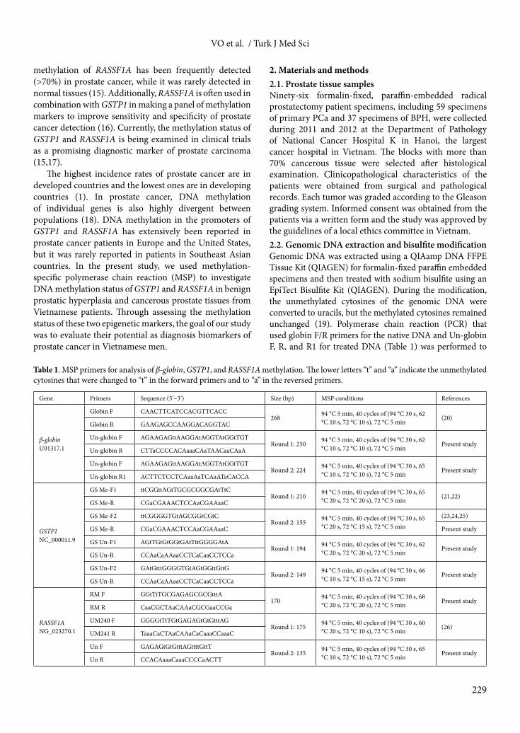
Table 1. MSP primers for analysis of β-globin, GSTP1, and RASSF1A methylation. The lower letters “t” and “a” indicate the unmethylated cytosines that were changed to “t” in the forward primers and to “a” in the reversed primers.
Gene Primers Sequence (5’–3’) Size (bp) MSP conditions References
β-globin U01317.1
Globin F CAACTTCATCCACGTTCACC268 94 °C 5 min, 40 cycles of (94 °C 30 s, 62
°C 10 s, 72 °C 10 s), 72 °C 5 min (20)Globin R GAAGAGCCAAGGACAGGTAC
Un-globin F AGAAGAGttAAGGAtAGGTAtGGtTGTRound 1: 250 94 °C 5 min, 40 cycles of (94 °C 30 s, 62
°C 10 s, 72 °C 10 s), 72 °C 5 min Present studyUn-globin R CTTaCCCCACAaaaCAaTAACaaCAaA
Un-globin F AGAAGAGttAAGGAtAGGTAtGGtTGTRound 2: 224 94 °C 5 min, 40 cycles of (94 °C 30 s, 65
°C 10 s, 72 °C 10 s), 72 °C 5 min Present studyUn-globin R1 ACTTCTCCTCAaaAaTCAaATaCACCA
GSTP1NC_000011.9
GS Me-F1 ttCGGttAGtTGCGCGGCGAtTtC Round 1: 210 94 °C 5 min, 40 cycles of (94 °C 30 s, 65
°C 20 s, 72 °C 20 s), 72 °C 5 min (21,22)GS Me-R CGaCGAAACTCCAaCGAAaaC
GS Me-F2 ttCGGGGTGtAGCGGtCGtCRound 2: 155 94 °C 5 min, 40 cycles of (94 °C 30 s, 65
°C 20 s, 72 °C 15 s), 72 °C 5 min
(23,24,25)
GS Me-R CGaCGAAACTCCAaCGAAaaC Present study
GS Un-F1 AGtTGtGtGGtGAtTttGGGGAtARound 1: 194 94 °C 5 min, 40 cycles of (94 °C 30 s, 62
°C 20 s, 72 °C 20 s), 72 °C 5 min Present studyGS Un-R CCAaCaAAaaCCTCaCaaCCTCCa
GS Un-F2 GAtGtttGGGGTGtAGtGGttGttGRound 2: 149 94 °C 5 min, 40 cycles of (94 °C 30 s, 66
°C 10 s, 72 °C 15 s), 72 °C 5 min Present studyGS Un-R CCAaCaAAaaCCTCaCaaCCTCCa
RASSF1ANG_023270.1
RM F GGtTtTGCGAGAGCGCGtttA170 94 °C 5 min, 40 cycles of (94 °C 30 s, 68
°C 20 s, 72 °C 20 s), 72 °C 5 min Present study RM R CaaCGCTAaCAAaCGCGaaCCGa
UM240 F GGGGtTtTGtGAGAGtGtGtttAGRound 1: 175 94 °C 5 min, 40 cycles of (94 °C 30 s, 60
°C 20 s, 72 °C 10 s), 72 °C 5 min (26)UM241 R TaaaCaCTAaCAAaCaCaaaCCaaaC
Un F GAGAGtGtGtttAGttttGttTRound 2: 135 94 °C 5 min, 40 cycles of (94 °C 30 s, 65
°C 10 s, 72 °C 10 s), 72 °C 5 min Present study Un R CCACAaaaCaaaCCCCaACTT

230
VO et al. / Turk J Med Sci
determine the efficiency of bisulfite conversion, and PCR that used MSP primers for the native DNA was performed to confirm the primer’s specificity only to methylated targets.2.3. Methylation-specific PCR (MSP)The methylation status of GSTP1 and RASSF1A was evaluated by using MSP for amplification of bisulfite-treated DNA with primers that distinguish methylated (M) from unmethylated (U) DNAs. Based on the primer designing tool for the MSP method (http://www.urogene.org/methprimer/index1.html), the primers for GSTP1 and RASSF1A were designed, and some of these primers were used in combination with the published ones (20–26). The primer sequences and amplicon lengths are shown in Table 1. Bisulfite-treated DNAs were subjected to single or nested PCR based on particular targeted genes. The PCR products were then subjected to electrophoresis on 12% acrylamide gel. All the PCR reactions were replicated at least three times.
DNA that was extracted from the lymphocytes of the healthy volunteers and then treated with bisulfite was used as a positive control for GSTP1 and RASSF1A unmethylation. DNA that was extracted from the PC3 cell line and then treated with bisulfite was used as a positive control for GSTP1 and RASSF1A methylation (27). Water without a DNA template was included in each PCR reaction as a control for any contamination. The methylation status was confirmed by sequencing the cloned MSP products for a subset of samples from each assay.2.4. Statistical analysisAssociations between clinicopathological characteristics and individual promoter methylation status were examined by using the chi-square test (SPSS Inc., Chicago, IL, USA). For all statistical analyses, P ≤ 0.05 was considered significant.
3. ResultsThe population in this study consisted of 59 patients with PCa and 37 patients with BPH, all of whom underwent radical prostatectomy. The clinicopathologic characteristics of all 96 patients are shown in Table 2. The median age of the cases was 71.65 years (range: 42–91), and most of the cases had tumors with Gleason grade IV or V (41/59 PCa, 69.4%).3.1. Verification of the specificity of MSP primersValidating the precision of the MSP primers specific only to the methylated target has been recommended in order to avoid false positive results due to coamplification of incompletely converted sequences (28). Thus, the bisulfite-untreated DNA and the bisulfite-treated DNA were separately subjected to MSP with the GSTP1 and RASSF1A primer sets that were specifically designed for the methylated targets. Efficient amounts of the DNA templates were checked by PCR with the globin primer sets that were designed from the native and unmethylated DNA targets (Figure 1A). No MSP products corresponding to the methylated GSTP1 and RASSF1A were amplified from untreated DNA extracted either from PC-3 cells or from the lymphocytes. Similarly, no MSP products corresponding to the methylated targets were amplified from treated DNA extracted from the lymphocytes, which was used as the positive control for unmethylated DNA. The methylated GSTP1 and RASSF1A were detected from only the treated DNA extracted from PC-3 cells (Figure 1B). The results confirmed the accuracy of the designed primer sets specific only to the methylated targets. These primers were subsequently subjected to analysis of the methylation status of GSTP1 and RASSF1A in prostate patients.
Table 2. Methylation frequencies of GSTP1 and RASSF1A in the samples of benign hyperplasia (BPH) and prostate cancer (Pca) patients.
Characteristics Overall, n = 96
GSTP1 RASSF1A
Un, n (%) Me, n (%) P-value Un, n (%) Me, n (%) P-value
Age (years)MedianRange
71.6542–91 92 43 94 29
Histological typePcaBPH
5937
55 (93.2)37 (100.0)
39 (66.1) 4 (10.8) <0.01 58 (98.3)
36 (97.3)19 (32.2)10 (27.0) 0.59
Histological grade (Gleason)I+IIIIIIV+V
51341
5 (100)10 (76.9)40 (97.6)
4 (80.0)10 (76.9)25 (61.0)
0.455 (100)12 (92.3)41 (100)
1 (20.0)5 (38.5)13 (41.5)
0.75
P-value: statistical analysis of the associations between clinicopathological characteristics and methylation status.

231
VO et al. / Turk J Med Sci
3.2. Methylation status of GSTP1 and RASSF1A in PCa and BPH tissuesThe genomic DNAs extracted from 59 PCa and 37 BPH specimens were treated with bisulfite and subjected directly to MSP. Representative results of the MSP products for methylation status of GSTP1 and RASSF1A are shown in Figures 2 and 3, respectively. Three patterns of M/M, M/U, and U/U signals of RASSF1A were observed in both PCa and BPH cases, but these patterns of GSTP1 were observed in cases of PCa only (Table 3). Biallelic unmethylation (U/U)
and monoallelic methylation (M/U) signals of GSTP1 were detected from BPH. Monoallelic M/M and biallelic M/U were count as the methylated status. MSP analysis revealed that the number of the methylated GSTP1 and RASSF1A was 39/59 (66.1%) and 19/59 (32.2%) patients with PCa, respectively (Table 2). MSP analysis also revealed that the methylation of GSTP1 and RASSF1A was detected in 4/37 (10.8%) and 10/37 (27%) patients with BPH, respectively. Forty-three out of 59 PCa (72.9%) samples showed methylation status of one or two genes.
Figure 1. A–B) The efficiency of bisulfite conversion of genomic DNA that was extracted from PC3 cell line (A) and from the lymphocytes (L) of the healthy volunteers (B). A band of 268 bp amplified from only the untreated DNAs (UT) by globin primers and a band of 244 bp amplified from only the treated DNAs (BT) by nested Un globin primer sets. C–D) Specificity of the GSTP1 and RASSF1A primer sets to only the methylated DNAs. A band of 155 bp and 170 bp amplified from only the treated genomic DNAs by the GSTP1 (C) and RASSF1A (D) primer sets specifically designed for methylated sequences. M: 100-bp DNA ladder. (–): Negative control without DNA template.
Figure 2. Representative results of the methylation analysis of GSTP1 in the prostate cancer (P1–P10) and benign hyperplasia (B1–B12) samples. The PCR products in lanes Me and Un indicate the presence of methylated (155 bp) and unmethylated (149 bp)GSTP1. L: lymphocytes of the healthy volunteers. PC3: prostate cell line. (–): Negative control without DNA template. M: 100-bp DNA ladder.
B2

232
VO et al. / Turk J Med Sci
The DNA methylation frequencies and clinical characteristics corresponding to surgical and pathological records of the cases were compared. There was a significant difference in the methylation rate between PCa and BPH for only GSTP1 (P < 0.01) (Table 2). No significant differences in the methylation frequencies of GSTP1 and RASSF1A were observed in terms of age and histological grade (Gleason) of the PCa patients (Table 2).
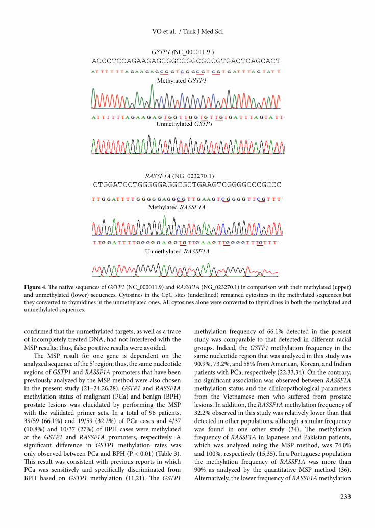
The methylation and unmethylation of GSTP1 and RASSF1A were confirmed by cloning and sequencing MSP products that were amplified from the treated DNAs extracted from the prostate cancer samples (Figure 4). The nucleotide sequences showed that all cytosine residues were converted to thymidines in the GSTP1 and RASSF1A unmethylated products, and that all cytosines in the CpG sites remained as cytosines and the cytosines that were not in the CpG sites were converted to thymidines in the GSTP1 and RASSF1A methylated products.
4. DiscussionAmong several DNA methylation markers associated with prostate cancer, GSTP1 and RASSF1A methylations captured the most interest because they were strongly associated with and considered as specific molecular
biomarkers of prostate cancer (5). Highly significant GSTP1 and RASSF1A methylation rates have been extensively reported from tissue biopsies and body fluids (plasma, serum, whole blood, urine, semen) from patients with prostate cancer (12,24,28) This evidence makes them the most promising commercial DNA methylation markers for early detection of this cancer (5).
DNA methylation profiles of thousands of genes or of a particular gene can be quantitatively assessed by technological approaches such as DNA microarrays or methylation-sensitive high-resolution melting (MS-HRM), which may not be accessible to many institutions in developing countries (29,30). The MSP method was chosen in the present study because of its sensitivity, specificity, and suitability in most moderately equipped laboratories (31). However, false positive results due to MSP primers unspecific to the methylated target have been reported (28). Thus, the standard controls were set up to test the accuracy of the primers specific to the methylated GSTP1 and RASSF1A through PCR, in which MSP products were amplified neither from the native DNA that was extracted from the lymphocytes or from PC3 cells, nor from the bisulfite-treated DNA that was extracted from the lymphocytes (Figure 1). This finding
Figure 3. Representative results of the methylation analysis of RASSF1A in the prostate cancer (P1–P10) and the benign hyperplasia (B1–B12) samples. The PCR products in lanes Me and Un indicate the presence of methylated (170 bp) and unmethylated (135 bp) RASSF1A. L: lymphocytes of the healthy volunteers. PC3: prostate cell line. (–): Negative control without DNA template. M: 100-bp DNA ladder.
Table 3. Status and frequency of methylation of GSTP1 and RASSF1A in PCa and BPH. Three patterns of M/M, M/U, and U/U.
GenesPCa BPH
GSTP1 RASSF1A GSTP1 RASSF1A
M/M 4 1 0 1
M/U 35 18 4 9
U/U 20 40 33 27
Methylation ratio 39/59 (66.1%) 19/59 (32.2%) 4/37 (10.8%) 10/37 (27%)
233
VO et al. / Turk J Med Sci
confirmed that the unmethylated targets, as well as a trace of incompletely treated DNA, had not interfered with the MSP results; thus, false positive results were avoided.
The MSP result for one gene is dependent on the analyzed sequence of the 5’ region; thus, the same nucleotide regions of GSTP1 and RASSF1A promoters that have been previously analyzed by the MSP method were also chosen in the present study (21–24,26,28). GSTP1 and RASSF1A methylation status of malignant (PCa) and benign (BPH) prostate lesions was elucidated by performing the MSP with the validated primer sets. In a total of 96 patients, 39/59 (66.1%) and 19/59 (32.2%) of PCa cases and 4/37 (10.8%) and 10/37 (27%) of BPH cases were methylated at the GSTP1 and RASSF1A promoters, respectively. A significant difference in GSTP1 methylation rates was only observed between PCa and BPH (P < 0.01) (Table 3). This result was consistent with previous reports in which PCa was sensitively and specifically discriminated from BPH based on GSTP1 methylation (11,21). The GSTP1
methylation frequency of 66.1% detected in the present study was comparable to that detected in different racial groups. Indeed, the GSTP1 methylation frequency in the same nucleotide region that was analyzed in this study was 90.9%, 73.2%, and 58% from American, Korean, and Indian patients with PCa, respectively (22,33,34). On the contrary, no significant association was observed between RASSF1A methylation status and the clinicopathological parameters from the Vietnamese men who suffered from prostate lesions. In addition, the RASSF1A methylation frequency of 32.2% observed in this study was relatively lower than that detected in other populations, although a similar frequency was found in one other study (34). The methylation frequency of RASSF1A in Japanese and Pakistan patients, which was analyzed using the MSP method, was 74.0% and 100%, respectively (15,35). In a Portuguese population the methylation frequency of RASSF1A was more than 90% as analyzed by the quantitative MSP method (36). Alternatively, the lower frequency of RASSF1A methylation
Figure 4. The native sequences of GSTP1 (NC_000011.9) and RASSF1A (NG_023270.1) in comparison with their methylated (upper) and unmethylated (lower) sequences. Cytosines in the CpG sites (underlined) remained cytosines in the methylated sequences but they converted to thymidines in the unmethylated ones. All cytosines alone were converted to thymidines in both the methylated and unmethylated sequences.

234
VO et al. / Turk J Med Sci
in this study might be due to the MSP primer’s specificity that was validated (Figure 1). Indeed, false positive results gave rise to an increase of 4 and 2 times the DNA methylation frequency (28).
A similar frequency of RASSF1A methylation in PCa (32.2%) and in BPH (27%) cases was observed in this study and in previous reports (25,34). The occurrence of methylation of RASSF1A in tumor and nontumor tissues from various cancers suggested that it is an early and premalignant sign (37). Thus, RASSF1A methylation in BPH has been considered as a sign of tumor progression. Indeed, a metaanalysis from 19 published studies on the association between RASSF1A promoter methylation and prostate cancer indicated that RASSF1A methylation was significantly associated with an increased risk of PCa (38).
Currently, the GSTP1 and RASSF1A methylation in body fluids is extensively studied because of its noninvasive
character and its ability to monitor prostate cancer (24,39). A high specificity of GSTP1 and RASSF1A methylation was found in these studies, regardless of methylation methods (12,14). Thus, the MSP method, which was supported by previous studies and was standardized in this study, is advantageous for further analyzing GSTP1 and RASSF1A methylation in body fluid specimens. Our study emphasized the authentic value of the MSP method that will allow the use of DNA methylation marker to quickly progress toward clinical application, especially in developing countries.
AcknowledgmentThis study was financially supported by grants KC.04.05/11-15 from the Ministry of Science and Technology of Vietnam.
References
1. Jamel A, Center MM, DeSantis C, Ward EM. Global patterns of cancer incidence and mortality rates and trends. Cancer Epidemiol Biomarkers Prev 2010; 19: 1893–1907.
2. Venderbos LD, Roobol MJ. PSA-based prostate cancer screening: the role of active surveillance and informed and shared decision making. Asian J Androl 2011; 13: 219–224.
3. Esserman LJ, Thompson IM, Reid B. Over diagnosis and over treatment in cancer: an opportunity for improvement. JAMA 2013; 310: 797–798.
4. Welch HG, Black WC. Overdiagnosis in cancer. J Natl Cancer Inst 2010; 102: 605–613.
5. Ahmed H. Promoter methylation in prostate cancer and its application for the early detection of prostate cancer using serum and urine samples. Biomark Cancer 2010; 2: 17–33.
6. Dawson MA, Kouzarides T. Cancer epigenetics: from mechanism to therapy. Cell 2012; 150:12–27.
7. Maxwell A, McCudden CR, Wians F, Monte S, Willis MS. Recent advances in the detection of prostate cancer using epigenetic markers in commonly collected laboratory samples. Lab Med 2009; 40: 171–178.
8. Majumdar S, Buckles E, Estrada J, Koochekpour S. Aberrant DNA methylation and prostate cancer. Cur Genomics 2011; 12: 486–505.
9. Lo HW, Stephenson L, Cao X, Milas M, Pollock R, Ali-Osman F. Identification and functional characterization of the human glutathione S-transferase P1 gene as a novel transcriptional target of the p53 tumor suppressor gene. Mol Cancer Res 2008; 6: 843–850.
10. Van Neste L, Herman JG, Otto G, Bigley JW, Epstein JI, Criekinge WV. The epigenetic promise for prostate cancer diagnosis. Prostate 2012; 72: 1248–1261.
11. Yoon HY, Kim YW, Kang HW, Kim WT, Yun SJ, Lee SC, Kim WJ, Kim YJ. DNA methylation of GSTP1 in human prostate tissues: pyrosequencing analysis. Korean J Urol 2012; 53: 200–205.
12. Wu T, Giovannucci E, Welge J, Mallick P, Tang WY, Mho S. Measurement of GSTP1 promoter methylation in body fluids may complement PSA screening: a meta-analysis. Br J Cancer 2011; 105: 65–73.
13. Dammann R, Takahashi T, Pfeifer GP. The CpG island of the novel tumor suppressor gene RASSF1A is intensely methylated in primary small cell lung carcinomas. Oncogenes 2001; 20: 3563–3577.
14. Pan J, Chen J, Zhang B, Chen X, Huang B, Zhuang J, Mo C, Qiu S. Association between RASSF1A promoter methylation and prostate cancer: a systematic review and Meta-analysis. PLoS ONE 2013; 8: e75283.
15. Kawamoto K, Okino ST, Place RF, Urakami S, Hirata H, Kikuno N, Kawakami T, Tanaka Y, Pookot D, Chen Z et al. Epigenetic modifications of RASSF1A gene through chromatin remodeling in prostate cancer. Clin Cancer Res 2007; 13: 2541–2548.
16. Rabiau N, Thiam MO, Satih S, Guy L, Kemeny JL, Boiteux JL, Fontana L, Bignon YJ, Bernard-Gallon D. Methylation analysis of BRCA1, RASSF1, GSTP1 and EPHB2 promoters in prostate biopsies according to different degrees of malignancy. In Vivo 2009; 23: 387–392.
17. Sunami E, Shinozaki M, Higano CS, Wollman R, Dorff TB, Tucker ST, Martinez SR, Singer FR, Hoon DSB. Multimarker circulating DNA assay for assessing blood of prostate cancer patients. Clin Chem 2009; 55: 559–567.
235
VO et al. / Turk J Med Sci
18. Addo BK, Wang S, Chung W, Jelinek J, Patierno SR, Wang BD, Andrawis R, Lee NH, Apprey V, Issa JP et al. Identification of differentially methylated genes in normal prostate tissues from African American and Caucasian men. Clin Cancer Res 2010; 16: 3539–3547.
19. Clark SJ, Statham A, Stirzaker C, Molloy PL, Frommer M. DNA methylation: bisulphite modification and analysis. Nat Protocol 2006; 1: 2355–2364.
20. Wilcox CB, Baysal BE, Gallion HH, Strange MA, DeLoia JA. High-resolution methylation analysis of the BRCA1 promoter in ovarian tumors. Cancer Genet Cytogen 2005; 159: 114–122.
21. Maruyama R, Toyooka S, Toyooka KO, Virmani AK, Zochbauer-Muller S, Farinas AJ, Minna JD, McConnell J, Frenkel EP, Gazdar AF. Aberrant promoter methylation profile of prostate cancers and its relationship to clinicopathological features. Clin Cancer Res 2002; 8: 514–519.
22. Nakayama M, Bennett CJ, Hicks JL, Epstein JI, Platz EA,
Nelson WG, De Marzo AM. Hypermethylation of the human glutathione S-transferase π gene (GSTP1) CpG island is present in a subset of proliferative inflammatory atrophy lesions but not in normal or hyperplastic epithelium of the prostate. Am J Pathol 2003; 163: 923–933.
23. Esteller M, Corn PG, Drena JM, Gabrielson E, Baylin SB, Herman JG. Inactivation of glutathione S-transferase P1 gene by promoter hypermethylation in human neoplasia. Cancer Res 1998; 58: 4515–4518.
24. Gonzalgo ML, Pavlovich CP, Lee SM, Nelson WG. Prostate cancer detection by GSTP1 methylation analysis of postbiopsy urine specimens. Clin Cancer Res 2003; 9: 2673–2677.
25. Dimitriadis E, Kalogeropoulos T, Velaeti S, Sotiriou S, Vassiliou E, Fasoulis L, Klapsas V, Synesiou M, Apostolaki A, Trangas T et al. Study of genetic and epigenetic alterations in urine samples as diagnostic markers for prostate cancer. Anticancer Res 2013; 33: 191–197.
26. Liu L, Yoon JH, Dammann R, Pfeifer GP. Frequent hypermethylation of the RASSF1A gene in prostate cancer. Oncogene 2002; 21: 6835–6840.
27. Vardi A, Bosviel R, Rabiau N, Adjakly M, Satih S, Dechelotte P, Boiteux J, Fontana L, Bignon YJ, Guy L et al. Soy phytoestrogens modify DNA methylation of GSTP1, RASSF1A, EPH2 and BRCA1 promoter in prostate cancer cells. In Vivo 2010; 24: 393–400.
28. Lan TV, Ha TN, Uyen QN, Duong TN, Huong TN, Thuan BT, Duong PA, To VT. Standardization of the methylation-specific PCR method for analysing BRCA1 and ER methylation. Mol Med Reports 2014; 9: 1844–1850.
29. Wojdacz TK, Dobrovic A. Methylation-sensitive high resolution melting (MS-HRM): a new approach for sensitive and high-throughput assessment of methylation. Nucl Acids Res 2007; 35: e41.
30. Houshdaran S, Hawley S, Palmer C, Campan M, Olsen MN, Ventura AP, Knudsen BS, Drescher CW, Urban ND, Brown PO et al. DNA methylation profiles of ovarian epithelial carcinoma tumors and cell lines. PLoS ONE 2010; 5: e9359.
31. Herman JG, Graff JR, Myohanen S, Nelkin BD, Baylin SB. Methylation-specific PCR: a novel PCR assay for methylation status of CpG islands. P Natl Acad Sci USA 1996; 93: 9821–9826.
32. Kristensen LS, Hansen L. PCR-based methods for detecting single-locus DNA methylation biomarkers in cancer diagnostics, prognostics, and response to treatment. Clin Chem 2009; 55: 1471–1483.
33. Cho NY, Kim BH, Choi M, Yoo EJ, Moon KC, Cho YM, Kim D, Kang GH. Hypermethylation of CpG island loci and hypomethylation of LINE-1 and Alu repeats in prostate adenocarcinoma and their relationship to clinicopathological features. J Pathol 2007; 211: 269–277.
34. Syeed N, Sameer AS, Hamid A, Shah ZA, Afroze D, Rasool R, Siddiqi MA. Promoter methylation profile of GSTP1 and RASSF1A in benign hyperplasia and metastatic prostate cancer patients in a Kashmiri population. Mol Med Rep 2010; 3: 883–887.
35. Yaqinuddin A, Qureshi SA, Pervez S, Bashir MU, Nazir R, Abbas F. Frequent DNA hypermethylation at the RASSF1A and APC gene loci in prostate cancer patients of Pakistani origin. ISRN Urology 2013; 2013: 627249.
36. Jeronimo C, Henrique R, Hoque MO, Mambo E, Ribeiro FR, Varzim G, Oliveira J, Teixeira MR, Lopes C, Sidransky D. A quantitative promoter methylation profile of prostate cancer. Clin Cancer Res 2004; 10: 8472–8478.
37. Dammann R, Schagdarsurengin U, Seidel C, Strunnikova M, Rastetter M, Baier K, Pfeifer GP. The tumor suppressor RASSF1A in human carcinogenesis: an update. Histol Histopathol 2005; 20: 645–653.
38. Ge YZ, Xu LW, Jia RP, Xu Z, Feng YM, Wu R, Yu P, Zhao Y, Gui ZL, Tan SJ et al. The association between RASSF1A promoter methylation and prostate cancer: evidence from 19 published studies. Tumor Biol 2014; 35: 3881–3890.
39. Bastian PJ, Palapattu GS, Yegnasubramanian S, Rogers CG, Lin X, Mangold LA, Trock B, Eisenberger MA, Partin AW, Nelson WG. CpG island hypermethylation profile in the serum of men with clinically localized and hormone refractory metastatic prostate cancer. J Urol 2008; 179: 529–535.