Embed Size (px)
Citation preview
1521-0081/65/1/171–222$25.00 http://dx.doi.org/10.1124/pr.111.005678PHARMACOLOGICAL REVIEWS Pharmacol Rev 65:171–222, January 2013Copyright © 2013 by The American Society for Pharmacology and Experimental Therapeutics
ASSOCIATE EDITOR: DAVID R. SIBLEY
The Pharmacology of L-DOPA-Induced Dyskinesia inParkinson’s Disease
Philippe Huot,1 Tom H. Johnston, James B. Koprich, Susan H. Fox, and Jonathan M. Brotchie
Toronto Western Research Institute, University Health Network, Toronto, Ontario, Canada (P.H., T.H.J, J.B.K, S.H.F., J.M.B.);and Division of Neurology, Movement Disorder Clinic, Toronto Western Hospital, University Health Network, Toronto, Ontario, Canada
(P.H., S.H.F.)
Abstract . . . . . . . . . . . . . . . . . . . . . . . . . . . . . . . . . . . . . . . . . . . . . . . . . . . . . . . . . . . . . . . . . . . . . . . . . . . . . . . . . 172I. Introduction. . . . . . . . . . . . . . . . . . . . . . . . . . . . . . . . . . . . . . . . . . . . . . . . . . . . . . . . . . . . . . . . . . . . . . . . . . . . . . . 173II. Review Terms of Reference and Presentation . . . . . . . . . . . . . . . . . . . . . . . . . . . . . . . . . . . . . . . . . . . . . . 173III. The Classic Model of the Functional Organization of the Basal Ganglia . . . . . . . . . . . . . . . . . . . . 174IV. The Dopaminergic System. . . . . . . . . . . . . . . . . . . . . . . . . . . . . . . . . . . . . . . . . . . . . . . . . . . . . . . . . . . . . . . . . 176
A. Striatal Dopamine Denervation . . . . . . . . . . . . . . . . . . . . . . . . . . . . . . . . . . . . . . . . . . . . . . . . . . . . . . . . 176B. Pulsatile Dopaminergic Therapy . . . . . . . . . . . . . . . . . . . . . . . . . . . . . . . . . . . . . . . . . . . . . . . . . . . . . . . 177C. Dopamine Receptors . . . . . . . . . . . . . . . . . . . . . . . . . . . . . . . . . . . . . . . . . . . . . . . . . . . . . . . . . . . . . . . . . . . 180
V. The Glutamatergic System . . . . . . . . . . . . . . . . . . . . . . . . . . . . . . . . . . . . . . . . . . . . . . . . . . . . . . . . . . . . . . . . 182A. NMDA Receptors . . . . . . . . . . . . . . . . . . . . . . . . . . . . . . . . . . . . . . . . . . . . . . . . . . . . . . . . . . . . . . . . . . . . . . 183
1. Postmortem Studies . . . . . . . . . . . . . . . . . . . . . . . . . . . . . . . . . . . . . . . . . . . . . . . . . . . . . . . . . . . . . . . . 1832. Pharmacological Studies. . . . . . . . . . . . . . . . . . . . . . . . . . . . . . . . . . . . . . . . . . . . . . . . . . . . . . . . . . . . 184
B. AMPA Receptors . . . . . . . . . . . . . . . . . . . . . . . . . . . . . . . . . . . . . . . . . . . . . . . . . . . . . . . . . . . . . . . . . . . . . . 1861. Postmortem Studies . . . . . . . . . . . . . . . . . . . . . . . . . . . . . . . . . . . . . . . . . . . . . . . . . . . . . . . . . . . . . . . . 1862. Pharmacological Studies. . . . . . . . . . . . . . . . . . . . . . . . . . . . . . . . . . . . . . . . . . . . . . . . . . . . . . . . . . . . 186
C. Metabotropic Glutamate Receptors. . . . . . . . . . . . . . . . . . . . . . . . . . . . . . . . . . . . . . . . . . . . . . . . . . . . . 1871. Metabotropic Glutamate Receptors Type 2/3. . . . . . . . . . . . . . . . . . . . . . . . . . . . . . . . . . . . . . 1872. Metabotropic Glutamate Receptors Type 5. . . . . . . . . . . . . . . . . . . . . . . . . . . . . . . . . . . . . . . . 188
D. Glutamate Release . . . . . . . . . . . . . . . . . . . . . . . . . . . . . . . . . . . . . . . . . . . . . . . . . . . . . . . . . . . . . . . . . . . . 188E. Altered Synaptic Plasticity. . . . . . . . . . . . . . . . . . . . . . . . . . . . . . . . . . . . . . . . . . . . . . . . . . . . . . . . . . . . . 189
VI. The Opioid System . . . . . . . . . . . . . . . . . . . . . . . . . . . . . . . . . . . . . . . . . . . . . . . . . . . . . . . . . . . . . . . . . . . . . . . . 190A. Preproenkephalin and Preprodynorphin. . . . . . . . . . . . . . . . . . . . . . . . . . . . . . . . . . . . . . . . . . . . . . . . 190B. Opioid Receptors . . . . . . . . . . . . . . . . . . . . . . . . . . . . . . . . . . . . . . . . . . . . . . . . . . . . . . . . . . . . . . . . . . . . . . 191
VII. The Serotonergic System . . . . . . . . . . . . . . . . . . . . . . . . . . . . . . . . . . . . . . . . . . . . . . . . . . . . . . . . . . . . . . . . . . 193A. 5-HT1A/1B Receptors . . . . . . . . . . . . . . . . . . . . . . . . . . . . . . . . . . . . . . . . . . . . . . . . . . . . . . . . . . . . . . . . . . . 193B. 5-HT2A Receptors. . . . . . . . . . . . . . . . . . . . . . . . . . . . . . . . . . . . . . . . . . . . . . . . . . . . . . . . . . . . . . . . . . . . . . 195C. Serotonin Transporter . . . . . . . . . . . . . . . . . . . . . . . . . . . . . . . . . . . . . . . . . . . . . . . . . . . . . . . . . . . . . . . . . 195
VIII. The GABAergic System . . . . . . . . . . . . . . . . . . . . . . . . . . . . . . . . . . . . . . . . . . . . . . . . . . . . . . . . . . . . . . . . . . . 196IX. The Adenosine System . . . . . . . . . . . . . . . . . . . . . . . . . . . . . . . . . . . . . . . . . . . . . . . . . . . . . . . . . . . . . . . . . . . . 197X. Cannabinoid Neurotransmission. . . . . . . . . . . . . . . . . . . . . . . . . . . . . . . . . . . . . . . . . . . . . . . . . . . . . . . . . . . 198
A. Cannabinoid Receptors . . . . . . . . . . . . . . . . . . . . . . . . . . . . . . . . . . . . . . . . . . . . . . . . . . . . . . . . . . . . . . . . 198B. Endogenous Cannabinoids . . . . . . . . . . . . . . . . . . . . . . . . . . . . . . . . . . . . . . . . . . . . . . . . . . . . . . . . . . . . . 199
XI. Adrenergic Neurotransmission. . . . . . . . . . . . . . . . . . . . . . . . . . . . . . . . . . . . . . . . . . . . . . . . . . . . . . . . . . . . . 199XII. Histaminergic Neurotransmission . . . . . . . . . . . . . . . . . . . . . . . . . . . . . . . . . . . . . . . . . . . . . . . . . . . . . . . . . 201
Address correspondence to: Jonathan M. Brotchie, Toronto Western Research Institute, MCL 11-419, Toronto Western Hospital, 399Bathurst Street, Toronto, Ontario, Canada, M5T 2S8. E-mail: [email protected]
This work was supported by The Cure Parkinson’s Trust and Krembil Neuroscience Fund. P.H. was supported by Fellowships from theEdmond J. Safra Philanthropic Foundation, the Parkinson Society Canada, and the Canadian Institutes of Health Research.
1Current affiliation: Baycrest Centre for Geriatric Care, Toronto, Ontario, Canada.There are no conflicts of interest. S.H.F. received consultancy fees from Merck, Merck Serono, and Teva. T.H.J., J.B.K., and J.M.B. received
consultancy fees from Atuka Ltd. and Atuka Inc., and hold equity positions in Atuka Inc. P.H. received consultancy fees from Atuka Ltd. andAtuka Inc.
dx.doi.org/10.1124/pr.111.005678.
171
by guest on July 7, 2018D
ownloaded from
XIII. Cholinergic Neurotransmission . . . . . . . . . . . . . . . . . . . . . . . . . . . . . . . . . . . . . . . . . . . . . . . . . . . . . . . . . . . . 202XIV. Tachykinin Neurotransmission . . . . . . . . . . . . . . . . . . . . . . . . . . . . . . . . . . . . . . . . . . . . . . . . . . . . . . . . . . . . 203XV. Transcription Factors and Intracellular Signaling . . . . . . . . . . . . . . . . . . . . . . . . . . . . . . . . . . . . . . . . . . 204
A. Transcription Factors. . . . . . . . . . . . . . . . . . . . . . . . . . . . . . . . . . . . . . . . . . . . . . . . . . . . . . . . . . . . . . . . . . 204B. DARPP-32, ERK1/2, and Histone Deacetylation . . . . . . . . . . . . . . . . . . . . . . . . . . . . . . . . . . . . . . . . 205C. Other Abnormal Intracellular Cascades . . . . . . . . . . . . . . . . . . . . . . . . . . . . . . . . . . . . . . . . . . . . . . . . 207
XVI. Emerging Concepts in Dyskinesia . . . . . . . . . . . . . . . . . . . . . . . . . . . . . . . . . . . . . . . . . . . . . . . . . . . . . . . . . 209A. Angiogenesis and Blood-Brain Barrier . . . . . . . . . . . . . . . . . . . . . . . . . . . . . . . . . . . . . . . . . . . . . . . . . 209B. Inflammation. . . . . . . . . . . . . . . . . . . . . . . . . . . . . . . . . . . . . . . . . . . . . . . . . . . . . . . . . . . . . . . . . . . . . . . . . . 209C. Fatty Acids. . . . . . . . . . . . . . . . . . . . . . . . . . . . . . . . . . . . . . . . . . . . . . . . . . . . . . . . . . . . . . . . . . . . . . . . . . . . 209D. Exocytosis . . . . . . . . . . . . . . . . . . . . . . . . . . . . . . . . . . . . . . . . . . . . . . . . . . . . . . . . . . . . . . . . . . . . . . . . . . . . . 210E. Thyrotropin Releasing Hormone . . . . . . . . . . . . . . . . . . . . . . . . . . . . . . . . . . . . . . . . . . . . . . . . . . . . . . . 210
XVII. Concluding Remarks . . . . . . . . . . . . . . . . . . . . . . . . . . . . . . . . . . . . . . . . . . . . . . . . . . . . . . . . . . . . . . . . . . . . . . 210References . . . . . . . . . . . . . . . . . . . . . . . . . . . . . . . . . . . . . . . . . . . . . . . . . . . . . . . . . . . . . . . . . . . . . . . . . . . . . . . . 211
Abstract——L-3,4-Dihydroxyphenylalanine (L-DOPA)remains the most effective symptomatic treatment ofParkinson’s disease (PD). However, long-term ad-ministration of L-DOPA is marred by the emergence ofabnormal involuntary movements, i.e., L-DOPA-induceddyskinesia (LID). Years of intensive research haveyielded significant progress in the quest to elucidatethe mechanisms leading to the development and ex-pression of dyskinesia and maintenance of thedyskinetic state, but the search for a complete un-derstanding is still ongoing. Herein, we summarizethe current knowledge of the pharmacology of LIDin PD. Specifically, we review evidence gatheredfrom postmortem and pharmacological studies, both
preclinical and clinical, and discuss the involvement ofdopaminergic and nondopaminergic systems, includingglutamatergic, opioid, serotonergic, g-aminobutyric acid(GABA)-ergic, adenosine, cannabinoid, adrenergic,histaminergic, and cholinergic systems. Moreover, wediscuss changes occurring in transcription factors,intracellular signaling, and gene expression in thedyskinetic phenotype. Inasmuch as a multitude ofneurotransmitters and receptors play a role in theetiology of dyskinesia, we propose that to op-timally alleviate this motor complication, it may benecessary to develop combined treatment approachesthat will target simultaneously more than oneneurotransmitter system. This could be achieved via
ABBREVIATIONS: 2-AG, 2-arachidonyl glycerol; ACP-103, pimavanserin; 5-HT, serotonin; 5-HT1A, serotonergic type 1A receptor; 5-HT1B,serotonergic type 1B receptor; 5-HT2A, serotonergic type 2A receptor; 6-OHDA, 6-hydroxydopamine; A1, adenosine 1 receptors; A2A, adenosine2A receptors; A2B, adenosine 2B receptors; A3, adenosine 3 receptors; AADC, aromatic L-amino acid decarboxylase; ACh, acetylcholine; AIMs,abnormal involuntary movements; Akt, protein kinase B; AMPA, 2-amino-3-(5-methyl-3-oxo-1,2-oxazol-4-yl)propionic acid; BrdU, 5-bromo-29-deoxyuridine; CalDAG-GEF1/2, calcium and diacylglycerol guanine nucleotide exchange factor types 1 and 2; CALM-PD, comparison of theagonist pramipexole versus levodopa on motor complications of Parkinson’s disease; cAMP, cyclic adenosine monophosphate; CB1,cannabinoid type 1 receptor; Cdk5, cyclin-dependent kinase 5; cGMP, cyclic guanosine monophosphate; COMT, catechol-O-methyltransferase;CREB, cAMP response element-binding protein; CSF, cerebrospinal fluid; D1, dopamine D1 receptor; D2, dopamine D2 receptor; D3, dopamineD3 receptor; D4, dopamine D4 receptor; D5, dopamine D5 receptor; DARPP-32, DARPP-32, dopamine- and cAMP-regulated phosphoprotein of32 kDa; DAT, dopamine transporter; DHA, docosohexaenoic acid; EGR1, early growth response protein 1; EMA, European Medicines Agency;ERK, extracellular signal-related protein kinase; ERK1/2, extracellular signal-regulated protein kinases 1 and 2; FAAH, fatty acid amidehydrolase; FDA, US Food and Drug Administration; FIRST-STEP, favorability of immediate-release carbidopa/levodopa versus stalevo short-term comparison in early Parkinson’s; FosB, FBJ murine osteosarcoma viral oncogene homolog B; GABA, g-aminobutyric acid; GAD, glutamicacid decarboxylase; GAD65, glutamic acid decarboxylase isoform 65; GAD67, glutamic acid decarboxylase isoform 67; GDNF, glial cell-linederived neurotrophic factor; GluR1, subunit GluR1 of AMPA receptors; GluR2, subunit GluR2 of AMPA receptors; GluR3, subunit GluR3 ofAMPA receptors; GP, globus pallidus; GPe, globus pallidus pars externa; GPi, globus pallidus pars interna; GPR18, G protein-coupled receptor18; GPR55, G protein-coupled receptor 55; GPR119, G protein-coupled receptor 119; GRK6, G protein-coupled receptor kinase 6; GSK3,glycogen synthase kinase 3; GTP, guanosine triphosphate; GTPase, guanosine triphosphatase; GTPgS, [35S]guanosine 59-O-[g-thio]triphosphate; H2, histamine receptor type 2; H3, histamine receptor type 3; HDAC, histone deacetylase; IC50, half-maximal inhibitoryconcentration; Ki, inhibition constant; L-, levo; L-DOPA, L-3,4-dihydroxyphenylalaine; LI-AIMs, L-DOPA-induced abnormal involuntarymovements; Leu, leucine; LID, L-DOPA-induced dyskinesia; LTD, long-term depression; LTP, long-term potentiation; M100,907, volinanserin;MDMA, 3,4-methylenedioxymethamphetamine; Met, methionine; mGlu2, metabotropic glutamate receptors type 2; mGlu3, metabotropicreceptors type 3; mGlu5, metabotropic glutamate receptors type 5; MPTP, 1-methyl-4-phenyl-1,2,3,6-tetrahydropyridine; mRNA, messengerribonucleic acid; MSK-1, mitogen- and stress-activated kinase 1; mTOR, mammalian target of rapamycin; NGF1-A, nerve growth factor-induced protein 1A; NHP, nonhuman primate; NMDA, N-methyl-D-aspartate; NO, nitric oxide; NOS, nitric oxide synthase; NR1, subunit NR1of NMDA receptors; NR2A, subunit NR2A of NMDA receptors; NR2B, subunit NR2B of NMDA receptors; pAkt, phosphorylated Akt; PD,Parkinson’s disease; PDE5, phosphodiesterase type 5; PET, positron emission tomography; PKA, protein kinase A; PKB, protein kinase B;PKC, protein kinase C; POMC, proopiomelanocortin; PPD, preprodynorphin; PPE-A, preproenkephalin-A; PPE-B, preproenkephalin-B; PPT,preprotachykinin; Ras-GRF1, Ras-guanine nucleotide-releasing factor 1; RBD, rapid-eye-movement sleep behavior disorder; Rhes, Rashomolog enriched in striatum; SERT, serotonin transporter; SN, substantia nigra; SNc, substantia nigra pars compacta; SNr, substantia nigrapars reticulata; STN, subthalamic nucleus; STRIDE-PD, stalevo reduction in dyskinesia evaluation in Parkinson’s disease; SV2A, synapticvesicle glycoprotein 2A; TRH, thyrotropin releasing hormone; TRPV1, transient receptor potential vanilloid type 1; VEGF, vascularendothelial growth factor.
172 Huot et al.
three ways as follows: 1) by developing compoundsthat will interact simultaneously to a multitude ofreceptors with the required agonist/antagonist effectat each target, 2) by targeting intracellular signaling
cascades where the signals mediated by multiplereceptors converge, and/or 3) to regulate geneexpression in a manner that has effects on signalingby multiple pathways
I. Introduction
In their ground-breaking study, Ehringer andHornykiewicz (1960) discovered that dopamine levelswere reduced in the striatum of patients suffering fromParkinson’s disease (PD). This led to the introductionof dopamine replacement therapy, in the early 1960s,with the dopamine precursor L-3,4-dihydroxyphenyla-lanine (L-DOPA) (Cotzias et al., 1967, 1969a,b; Barbeau,1969a,b; Cotzias, 1969). At first, L-DOPA was admin-istered without a peripherally acting aromatic L-aminoacid decarboxylase (AADC) inhibitor, such as carbi-dopa or benserazide, and the doses of L-DOPA requiredto achieve therapeutic efficacy were higher than thoseused today. AADC inhibitors were added to L-DOPAduring the second half of the 1960s and first half of the1970s (Barbeau et al., 1971; Marsden et al., 1973a,b).AADC inhibitors allowed for the reduction of L-DOPAdoses, a faster onset of an antiparkinsonian benefit,and a decrease in side effects of dopamine on thecardiovascular and gastrointestinal systems (Cotziaset al., 1969b; Anonymous, 1974). However, shortlyafter the introduction of L-DOPA as a therapy for PD,motor complications such as dyskinesia and wearing-off were observed following repeated administration ofthe dopamine precursor (Barbeau, 1971; Parkes et al.,1971; Weiss et al., 1971; Marsden et al., 1973a,b). Ina recent community-based study, the mean time toonset of dyskinesia was 6.6 years (Evans et al., 2011).However, other groups have reported that up to 56% ofPD patients experience dyskinesia as early as 2.9 yearsafter introduction of L-DOPA (Blanchet et al., 1996a),and this percentage climbs to 95% after 15 years oftherapy (Hely et al., 2005). Patients with earlier-onsetPD tend to be more prone to develop L-DOPA-induceddyskinesia (LID) than patients with later onset(Kumar et al., 2005). Dyskinesia has a negative impacton quality of life (Dodel et al., 1998; Damiano et al.,2000; Pechevis et al., 2005), sometimes being moredisabling than PD itself (Fahn, 2000). Although somepatients often prefer experiencing dyskinesia thanbeing in the off-state and unable to move (Hunget al., 2010), new, more effective therapies are stillrequired for severe disabling dyskinesia to affordpatients an improved quality of benefit while in theon-state.The clinical phenomenology of dyskinesia is complex,
and a wide range of presentations have been described,including neck, truncal, facial, and limb chorea anddystonia (Nutt, 1990; Luquin et al., 1992b). Thepattern of dyskinesia varies with respect to the timeof onset in relation to L-DOPA intake. Peak-dose
dyskinesia occurs when plasma levels of L-DOPA arehigh and tends to be predominantly chorea with somedystonia (Muenter and Tyce, 1971; Lees et al., 1977).Diphasic dyskinesia, or “onset and end-of-dose dyski-nesia,” occurs when plasma levels of L-DOPA are eitherrising or falling (“low-dose dyskinesia”), but not whenthey are stable, and tend to be predominantly dystonic(Muenter et al., 1977; Lhermitte et al., 1978).
Therefore, LID appears to be a complex set ofphenomena, in terms of both clinical presentationand pharmacokinetics. This complexity perhaps ex-plains why, despite extensive preclinical and clinicalresearch focused on increasing the pharmacotherapeu-tic arsenal, few agents have successfully been shown toreduce dyskinesia or to successfully translate frompreclinical to clinical settings. One reason has beenthat the majority of research to date have focused onpeak dose (high L-DOPA dose) dyskinesia, and manyPD subjects may experience a mixture of peak anddiphasic dyskinesia. Another reason might be that, aswill be seen in this review article, there is evidence forabnormalities in several neurotransmitter systems indyskinesia and that the traditional approaches, whichusually focus on a single target, may not be appropri-ate. The following sections will summarize the currentknowledge on the pharmacology of LID. It is importantto bear in mind that although all of the mechanismsdescribed herein probably contribute, to a certainextent, to the etiology of LID, no single factor has beenidentified as a requisite culprit in the pathophysiologyof dyskinesia.
II. Review Terms of Reference and Presentation
The data from animal models reviewed in thepresent article include only those from the 1-methyl-4-phenyl-1,2,3,6-tetrahydropyridine (MPTP)-lesionednonhuman primate (NHP) and the 6-hydroxydopamine(6-OHDA)-lesioned rat and mouse. Both chorea anddystonia are considered in the NHP, whereas abnormalinvoluntary movements (AIMs), the rodent correlate ofdyskinesia (Lee et al., 2000; Cenci and Lundblad,2007), are considered when discussing dyskinesia inthe rodent.
Mechanisms underlying sensitized rotational behav-ior occurring upon repeated administration of L-DOPAor dopamine agonists in the hemi-parkinsonian rodentare not included in the present article, becauseinterpretation of this behavior is controversial (Laneet al., 2006; Marin et al., 2006b), or at least difficult,being regarded both a marker of antiparkinsonianefficacy (Duvoisin et al., 1982; Traub et al., 1985;
L-DOPA-Induced Dyskinesia Pharmacology 173

Prikhojan et al., 2000) and priming/sensitization ofa hyperkinetic response, part of which could be relatedto dyskinesia (Carey, 1991; Konitsiotis and Tsironis,2006). Moreover, compounds such as yohimbine, clo-zapine, and naloxone, all of which effectively alleviateAIMs, do not reduce rotational behavior, whereasvolinanserin (M100,907) effectively reduces rotationswithout alleviating AIMs (Taylor et al., 2006), high-lighting differences between the pharmacology of thetwo phenomena (Lundblad et al., 2002).Additionally, with a few exceptions that are specif-
ically noted, studies performed in animal models of PDthat describe molecular changes without behavioralcorrelates are excluded from this article, because it isimpossible to correlate the postmortem findings withdyskinesia. For the same reasons, studies performed inidiopathic PD patients with motor complications, butwhere such complications are limited to wearing-offand do not include dyskinesia, are not reviewed. Inaddition, several postmortem studies performed usingthe brains of dyskinetic PD patients are reviewed,although critical information may be missing in each ofthese studies, such as the time of last administration ofL-DOPA and/or dopamine agonist prior to death.Indeed, because the patients may not have died attime of peak dyskinesia expression, it is unclearwhether the molecular changes reported are represen-tative of the on-state or the off-state of chronic L-DOPAtreatment.Throughout this article, the term “priming” refers to
the behavioral and molecular sensitization phenomenaoccurring after the first dose of L-DOPA. Priming is notnecessarily associated with the expression of dyskine-sia but by definition leads to changes in response tosubsequent L-DOPA challenges that result in theemergence of overt dyskinesia and its subsequentdevelopment into a more severe phenotype, as well asthe maintenance of the brain in the dyskinetic state,even when off treatment (Jenner, 2002; Nadjar et al.,2009). Once priming has occurred, lower doses ofL-DOPA or dopamine agonists are sufficient to elicitdyskinesia, and their therapeutic window is narrowed(Mouradian et al., 1989; Verhagen Metman et al.,1997).Only peer-reviewed articles are included in the
current review. Data presented as abstracts in scien-tific meetings have therefore been excluded.Throughout the article, unless specified otherwise,
the term L-DOPA refers to the combination of L-DOPAand an AADC inhibitor. Lastly, several pharmacolog-ical molecules are discussed in this review. It isimportant to bear in mind that, although they exhibitselectivity for a certain target, some of them are notvery selective and their biologic effect, althoughusually attributed to a specific pharmacological target,might in fact come from an interaction with off-targetreceptors.
III. The Classic Model of the FunctionalOrganization of the Basal Ganglia
To understand the mechanisms potentially underly-ing the pharmacological actions reviewed below, it isfirst helpful to review the anatomic circuitry underlingLID and consider neurotransmitter and receptor distri-bution among the components of that circuit. Accordingto the classic cortico-basal ganglia-thalamo-corticalmodel of the organization of the basal ganglia (Figs. 1and 2), the cerebral cortex sends excitatory glutama-tergic inputs to the striatum. These inputs aremodulated by nigrostriatal dopaminergic projectionsthat exert an excitatory effect, via dopamine D1
receptors, on the substance P/dynorphin-containingstriatofugal neurons of the direct pathway and aninhibitory effect, via dopamine D2 receptors, on theenkephalin-containing striatofugal neurons of the in-direct pathway. Striatofugal neurons of the directpathway send inhibitory g-aminobutyric acid (GABA)-ergic projections to the output structures of the basalganglia, the globus pallidus (GP) pars interna (GPi),and substantia nigra (SN) pars reticulata (SNr), whichalso emit GABAergic fibers, toward the ventral tier(ventrolateral and ventroanterior nuclei) of the thala-mus. In the indirect pathway, striatofugal GABAergicaxons contact the globus pallidus pars externa (GPe),which sends GABAergic fibers toward the subthalamicnucleus (STN). The STN then emits glutamatergicaxons toward the output structures of the basalganglia. Glutamatergic thalamocortical fibers completethe loop. Reciprocal GABAergic fibers connect the GPewith the GPi/SNr. The direct and indirect pathwaysexert opposing effects on movements and an imbalancein their activity is believed to lead to hypokinetic(i.e., parkinsonism) or hyperkinetic (i.e., dyskinesia)movement disorders. Although not traditionally part ofthe classic model of the basal ganglia, cortico-STNprojections, sometimes referred to as the “hyperdirectpathway,” have been recognized (DeLong, 1990;Parent, 1990; Parent and Hazrati, 1995a,b; Parentet al., 2000; Nambu et al., 2002; DeLong andWichmann, 2007; Koprich et al., 2009).
According to the model of the organization of thebasal ganglia described above, in the dyskinetic state,administration of L-DOPA leads to excessive activity ofstriatal projection neurons of the direct pathway. Thismodel is based very simply upon a consideration ofaverage frequency of firing, over time courses ofminutes and hours, of projections between the compo-nent nuclei (Eusebio and Brown, 2007; Hammondet al., 2007). Thus, D1-mediated transmission along thedirect pathway becomes overactive, which leads toexcessive activity along the direct pathway; D2-medi-ated activity along the indirect pathway also becomesoveractive, but this results in a reduction in activi-ty along the indirect pathway. Overactivity of the
174 Huot et al.

direct pathway and underactivity of the indirect path-way ultimately result in cortical overexcitation anddyskinesia (DeLong, 1990; Parent, 1990; Parent andHazrati, 1995a,b; Parent et al., 2000; DeLongand Wichmann, 2007; Koprich et al., 2009). In ac-cordance with a cortical overexcitation in the dyski-netic state, metabolic neuroimaging studies havedemonstrated overactivity of the supplementary motorarea and motor cortex when dyskinetic PD patientswere compared with nondyskinetic PD patients andnormal subjects (Rascol et al., 1994b, 1998; Brookset al., 2000).However, this classic explanation of dyskinesia
pathophysiology is too simplistic, and we consider itmore a metaphor to allow hypothesis building thana model that captures the true processing nature of thecircuit, and indeed, it revolves around dopamine,a subset of dopamine receptors, glutamate, GABA,and does not fit with several pieces of experimentaldata. For example, in the dyskinetic MPTP-lesionedNHP and in dyskinetic PD patients, there is noevidence of hypoactivity of the GPe (Vila et al., 1997),which argues against gross hypoactivity of the indirectpathway in the dyskinetic state. Moreover, according tothe classic model of the basal ganglia, lesions or deepbrain stimulation of the GPi should suppress thalamo-cortical inhibition, thereby resulting in an exacerbationof dyskinesia severity, whereas the opposite is seen inthe NHP (Iravani et al., 2005) and in clinic (Parkinet al., 2002). These caveats aside, it is remarkable howuseful such a simple metaphor has proved. In agree-ment with the opposite reduction in activity of the
output structures in the dyskinetic state, the admin-istration of the dopamine agonist apomorphine to theMPTP-lesioned NHP inhibits firing of the GPi (Filion,1979) and similar findings have been reported in PDpatients (Hutchinson et al., 1997). Moreover, as will bedescribed in detail below, the distribution of neuro-transmitter receptors and their function within differ-ent components of the circuits has led to theidentification of novel targets for LID and many ofthese have generated hypotheses that have been testedin pharmacological studies that have led to clinicaltrials across several classes of drug.
These successes notwithstanding, to reconcile theseconflicting data with the classic average frequencymodel of the basal ganglia, it has been proposed thatboth the frequency and pattern of firing play a criticalrole in dyskinesia genesis and relief of parkinsonism,dyskinesia genesis being more related to the pattern offiring and the improvement of parkinsonism beingmore related to the frequency of firing (Boraud et al.,2001). However, although an advance, this may not becompletely sufficient to explain the pathophysiology,because frequency of firing also appears to be animportant etiological contributor to dyskinesia, asa frequency firing of 4–10 Hz in the STN is associatedwith dyskinesia in PD patients (Alonso-Frech et al.,2006). Similar results were obtained in the 6-OHDA-lesioned rat, where in vivo recording of neurons of theSNr disclosed an increase in 4–10 Hz local-fieldpotential during dyskinesia expression (Meissneret al., 2006). In addition to frequency of firing,abnormal synchronization of the output structures ofthe basal ganglia may play a role in dyskinesiapathophysiology, and interruption of this abnormalsynchronization is thought to mediate the antidyski-netic efficacy of internal pallidotomy or deep-brainstimulation of the GPi (Brown and Eusebio, 2008;Obeso and Lanciego, 2011).
Additional criticisms relating to the classic model ofthe organization of the basal ganglia come from severalanatomic studies that have discovered an importantcollateralization in the inner basal ganglia circuitry,thus arguing against the simple functional dichotomi-zation highlighted above (Parent et al., 2000; Levesqueand Parent, 2005b; Nadjar et al., 2006). Also againstthe classic view of the basal ganglia model is the factthat D1 and D2 receptors are not completely segregatedbut appear to be coexpressed on 15–20% of striatalprojection neurons (Surmeier et al., 1996; Aizmanet al., 2000; Deng et al., 2006b). In addition, the modeldoes not take into account the interneurons present incore structures of the basal ganglia such as thestriatum and the STN (Cicchetti et al., 2000; Levesqueand Parent, 2005a; Huot and Parent, 2007) nor theinvolvement of many functionally important striatalafferents that employ several nondopaminergic trans-mitters such as serotonin (Lavoie and Parent, 1990,
Fig. 1. See text for description of the model of the organization of thebasal ganglia and for a description of the changes occurring in thedyskinetic state and references. Green lines indicate glutamatergicexcitatory transmission, whereas red lines indicate GABAergic inhibitorytransmission. Figure 1 is modified from Huot et al. (2011a).
L-DOPA-Induced Dyskinesia Pharmacology 175
1991), acetylcholine (ACh) (Heckers et al., 1992;Mesulam et al., 1992), or histamine (Steinbuschet al., 1986; Panula et al., 1989). However, as will beseen below, the classic model can be used to predictdrug effects and identify potential therapeutic benefitif its net influence on overall output of a pathway in themodel is known. For instance, although cholinergicinterneurons are not represented, the effect of cholin-ergic drugs on the direct and indirect pathways can bemodeled and the therapeutic benefits of such predicted(Pisani et al., 2007). Moreover, the classic modelfocuses exclusively on the sensorimotor territory ofthe basal ganglia, whereas metabolic changes in theassociative and limbic subdivisions of the basal gangliawere demonstrated at peak dyskinesia in the MPTP-lesioned macaque (Guigoni et al., 2005b). Therefore,although several references will be made to the modelof the basal ganglia organization throughout this
article, and we believe it remains an important andlikely the most useful model for the generation ofhypotheses, these limitations must be kept in mind.
IV. The Dopaminergic System
A. Striatal Dopamine Denervation
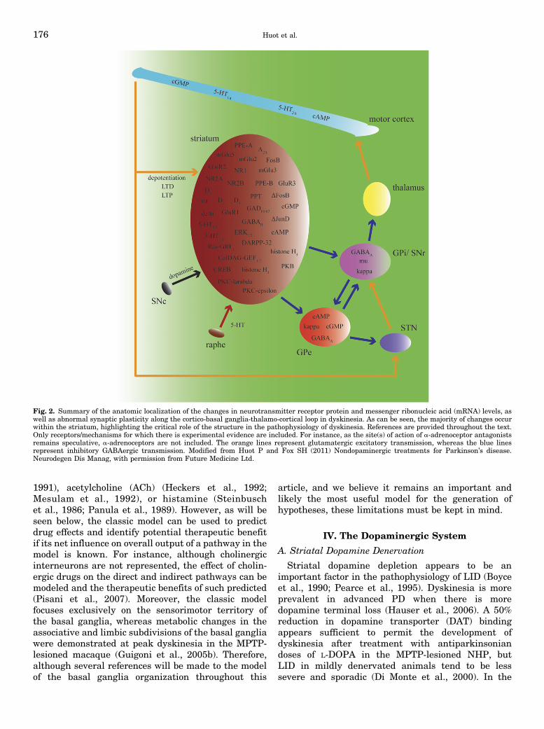
Striatal dopamine depletion appears to be animportant factor in the pathophysiology of LID (Boyceet al., 1990; Pearce et al., 1995). Dyskinesia is moreprevalent in advanced PD when there is moredopamine terminal loss (Hauser et al., 2006). A 50%reduction in dopamine transporter (DAT) bindingappears sufficient to permit the development ofdyskinesia after treatment with antiparkinsoniandoses of L-DOPA in the MPTP-lesioned NHP, butLID in mildly denervated animals tend to be lesssevere and sporadic (Di Monte et al., 2000). In the
Fig. 2. Summary of the anatomic localization of the changes in neurotransmitter receptor protein and messenger ribonucleic acid (mRNA) levels, aswell as abnormal synaptic plasticity along the cortico-basal ganglia-thalamo-cortical loop in dyskinesia. As can be seen, the majority of changes occurwithin the striatum, highlighting the critical role of the structure in the pathophysiology of dyskinesia. References are provided throughout the text.Only receptors/mechanisms for which there is experimental evidence are included. For instance, as the site(s) of action of a-adrenoceptor antagonistsremains speculative, a-adrenoceptors are not included. The orange lines represent glutamatergic excitatory transmission, whereas the blue linesrepresent inhibitory GABAergic transmission. Modified from Huot P and Fox SH (2011) Nondopaminergic treatments for Parkinson’s disease.Neurodegen Dis Manag, with permission from Future Medicine Ltd.
176 Huot et al.
MPTP-lesioned marmoset, intraventricular adminis-tration of glial cell-line derived neurotrophic factor(GDNF) led to the reversal of established dyskinesia;although the antidyskinetic mechanism of GDNF hasyet to be established, an increase in nigral dopaminer-gic cells occurred in that study, suggesting thata reversal of nigrostriatal denervation was involved(Iravani et al., 2001). In agreement with the impor-tance of striatal dopamine depletion in the etiology ofdyskinesia, a postmortem study found lower dopaminelevels in the putamen of dyskinetic when comparedwith nondyskinetic L-DOPA-treated PD patients (Rajputet al., 2004).On the other hand, some severely parkinsonian
macaques (Guigoni et al., 2005a) and 6-OHDA-lesioned rats (Cenci et al., 1998; Lee et al., 2000) neverdevelop LID, suggesting that, although the extent oflesion is an important determinant in the emergence ofdyskinesia, other factors are also involved. One of thesefactors might be the magnitude of L-DOPA doses ad-ministered. Hence, normal, non-MPTP-lesioned NHPsdevelop dyskinesia following treatment with high,nontherapeutically relevant, doses of L-DOPA (Pearceet al., 2001; Togasaki et al., 2001, 2005). Total dailydose of L-DOPA was also identified, in clinical settings,as a risk factor to the development of LID in PDpatients (Sharma et al., 2008).These data suggest that, although dopamine de-
nervation may not be required for LID, it may bea permissive factor that, when present, leads to theemergence of LID more rapidly, with lower L-DOPAdoses and with a more severe phenotype.
B. Pulsatile Dopaminergic Therapy
The short half-life of L-DOPA, approximately 1.5–2hours, leading to alternating peaks and troughs of highand low plasma levels (Cedarbaum, 1987; Gancheret al., 1987), is believed to play an important role in thedevelopment of dyskinesia and underlies the concept ofcontinuous dopaminergic stimulation to avoid dyski-nesia (Olanow et al., 2000; Stocchi and Olanow, 2004).In the normal state, dopamine release is both tonic andphasic, implying that basal levels of dopamine neverfall below a certain threshold (Grace, 1995; Goto et al.,2007). In the parkinsonian state, especially in latestages of the disease where the “buffering capacity” ofthe DAT has disappeared (Sohn et al., 1994), tonicdopamine release eventually disappears and dopaminerelease becomes exclusively phasic, i.e., pulsatile,following each dose of L-DOPA.Changes in dopamine vesicular release and reuptake
with disease progression might contribute to exacer-bate the fluctuation of dopamine levels (de la Fuente-Fernandez et al., 2004a). Hence, DAT binding levels inthe putamen of dyskinetic PD patients are lower whencompared with nondyskinetic PD patients (Troianoet al., 2009). Although DAT downregulation might lead
to enhanced synaptic levels of dopamine and thusa greater antiparkinsonian benefit, it will also exacer-bate peak dopamine levels associated with eachL-DOPA administration (de la Fuente-Fernandezet al., 2004b) and might thus be a key determinant inthe etiology of LID. However, although LID severitycorrelates with peak L-DOPA plasma concentration,there is a ceiling above which LID severity does notincrease despite higher L-DOPA dose, while duration ofantiparkinsonian benefit further increases (Metmanet al., 1997).
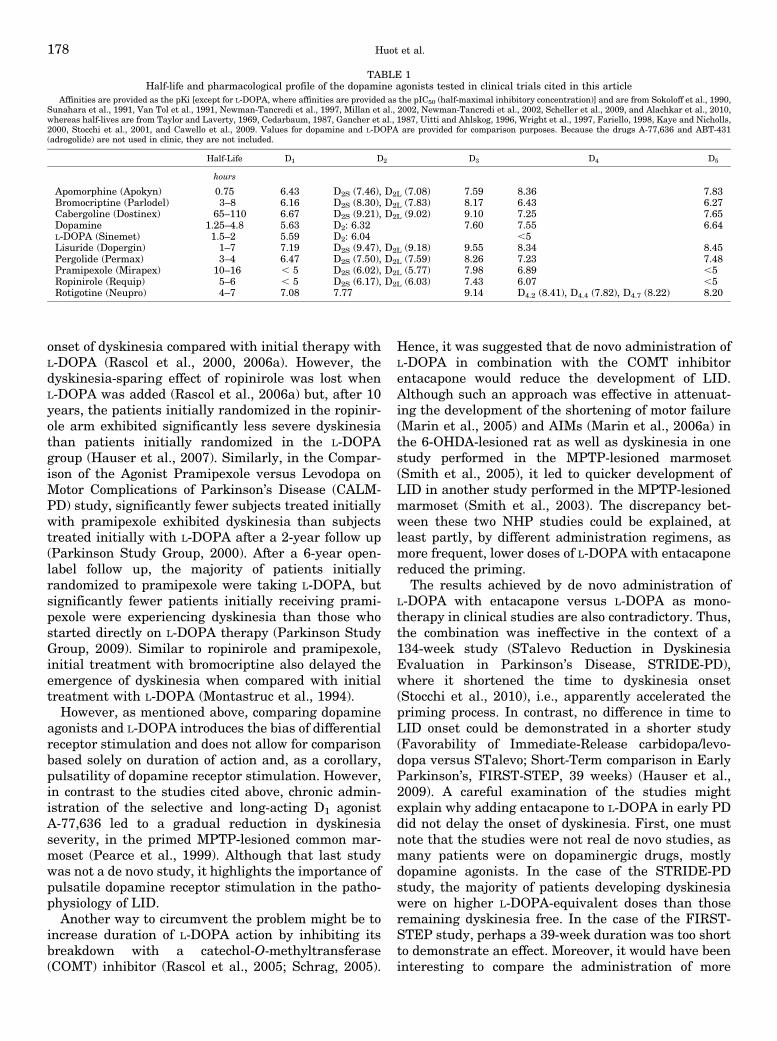
To elucidate the contribution of pulsatile dopaminereceptor stimulation in the etiology of dyskinesia,several preclinical and clinical studies compared thetime to onset of dyskinesia upon administration ofL-DOPA or dopamine agonists or upon administrationof dopamine agonists with variable half-lives. Aspresented in Table 1, with the exceptions of apomor-phine and lisuride, dopamine agonists have longerhalf-lives than L-DOPA, and their administrationtherefore results in more continuous stimulation ofdopamine receptors. As will be seen in the nextparagraphs, there is now evidence, although somecontroversy persists, that starting therapy with a do-pamine agonist delays the onset of dyskinesia com-pared with L-DOPA, but the data cannot be attributedsimply to their longer duration of action, becausedopamine agonists all exhibit greater potency for themembers of the dopamine D2 receptors family overmembers of the D1 receptors family (Table 1). As willbe seen in the next subsection, it is believed thatstimulation of D1 receptors is critical in the primingprocess, leading to the expression of dyskinesia.
In accordance with a role for pulsatile dopaminergictherapy in the induction of dyskinesia, the short-actingdopamine agonist apomorphine induced more AIMsthan the longer-acting dopamine agonists pramipexoleand pergolide in the 6-OHDA-lesioned rat (Papathanouet al., 2011). Perhaps quite surprisingly though, in theMPTP-lesioned marmoset, de novo administration ofapomorphine or pergolide led to dyskinesia of the sameseverity (Maratos et al., 2003). De novo continuousadministration of the dopamine receptor agonistsapomorphine and rotigotine induced less dyskinesiathan de novo once or twice daily administration ofthese drugs in the MPTP-lesioned NHP (Bibbianiet al., 2005a; Stockwell et al., 2009). Moreover, in theMPTP-lesioned NHP, continuous administration ofrotigotine alleviated dyskinesia induced by chronicpulsatile therapy with rotigotine or L-DOPA (Stockwellet al., 2010). De novo administration of ropinirole(Maratos et al., 2001; Jackson et al., 2007) or prami-pexole (Tayarani-Binazir et al., 2010) also led to lessdyskinesia than de novo therapy with L-DOPA in theMPTP-lesioned marmoset.
Similar results were achieved in PD patients treatedinitially with ropinirole, which successfully delayed the
L-DOPA-Induced Dyskinesia Pharmacology 177
onset of dyskinesia compared with initial therapy withL-DOPA (Rascol et al., 2000, 2006a). However, thedyskinesia-sparing effect of ropinirole was lost whenL-DOPA was added (Rascol et al., 2006a) but, after 10years, the patients initially randomized in the ropinir-ole arm exhibited significantly less severe dyskinesiathan patients initially randomized in the L-DOPAgroup (Hauser et al., 2007). Similarly, in the Compar-ison of the Agonist Pramipexole versus Levodopa onMotor Complications of Parkinson’s Disease (CALM-PD) study, significantly fewer subjects treated initiallywith pramipexole exhibited dyskinesia than subjectstreated initially with L-DOPA after a 2-year follow up(Parkinson Study Group, 2000). After a 6-year open-label follow up, the majority of patients initiallyrandomized to pramipexole were taking L-DOPA, butsignificantly fewer patients initially receiving prami-pexole were experiencing dyskinesia than those whostarted directly on L-DOPA therapy (Parkinson StudyGroup, 2009). Similar to ropinirole and pramipexole,initial treatment with bromocriptine also delayed theemergence of dyskinesia when compared with initialtreatment with L-DOPA (Montastruc et al., 1994).However, as mentioned above, comparing dopamine
agonists and L-DOPA introduces the bias of differentialreceptor stimulation and does not allow for comparisonbased solely on duration of action and, as a corollary,pulsatility of dopamine receptor stimulation. However,in contrast to the studies cited above, chronic admin-istration of the selective and long-acting D1 agonistA-77,636 led to a gradual reduction in dyskinesiaseverity, in the primed MPTP-lesioned common mar-moset (Pearce et al., 1999). Although that last studywas not a de novo study, it highlights the importance ofpulsatile dopamine receptor stimulation in the patho-physiology of LID.Another way to circumvent the problem might be to
increase duration of L-DOPA action by inhibiting itsbreakdown with a catechol-O-methyltransferase(COMT) inhibitor (Rascol et al., 2005; Schrag, 2005).
Hence, it was suggested that de novo administration ofL-DOPA in combination with the COMT inhibitorentacapone would reduce the development of LID.Although such an approach was effective in attenuat-ing the development of the shortening of motor failure(Marin et al., 2005) and AIMs (Marin et al., 2006a) inthe 6-OHDA-lesioned rat as well as dyskinesia in onestudy performed in the MPTP-lesioned marmoset(Smith et al., 2005), it led to quicker development ofLID in another study performed in the MPTP-lesionedmarmoset (Smith et al., 2003). The discrepancy bet-ween these two NHP studies could be explained, atleast partly, by different administration regimens, asmore frequent, lower doses of L-DOPA with entacaponereduced the priming.
The results achieved by de novo administration ofL-DOPA with entacapone versus L-DOPA as mono-therapy in clinical studies are also contradictory. Thus,the combination was ineffective in the context of a134-week study (STalevo Reduction in DyskinesiaEvaluation in Parkinson’s Disease, STRIDE-PD),where it shortened the time to dyskinesia onset(Stocchi et al., 2010), i.e., apparently accelerated thepriming process. In contrast, no difference in time toLID onset could be demonstrated in a shorter study(Favorability of Immediate-Release carbidopa/levo-dopa versus STalevo; Short-Term comparison in EarlyParkinson’s, FIRST-STEP, 39 weeks) (Hauser et al.,2009). A careful examination of the studies mightexplain why adding entacapone to L-DOPA in early PDdid not delay the onset of dyskinesia. First, one mustnote that the studies were not real de novo studies, asmany patients were on dopaminergic drugs, mostlydopamine agonists. In the case of the STRIDE-PDstudy, the majority of patients developing dyskinesiawere on higher L-DOPA-equivalent doses than thoseremaining dyskinesia free. In the case of the FIRST-STEP study, perhaps a 39-week duration was too shortto demonstrate an effect. Moreover, it would have beeninteresting to compare the administration of more
TABLE 1Half-life and pharmacological profile of the dopamine agonists tested in clinical trials cited in this article
Affinities are provided as the pKi [except for L-DOPA, where affinities are provided as the pIC50 (half-maximal inhibitory concentration)] and are from Sokoloff et al., 1990,Sunahara et al., 1991, Van Tol et al., 1991, Newman-Tancredi et al., 1997, Millan et al., 2002, Newman-Tancredi et al., 2002, Scheller et al., 2009, and Alachkar et al., 2010,whereas half-lives are from Taylor and Laverty, 1969, Cedarbaum, 1987, Gancher et al., 1987, Uitti and Ahlskog, 1996, Wright et al., 1997, Fariello, 1998, Kaye and Nicholls,2000, Stocchi et al., 2001, and Cawello et al., 2009. Values for dopamine and L-DOPA are provided for comparison purposes. Because the drugs A-77,636 and ABT-431(adrogolide) are not used in clinic, they are not included.
Half-Life D1 D2 D3 D4 D5
hours
Apomorphine (Apokyn) 0.75 6.43 D2S (7.46), D2L (7.08) 7.59 8.36 7.83Bromocriptine (Parlodel) 3–8 6.16 D2S (8.30), D2L (7.83) 8.17 6.43 6.27Cabergoline (Dostinex) 65–110 6.67 D2S (9.21), D2L (9.02) 9.10 7.25 7.65Dopamine 1.25–4.8 5.63 D2: 6.32 7.60 7.55 6.64L-DOPA (Sinemet) 1.5–2 5.59 D2: 6.04 ,5Lisuride (Dopergin) 1–7 7.19 D2S (9.47), D2L (9.18) 9.55 8.34 8.45Pergolide (Permax) 3–4 6.47 D2S (7.50), D2L (7.59) 8.26 7.23 7.48Pramipexole (Mirapex) 10–16 , 5 D2S (6.02), D2L (5.77) 7.98 6.89 ,5Ropinirole (Requip) 5–6 , 5 D2S (6.17), D2L (6.03) 7.43 6.07 ,5Rotigotine (Neupro) 4–7 7.08 7.77 9.14 D4.2 (8.41), D4.4 (7.82), D4.7 (8.22) 8.20
178 Huot et al.
frequent, smaller doses of L-DOPA/entacapone to thethree and four times a day regimens that were used inboth studies, as perhaps these administration para-digms did not allow sufficient continuous stimulationof dopamine receptors, although adherence to suchmore frequent regimens might have been complicated.Pulsatile L-DOPA delivery is also likely involved in
the maintenance of the dyskinetic state once priminghas occurred. Thus, in the MPTP-lesioned NHP, a re-duction in severity of established dyskinesia wasachieved following continuous administration of roti-gotine (Stockwell et al., 2010) or by administration ofthe long-acting dopamine agonist cabergoline (HadjTahar et al., 2000b). In clinical settings, continuousdaytime delivery of intraduodenal L-DOPA, over a 6-month period to PD patients with severe motorcomplications led to a reduction in previously estab-lished dyskinesia compared with intermittent admin-istration (Stocchi et al., 2005). Similar results wereachieved with continuous intravenous infusion of thedopamine agonist lisuride over a 3-month period(Baronti et al., 1992). The minimal amount of timerequired for continuous dopaminergic stimulation toalleviate established LID is unknown, but 48 hours ofuninterrupted i.v. L-DOPA administration seems tooshort (Juncos et al., 1987c), whereas 7–12 days isenough to modulate the dyskinesia threshold dose(Mouradian et al., 1990).Similar results were obtained in a study in which
ropinirole prolonged release was added to L-DOPA inpatients not optimally treated 3 years after diseaseonset; in that study, the addition of ropiniroleprolonged release, in contrast to increasing the doseof L-DOPA, led to a reduced appearance of dyskinesia(Watts et al., 2010). In contrast, in studies in whichcontrolled-release forms of L-DOPA were administered,leading to constant L-DOPA plasma levels over 4–6hours, there was no reduction in dyskinesia severityafter chronic administration compared with standardrelease L-DOPA (Juncos et al., 1987a, b), and theapparent reduction in dyskinesia achieved with ropi-nirole might in fact be due to preferential stimulationof D2/D3 receptors rather than its longer duration ofaction. The results of these two studies with controlled-release forms of L-DOPA pave the way to an important,as yet unanswered question: what does the term“pulsatility” mean and what duration of action shouldany antiparkinsonian compound have to lead to “non-pulsatile” stimulation of dopamine receptors? Althoughwidely referred to in the dyskinesia literature, theconcept of pulsatility remains poorly defined, but froma physiologic point of view, one might suggest that anymode of dopamine replacement therapy that is notcontinuous is necessarily pulsatile, regardless of thehalf-life of the molecule. Accordingly, and despite theresults of the studies cited above, it would be mis-leading to pretend that administration of L-DOPA
controlled release leads to continuous dopaminergicadministration, its duration of action being onlyslightly longer than that of L-DOPA standard release(Yeh et al., 1989; LeWitt, 1992; Hammerstad et al.,1994).
Although very few studies have specifically ad-dressed this issue, the moment of initiation of do-paminergic therapy might also have an impact on thedevelopment, but not severity, of dyskinesia. Thus, inthe 6-OHDA-lesioned rat, when L-DOPA was initiated4 weeks after lesion, significantly fewer animalsdeveloped AIMs compared with when L-DOPA wasinitiated 12 weeks after lesion, but AIMs severity indyskinetic animals did not differ between groups whenassessed 22 days after the initial dose of L-DOPA(Marin et al., 2009a). This issue was never assessed incontrolled clinical studies, and it is therefore difficult topredict whether the same phenomenon would occur inidiopathic PD patients, but long-term follow up ofpatients from the ELLDOPA study (Fahn et al., 2004)might eventually provide an answer to this question,as with disease progression all PD patients eventuallyrequire L-DOPA (Nutt et al., 2000). However, startingdopaminergic therapy early in the disease processmight not be a promising approach to delay dyskinesiaonset, as cumulative L-DOPA dose and L-DOPAequivalent dose have been established as clinicalvariables associated with dyskinesia development(Hauser et al., 2006). However, although cumulativeL-DOPA dose is an important contributory factor in theetiology of LID, the frequency of L-DOPA administra-tion, hence pulsatility of dopamine replacement ther-apy, might be more important. Thus, in a studyperformed in the 6-OHDA-lesioned rat, rats treatedwith higher L-DOPA dose developed AIMs quicker thanrats treated chronically with lower doses and that hadreceived more cumulated L-DOPA when AIMs firstappeared (Tsironis et al., 2008).
Thus, despite some of the limitations mentionedabove in the interpretation of the available data, itappears that longer-acting agents or continuous de-livery of L-DOPA are associated with either a delay inLID onset or a reduction in established LID severity.Unfortunately, the antiparkinsonian benefit conferredby dopamine agonists is not as potent as the oneobtained with L-DOPA and dopamine agonists cannotbe administered as monotherapy in advanced PD, andas such, initiating dopaminergic therapy with a dopa-mine agonist may only delay dyskinesia onset. On theother hand, the limitations of continuous delivery of L-DOPA at the moment are obvious, because no pillexists that would provide round-the-clock delivery ofthe drug. Intravenous administration of the drug canhardly be envisioned outside of hospital-based settings,and intraduodenal infusion is an invasive approachthat would not be justifiable in early PD where goodcontrol of the symptoms is easily achieved with two of
L-DOPA-Induced Dyskinesia Pharmacology 179
three times daily administration of dopaminergicagents. As such, research must continue to developan oral formulation that will allow constant plasmaL-DOPA levels throughout the day.
C. Dopamine Receptors
Dopamine exerts its effects by interacting with fivesubtypes of receptors, D1–5 (Missale et al., 1998;Beaulieu and Gainetdinov, 2011). D1 and D5 receptorsstimulate adenylyl cyclase activity and constitute theD1-like family of dopamine receptors (Civelli et al.,1993). In contrast, D2, D3, and D4 receptors reduce theactivity of adenylyl cyclase and form the D2-likereceptor family (Gingrich and Caron, 1993). The D2
receptor has two variants, D2S and D2L, whereas theD4 receptor has three variants, D4.2, D4.4, and D4.7
(Seeman and Van Tol, 1994). D1 and D2 receptors arethe most abundant of dopamine receptors within thestriatum (Sokoloff and Schwartz, 1995) and are themost extensively studied in PD and LID.Dopamine receptor supersensitivity was initially
proposed as a mechanism underlying LID. Severalautoradiographic receptor binding studies investigatedD1 and D2 receptor levels in the parkinsonian anddyskinetic states. However, they provided little in-formation to support a potential involvement of thesereceptors in dyskinesia. Thus, D2 receptor bindinglevels were increased in the striatum of L-DOPA-naive6-OHDA-lesioned rats (Creese et al., 1977), MPTP-lesioned NHPs (Graham et al., 1993), and PD patients(Lee et al., 1978). This increase in D2 receptors was notmaintained after chronic L-DOPA therapy in theMPTP-lesioned NHP (Graham et al., 1993) and PDpatients (Lee et al., 1978). Unlike the D2 receptor, D1
receptor binding levels did not change in PD (Lee et al.,1978; Shinotoh et al., 1993) or in the MPTP-lesionedNHP (Aubert et al., 2005), regardless of L-DOPAtreatment. In contrast, in the 6-OHDA-lesioned rat,D1 receptor mRNA was reduced in the L-DOPA-naiveparkinsonian state, but the reduction was reversedfollowing treatment with the D1 agonist SKF-38,393(Gerfen et al., 1990). Similar findings were encoun-tered in positron emission tomography (PET) studies.Thus, D1 binding levels were unchanged when PDpatients, with and without dyskinesia, were comparedwith healthy controls 4 hours after last dose ofL-DOPA, whereas D2 binding levels were reduced inboth dyskinetic and nondyskinetic PD patients com-pared with normal subjects, again 4 hours after lastdose of L-DOPA (Turjanski et al., 1997).More insight can be gathered from studies looking at
the subcellular distribution of D1 and D2 receptors,which indicate specific abnormalities in subcellulardistribution of D1 but not D2 receptors in dyskinesia.Thus, in the MPTP-lesioned NHP killed 1 hour afterthe last dose of L-DOPA, in nondyskinetic animals, D1
receptors were recruited at the synaptic membrane of
striatal neurons, whereas they were recruited at boththe synaptic membrane and the cytoplasmic compart-ment in dyskinetic animals (Guigoni et al., 2007).Hypoactivity of the 20S proteasomal subunit of striatalmedium spiny neurons could be an important de-terminant in this altered distribution of D1 receptors indyskinesia (Berthet et al., 2012). Indeed, hypoactivityof the 20S proteasomal subunit would impair degra-dation of D1 receptors, which might account for theirrecruitment at both the synaptic membrane and thecytoplasmic compartment (Berthet et al., 2012).
However, in the 6-OHDA-lesioned rat killed 45minutes after the last dose of L-DOPA, treatment withL-DOPA induced an internalization of D1 receptors(Muriel et al., 2002). These findings in the rat weresimilar to those encountered in idiopathic PD, wherechronic L-DOPA treatment was associated with a pref-erential cytoplasmic localization of D1 receptors com-pared with healthy control individuals (Muriel et al.,1999). Therefore, if it seems clear that subcellulardistribution of D1 receptors is altered in the dyskineticstate, the type of alteration seems to be variable acrossspecies and it is not clear whether this abnormaldistribution is a mechanism responsible for the de-velopment, expression, or maintenance of LID. Toelucidate this question, it would be important to repeatthose experiments and to include a group of animalsthat would be killed at different times during thepriming phase and a group of dyskinetic animals thatwould be killed while in the off-state. Similarly, theinformation provided by human studies with respect tothe expression of the dyskinetic phenotype remainslimited. Indeed, the majority of PD patients included inthese studies did not die at peak dyskinesia expressionand as such, the postmortem changes described are notthose encountered when dyskinesia is being acutelyexpressed but rather reflect the off-state of PD patientstreated chronically with L-DOPA. Nevertheless, aninternalization of D1 receptors in the dyskinetic statemight represent an endogenous compensatory mecha-nism aimed at alleviating dyskinesia.
There is also evidence for overactive D1-mediatedsignaling in dyskinesia. Thus, in brain slices fromMPTP-lesioned macaques killed at peak expression ofdyskinesia, D1 agonist-induced [35S]guanosine 59-O-[g-thio]triphosphate (GTPgS) binding correlated line-arly with dyskinesia severity (Aubert et al., 2005).
In agreement with abnormal cellular distribution ofD1 receptors and overactive D1-mediated transmissionin dyskinesia, internalization of D1 receptors isaccompanied by a reduction of dyskinesia severity.Thus, lentiviral-induced striatal overexpression of Gprotein-coupled receptor kinase 6 (GRK6) led to aninternalization of D1 receptors in the dyskinetic6-OHDA-lesioned rat and the MPTP-lesioned ma-caque (Ahmed et al., 2010); this was associated witha reduction of established L-DOPA-induced-AIMs
180 Huot et al.

(LI-AIMs) and LID in rat and macaque, respectively(Ahmed et al., 2010).D1 receptors also appear to play a role in the priming
process leading to the expression of dyskinesia. Thus,de novo treatment with the D1 agonist SKF-81,297 ledto the development of AIMs in the 6-OHDA-lesionedrat (Dupre et al., 2007; Jaunarajs et al., 2009), and denovo administration of the D1 agonist SFK-82,958 ledto the development of dyskinesia in the MPTP-lesionedmacaque (Blanchet et al., 1996b; Goulet et al., 1996).To our knowledge, only one de novo study witha selective D1 agonist was performed in PD patients;in that study, no dyskinesia was reported aftertreatment with the D1 partial agonist CY-208,243(Emre et al., 1992).D2 receptors also appear to be involved in dyskine-
sia, although they are traditionally regarded as beingless involved than D1 receptors, perhaps because of thede novo studies with dopamine agonists and of thepostmortem data discussed above. However, althoughD1 receptors are traditionally considered as the pillarsof dyskinesia, D2 receptors play an important role.Thus, once priming has occurred, both D1 and D2
agonists can induce AIMs in the 6-OHDA-lesioned rat(Dupre et al., 2007) and dyskinesia in the MPTP-lesioned NHP (Pearce et al., 1995; Fasano et al., 2010)and in idiopathic PD patients (Olanow et al., 1994;Blanchet et al., 1998a; Rascol et al., 1999, 2001b;Schapira et al., 2011). Moreover, viral vector-inducedoverexpression of RGS2-9, a guanosine triphosphatase(GTPase) protein that inhibits D2 receptor downstreamsignaling, alleviated established LID in the MPTP-lesioned macaque and LI-AIMs in the 6-OHDA-lesioned rat (Gold et al., 2007). Additionally, inMPTP-lesioned NHPs with established LID, selectivestimulation of D1 receptors with A-77636 (Pearce et al.,1999) and A-86929 (Grondin et al., 1997; Pearce et al.,1999) induced less severe dyskinesia than L-DOPA,suggesting that simultaneous stimulation of both andD1 and D2 receptors leads to more severe dyskinesiaexpression than selective stimulation of either D1 or D2
receptors. A study performed in the MPTP-lesionedNHP with established LID, where several D1-selectiveand D2-selective agonists were administered, has alsosuggested that selective stimulation of D2 receptorselicits more severe dyskinesia than selective stimula-tion of D1 receptors (Blanchet et al., 1993).Moreover, selective stimulation of D2 receptors with
(+)-PHNO led to the development of dyskinesia in theMPTP-lesioned NHP and, once the priming hadoccurred, administration of (+)-PHNO in the presenceof the D1 antagonist SCH-23,390 was sufficient to elicitdyskinesia (Luquin et al., 1992a). Additionally, follow-ing induction of dyskinesia with (+)-PHNO, stimulationof D1 receptors with CY-208,243 induced dyskinesia assevere as these induced by (+)-PHNO in the MPTP-lesioned macaque (Gomez-Mancilla and Bedard, 1992a).
These data are of critical importance in the un-derstanding of dyskinesia, as de novo administration ofeither D1-selective or D2-selective dopamine agonistsseems sufficient to lead to the development of dyski-nesia and, once the priming has occurred, administra-tion of either D1-selective or D2-selective dopamineagonists is also sufficient to elicit dyskinesia. There-fore, it may well be that dopamine agonists used denovo in clinical trials generated less dyskinesia than L-DOPA because of their longer half-life rather thantheir selectivity for D2 receptors. Accordingly, de novopulsatile administration of the D2 agonist U-91356Aled to the development of more severe dyskinesia thancontinuous administration of U-91356A in the MPTP-lesioned NHP (Blanchet et al., 1995). As such, wepropose that duration of dopamine receptor stimula-tion, reflected by the half-life of the drug, is at least asimportant as selectivity of receptor stimulation anddevelopment of novel pharmacotherapeutics shouldtherefore take these observations into consideration.
D3 receptors also appear to play a role in theinduction and expression of LID, although they havebeen far less studied than D1 and D2 receptors. Thus,administration of the selective D3 agonist PD-128,907to primed MPTP-lesioned macaques induced dyskine-sia, which was comparable to apomorphine in terms ofseverity (Blanchet et al., 1997). Additionally, chronicde novo treatment with the highly selective D3 receptorantagonist S-33,084 attenuated the development ofdyskinesia without affecting L-DOPA antiparkinsonianbenefit in the MPTP-lesioned marmoset (Visanji et al.,2009a); however, in the same study, S-33,084 did notreduce established LID, and, in another study, S-33,084 was ineffective against ropinirole-elicited dys-kinesia in primed marmosets (Silverdale et al., 2004).In another study, performed in the MPTP-lesionedmacaque, antagonizing D3 receptors with nafadotrideled to a reduction of previously established LID,although this was associated with a reduction inL-DOPA antiparkinsonian action (Bezard et al., 2003),whereas treatment with the D3 partial agonist BP897reduced dyskinesia severity without adversely affect-ing parkinsonism (Bezard et al., 2003). In contrast, inthe MPTP-lesioned squirrel monkey, BP897 onlyexerted a mild antidyskinetic effect and impairedL-DOPA antiparkinsonian action (Hsu et al., 2004).Part of the discrepancy between these studies mayresult from the pharmacology of the agents used,because there has been some discussion as to theselectivity of nafadotride (Bezard et al., 2003) andBP897 (Pilla et al., 1999), whereas part may resultfrom the models used. More evidence for a role of D3
receptors in dyskinesia and sensitized behavior comesfrom a study performed in the 6-OHDA-lesioned rat,where intrastriatal administration of oligonucleotideantisense to D3 receptor mRNA reduced rotationalbehavior (van Kampen and Stoessl, 2003).
L-DOPA-Induced Dyskinesia Pharmacology 181

The fate of D3 receptors in LID has also beeninvestigated in postmortem studies. Thus, in thecommon marmoset killed 3 hours after last L-DOPAadministration, D3 receptor binding levels were un-changed following MPTP administration and chronic L-DOPA therapy (Hurley et al., 1996a). D3 receptorbinding levels also remained stable in the striatum ofPD patients treated with L-DOPA (Hurley et al.,1996b). In contrast, chronic L-DOPA treatment led toan increase in D3 receptor levels in the striatum of the6-OHDA-lesioned rat and in the putamen and GPi ofthe MPTP-lesioned macaque (although time of deathwith respect to drug administration was not mentionedin the article, this group historically has typicallykilled animals at time of peak dyskinesia expression)(Bezard et al., 2003). Given that the marmoset appearsto model changes in D3 receptors seen in PD patientsand that the pharmacological characterization of S-33,084 is better established, D3 antagonists likely havepotential to prevent development of LID, whereas theireffect in reducing established LID may be minimal.However, if antagonizing D3 receptors might be aneffective way to prevent the priming leading to theexpression of LID, it is unknown whether selectivelystimulating D3 receptors would itself be sufficient fordyskinesia to develop. Further studies are needed toanswer that question, which in turn will refine ourunderstanding of the role of D3 receptors in LID.Although the mechanisms underlying the involve-
ment of D3 receptors in dyskinesia have been far lessstudied than those of D1 and D2 receptors, it seemsthat a cross-talk with D1 receptors is involved, at leastaccording to studies performed in the 6-OHDA-lesionedrat which used rotations as the behavioral end point(Bordet et al., 1997, 2000). A cross-talk between D1 andD3 receptors was suggested in these two studies,because stimulation of D1 receptors led to increasesin striatal D3 mRNA and binding levels (Bordet et al.,1997, 2000). Accordingly, in a study that used AIMs asthe behavioral end point, antagonizing D3 receptorswith ST-198 restored normal levels of membrane-bound D1 receptors in the dyskinetic 6-OHDA-lesioned rat killed 60 minutes after last treatmentadministration (Berthet et al., 2009). In this way, D3
signaling might be permissive for D1 stimulation tolead to overactivity of the direct pathway, as discussedabove, a critical component of the pathophysiology ofLID, and as such may be a more attractive target for anantidyskinetic therapy than D1 antagonists, whichwould also likely reduce antiparkinsonian benefit.Very few studies have assessed the effect of antag-
onizing D4 receptors on LID. Early evidence ofa potential antidyskinetic efficacy comes from a studyperformed in the MPTP-lesioned NHP, in which theclozapine analog JL-18 effectively alleviated LID (HadjTahar et al., 2000a). However, JL-18 is not selective forD4 receptors but also exhibits high affinity for
serotonergic type 2 (5-HT2), muscarinic, and D2
receptors (Liegeois et al., 1995), and the contributionof these off-target sites could not be excluded in thatstudy. Moreover, JL-18 impaired L-DOPA antiparkin-sonian action at high dose, an effect that might wellhave been mediated by an interaction with D2
receptors. More recently, the potent and selective D4
antagonist L-745,870 significantly reduced LID sever-ity, while increasing duration of on-time withoutdisabling dyskinesia, in the MPTP-lesioned macaque(Huot et al., 2012a). That last study determinedpharmacokinetic parameters and brain levels of L-745,870 associated with an antidyskinetic effect andestablished that L-745,870 alleviated LID at brainlevels at which it blocked . 90% of D4 receptors whileremaining selective for the primary target.
To our knowledge, no postmortem study measuringD4 receptor levels in animal models of PD experiencingLID or in dyskinetic PD patients has been performed,and the site(s) and mechanism(s) underlying theantidyskinetic effects of D4 receptor blockade haveyet to be determined. Whether D4 receptors are alsoinvolved in the priming process leading to LIDexpression is also unknown.
Table 2 summarizes some of the key concepts putforth in the dopamine receptors subsection.
V. The Glutamatergic System
Glutamate is the most abundant excitatory neuro-transmitter within the brain (McEntee and Crook,1993). Glutamate exerts its effects via ionotropic andmetabotropic receptors and plays an important role insynaptic plasticity (Raiteri, 2006) (vide infra). Asshown in Figs. 1 and 2 and discussed above, glutamateis a key player in the physiology of the basal gangliaand has been, for that reason, extensively studied inPD and LID.
Several postmortem and pharmacological studieshave investigated the involvement of the glutamatergicsystem in dyskinesia. Overactive glutamatergic trans-mission is believed to be an important contributor toboth the development and expression of LID. Inparticular, the classic model suggests overactivity ofglutamatergic corticostriatal projections as being crit-ical to overactivity of the direct pathway. That said,there are other areas where increased glutamatergictransmission is involved, e.g., subthalamic efferents,and receptor distribution across the circuit or pharma-codynamic differences may underlie the ability of someanti-glutamate agents to reduce LID whereas others donot. As will be seen in this section, although it is clearthat N-methyl-D-aspartate (NMDA) and metabotropicglutamate (mGlu) receptors type 5 (mGlu5) playa significant role in dyskinesia, the involvement ofa-amino-3-hydroxyl-5-methyl-4-isoxazole-propionic acid(AMPA) and kainate receptors remains uncertain. The
182 Huot et al.
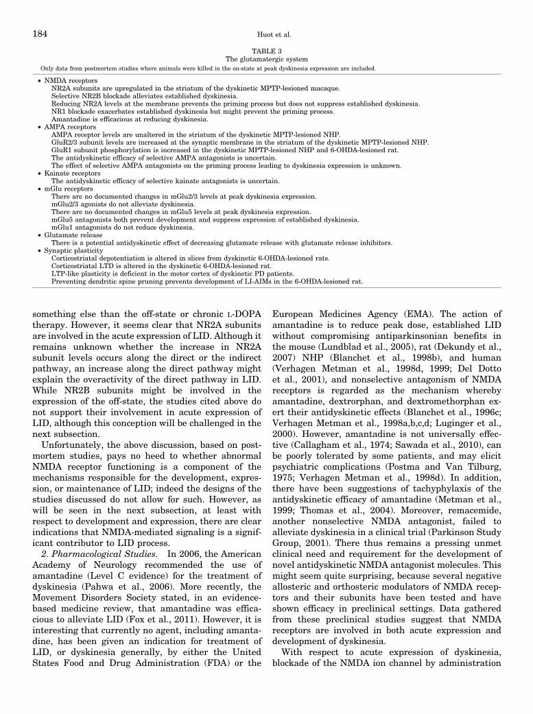
key points put forth in this section are summarized inTable 3.
A. NMDA Receptors
1. Postmortem Studies. NMDA receptors consist oftetramers comprising at least one NR1 and one NR2(NR2A-D) subunit, usually two of each (Dingledineet al., 1999), although they may also contain one NR3subunit (Sasaki et al., 2002). Glutamate binds to theNR2 subunits, whereas the positive allosteric modula-tor glycine binds to the NR1 subunit (Paoletti andNeyton, 2007). Although all of these glutamate sub-units, especially NR1, can be divided into multiplevariants due to splicing (Stephenson, 2006), attentionhas been paid mostly to the NR2A and NR2B subunitsin LID. NR2A and NR2B subunits lead to differentreceptor-mediated current kinetics, NR2B-mediatedcurrent decaying slower than that mediated by NR2A(Lu et al., 2001).Changes in NMDA receptor levels have been
documented in the dyskinetic state. Thus, in theL-DOPA-treated dyskinetic MPTP-lesioned squirrelmonkey killed after a 3-day drug washout, NMDAbinding levels were decreased in the striatum com-pared with MPTP-lesioned, nondyskinetic animals (Heet al., 2000). Changes in NMDA subunit levels havealso been reported in the dyskinetic state. Thus, in thestriatum of L-DOPA-naive MPTP-lesioned macaques,NR1 and NR2B subunit levels were reduced, where-as NR2A subunit levels were unchanged comparedwith normal macaques (Hallett et al., 2005). How-ever, in macaques with established LID killed 60minutes following last drug administration, NR1 andNR2B subunit levels were normalized, and NR2Asubunit levels were upregulated compared withnonparkinsonian macaques (Hallett et al., 2005). Inthe dyskinetic MPTP-lesioned common marmosetkilled 8 days following last L-DOPA administration,NR2B subunit levels were increased in the striatumbut unchanged in the GP, whereas NR1 subunit levelswere unaltered compared with nonparkinsonian ani-mals (Hurley et al., 2005). In the MPTP-lesionedmacaque rendered dyskinetic with the D1 agonistSKF-82,958 and killed in the off-state, striatal levelsof NR1/NR2A but not NR1/NR2B heteromers wereincreased compared with nonparkinsonian animals
(Calon et al., 2002b). A study examining absolutelevels of NR1/NR2A and NR1/NR2B heteromers in thestriatum of dyskinetic PD patients found no differencescompared with healthy controls or PD patients withoutmotor complications (Calon et al., 2003b).
In addition to numerical regulation of NMDAreceptors and their subunits, there is also evidence ofaltered phosphorylation and cellular distribution in thedyskinetic state. In the striatum of the 6-OHDA-lesioned rat treated chronically with L-DOPA (dyski-netic status unknown but the animals displayedevidence of sensitization) and killed 1 hour after lastL-DOPA injection, the number of phosphorylated NR1subunits was decreased compared with control animalsbut increased compared with 6-OHDA-lesioned ratsnot exposed to L-DOPA (Napolitano et al., 2006). In thestriatum of the dyskinetic 6-OHDA-lesioned rat (timeof sacrifice with respect to last L-DOPA dose notmentioned), there was a redistribution of NR2Bsubunits from synaptic to extrasynaptic sites in theplasma membrane and the number of postsynapticNR2A subunits was increased (Gardoni et al., 2006). Inthe striatum of the 6-OHDA-lesioned rat killed 12hours after last L-DOPA administration, NR1/NR2Bheteromers were redistributed toward subcellularcompartments, and, after chronic L-DOPA therapy,their membrane levels were normalized but they wereabnormally phosphorylated (Oh et al., 1998; Dunahet al., 2000). Stimulating D1 receptors was demon-strated to play a key role in the redistribution andphosphorylation of NR1/2A/2B subunits (Dunah andStandaert, 2001). Although the cellular pathwaysultimately leading to abnormal phosphorylation ofglutamate subunits remain largely unknown, Src andLyn kinases are unlikely to mediate it, because theirmRNA and protein levels were reduced in the striatumof the 6-OHDA-lesioned rat treated chronically withL-DOPA killed at peak dyskinesia compared withunlesioned and L-DOPA-naive 6-OHDA-lesioned rats(Napolitano et al., 2006).
A major issue that complicates the interpretation ofdata are that the majority of the postmortem studiescited above were performed with animals killed in theoff-state or in studies in which the behavioral correlatewas not described, and it is therefore impossible toattribute many of the changes enumerated to
TABLE 2The dopaminergic system in dyskinesia
• Striatal dopamine depletion is not a prerequisite for development of dyskinesia, if high doses of L-DOPA are administered.• Pulsatile dopamine replacement therapy appears as a key etiological factor in the development of LID.• Initial therapy with longer-lasting dopaminergic agents can delay the onset of dyskinesia.• Continuous delivery of dopamine agonists or L-DOPA can reduce the severity of established dyskinesia.• Dopamine receptor supersensitivity.• Overactive D1-mediated neurotransmission.• Recruitment of D1 receptors at the synaptic membrane.• Lentiviral-induced internalization of D1 receptors alleviates LI-AIMs in the 6-OHDA-lesioned rat and LID in the MPTP-lesioned NHP.• Each of D1, D2, and D3 receptors are involved in the development and expression of dyskinesia.• Cross-talk between D1 and D3 receptors.
L-DOPA-Induced Dyskinesia Pharmacology 183
something else than the off-state or chronic L-DOPAtherapy. However, it seems clear that NR2A subunitsare involved in the acute expression of LID. Although itremains unknown whether the increase in NR2Asubunit levels occurs along the direct or the indirectpathway, an increase along the direct pathway mightexplain the overactivity of the direct pathway in LID.While NR2B subunits might be involved in theexpression of the off-state, the studies cited above donot support their involvement in acute expression ofLID, although this conception will be challenged in thenext subsection.Unfortunately, the above discussion, based on post-
mortem studies, pays no heed to whether abnormalNMDA receptor functioning is a component of themechanisms responsible for the development, expres-sion, or maintenance of LID; indeed the designs of thestudies discussed do not allow for such. However, aswill be seen in the next subsection, at least withrespect to development and expression, there are clearindications that NMDA-mediated signaling is a signif-icant contributor to LID process.2. Pharmacological Studies. In 2006, the American
Academy of Neurology recommended the use ofamantadine (Level C evidence) for the treatment ofdyskinesia (Pahwa et al., 2006). More recently, theMovement Disorders Society stated, in an evidence-based medicine review, that amantadine was effica-cious to alleviate LID (Fox et al., 2011). However, it isinteresting that currently no agent, including amanta-dine, has been given an indication for treatment ofLID, or dyskinesia generally, by either the UnitedStates Food and Drug Administration (FDA) or the
European Medicines Agency (EMA). The action ofamantadine is to reduce peak dose, established LIDwithout compromising antiparkinsonian benefits inthe mouse (Lundblad et al., 2005), rat (Dekundy et al.,2007) NHP (Blanchet et al., 1998b), and human(Verhagen Metman et al., 1998d, 1999; Del Dottoet al., 2001), and nonselective antagonism of NMDAreceptors is regarded as the mechanism wherebyamantadine, dextrorphan, and dextromethorphan ex-ert their antidyskinetic effects (Blanchet et al., 1996c;Verhagen Metman et al., 1998a,b,c,d; Luginger et al.,2000). However, amantadine is not universally effec-tive (Callagham et al., 1974; Sawada et al., 2010), canbe poorly tolerated by some patients, and may elicitpsychiatric complications (Postma and Van Tilburg,1975; Verhagen Metman et al., 1998d). In addition,there have been suggestions of tachyphylaxis of theantidyskinetic efficacy of amantadine (Metman et al.,1999; Thomas et al., 2004). Moreover, remacemide,another nonselective NMDA antagonist, failed toalleviate dyskinesia in a clinical trial (Parkinson StudyGroup, 2001). There thus remains a pressing unmetclinical need and requirement for the development ofnovel antidyskinetic NMDA antagonist molecules. Thismight seem quite surprising, because several negativeallosteric and orthosteric modulators of NMDA recep-tors and their subunits have been tested and haveshown efficacy in preclinical settings. Data gatheredfrom these preclinical studies suggest that NMDAreceptors are involved in both acute expression anddevelopment of dyskinesia.
With respect to acute expression of dyskinesia,blockade of the NMDA ion channel by administration
TABLE 3The glutamatergic system
Only data from postmortem studies where animals were killed in the on-state at peak dyskinesia expression are included.
• NMDA receptorsNR2A subunits are upregulated in the striatum of the dyskinetic MPTP-lesioned macaque.Selective NR2B blockade alleviates established dyskinesia.Reducing NR2A levels at the membrane prevents the priming process but does not suppress established dyskinesia.NR1 blockade exacerbates established dyskinesia but might prevent the priming process.Amantadine is efficacious at reducing dyskinesia.
• AMPA receptorsAMPA receptor levels are unaltered in the striatum of the dyskinetic MPTP-lesioned NHP.GluR2/3 subunit levels are increased at the synaptic membrane in the striatum of the dyskinetic MPTP-lesioned NHP.GluR1 subunit phosphorylation is increased in the dyskinetic MPTP-lesioned NHP and 6-OHDA-lesioned rat.The antidyskinetic efficacy of selective AMPA antagonists is uncertain.The effect of selective AMPA antagonists on the priming process leading to dyskinesia expression is unknown.
• Kainate receptorsThe antidyskinetic efficacy of selective kainate antagonists is uncertain.
• mGlu receptorsThere are no documented changes in mGlu2/3 levels at peak dyskinesia expression.mGlu2/3 agonists do not alleviate dyskinesia.There are no documented changes in mGlu5 levels at peak dyskinesia expression.mGlu5 antagonists both prevent development and suppress expression of established dyskinesia.mGlu1 antagonists do not reduce dyskinesia.
• Glutamate releaseThere is a potential antidyskinetic effect of decreasing glutamate release with glutamate release inhibitors.
• Synaptic plasticityCorticostriatal depotentiation is altered in slices from dyskinetic 6-OHDA-lesioned rats.Corticostriatal LTD is altered in the dyskinetic 6-OHDA-lesioned rat.LTP-like plasticity is deficient in the motor cortex of dyskinetic PD patients.Preventing dendritic spine pruning prevents development of LI-AIMs in the 6-OHDA-lesioned rat.
184 Huot et al.
of magnesium sulfate alleviated established LID in theMPTP-lesioned NHP (Chassain et al., 2003). Antago-nizing the glycine-binding site by increasing kynurenicacid levels with Ro-61-8048 also alleviated establishedLID, although modestly, in the MPTP-lesioned ma-caque, without affecting L-DOPA antiparkinsonianaction (Samadi et al., 2005). Accordingly, the glycineprodrug milacemide had no effect on established LIDin a small clinical trial (Giuffra et al., 1993). A studyperformed in the MPTP-lesioned NHP suggested thatnonselective blockade of NMDA receptors might havedifferent effects on chorea and dystonia, chorea beingimproved with high dose of LY-235,959, whereasdystonia was exacerbated (Papa and Chase, 1996).However, this preferential antichorea effect was notencountered in another study in which the nonselectiveNMDA antagonist, MK-801, was administered to theMPTP-lesioned NHP, although a reduction in LIDseverity was achieved along with a reduction in L-DOPA antiparkinsonian action (Gomez-Mancilla andBedard, 1993). It should also be pointed out that thepreferential antichorea effect of nonselective NMDAantagonists was not encountered in the clinical trialsthat studied amantadine, dextromethorphan, or dex-torphan, and as such, the data suggesting preferentialantichorea effect of nonselective antagonists are tooweak to make any recommendations.The data from the pharmacological studies cited in
the previous paragraphs almost uniformly indicate anantidyskinetic effect of nonselective NMDA antago-nists. However, based on the results obtained in clinicwith amantadine and remacemide, their efficacy isclearly limited and the loss of L-DOPA antiparkinso-nian efficacy upon administration of MK-801 remainsa concern, because it suggests that such moleculesmight have a very narrow therapeutic window.Studies have also examined the effect of antagonists
at subunits of NMDA receptors and these are veryinformative in terms of determining which NMDAsubunits are critical to be blocked to acutely alleviateor prevent the development of LID, which willultimately lead to the development of more selectiveand better tolerated antidyskinetic agents. Thus,negative allosteric modulation of NMDA receptorswith the NR2B antagonist Co-101,244/PD-174,494alleviated LID in the MPTP-lesioned macaque (Blan-chet et al., 1999). The results obtained with dual NR1/NR2B antagonists are more difficult to interpret, astraxoprodil exerted a mild antidyskinetic effect ina phase II clinical trial (Nutt et al., 2008), whereas itexacerbated dyskinesia severity in the MPTP-lesionedNHP (Nash et al., 2004). Likewise, dual blockade of theNR1A/2A subunits with MDL-100,453 exacerbatedLID in the MPTP-lesioned NHP (Blanchet et al.,1999). In the 6-OHDA-lesioned rat, selective reductionof the number of NR2A subunits at the synapticmembrane by intrastriatal delivery of the peptide
TAT2A did not lead to a suppression of well-established AIMs (Gardoni et al., 2011).
With respect to interference with the priming pro-cess, de novo treatment with the dual NR1A/2Bantagonist Cl-1041 prevented the development of LIDin the MPTP-lesioned NHP (Hadj Tahar et al., 2004;Morissette et al., 2006b). Antagonizing the glycine-binding site by increasing kynurenic acid levels withRo-61-8048 also reduced development of LID in theMPTP-lesioned macaque (Gregoire et al., 2008). Inthe 6-OHDA-lesioned rat, chronic treatment with theNR2B antagonist Ro-631,908 in combination withL-DOPA did not prevent the emergence of AIMs(Rylander et al., 2009), whereas reducing the numberof NR2A subunits at the synaptic membrane bysystemic injection of the peptide TAT2A reduced thedevelopment of LI-AIMs (Gardoni et al., 2011).
By taking a step back and examining the data fromthe studies performed with selective antagonists ofspecific NMDA subunits, a certain number of conclu-sions can be drawn with respect to the role played byeach subunit in LID. Thus, it appears that, unlike whatmight be expected based on the findings of thepostmortem studies cited in the previous subsections,selective blockade of NR2B subunits is effective atreducing established LID but is of little value inaltering the priming process. In contrast, reducingthe number of NR2A subunits at the synapticmembrane—similar effects would likely be achievedupon selective NR2A blockade—appears to be effectivein preventing the priming process but does noteffectively suppress established LID, which is also incontrast to what might be expected based on thefindings of the postmortem studies cited above.Antagonizing NR1 subunits appears to play a deleteri-ous effect on established LID, as the antidyskineticeffect of selective NR2B antagonists is no longerpresent with dual NR1/NR2B antagonists. However,NR1 subunits might play a role in the priming process,as dual blockade of NR1/NR2B subunits effectivelyprevents the development of dyskinesia, whereasselective NR2B blockade has no effect. We thereforepropose that development of future NMDA antagonistsshould focus on blocking NR2B subunits for reductionof established LID while avoiding antagonizing bothNR1 and NR2A subunits. In contrast, development offuture NMDA antagonists to counteract the primingprocess should be centered on blocking NR1 and NR2Asubunits while avoiding NR2B subunits. It is atpresent unknown if dual blockade of NR1/NR2Asubunits would be more effective than selectiveblockade of either to prevent the priming.
The data from the pharmacological studies, withoutcontradicting them, raise serious questions on how tointerpret findings from the postmortem studies. Hence,at peak dyskinesia expression, NR2B subunit levelswere normal in the NHP, whereas NR2A subunits
L-DOPA-Induced Dyskinesia Pharmacology 185
were upregulated. On the basis of pharmacologicalstudies, it seems that antagonizing NR2B subunitsprovides the most consistent antidyskinetic benefit,whereas NR2A blockade seems of little value in thecontext of established LID. Perhaps a reappraisal ofour interpretation of the postmortem studies is needed,because a crude alteration of receptor levels is notnecessarily predictive of the antidyskinetic efficacy ofmodulating this receptor.
B. AMPA Receptors
AMPA receptors consist of double dimers, eachcontaining one GluR2 subunit and either one GluR1,GluR2, GluR3, or GluR4 subunit (Greger et al., 2007),although the GluR2 subunit is sometimes absent, inwhich case AMPA receptors are permeable to calciumions (Dingledine et al., 1999). GluR2 homomers arepermeable to chloride anions (Burnashev et al., 1996).The ionic conductance of AMPA receptors variesaccording to their subunit composition and the editingof the GluR2 subunit (Dingledine et al., 1999).1. Postmortem Studies. The fate of AMPA receptors
in the dyskinetic state is unclear, because one studydemonstrated that AMPA receptor levels were notaltered in MPTP-lesioned macaques killed 20 minutesafter apomorphine administration compared withcontrols (Silverdale et al., 2002), whereas anotherstudy examined MPTP-lesioned macaques killed inthe off-state and revealed decreases in AMPA bindinglevels in the anterior striatum but increases in theposterior striatum in both L-DOPA-naive and L-DOPAtreated dyskinetic states (Ouattara et al., 2010b). Indyskinetic PD patients, AMPA receptor binding levelswere decreased in the caudate but not in the putamencompared with healthy controls (Calon et al., 2003b).As for NMDA receptors, the discrepancy between thestudies might relate to the fact that data were collectedin both the on- and off-states, as well as the fact thatapomorphine instead of L-DOPA was used in the solestudy that did not disclose numerical alterations ofAMPA receptors in dyskinesia.Similar to NMDA receptors, there seem to be
changes in the subcellular distribution of AMPAreceptor subunits in the dyskinetic state. Thus, in thestriatum of the dyskinetic MPTP-lesioned NHP killedat peak dyskinesia expression, GluR2/3 subunit levelswere increased at the synaptic membrane, whereasGluR1 subunit levels were unchanged compared withboth nonparkinsonian and parkinsonian untreatedanimals (Santini et al., 2010; Silverdale et al., 2010).However, GluR1 phosphorylation at Ser845 wasmarkedly increased in the striatum of the L-DOPA-naive and the dyskinetic MPTP-lesioned macaque(Santini et al., 2010). These data suggest that GluR2/3-mediated neurotransmission is enhanced when dys-kinesia is being maximally expressed.
In the striatum of the L-DOPA-naive 6-OHDA-lesioned rat, GluR1 subunit levels were decreased atthe synaptic membrane, and the number of phosphor-ylated GluR1 subunits at Ser831 was decreased;chronic treatment with L-DOPA significantly increasedthe number of GluR1 subunits and the number ofphosphorylated GluR1 subunits at Ser831 at theplasma membrane, whether the animals were killed3 or 24 hours following last L-DOPA administration(Ba et al., 2006). These discrepancies in GluR1absolute numbers between the rat and NHP LIDmodels are important to point out, as they suggestthat selectively antagonizing GluR1 subunits mightalleviate LID in the rat but not necessarily in the NHP.However, 3 hours post-L-DOPA administration, ratsare often wearing off, if not completely off and, as such,findings of abnormal GluR1 may be more reminiscentof the wearing-off state than the dyskinetic state.Taken together, these postmortem studies suggesta lack of involvement of GluR1 subunit in LID.
Thus, although the fate of AMPA receptors indyskinesia has been much less studied than the fateof NMDA receptors, a few important conclusions can bedrawn. First, as mentioned above, they do not supportan involvement of GluR1 subunits in established LID.Second, abnormal GluR2/3 transmission appears to bepresent in established dyskinesia. However, severalholes remain in our comprehension of the role of AMPAreceptors in dyskinesia. For instance, although AMPAbinding levels are increased in the acutely dyskineticapomorphine-treated NHP, it remains unknownwhether these changes occur along the direct or theindirect pathway. An increase in AMPA-mediatedglutamatergic transmission along the direct pathwaymight account for its overactivity in dyskinesia.Moreover, by putting data from all NHP studiestogether, the increase in AMPA receptors might wellbe predominantly made of GluR2 subunits and perhapsGluR2 homomers that would have implications onchloride conductance of the AMPA channels. Thisopens the way to speculate that antidyskinetic thera-pies based on modulation of ionic channels mightrepresent a new approach against LID and severalsuch molecules are used in clinic; as such it would notbe complicated to move forward to clinical trialsrapidly. In agreement with this possibility, the potas-sium Kv7.2-7.5 channel openers retigabine and flupir-tine, two clinically available molecules (Porter et al.,2007; Stoessel et al., 2010), alleviated the severity ofestablished LI-AIMs in the 6-OHDA-lesioned rat(Sander et al., 2012).
2. Pharmacological Studies. In agreement with thefindings of the postmortem studies cited above, phar-macological studies have suggested an antidyskineticeffect of dampening AMPA-mediated transmission,although some doubts persist on the antidyskineticpotential of selective AMPA antagonists, because the
186 Huot et al.

molecules studied were not selective for AMPA recep-tors. Indeed, in the MPTP-lesioned NHP, the GluR2/NMDA antagonist IEM-1460 (Kobylecki et al., 2010)and the AMPA/kainate antagonists LY-300,164 (Konit-siotis et al., 2000) and topiramate (Silverdale et al.,2005) reduced the severity of established LID. In-terestingly, administration of subthreshold doses, in-effective as monotherapy, of the AMPA/kainateantagonist GYK-47,261 (Abraham et al., 2000) incombination with low doses of either of the nonselectiveNDMA antagonists MK-801 or amantadine led toa significant reduction in LID in the MPTP-lesionedNHP (Bibbiani et al., 2005b).At the clinical level though, the only AMPA
antagonist evaluated and reported, perampanel, didnot alleviate dyskinesia in a phase II trial (Eggertet al., 2010). Those results shed serious doubt on theantidyskinetic efficacy of AMPA antagonists, becauseperampanel is highly selective for AMPA receptors(Rogawski, 2011), indeed more selective than any ofthe drugs cited in the previous paragraph. Two phase I/II trials were performed with the AMPA antagonisttalampanel (Clinical trials NCT00036296 andNCT00108667), but the results have not been reported.To our knowledge, only one study examined the
effect of AMPA antagonists, in that case dual GluR2/NMDA blockade, on the development of LID. In thatstudy, concomitant administration of IEM-1460 and L-DOPA during the priming phase reduced the inductionof AIMs, without impairing L-DOPA antiparkinsonianaction, in the 6-OHDA-lesioned rat (Kobylecki et al.,2010).These pharmacological data need be reinterpreted in
light of the postmortem studies discussed in theprevious section. However, before doing so, it isimportant to state that a molecule acting on a specifictarget might affect downstream signaling processessufficiently to alleviate dyskinesia, irrespective of theabsolute number of binding sites or the phosphoryla-tion status of these binding sites, usually a receptor. Assuch, changes in binding levels might be an indicator,although might not necessarily be predictive of theaction of a pharmacological agent. Thus, at peakdyskinesia expression, no changes were encounteredin AMPA binding levels in the MPTP-lesioned NHP,and as such, the lack of antidyskinetic effect ofperampanel is perhaps not surprising. This highlightsthe importance of killing the animals in the on-state atpeak dose dyskinesia when postmortem studies areexamining a receptor potentially involved in LID;indeed, the postmortem study performed in the NHPin which animals were killed in the off-state was notpredictive of the lack of efficacy of perampanel, becausechanges in AMPA binding levels were detected in theoff-state, but these likely have little to do with theexpression of the dyskinetic phenotype. However, atpeak dyskinesia, synaptic levels of the GluR2/3
subunits were increased in the striatum of theMPTP-lesioned NHP. The failure of perampanel toalleviate LID might be due to the fact that it is anAMPA antagonist that does not discriminate betweensubunits, and it may well be that a selective blockadeof the GluR2/3 subunits is required to alleviate LIDwhile antagonizing AMPA receptors. The antidyski-netic efficacy of such selective compounds has not beentested at present, but we propose that future AMPAantagonists being developed as antidyskinetic agentsshould focus on dual blockade of these two subunits.The effects of selective AMPA receptor blockade on thedevelopment of LID in the context of de novo studiesremain largely unexplored.
C. Metabotropic Glutamate Receptors
mGlu receptors are seven transmembrane domain Gprotein-coupled receptors (Conn and Pin, 1997;Kunishima et al., 2000). Eight types of mGlu receptorshave been identified, mGlu1–8 (Foord et al., 2005).mGlu receptors have been further divided in threegroups, based in part on similar signal transductionmechanisms. Thus, mGlu1 and mGlu5 receptors formGroup I, mGlu2 and mGlu3 form Group II, whilemGlu4, mGlu6, mGlu7, and mGlu8 receptors formGroup III (Swanson et al., 2005; Gladding et al., 2009).
As will be seen in this section, few postmortemstudies have been performed that investigated the fateof mGlu receptors in dyskinesia. Hence, only receptorbinding studies have investigated the numericalregulation of mGlus in dyskinesia and several impor-tant questions remain, such as the phosphorylationstate or the membrane/subcellular distribution of thesereceptors in dyskinesia. Moreover, the postmortemstudies were mostly performed when animals orpatients were in the off-state and thus provide onlylimited information about the role played by thesereceptors in the expression of dyskinesia. More insightcan be garnered from pharmacological studies thatmodulated mGlu-mediated transmission in LID.
1. Metabotropic Glutamate Receptors Type 2/3.In dyskinetic L-DOPA-treated MPTP-lesioned maca-ques killed 24 hours after treatment, mGlu2/3 bindinglevels were increased in the putamen and GPe butunchanged in the caudate, STN, GPi, and SNrcompared with MPTP-lesioned, L-DOPA-naive NHPs;no alterations in mGlu2/3 levels have been reported inL-DOPA-naive MPTP-lesioned NHPs (Samadi et al.,2008a). In PD patients, with and without dyskinesia,mGlu2/3 receptor levels were reduced in the caudateand GPi, but not in the putamen and GPe, comparedwith controls (Samadi et al., 2009). These discrepanciesbetween mGlu2/3 levels encountered in dyskineticidiopathic PD patients and dyskinetic MPTP-lesionedNHPs are quite surprising, because both studies weredone by the same group with the same radioligand andboth involved subjects in the off-state. Regardless of
L-DOPA-Induced Dyskinesia Pharmacology 187

these discrepancies, these studies did not establish aninvolvement of mGlu2/3 receptors in LID. In agree-ment with a lack of altered mGlu2/3-mediated trans-mission in LID, mGlu2/3-mediated transmission at thecorticostriatal synapse in striatal slices from 6-OHDA-lesioned rats treated chronically with L-DOPA was notdifferent to that from normal, nonparkinsonian rats(Picconi et al., 2002).The lack of involvement of mGlu2/3 receptors in LID
was further suggested by a pharmacological studyconducted in the 6-OHDA-lesioned rat where themGlu2 agonist LY-379,268 had no effect in preventingthe development or acutely suppressing the expressionof LI-AIMs (Rylander et al., 2009). The antidyskineticefficacy of mGlu2/3 receptor agonists has not beentested in the MPTP-lesioned NHP or in clinicalsettings, but it is unlikely that this avenue will bepursued further.2. Metabotropic Glutamate Receptors Type 5.
mGlu5 receptors are abundant within the striatum,where they are located postsynaptically on striatalprojection neurons (Testa et al., 1995). mGlu5 andNMDA receptors appear to be intimately con-nected, with a reciprocal positive-feedback interaction(Alagarsamy et al., 1999a,b), and as such, antagonizingmGlu5 receptors is another way to reduce overactiveglutamatergic transmission in LID.In the L-DOPA-naive MPTP-lesioned NHP, mGlu5
binding levels were increased in the striatum andincreased further following L-DOPA treatment, the lastdose being administered 24 hours prior to sacrifice(Samadi et al., 2008b; Ouattara et al., 2011). mGlu5mRNA levels were also increased in the striatum ofdyskinetic MPTP-lesioned macaques compared withL-DOPA-naive MPTP-lesioned macaques or nonpar-kinsonian macaques when they were killed 4 hoursafter last L-DOPA dose (Ouattara et al., 2010a). In PDpatients, mGlu5 binding levels were elevated in theputamen and both segments of the GP when dyskineticsubjects were compared with nondyskinetic patients(Ouattara et al., 2009). Results of these postmortemstudies argue for overactive mGlu5-mediated trans-mission in the dyskinetic state.Pharmacological studies have confirmed mGlu5
receptors in both development and acute expressionof LID. Hence, in studies performed in the 6-OHDA-lesioned rat where the mGlu5 antagonist MTEP wasadministered de novo with L-DOPA, MTEP adminis-tration led to significantly less severe AIMs thanvehicle treatment (Mela et al., 2007; Rylander et al.,2009). In another study, acute challenges of MTEPeffectively suppressed established AIMs, without al-tering the antiparkinsonian action of L-DOPA(Dekundy et al., 2006). More recently, the mGlu5negative allosteric modulator MRZ-8676 was shown tosignificantly reduce established LI-AIMs in the rat, theeffects of which showed no tolerance over a 6-day
treatment period (Dekundy et al., 2011). In the MPTP-lesioned NHP, the mGlu5 antagonists AFQ056(Gregoire et al., 2011), ADX-48,621 (Hill et al., 2010),fenobam (Rylander et al., 2010a), MPEP (Morin et al.,2010), and MTEP (Johnston et al., 2010b) reduced theseverity of previously established LID. With MTEP,however, an antidyskinetic effect was obtained only athigh dose and was accompanied by a reduction of L-DOPA antiparkinsonian efficacy (Johnston et al.,2010b), suggesting that although negative allostericmodulation of mGlu5 receptors alleviates LID, thecompounds might have a narrow therapeutic windowand the antidyskinetic benefit might come at theexpense of a reduction in L-DOPA antiparkinsonianaction. At the clinical level, AFQ056 is the only mGlu5antagonist that has been reported on to date tosuccessfully alleviate established LID (Berg et al.,2011). A phase IIa trial with ADX-48,621 is currentlyrecruiting participants (Clinical trial NCT01336088).
Although it remains to be established if the increasein striatal mGlu5 receptor levels described above in thedyskinetic state occurs predominantly on striatofugalneurons of the direct pathway, an increase in striatalmGlu5 levels along the direct pathway would explainits overactivity in the dyskinetic state and provide ananatomic rationale for antagonizing mGlu5 receptorsin LID. A second mechanism underlying the anti-dyskinetic efficacy of mGlu5 antagonists might bea reduction in GABA release within the SNr (Melaet al., 2007), which would ultimately lead to a greaterinhibition of the thalamus and a reduction of corticalexcitation.
As seen above, along with mGlu5 receptors, mGlu1receptors form the Group I family of mGlu receptors(Bonsi et al., 2005). In contrast to mGlu5 receptors,antagonizing mGlu1 receptors did not prevent thedevelopment (Rylander et al., 2009) nor alleviate theexpression of established (Dekundy et al., 2006) AIMsin the 6-OHDA-lesioned rat. The antidyskinetic poten-tial of mGlu1 antagonists has not been assessed in theMPTP-lesioned NHP or in PD patients and, based onthe negative results obtained in rat studies, mGlu1receptors do not appear to represent a promisingantidyskinetic target.
D. Glutamate Release
In agreement with overactive glutamatergic trans-mission in LID and the antidyskinetic effect of variousglutamate receptor antagonists, studies have demon-strated increased levels of glutamate in the striatumand increased levels of the GLT1 glutamate trans-porter in the basal ganglia of the dyskinetic 6-OHDA-lesioned rat (Jonkers et al., 2002; Robelet et al., 2004;Dupre et al., 2011). Accordingly, decreasing glutamaterelease was proposed to be an effective way to alleviatedyskinesia. Thus, the glutamate release inhibitorriluzole alleviated established AIMs in the 6-OHDA-
188 Huot et al.

lesioned mouse (Lundblad et al., 2005) and rat(Dekundy et al., 2007) and decreased duration of on-time with severe dyskinesia in a small pilot study(Merims et al., 1999) but was ineffective in another(Bara-Jimenez et al., 2006). Similarly, another gluta-mate release inhibitor, naftazone, alleviated estab-lished dyskinesia in a N-of-1 trial (Rascol et al., 2011).Neither riluzole nor naftazone impaired L-DOPAantiparkinsonian benefit. Although promising, theseresults remain largely preliminary and additionalbetter controlled studies are needed to confirm thevalue of reducing glutamate release in LID.
E. Altered Synaptic Plasticity
In accordance with changes in glutamatergic trans-mission, there is evidence for altered synaptic plastic-ity at the corticostriatal synapse in dyskinesia. Thus,in striatal slices prepared from dyskinetic 6-OHDA-lesioned rats, long-term potentiation (LTP) appearednormal, but subsequent depotentiation was impaired(Picconi et al., 2003, 2008). In contrast, depotentiationwas not impaired in striatal slices from 6-OHDA-lesioned rats treated chronically with low dose L-DOPAthat did not develop AIMs (Picconi et al., 2008). Thereis also evidence of altered corticostriatal long-termdepression (LTD) in the dyskinetic 6-OHDA-lesionedrat (Picconi et al., 2011). However, these data, exceptthose from Picconi et al., (2008) in which the animalswere killed while in the on-state, were obtained ex vivoin striatal slices without L-DOPA or dopamine added tobath and thus probably represent a correlate ofa primed patient off L-DOPA treatment. However,even in the study in which animals were killed in theon-state, the time required to prepare the slices andthe subsequent perfusion with artificial cerebrospinalfluid (CSF) might lead to a condition akin to the off-state. The mechanisms defined might thus representa component of the process responsible for priming andmaintaining the striatum in the primed state, as theyare consistent with corticostriatal overactivity. How-ever, the situation is not analogous to that whenL-DOPA is present and LID observable. Althoughtechnically challenging, further studies are requiredto assess plasticity in vivo in the on-state to assessa role for abnormal plasticity in the expression of LID.Indirect evidence of altered LTD was also provided
by pharmacological studies that targeted the nitricoxide synthase (NOS). Nitric oxide (NO) is involved incorticostriatal LTD (Calabresi et al., 1999), andstimulation of D1 receptors increases NO efflux in thestriatum (Sammut et al., 2006). NOS inhibitors weredemonstrated to reduce established LID in the MPTP-lesioned marmoset (Jenner, 2008) and the 6-OHDA-lesioned rat (Padovan-Neto et al., 2011). Although noformal assessment of neuronal plasticity was per-formed in those studies, restoring normal cortico-striatal LTD might be a mechanism whereby NOS
inhibitors alleviated LID and LI-AIMs, though furtherstudies are needed to confirm that hypothesis.
Very few clinical studies have evaluated synapticplasticity in LID in PD patients, possibly because thetools to do so are, at present, limited, and invasiveapproaches would undoubtedly raise ethical questions.However, repetitive transcranial magnetic stimulation(Barker et al., 1985) is a technique that is beingincreasingly used and that has the ability to provideinsights on synaptic plasticity, though it is limited tothe processes occurring in the cortex, thus beinginsensitive to subcortical changes (Fitzgerald et al.,2006). A study using repetitive transcranial magneticstimulation has demonstrated deficient LTP-like plas-ticity in the motor cortex of dyskinetic PD patients(Morgante et al., 2006). In that study, subjects weretested 1 hour after L-DOPA intake and were in the on-state. The results of that study are difficult to reconcilewith the general idea of overactivity of the cortex inLID expanded above, and further studies are needed tosee how reduced LTP in the motor cortex mightcontribute to LID.
The results of the aforementioned studies are veryinformative in showing altered plasticity in LID, butthey do not establish abnormal synaptic plasticity asan etiological factor or a consequence of LID. There is,however, evidence that altered synaptic plasticitymight be a critical condition underlying the develop-ment of dyskinesia. Thus, it appears that synapticpruning is a necessary condition for AIMs to develop.Indeed, antagonizing the calcium channels Cav1.2–1.3with isradipine as early as the day after administrationof 6-OHDA prevented the pruning of dendritic spineson striatal medium spiny neurons as well as theemergence of AIMs upon chronic L-DOPA treatment(Schuster et al., 2009). In contrast, initiating isradipinetreatment at a later stage, where significant pruning ofdendritic spines had occurred, was insufficient toprevent AIMs development upon chronic dopaminergictherapy (Schuster et al., 2009). Starting isradipineconcomitantly with L-DOPA was also ineffective atpreventing the development of AIMs (Rylander et al.,2009). At the moment, the results linking synapticpruning and LID are limited to the 6-OHDA-lesionedrat model of PD, but should these results to bereplicated in a higher model such as the MPTP-lesioned NHP, they might have some importanttherapeutic implications, as nondopaminergic medica-tions such as dihydropyridine calcium channel blockersmight have to be started as soon as diagnosis is madeto preserve as many dendritic spines as possible anddelay or prevent the development of dyskinesia. Assuch medications are widely used for other indications,they would be readily amenable to clinical trials.However, as dihydropyridine calcium channel blockersare effective antihypertensive agents (Cutler, 1998),their efficacy might be limited in advanced PD, where
L-DOPA-Induced Dyskinesia Pharmacology 189
orthostatic hypotension is frequent (Senard et al.,2001).
VI. The Opioid System
A. Preproenkephalin and Preprodynorphin
The high molecular weight opioid precursorpreproenkephalin-A (PPE-A) is expressed by striato-pallidal neurons of the indirect pathway, whereaspreproenkephalin-B (PPE-B; sometimes termed pre-prodynorphin, PPD) is expressed on striatonigralneurons of the direct pathway (see below). PPE-A canbe processed to generate leucine (Leu)-enkephalin andmethionine (Met)-enkephalin (Noda et al., 1982),whereas PPE-B contains the determinants for a- andb-neoendorphin, dynorphin-A, and leumorphin(dynorphin-B and related peptides of shorter length)(Horikawa et al., 1983). Proopiomelanocortin (POMC)is another opioid precursor and codes for b-endorphin(Chretien et al., 1979).Endogenous opioid peptides exert their biologic
effects through binding to opioid receptors. Leu- andMet-enkephalin are considered the endogenous ligandsof d-opioid receptors (Quock et al., 1999), whereas thedynorphins, the neoendorphins, and leumorphin bindprimarily to k-opioid receptors (Chavkin et al., 1982),and b-endorphin exhibits high affinity for m-opioidreceptors (Wang et al., 1993). The endomorphins(endomorphin-1 and -2) are endogenous opioid peptidesthat exhibit high affinity for the m-opioid receptors(Fichna et al., 2007), but their opioid precursor has notbeen identified yet, and their role in parkinsonism anddyskinesia, if any, has yet to be determined. It isnoteworthy that all endogenous opioids exhibit a pref-erential affinity for one type of receptor, but also bindto the others (Corbett et al., 2006). Nociceptin(orphanin FQ) is a neuropeptide that binds to thenociceptin receptor (also referred to as the orphaninFQ receptor or k-type 3 opioid receptor) (Mollereauet al., 1996). The nociceptin receptor has a high degreeof homology to each of m-, d-, and k-opioid receptors, butendogenous opioids, with the exception of nociceptin,exhibit little or no affinity for it (Henderson andMcKnight, 1997). Other opioid receptors have beenidentified, such as the opioid growth factor receptor(initially termed zeta-opioid receptor) (Zagon et al.,2000), whereas whether « and l receptors belong tothe opioid receptors remains unsettled (http://www.iuphar-db.org/DATABASE/FamilyIntroductionForward?familyId=50); their role in parkinsonism and LID, if any,has yet to be determined.In PD and dyskinesia, several postmortem studies
have assessed levels of PPE-A and PPE-B mRNA.Given their respective patterns of expression, PPE-AmRNA is used as a marker of activity of the indirectpathway, whereas PPE-B mRNA is used to study thedirect pathway (Bezard et al., 2001; Ravenscroft et al.,
2004). In contrast, POMC has been relatively un-explored (Halabe Bucay, 2008). Indeed, the fate ofb-endorphin in PD and dyskinesia is unclear, as onestudy found higher plasma levels of b-endorphin in PDpatients treated with L-DOPA or not (Franceschi et al.,1986), whereas another study found unchanged plasmalevels and reduced CSF levels of b-endorphin (Nappiet al., 1985). Neither of these two studies establishedcorrelations with dyskinesia. Many pharmacologicalstudies have studied modulation of opioid receptors;they will be summarized in the next subsection. Themajor points discussed in this section are summarizedin Table 4.
Data regarding PPE-A expression following chronicL-DOPA administration and development of AIMs inrodents or LID in NHPs or PD patients can initiallyappear contradictory, although it is now clear that theyneed to be interpreted in light of information on thetime of death with respect to last dose of L-DOPA. Inthe rat, 6-OHDA lesion increased levels of PPE-AmRNA and subsequent treatment with L-DOPA—lastdose given 24 hours before death—reversed thisincrease (Zeng et al., 1995). In contrast, in anotherstudy in which rats were killed 2 days after last dose,chronic treatment with L-DOPA resulted in a furtherincrease in PPE-A mRNA (Westin et al., 2001).
In the common marmoset, MPTP administration ledto increased striatal levels of PPE-A mRNA (Tel et al.,2002). Following chronic treatment with L-DOPA andthe development of dyskinesia, PPE-A mRNA levelswere reduced (but time of administration with respectto killing was not defined) (Tel et al., 2002). Similarchanges in PPE-A mRNA levels were encountered inone study in the macaque following MPTP lesioning,being increased, and subsequent treatment with L-DOPA, decreased (although again, the time of admin-istration with respect to killing was not defined)(Herrero et al., 1995). In contrast, another studyperformed in the macaque in which the animals werekilled in the off-state, found increased striatal PPE-AmRNA levels in L-DOPA-treated dyskinetic MPTP-lesioned macaques compared with L-DOPA-naive,parkinsonian macaques and nonparkinsonian animals(Tamim et al., 2010). In contrast, in the MPTP-lesionedsquirrel monkey, killed 3–4 hours or 3 days after L-DOPA administration, PPE-A mRNA levels wereincreased in the striatum compared with unlesionedanimals, and levels remained high after L-DOPAtreatment and emergence of dyskinesia (Quik et al.,2002). The situation in PD patients thus appearssimilar to that in the squirrel monkey study, becausePPE-A mRNA was increased in the putamen ofdyskinetic L-DOPA-treated subjects (Calon et al.,2002a), presumably because there was significantdelay in both studies between last treatment andanalysis of expression. Interpreting data from thesestudies with respect to dyskinesia is thus difficult, but
190 Huot et al.
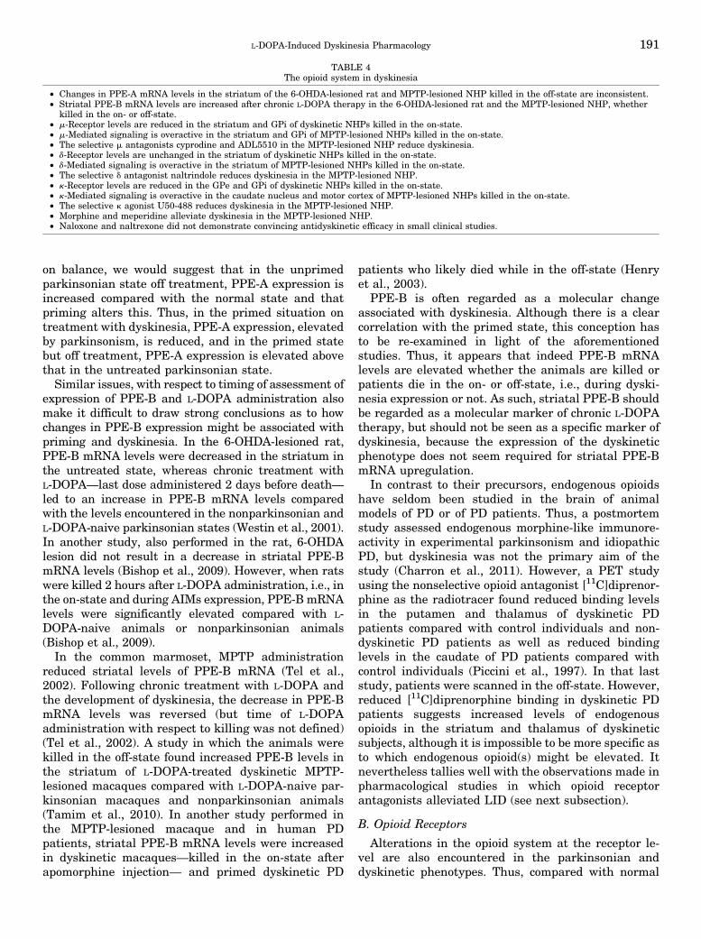
on balance, we would suggest that in the unprimedparkinsonian state off treatment, PPE-A expression isincreased compared with the normal state and thatpriming alters this. Thus, in the primed situation ontreatment with dyskinesia, PPE-A expression, elevatedby parkinsonism, is reduced, and in the primed statebut off treatment, PPE-A expression is elevated abovethat in the untreated parkinsonian state.Similar issues, with respect to timing of assessment of
expression of PPE-B and L-DOPA administration alsomake it difficult to draw strong conclusions as to howchanges in PPE-B expression might be associated withpriming and dyskinesia. In the 6-OHDA-lesioned rat,PPE-B mRNA levels were decreased in the striatum inthe untreated state, whereas chronic treatment withL-DOPA—last dose administered 2 days before death—led to an increase in PPE-B mRNA levels comparedwith the levels encountered in the nonparkinsonian andL-DOPA-naive parkinsonian states (Westin et al., 2001).In another study, also performed in the rat, 6-OHDAlesion did not result in a decrease in striatal PPE-BmRNA levels (Bishop et al., 2009). However, when ratswere killed 2 hours after L-DOPA administration, i.e., inthe on-state and during AIMs expression, PPE-BmRNAlevels were significantly elevated compared with L-DOPA-naive animals or nonparkinsonian animals(Bishop et al., 2009).In the common marmoset, MPTP administration
reduced striatal levels of PPE-B mRNA (Tel et al.,2002). Following chronic treatment with L-DOPA andthe development of dyskinesia, the decrease in PPE-BmRNA levels was reversed (but time of L-DOPAadministration with respect to killing was not defined)(Tel et al., 2002). A study in which the animals werekilled in the off-state found increased PPE-B levels inthe striatum of L-DOPA-treated dyskinetic MPTP-lesioned macaques compared with L-DOPA-naive par-kinsonian macaques and nonparkinsonian animals(Tamim et al., 2010). In another study performed inthe MPTP-lesioned macaque and in human PDpatients, striatal PPE-B mRNA levels were increasedin dyskinetic macaques—killed in the on-state afterapomorphine injection— and primed dyskinetic PD
patients who likely died while in the off-state (Henryet al., 2003).
PPE-B is often regarded as a molecular changeassociated with dyskinesia. Although there is a clearcorrelation with the primed state, this conception hasto be re-examined in light of the aforementionedstudies. Thus, it appears that indeed PPE-B mRNAlevels are elevated whether the animals are killed orpatients die in the on- or off-state, i.e., during dyski-nesia expression or not. As such, striatal PPE-B shouldbe regarded as a molecular marker of chronic L-DOPAtherapy, but should not be seen as a specific marker ofdyskinesia, because the expression of the dyskineticphenotype does not seem required for striatal PPE-BmRNA upregulation.
In contrast to their precursors, endogenous opioidshave seldom been studied in the brain of animalmodels of PD or of PD patients. Thus, a postmortemstudy assessed endogenous morphine-like immunore-activity in experimental parkinsonism and idiopathicPD, but dyskinesia was not the primary aim of thestudy (Charron et al., 2011). However, a PET studyusing the nonselective opioid antagonist [11C]diprenor-phine as the radiotracer found reduced binding levelsin the putamen and thalamus of dyskinetic PDpatients compared with control individuals and non-dyskinetic PD patients as well as reduced bindinglevels in the caudate of PD patients compared withcontrol individuals (Piccini et al., 1997). In that laststudy, patients were scanned in the off-state. However,reduced [11C]diprenorphine binding in dyskinetic PDpatients suggests increased levels of endogenousopioids in the striatum and thalamus of dyskineticsubjects, although it is impossible to be more specific asto which endogenous opioid(s) might be elevated. Itnevertheless tallies well with the observations made inpharmacological studies in which opioid receptorantagonists alleviated LID (see next subsection).
B. Opioid Receptors
Alterations in the opioid system at the receptor le-vel are also encountered in the parkinsonian anddyskinetic phenotypes. Thus, compared with normal
TABLE 4The opioid system in dyskinesia
• Changes in PPE-A mRNA levels in the striatum of the 6-OHDA-lesioned rat and MPTP-lesioned NHP killed in the off-state are inconsistent.• Striatal PPE-B mRNA levels are increased after chronic L-DOPA therapy in the 6-OHDA-lesioned rat and the MPTP-lesioned NHP, whether
killed in the on- or off-state.• m-Receptor levels are reduced in the striatum and GPi of dyskinetic NHPs killed in the on-state.• m-Mediated signaling is overactive in the striatum and GPi of MPTP-lesioned NHPs killed in the on-state.• The selective m antagonists cyprodine and ADL5510 in the MPTP-lesioned NHP reduce dyskinesia.• d-Receptor levels are unchanged in the striatum of dyskinetic NHPs killed in the on-state.• d-Mediated signaling is overactive in the striatum of MPTP-lesioned NHPs killed in the on-state.• The selective d antagonist naltrindole reduces dyskinesia in the MPTP-lesioned NHP.• k-Receptor levels are reduced in the GPe and GPi of dyskinetic NHPs killed in the on-state.• k-Mediated signaling is overactive in the caudate nucleus and motor cortex of MPTP-lesioned NHPs killed in the on-state.• The selective k agonist U50-488 reduces dyskinesia in the MPTP-lesioned NHP.• Morphine and meperidine alleviate dyskinesia in the MPTP-lesioned NHP.• Naloxone and naltrexone did not demonstrate convincing antidyskinetic efficacy in small clinical studies.
L-DOPA-Induced Dyskinesia Pharmacology 191

animals, m-opioid receptor levels were significantlyreduced in the ventrolateral putamen and GPi ofMPTP-lesioned NHPs treated chronically with L-DOPA—last dose administered 1 hour before beingkilled—regardless of their dyskinetic status (Aubertet al., 2007), whereas they were unaffected by MPTPadministration alone. m-Opioid receptors were de-creased in the GPe of untreated MPTP-lesioned NHPsand this decrease was partly reversed by L-DOPAadministration, regardless of the occurrence of dyski-nesia (Aubert et al., 2007). In the 6-OHDA-lesioned rat,m receptors were increased in the premotor and motorcortex of dyskinetic animals when compared withL-DOPA-treated nondyskinetic animals— last doseadministered 3 days before death— whereas theirlevels in the basal ganglia were reduced in theparkinsonian state but unaltered by L-DOPA, whetherAIMs were present or not (Johansson et al., 2001). InPD patients treated chronically with L-DOPA, m-re-ceptor binding levels were reduced in both the caudateand putamen compared with nonparkinsonian individ-uals (Fernandez et al., 1994). Thus, there appears to bevariability in m-receptor levels depending on the areaof the brain studied and the time of death, and it isdifficult to correlate m-receptor levels with the dyski-netic phenotype.However, a postmortem study that used [35S]GTPgS
as a marker of G protein-coupled receptor signalingfound hyperactive m-mediated opioid signal transduc-tion in the striatum and GPi of dyskinetic MPTP-lesioned NHPs killed 3 hours after last administrationof L-DOPA (Chen et al., 2005). This functional studypoints out that absolute levels of m- opioid receptors arenot necessarily a good marker of their activity in thedyskinetic state and suggests that other factors, suchas phosphorylation status or membrane versus sub-cellular distribution, must also be altered in thedyskinetic state. Pharmacological studies have pro-vided supportive evidence of increased m-mediatedopioid transmission in the dyskinetic state. Thus, them-receptor antagonists cyprodine and ADL5510 bothalleviated LID in the MPTP-lesioned NHP withoutaffecting L-DOPA antiparkinsonian efficacy (Henryet al., 2001; Koprich et al., 2011).As with m receptors, d-receptor numbers are not
found to be altered in any consistent way across brainregions in LID. However, enhanced signaling at dreceptors appears to be a mechanism by which theycould contribute to the expression of LID. Thus, in theMPTP-lesioned NHP killed in the on-state, in no brainregion assessed were d-opioid receptor levels signifi-cantly different to those in control animals, regardlessof L-DOPA administration (Aubert et al., 2007; Hallettand Brotchie, 2007). However, d-receptor levels wereincreased in both the motor and premotor cortex ofdyskinetic 6-OHDA-lesioned rats killed in the off-statecompared with nondyskinetic animals (Johansson
et al., 2001). Those findings in both the NHP and therat are in contrast to those in PD patients treatedchronically with L-DOPA where d-receptor bindinglevels were decreased in the putamen compared withnonparkinsonian individuals (Fernandez et al., 1994).However, in the MPTP-lesioned NHP, there is evidenceof increased d-mediated signaling in the dyskineticstate. Thus, using [35S]GTPgS, a postmortem study inwhich the animals were killed 3 hours after L-DOPAadministration found enhanced d-mediated opioidsignaling in the striatum of dyskinetic MPTP-lesioned NHPs (Chen et al., 2005). In agreement withhyperactive d-mediated transmission in dyskinesia,the d-receptor antagonist naltrindole reduced LID inthe MPTP-lesioned NHP without affecting L-DOPAantiparkinsonian efficacy (Henry et al., 2001).
k Receptors were unaltered in the untreated MPTP-lesioned NHP, but their levels were reduced in both theGPe and GPi in animals treated chronically with L-DOPA and killed 1 hour after last dose (Aubert et al.,2007). In contrast, in the dyskinetic 6-OHDA-lesionedrat killed 3 days after last administration of L-DOPA,k-receptor levels were decreased in the striatum andSN of dyskinetic animals, but unaltered in the GP,compared with nondyskinetic 6-OHDA-lesioned rats(Johansson et al., 2001). As for m and d receptors, in theNHP, [35S]GTPgS binding suggested hyperactivek-mediated signaling in the caudate nucleus and motorcortex of dyskinetic MPTP-lesioned NHPs killed 3hours after L-DOPA administration (Chen et al., 2005).Paradoxically, despite overactive k-mediated signalingin LID, antagonizing k receptors with norbinaltorphi-mine did not reduce LID in the MPTP-lesioned NHP(Henry et al., 2001), whereas stimulation of k receptorswith U50-488 reduced established AIMs in the 6-OHDA-lesioned rat and dyskinesia in the MPTP-lesioned squirrel monkey, although at the expense ofimpairing L-DOPA antiparkinsonian action (Cox et al.,2007).
Perhaps highlighting the importance of k-mediatedneurotransmission over m and d transmissions in theacute expression of LID, nonsubtype selective stimu-lation of opioid receptors with morphine alleviatedestablished LID as well as dyskinesia elicited byselective stimulation of either D1 or D2 receptors inthe MPTP-lesioned NHP (Samadi et al., 2004),whereas meperidine alleviated LID in the MPTP-lesioned NHP (Gomez-Mancilla and Bedard, 1993).Neither morphine nor meperidine adversely affectedthe antiparkinsonian action of L-DOPA.
The antidyskinetic efficacy of nonsubtype selectiveopioid receptor antagonists has also been evaluated inpreclinical and clinical studies. Thus, the nonselectiveopioid receptor antagonist naloxone alleviated estab-lished LI-AIMs in the 6-OHDA-lesioned rat (Lundbladet al., 2002), although it was ineffective in the MPTP-lesioned macaque (Gomez-Mancilla and Bedard, 1993),
192 Huot et al.

whereas naltrexone, another nonselective opioid an-tagonist, alleviated LID in the MPTP-lesioned marmo-set (Henry et al., 2001). In clinical settings, naltrexone(Rascol et al., 1994a; Manson et al., 2001) did notproduce antidyskinetic effects consistent between stu-dies, whereas naloxone was ineffective in a controlledstudy (Fox et al., 2004). The lack of clear efficacy ofnonsubtype selective opioid receptor antagonists in theclinic may reflect the fact that the antidyskineticbenefit conferred by blocking m and d receptors wasoffset by blockade of k receptors.If the failure of nonsubtype selective opioid antago-
nists to alleviate LID in clinical trials is indeed due totheir blockade of k receptors and if the antidyskineticefficacy of the nonsubtype selective opioid agonists isprimarily mediated via an agonist effect at k receptors,then selective stimulation of k receptors might bea promising, yet largely unexplored, antidyskinetictarget.To our knowledge, only one study examined blockade
of the nociceptin/orphanin FQ receptor to alleviateLID. In that study, J-113397 worsened LID in theMPTP-lesioned NHP (Visanji et al., 2008).Thus, selective modulation of the opioid system
appears a promising way to alleviate LID by antago-nizing m and d receptors or perhaps by stimulating kreceptors, yet several questions remain. For instance,although postmortem studies have suggested over-active opioid transmission within the basal ganglia inthe dyskinetic state, these studies do not tally wellwith other postmortem studies that have demon-strated reduced receptor levels, thereby suggestingthat changes beyond numerical regulation of opioidreceptors such as abnormal phosphorylation or neuro-nal distribution are also likely to contribute to thedyskinetic phenotype. Moreover, why would stimula-tion of k receptors alleviate LID when there is evidenceof enhanced striatal k-mediated signaling in dyskine-sia? It is possible, but unproven, that increasedk-mediated transmission in LID is in fact an endoge-nous compensatory mechanism that is ineffectiveunder normal circumstances, but which becomeseffective when enhanced with k agonists. Additionally,the precise effects of modulating the different opioidreceptors within the basal ganglia on GABAergic,glutamatergic, and dopaminergic transmissions re-main unknown.
VII. The Serotonergic System
The serotonergic system has been far less studiedthan the dopaminergic and glutamatergic systems inLID, although it is believed to play a key role in thepathophysiology of LID. However, serotonin (5-HT)itself does not appear to play a determinant role in theetiology of LID, and the serotonergic system appearsmore as a vector whereby abnormal dopaminergic and
glutamatergic transmissions occur in the striatum. Assuch, modulation of the 5-HT system in LID appears tobe a way to modulate the two neurotransmitters at theheart of the dyskinetic phenotype, dopamine andglutamate. Moreover, if there is pharmacological andpostmortem evidence indicating an involvement of the5-HT system in the pathophysiology of LID, themechanisms underlying this involvement are largelyunknown. Indeed, very little is known regarding themembrane versus intracellular localization or thephosphorylation state of the different 5-HT receptorsor the 5-HT transporter (SERT) in dyskinesia. Like-wise, the intracellular signaling after binding to 5-HTreceptors or transporter in the dyskinetic state alsoremains unexplored, and forthcoming studies investi-gating these mechanisms are needed. An exhaustivereview article about the serotonergic system in PD anddyskinesia was recently published (Huot et al., 2011a).
A. 5-HT1A/1B Receptors
Both serotonergic type 1A and type 1B (5-HT1A and5-HT1B) receptors are coupled to a Gi/o protein (Nicholsand Nichols, 2008). They have been extensively studiedin PD and dyskinesia, and anatomic sites of action andpharmacological mechanisms underlying their antidy-skinetic effect have been identified. Thus, serotonergicraphestriatal fibers are equipped with the enzymeAADC (Arai et al., 1996) and can therefore synthesizedopamine from L-DOPA (Gershanik et al., 1978; Tisonet al., 1991; Arai et al., 1994, 1995; Tanaka et al., 1999;Maeda et al., 2005). 5-HT terminals also participate inthe reuptake of synaptic dopamine (Berger, 1978;Berger and Glowinski, 1978). Following degenerationof the nigrostriatal system, raphestriatal serotonergicterminals were demonstrated to release dopamine,which acts as a “false neurotransmitter,” i.e., in anenvironment devoid of the autoregulatory mechanismsrequired for physiologic dopaminergic transmission,and this phenomenon was demonstrated to be a keydeterminant in the development and expression of LID(Carta et al., 2007, 2008a, b; Nevalainen et al., 2011).Accordingly, in the 6-OHDA-lesioned rat, administra-tion of L-DOPA to animals with dual lesions of themedial forebrain bundle and rostral raphe nucleus didnot lead to the development of AIMs (Eskow et al.,2009). Further experimental evidence as to the in-volvement of the 5-HT system in LID came fromexperiments performed in the 6-OHDA-lesioned rat inwhich intrastriatal grafts of serotonergic neuronsexacerbated established AIMs in primed animals(Carlsson et al., 2007). In agreement with an abnormalrelease of dopamine in the striatum in motor compli-cations, neuroimaging studies performed in PDpatients have provided evidence in favor of nonphysio-logic dopaminergic transmission and altered pharma-cokinetics of L-DOPA (de la Fuente-Fernandez et al.,2001). Stimulation of 5-HT1A receptors alone or in
L-DOPA-Induced Dyskinesia Pharmacology 193
combination with 5-HT1B receptors reduces raphes-triatal dopamine release (Eskow et al., 2009; Lindgrenet al., 2010) and is believed to be a key mechanismwhereby 5-HT1A agonists alleviate dyskinesia.However, the antidyskinetic actions of 5-HT1A
agonists are not solely mediated by their effect onraphestriatal dopamine release. Thus, in the 6-OHDA-lesioned rat, the antidyskinetic effect of 5-HT1A re-ceptor stimulation was paralleled by a reduction ofstriatal glutamate levels (Dupre et al., 2011), therebylinking 5-HT1A-mediated and glutamatergic transmis-sions. Direct stimulation of cortical 5-HT1A receptorsalso alleviated LI-AIMs in the 6-OHDA-lesioned rat(Ostock et al., 2011), so reduction of corticostriatalglutamate release likely represents a second mecha-nism by which 5-HT1A agonists alleviate dyskinesia. Atthe postmortem level, a study performed in the MPTP-lesioned macaque found altered 5-HT1A receptor levelsin the cortex and striatum of dyskinetic macaquestreated chronically with L-DOPA killed at peakdyskinesia expression, also suggesting altered cortico-striatal 5-HT1A-mediated neurotransmission in thedyskinetic state (Huot et al., 2012c).Experimental evidence points toward an additional
mechanism or site of action for the antidyskinetic effectof 5-HT1A agonists. Thus, stimulation of 5-HT1A
receptors alleviated AIMs induced by the D1 agonistSKF-81,297 independently of striatal glutamate levels(Dupre et al., 2011). As dopamine agonists directlystimulate dopamine receptors, a reduction in raphes-triatal L-DOPA-derived dopamine release is unlikely toplay a key role in 5-HT1A-mediated reduction of SKF-81,297-induced AIMs. Those data suggest differentpathophysiological mechanisms underlying L-DOPA-induced and dopamine agonist-induced dyskinesia(Huot and Brotchie, 2011) or an additional site ofaction of 5-HT1A agonists within the basal gangliacircuit, for instance the thalamocortical synapse (Huotet al., 2011b). The STN might well be another site ofaction of 5-HT1A agonists, as suggested by a studyconducted in the 6-OHDA-lesioned rat in which in-jection of the nonselective 5-HT1A agonist sarizotan inthe STN alleviated LI-AIMs (Marin et al., 2009b).However, an intra-STN site of action is hard toreconcile with the classic model of the basal ganglia,as it would ultimately disinhibit glutamatergic thala-mocortical transmission, thereby exacerbating LID, yeta reduction was observed experimentally.However, despite the strong antidyskinetic mecha-
nisms highlighted above, the antidyskinetic efficacy of5-HT1A agonists has been rather disappointing inclinical settings, and there is also evidence that theymay impair L-DOPA antiparkinsonian action. Indeed,a reduction of L-DOPA antiparkinsonian action wasencountered in the NHP with R-(+)-8-OH-DPAT (Ira-vani et al., 2006) and sarizotan (Gregoire et al., 2009)and in clinical trials with tandospirone (Kannari et al.,
2002) and sarizotan (Goetz et al., 2007). Moreover, twophase III studies performed with sarizotan failed toshow any antidyskinetic efficacy when compared withplacebo (Muller et al., 2006; Rascol et al., 2006b; Goetzet al., 2008); one of several reasons for this may be thatdoses were selected low to avoid loss of antiparkinsonianbenefit and in so-doing reduced antidyskinetic efficacy toa level that could not be clearly demonstrated. Similarly,the antidyskinetic efficacy of the 5-HT1A agonist and D2
antagonist buspirone has been conflicting in clinicalstudies, and the antiparkinsonian efficacy of L-DOPAhas been impaired in several patients (Hammerstadet al., 1986; Kleedorfer et al., 1991).
It has been suggested recently that to effectivelyalleviate dyskinesia without exacerbating parkinson-ism with 5-HT1A agonists, it might be necessary tostimulate specific subpopulations of 5-HT1A receptors.Indeed, stimulating postsynaptic corticostriatal5-HT1A receptors along the direct pathway wouldalleviate LID without impairing L-DOPA antipar-kinsonian efficacy, whereas stimulating presynaptic5-HT1A receptors at the thalamocortical synapse wouldresult in reduced cortical excitation, which mightimpair L-DOPA antiparkinsonian action (Huot et al.,2011b). 5-HT1A agonists displaying such anato-mic selectivity are currently under development(Newman-Tancredi, 2011), although none is availableclinically. Another approach to alleviate LID using5-HT1A agonists and possibly minimizing the effect onL-DOPA antiparkinsonian action might be to combinesubthreshold doses of 5-HT1A and 5-HT1B agonists,which was demonstrated to be more effective thanstimulation of each receptor alone to reduce LID in theMPTP-lesioned macaque (Munoz et al., 2008).
There is also evidence that stimulation of 5-HT1A/1B
receptors interferes with the priming process, leadingto the expression of LID. Thus, de novo administrationof combined 5-HT1A/1B agonists with L-DOPA reducedthe development of AIMs in the 6-OHDA-lesioned rat(Munoz et al., 2008). The mechanisms underlying thisreduction in priming are not fully elucidated but arelikely to be related, although not necessarily exclusive,to an interference with the dopamine- and glutamate-mediated priming processes mentioned in the previoussections.
Although mechanistic studies assessing 5-HT1B
receptors in LID are fewer than those for 5-HT1A
receptors, the adaptor protein p11, a component in-volved in the expression of 5-HT1B receptors at cellsurface (Svenningsson and Greengard, 2007), wasdemonstrated to play an important role in theantidyskinetic action of stimulating 5-HT1B receptorsin the 6-OHDA-lesioned mouse with established AIMs(Zhang et al., 2008b). To conclude this section, it isimportant to point out that, despite sharing 93% aminoacid homology, primate and rodent 5-HT1B receptorsexhibit different pharmacological profiles (Adham
194 Huot et al.
et al., 1992). Such different pharmacological profilesmay make the transition difficult from rodent to NHPor human.
B. 5-HT2A Receptors
Serotonergic type 2A (5-HT2A) receptors are Gaqprotein-coupled receptors (Nichols and Nichols, 2008).5-HT2A receptors have been extensively studied in PDand LID, and postmortem and pharmacological studieshave provided indirect evidence of altered 5-HT2A-mediated neurotransmission in the dyskinetic state.Thus, in the MPTP-lesioned macaque killed 1 hourafter L-DOPA administration, 5-HT2A receptor levelswere increased in the motor cortex and striatum ofdyskinetic animals (Huot et al., 2012b). Moreover,antagonizing 5-HT2A receptors with the atypical anti-psychotics clozapine and quetiapine led to a reductionof established AIMs severity in the 6-OHDA-lesionedrat (Lundblad et al., 2002) and LID in the MPTP-lesioned primate (Oh et al., 2002; Visanji et al., 2006).It should be noted though that both clozapineand quetiapine bind to several other serotonergic andnonserotonergic receptors (Huot et al., 2011a) and that,although their antidyskinetic efficacy is usually attrib-uted to antagonizing 5-HT2A receptors, a contributionof these other targets cannot be excluded, thereforelimiting the interpretation of these pharmacologicalstudies. More selective compounds such as the R-enantiomer of 3,4-methylenedioxymethamphetamine(MDMA) (Huot et al., 2011c) and pimavanserin (ACP-103) (Vanover et al., 2008) also alleviated LID in theMPTP-lesioned NHP. In clinical settings, clozapine(Durif et al., 2004) as well as the more selective 5-HT2A
antagonists ritanserin (Maertens de Noordhout andDelwaide, 1986; Meco et al., 1988) and pimavanserin(Roberts, 2006) all alleviated dyskinesia.Unlike 5-HT1A receptors, the mechanisms underly-
ing the antidyskinetic action of 5-HT2A antagonistsremain largely unexplored, but it has been suggestedthat modulation of nigrostriatal dopamine release andcorticostriatal glutamate release might both play a role(Huot et al., 2011b, 2012b). With respect to nigros-triatal dopaminergic transmission, 5-HT2A receptoractivation increases nigrostriatal dopamine release(Lucas and Spampinato, 2000), and, as a corollary,blocking 5-HT2A receptors should decrease nigrostria-tal dopamine release. In vivo microdialysis studies inwhich a selective 5-HT2A antagonist, for instancevolinanserin, would be injected systemically in theSNc and in the striatum are needed to validate thishypothesis. The obvious problem of this hypotheticalmechanism though is that reducing nigrostriataldopamine transmission might lead to a reduction ofL-DOPA antiparkinsonian action and, as such, 5-HT2A
antagonists might theoretically impair L-DOPA anti-parkinsonian action. In clinical settings, both clozapine(Wolters et al., 1990) and quetiapine (Reddy et al.,
2002), as well as the more selective 5-HT2A antagonistritanserin, (Maertens de Noordhout and Delwaide,1986; Meco et al., 1988) were reported to impairL-DOPA antiparkinsonian action and, as for 5-HT1A
agonists, 5-HT2A antagonists might have a very narrowantidyskinetic window that might eventually limittheir usefulness as antidyskinetic molecules. Studieswith more selective 5-HT2A antagonists are needed toinvestigate that possibility and, as for 5-HT1A agonists,perhaps the ideal 5-HT2A antagonist will have to blockselectively certain subpopulations of 5-HT2A receptors,such as those located on corticostriatal terminals (Huotet al., 2011b) to alleviate LID without impairingL-DOPA antiparkinsonian action. With respect tocorticostriatal glutamate release, there is evidence that5-HT2A-mediated neurotransmission increases presyn-aptic glutamate release (Aghajanian and Marek, 1997)and postsynaptic NMDA-mediated depolarization(Neuman and Rahman, 1996). Accordingly, intrastria-tal administration of the selective 5-HT2A antagonistvolinanserin to L-DOPA-naive MPTP-lesioned miceresulted in a reduction of striatal glutamate levels(Ansah et al., 2011). However, such a 5-HT2A-mediatedreduction in striatal glutamate levels has yet to bedemonstrated in the L-DOPA-treated dyskinetic state.
C. Serotonin Transporter
The SERT could play a role in the expression of LID,although the data presented here may also beinterpreted differently, as they suggest that manipu-lation of presynaptic and/or synaptic levels of 5-HTand/or dopamine (via SERT modulation or othermechanisms) plays a role in the expression of LID.Hence, chronic administration of the selective SERTinhibitor citalopram (Celexa) alleviated LI-AIMs in the6-OHDA-lesioned rat (Kuan et al., 2008), whereasacute challenges of fluoxetine (Prozac) could notsuppress established AIMs (Dekundy et al., 2007). Atthe clinical level, daily administration of fluoxetineover a fortnight alleviated apomorphine-induced dys-kinesia compared with baseline (Durif et al., 1995). Itremains unknown why only subchronic/chronic SERTmodulation may exert an antidyskinetic effect, but thismay have to do with depletion of presynaptic vesicles.Indeed, in the 6-OHDA-lesioned rat, there is a correla-tion between striatal SERT levels and AIMs severity(Rylander et al., 2010b) and in the MPTP-lesionedmacaque, development of LID is associated withincreased striatal SERT levels (Rylander et al.,2010b). In addition to participating in L-DOPA-derived dopamine release in the dyskinetic state,raphestriatal terminals, via a SERT-mediated mecha-nism, also participate in dopamine reuptake in thepresynaptic terminal (Berger, 1978; Berger and Glo-winski, 1978). It is therefore conceivable that acuteinhibition of SERT did not alleviate AIMs, as it wouldincrease synaptic levels of dopamine. However,
L-DOPA-Induced Dyskinesia Pharmacology 195
without SERT inhibition, dopamine would normally betaken up by the presynaptic terminals and stored insynaptic vesicles and eventually be released. Withchronic SERT inhibition, this vesicle repackagingprocess cannot happen, because dopamine is degradedin the synaptic cleft and it is possible that that chronicSERT inhibition leads in fact to a relative depletion ofpresynaptic dopamine vesicles within raphestriatalterminals, thereby accounting for the antidyskineticefficacy of chronic treatment with a SERT inhibitor.However, it should be emphasized that this explana-tion remains hypothetical and electron microscopicstudies are needed to verify this hypothesis. Anotherexplanation might involve a reduction in dopaminerelease mediated by presynaptic 5-HT1A receptors(Yamato et al., 2001). These explanations might alsoaccount for the concerns of impairing L-DOPA anti-parkinsonian action upon administration of selectiveSERT inhibitors (van de Vijver et al., 2002), althoughthat issue remains controversial (Gony et al., 2003). Toconclude on SERT inhibitors and dyskinesia, it shouldbe emphasized that many trials have been performedwith SERT inhibitors in PD and that only rarely havethese reported an improvement in dyskinesia, al-though dyskinesia reduction was not necessarily anendpoint of these studies.
VIII. The GABAergic System
GABA is the major inhibitory neurotransmitter inthe mammalian brain (Watanabe et al., 2002). GABA issynthesized from glutamate (Roberts and Frankel,1950) via the enzyme glutamic acid decarboxylase(GAD) (Roberts and Frankel, 1951) and exerts itseffects via two types of receptors, GABAA and GABAB.GABAA receptors are present within the brain and areessentially ligand-gated ion channels permeable to thechloride anion (Mehta and Ticku, 1999), whereasGABAB receptors are G protein-coupled receptors thatare widely expressed in the brain (Bettler et al., 2004).As discussed above, GABA transmission is central to
the basal ganglia physiology. As such, modulatingGABAergic transmission represents an attractive wayto alleviate LID. For instance, enhancing GABAergictransmission from the GPi/SNr to the thalamus wouldresult in inhibition of the thalamocortical pathway,which would decrease cortical excitation and alleviateLID. However, the ubiquity of GABA within the brainmight lead to adverse effects or even mitigate possibleantidyskinetic benefit while modulating this system.Several postmortem studies that examined the
GABAergic system in the dyskinetic state wereperformed. Unfortunately, in none of them were theanimals killed in the on-state in which dyskinesia wasmaximal. As such, they are more informative about thefate of the GABA synthesizing enzyme or GABAergicreceptors in the off-state following chronic L-DOPA
treatment and provide no information related to theacute expression of dyskinesia.
Changes in mRNA levels of the isoform 67 of theenzyme GAD (GAD67) have been documented in thedyskinetic 6-OHDA-lesioned rat (Cenci et al., 1998).Thus, 3 days after last L-DOPA dose, GAD67 levelswere increased in the striatum and GP when dyski-netic rats were compared with nondyskinetic 6-OHDA-lesioned animals and control nonlesioned rats (Cenciet al., 1998). In contrast, in the dyskinetic macaquekilled 24 hours after last dose of L-DOPA and inL-DOPA-treated PD patients, GAD67 mRNA levelswithin the GP were not different from those encoun-tered in nonparkinsonian animals and subjects(Herrero et al., 1996). In the striatum of the MPTP-lesioned macaque killed in the off-state, GAD67 mRNAlevels were increased and remained increased follow-ing chronic L-DOPA therapy and induction of dyskine-sia compared with nonparkinsonian macaques (Levyet al., 1995a); in PD patients chronically treated withL-DOPA, GAD67 mRNA levels were reduced in thestriatum compared with healthy control subjects (Levyet al., 1995a). GAD isoform 65 (GAD65) mRNA levelswere also increased in the striatum of the MPTP-lesioned macaque treated chronically with L-DOPAkilled in the off-state compared with normal controlsand L-DOPA-untreated parkinsonian animals (Sogho-monian et al., 1996).
Changes in GABA receptor levels have also beenfound in animals expressing dyskinesia when on butthat were killed in the off-state. Thus, in the dyskineticMPTP-lesioned NHP treated chronically with L-DOPAand killed in the off-state, GABAA receptor levels wereincreased in the caudate and GPi (Calon et al., 1995)but decreased in the SNr (Samadi et al., 2008c).Chronic selective stimulation of D1 or D2 receptorswith SKF-82,958 or cabergoline, respectively, did notchange GABAA binding levels in the striatum ofMPTP-lesioned macaques, which remained low com-pared with controls, although, here again, the animalswere killed when off-therapy (Calon et al., 1999). In theNHP, MPTP administration increased GABAB recep-tor levels in the GPi, and this increase was maintainedbut not altered following treatment with the D1 agonistSKF-82,958 and reduced following treatment withcabergoline (Calon et al., 2000). Changes in GABAreceptors have been described in PD patients withmotor complications. Thus, GABAA receptor levelswere increased in the GPe and GABAB receptor levelswere increased in the striatum of dyskinetic PDpatients (Calon et al., 2003a).
Taken together, these studies are difficult to in-terpret and do not define a single important role ofabnormal GABAergic transmission in LID. As men-tioned above, results of these studies do not reflectchanges associated with the acute expression ofdyskinesia and, as such, further studies with different
196 Huot et al.

terminal time points with respect to L-DOPA admin-istration are needed. Moreover, they are somewhatcontradictory with respect to our understanding of thebasal ganglia organization. For instance, what mightbe the resulting effect of an increase in GABAA
receptor levels in the GPi coupled with a decrease inGABAA receptor levels in the SNr with respect toinhibition of the thalamus? In addition, several ques-tions remain unanswered. For example, is the increasein GABAA receptor levels within the caudate nucleuson neurons forming the direct or the indirect pathway?Because the two pathways exert opposite effects onmovement, this is an important missing piece ofinformation. Additionally, very little is known aboutthe membrane versus subcellular distribution or thephosphorylation status of GABA receptors in LID.Very few pharmacological studies assessing the
antidyskinetic potential of modulating GABAergictransmission in LID have been performed. In theMPTP-lesioned NHP, the positive GABAA allostericmodulator diazepam alleviated LID without impairingL-DOPA antiparkinsonian action (Gomez-Mancilla andBedard, 1993), whereas the GABAB agonist baclofendid not alter LID severity, but reduced L-DOPAantiparkinsonian efficacy (Gomez-Mancilla andBedard, 1993). Diazepam also exerted a mild anti-dyskinetic effect in a pilot clinical study (Pourcheret al., 1989). In a case report, zolpidem, a GABAA-positive allosteric modulator (Smith et al., 2001),significantly alleviated LID (Ruzicka et al., 2000),although antidyskinetic effects of zolpidem were notreported in another clinical study (Daniele et al., 1997).Although the benzodiazepine diazepam effectively re-duced dyskinesia in the MPTP-lesioned NHP and ina small clinical study, the use of benzodiazepines toalleviate LID might be complicated by the fact thatthese drugs are susceptible to tolerance, can causesedation and dependence (Lader, 2008), and for thesereasons, one might be reluctant to initiate them toalleviate LID. Additionally, the benzodiazepine clona-zepam is frequently administered to PD patients toalleviate rapid eye movement sleep behavior disorder(RBD) (Boeve et al., 2004), and no improvement in LIDhas been reported following its administration. Al-though clonazepam administration in RBD is usuallyat bedtime, its half-life is between 19 and 60 hours inhumans (Riss et al., 2008), and were benzodiazepineseffective antidyskinetic agents, one might haveexpected clonazepam to reduce LID, yet this has notbeen reported. Accordingly, the anticonvulsant zonisa-mide, which enhances GABAergic transmission (Yosh-ida et al., 2005) and interacts weakly with GABAA
receptors (Mimaki et al., 1990), did not alleviate LID ina phase II study (Murata et al., 2007). However, it isnoteworthy that zonisamide does not solely interactwith the GABAergic system (Leppik, 2004) and its lackof effect on dyskinesia might be attributed to actions at
other sites. Nevertheless, on the basis of the literaturesummarized herein, stimulating the GABAergic sys-tem with the currently available agents does notappear as a promising antidyskinetic therapy.
IX. The Adenosine System
Adenosine is a purinergic neurotransmitter thatexerts its effects via four types of receptors, adenosine1, 2A, 2B, and 3 (A1, A2A, A2B, and A3, respectively)(Fredholm et al., 2001; Hasko et al., 2008). In PD anddyskinesia, attention has been drawn to A2A receptors,which will be the primary focus of this section. A2A
receptors are primarily coupled to a Gs protein (Olah,1997). A2A receptors are located on striatal neuronsforming the indirect pathway (Morelli et al., 2007;Schiffmann et al., 2007) and can form heterodimers withboth D2 (Canals et al., 2003) and mGlu5 (Ferre et al.,2002) receptors. A2A receptors can modulate NMDA-mediated neurotransmission in striatal slices (Nash andBrotchie, 2000). Thus, A2A receptors can modulate bothdopaminergic and glutamatergic transmissions alongthe indirect pathway, whereas their influence on theactivity of the direct pathway is limited.
In normal macaques chronically exposed to highdoses of L-DOPA, thereby evoking severe dyskinesiaeven in the nonparkinsonian state, but killed 5 hoursafter L-DOPA administration when dyskinesia was notpresent, A2A receptor mRNA levels were increased inthe striatum compared with nondyskinetic animals(Zeng et al., 2000), suggesting an overactivity of theindirect pathway. However, as that study was per-formed in nonparkinsonian animals, its relevance todyskinesia in chronic, therapeutically relevant admin-istration of L-DOPA in the parkinsonian state remainsto be established, a fortiori because the animals werenot dyskinetic at the time of death.
In a study performed in the MPTP-lesioned macaquewhere dyskinetic animals were killed in the off-state,A2A receptor binding levels were unchanged in thestriatum of L-DOPA-treated, dyskinetic MPTP-lesionedmacaques compared with L-DOPA-naive parkinsonianmacaques or to nonparkinsonian animals (Morissetteet al., 2006a). In idiopathic PD, A2A receptor mRNA andreceptor binding levels were increased in the putamenof dyskinetic patients compared with nondyskineticpatients (Calon et al., 2004) and were increased in theSNr when PD patients treated chronically with L-DOPA(dyskinetic status unknown) were compared withhealthy controls (Hurley et al., 2000). A recent PETstudy found slightly different results. Indeed, A2A
binding levels were increased in the putamen ofdyskinetic PD patients in the off-state compared withhealthy individuals but not compared with drug-naivePD patients. A2A binding levels were also elevated inthe head of the caudate nucleus of dyskinetic PD
L-DOPA-Induced Dyskinesia Pharmacology 197
patients, but the increase did not reach statisticaldifference (Mishina et al., 2011).Pharmacological studies with A2A antagonists have
focused on their potential to extend on-time duration,but a few studies have examined their potentialantidyskinetic effect. Thus, in the MPTP-lesionedNHP, de novo administration of apomorphine com-bined with istradefylline attenuated development ofapomorphine-induced dyskinesia (Bibbiani et al.,2003). In clinical settings, istradefylline enhanced theantiparkinsonian action of low-dose i.v. L-DOPA whileeliciting significantly less dyskinesia than L-DOPAalone (Bara-Jimenez et al., 2003; Bibbiani et al., 2003).However, istradefylline slightly exacerbated dyskine-sia severity in placebo-controlled studies (Hauser et al.,2008; LeWitt et al., 2008), but another A2A antagonist,preladenant, significantly increased duration of on-time without troublesome dyskinesia in a phase IIstudy (Hodgson et al., 2010).Taken together, those studies do not point toward
a critical involvement of A2A receptors in the acuteexpression of LID, although they may be effective inreducing the priming occurring upon chronic adminis-tration of dopaminergic agents, perhaps because ofa “sparing effect” whereby lower doses of dopaminergicdrugs can be administered.
X. Cannabinoid Neurotransmission
A. Cannabinoid Receptors
Cannabinoid type 1 and 2 (CB1 and CB2) receptorsmediate the actions of endogenous cannabinoids(Pertwee, 2006). CB1 receptors are found mostly withinthe nervous system (Matsuda et al., 1990), whereasCB2 receptors predominate outside of the nervoussystem (Munro et al., 1993). G protein-coupled receptor18 (GPR18) (McHugh et al., 2010), G protein-coupledreceptor 55 (GPR55) (Hiley and Kaup, 2007), and Gprotein-coupled receptor 119 (GPR119) (Overton et al.,2006) are also receptors that can be activated byendogenous cannabinoids, but they have been muchless studied than CB1 and CB2 receptors. With respectto PD and LID, studies have focused on modulation ofCB1 receptors, which are abundant within the basalganglia (Pacher et al., 2006), where they can modulaterelease of glutamate and GABA (Howlett et al., 2002).The transient receptor potential vanilloid type 1(TRPV1) also displays affinity for endocannabinoids(Ross, 2003) and has mostly been studied in the contextof pharmacological experiments (see below).In the rat, striatal levels of CB1 receptor mRNA were
unchanged following 6-OHDA lesion but increasedfollowing chronic L-DOPA therapy when the animalswere killed 3 hours after last L-DOPA dose (Zeng et al.,1999). As no behavioral correlate was provided, it isdifficult to link the increase in CB1 receptor mRNA tothe dyskinetic phenotype (Zeng et al., 1999). In the
MPTP-lesioned marmoset (time of death with respectto last dose of L-DOPA not mentioned), striatal CB1
binding levels were not different to those in controlanimals but were lower than those encountered inMPTP-lesioned, L-DOPA-naive animals (Lastres-Becker et al., 2001). In PD patients treated chronicallywith L-DOPA, striatal CB1 binding levels were in-creased compared with control subjects (Lastres-Becker et al., 2001). In another study, CB1 mRNAlevels were decreased in the striatum and GPe when L-DOPA-treated PD patients were compared withhealthy controls (Hurley et al., 2003). Unfortunately,the dyskinetic status of the PD patients was notmentioned in these two studies, and it is difficult todraw strong conclusions related to dyskinesia fromeither. Moreover, in the case of cannabinoid receptors,their coupling to G proteins seems more importantthan their absolute levels to initiate a biologic response(Breivogel et al., 1997), and the results of thesepostmortem studies, which globally suggested but didnot demonstrate enhanced CB1-mediated transmissionin the striatum in dyskinesia, may not accuratelyreflect changes in cannabinoid transmission in LID.Therefore, studies focusing on the coupling of CB1
receptors to G proteins or on the intracellular cascadesinitiated by CB1 receptors in which animals would bekilled in the on-state, are likely to be more informativeas to the site of CB1 involvement in LID.
Despite the inconclusive results of the studiesdiscussed in the previous paragraph, pharmacologicalstudies have suggested an involvement of the canna-binoid system in the expression of LID, although theirinterpretation is not straightforward. Thus, in theMPTP-lesioned NHP, the CB1 antagonist rimonabantreduced established dyskinesia without impairing L-DOPA antiparkinsonian action (van der Stelt et al.,2005). However, another CB1 antagonist, CE, did notalleviate LID in the MPTP-lesioned NHP (Cao et al.,2007). Paradoxically, stimulation of CB1 receptors withthe CB1 agonist nabilone also alleviated LID in theMPTP-lesioned NHP (Fox et al., 2002a) and in a smallclinical pilot study (Sieradzan et al., 2001). However,stimulation of CB1 receptors with an ethanolic extractof Cannabis sativa produced no antidyskinetic effect ina small randomized-controlled trial (Carroll et al.,2004).
The mechanisms whereby CB1 agonists mightalleviate dyskinesia remain hypothetical but includea reduction of corticostriatal glutamatergic transmis-sion (Brown et al., 2003) and a reduction of GABAreuptake in the GPi (Maneuf et al., 1996; Sieradzanet al., 2001; Fox et al., 2002b), although not allendogenous cannabinoids appear to mediate this lattereffect (Venderova et al., 2005). However, these pro-posed antidyskinetic mechanisms for CB1 agonists aredifficult to reconcile with the antidyskinetic efficacy ofthe CB1 antagonist rimonabant. The antidyskinetic
198 Huot et al.
mechanism(s) of rimonabant remains unknown, but itwas suggested that it could antagonize the effects ofthe endocannabinoid 2-arachidonyl glycerol (2-AG), thelevels of which are elevated in the striatum of thedyskinetic NHP (van der Stelt et al., 2005). However,the mechanism(s) by which elevated striatal levels of2-AG leads to dyskinesia is itself unclear and furtherresearch is needed to resolve the paradox of how bothCB1 agonists and antagonists appear to effectivelyreduce dyskinesia severity. Another paradox of thestudies cited above is the variable antidyskineticefficacy of different CB1 agonists and antagonists.Indeed, CB1 blockade with rimonabant but not withCE alleviates dyskinesia, whereas CB1 stimulationwith nabilone but not with an ethanolic extract of C.sativa reduces dyskinesia.A potential explanation of these two paradoxes may
be related to the importance of G protein coupling ofCB1 receptors in their biologic action highlightedabove. Thus, it is possible that the different CB1
modulators tested so far exhibit functional selectivityfor different types of G proteins, a phenomenon re-ferred to as “biased agonism” (Huot et al., 2011b;Kenakin, 2011; Newman-Tancredi, 2011). For in-stance, although they are both CB1 agonists, nabiloneand the ethanolic extract of C. sativa might welldisplay affinity for different subpopulations of CB1
receptors within the basal ganglia, leading to a re-duction of LID in one case (nabilone) but not in theother (C. sativa). Functional selectivity might alsoexplain the antidyskinetic efficacy of rimonabant andthe lack of antidyskinetic efficacy of CE, two CB1
antagonists, that may well turn out to block certainpathways but not others, with different effects ondyskinesia in the parkinsonian brain. Other pharma-cological factors, such as different collateral efficacy orpermissive antagonism (Kenakin, 2005) of the CB1
ligands in the parkinsonian brain, might also accountfor these differences. Further research is needed toconfirm these hypotheses. Another possibility, butvalid only for the ethanolic extract of C. sativa, is thatC. sativa comprises two active ingredients, D9-tetrahy-drocannabinol, which acts as a CB1 agonist, andcannabidiol, which is a CB1 antagonist (Mechoulamet al., 2007); simultaneous stimulation and blockade ofCB1 receptors with C. sativa might thus account for thelack of antidyskinetic effect.
B. Endogenous Cannabinoids
Levels of endocannabinoids in the dyskinetic statehave been assessed by only a few studies. Asmentioned in the previous subsection, levels of 2-AGare elevated in the striatum of the dyskinetic NHPkilled in the on-state, but how such elevated levels of2-AG would lead to dyskinesia remains unknown (vander Stelt et al., 2005). In the 6-OHDA-lesioned rat(time of death following L-DOPA administration not
defined), levels of the endogenous cannabinoid anan-damide in the striatum in L-DOPA-treated rats werenot different from those of control animals (Gubelliniet al., 2002; Maccarrone et al., 2003).
However, increasing levels of the endocannabinoidanandamide might be a promising way to alleviate LID,although evidence is currently conflicting. Indeed,elevating anandamide levels with the fatty acid amidehydrolase (FAAH) inhibitor URB597 did not reduce LIDseverity in the MPTP-lesioned marmoset (Johnstonet al., 2011), whereas is did so in the MPTP-lesionedmacaque (Johnston et al., unpublished). It is believedthat activation of TRPV1 by anandamide is the reasonwhy URB597 might not consistently alleviate dyskine-sia. Accordingly, the combination of URB597 and theTRPV1 antagonist capsazepine effectively alleviated LI-AIMs in the 6-OHDA-lesioned rat (Morgese et al., 2007)and the MPTP-lesioned macaque (Johnston et al., un-published), although clearly this explanation is onlypartial because URB597 as monotherapy also effectivelyalleviated LID in the parkinsonian macaque. Themechanism(s), whereby elevating anandamide levelsalleviates LID, is presently unknown, but the mecha-nisms speculated above for CB1 agonists might well playa role. The antidyskinetic potential of FAAH inhibitorshas not been explored in the clinic yet, but in agreementwith a potential antidyskinetic benefit of elevatinganandamide, a recent study that measured CSF levelsof anandamide found decreased levels in PD patientstreated with either L-DOPA or dopamine agonistscompared with de novo PD patients or with patientswith long-standing PD who underwent a medicationwashout (Pisani et al., 2010). Unfortunately, thedyskinetic status was not mentioned in the study norwas the time of last drug administration.
XI. Adrenergic Neurotransmission
Adrenaline and noradrenaline exert their effects byactivating the adrenergic receptors, also termed adre-noceptors (Foord et al., 2005). Adrenoceptors are Gprotein-coupled receptors (Vassilatis et al., 2003) andcan be divided into a and b receptors. a and bAdrenoceptors can be further divided into nine sub-types. Thus, a adrenoceptors encompass a1A, a1B, a1D,a2A, a2B, and a2C receptors, while b-adrenoceptors en-compass b1, b2, and b3 receptors (Kobilka, 2011).
To our knowledge, no postmortem study has in-vestigated the fate of adrenoceptors in dyskinesia,although one study that used rotations as the behav-ioral marker, investigated a2A and a2C mRNA levels inL-DOPA-treated 6-OHDA-lesioned rats (Alachkaret al., 2012). As such, the current knowledge support-ing altered noradrenergic transmission in dyskinesia isbased exclusively on pharmacological studies.
a Adrenoceptors appear to play an important role inthe expression of the dyskinetic phenotype. Thus, the
L-DOPA-Induced Dyskinesia Pharmacology 199
nonselective a-adrenergic antagonist yohimbine re-duced the severity of established LI-AIMs in the 6-OHDA-lesioned rat, although at the expense ofimpairing the antiparkinsonian efficacy of L-DOPA(Lundblad et al., 2002; Dekundy et al., 2007). In theMPTP-lesioned NHP, idazoxan, as well as yohimbineand rauwolscine (two nonselective a1 and a2 antago-nists that also act as 5-HT1A agonists and 5-HT2A
antagonists) effectively alleviated LID (Henry et al.,1999); idazoxan also enhanced L-DOPA antiparkinso-nian action (Henry et al., 1999). The selective a2-receptor antagonist fipamezole also alleviated LIDwhile enhancing L-DOPA antiparkinsonian efficacy inthe MPTP-lesioned common marmoset (Savola et al.,2003) and the MPTP-lesioned macaque (Johnstonet al., 2010c). At the clinical level, idazoxan effectivelyalleviated LID (Rascol et al., 2001a) but had no effecton apomorphine-induced dyskinesia (Manson et al.,2000), whereas fipamezole was also effective atrelieving LID (Dimitrova et al., 2009; Lewitt et al.,2012).While a2-receptor blockade appears to effectively
alleviate LID without impairing L-DOPA antiparkin-sonian action, stimulation of a2 receptors also seemseffective to reduce LID, although it may have delete-rious effects on L-DOPA antiparkinsonian benefit.Thus, clonidine, an a2-receptor agonist, alleviated LI-AIMs in the 6-OHDA-lesioned rat (Dekundy et al.,2007) and LID in the MPTP-lesioned NHP (Gomez-Mancilla and Bedard, 1993) but decreased L-DOPAantiparkinsonian efficacy in both models.The mechanism(s) underlying the antidyskinetic
action of a2-receptor antagonists remains hypothetical.Although idazoxan effectively reduced LID, it wasineffective against apomorphine-induced dyskinesia(Fox et al., 2001), suggesting that a direct competitionwith L-DOPA-derived dopamine and/or noradrenalineplays a role in the antidyskinetic action of a2
antagonists (Alachkar et al., 2006). This competitionwould prevent activation of a2C receptors located onmedium-sized spiny neurons of the striatonigralpathway and would normalize the activity of the directpathway, with a resulting reduction of dyskinesia(Johnston et al., 2010c). These actions could be on cellbodies in the striatum, reducing excitability, or onterminals of the direct pathway in the output regions ofthe basal ganglia, reducing GABA release (Fox et al.,2001; Johnston et al., 2010c). GABA release is reducedindirectly by blockade of a2 autoreceptors, increasingnoradrenaline release and thereby leading to stimula-tion of a2 heteroreceptors on GABAergic terminal,which reduces GABA release (Fox et al., 2001;Johnston et al., 2010c). Further studies are needed toconfirm these mechanistic hypotheses and to deter-mine if other mechanisms, such an interaction withglutamatergic transmission, are also involved. Reduc-tion of striatal L-DOPA-derived dopamine release by
idazoxan was recently demonstrated in the primed 6-OHDA-lesioned rat (Buck et al., 2010) and maycontribute to the antidyskinetic action of a2 antago-nists, although if such an action is functionallyimportant it is surprising that this class of compoundhas never been associated with a reduction in anti-parkinsonian benefit (in the case of yohimbine, loss ofantiparkinsonian efficacy could be attributed to anagonist action at 5-HT1A receptors, see above).
Although antagonizing a2 adrenoceptors appears asan effective way to alleviate dyskinesia, the role of a1
adrenoceptors is less clear. Thus, in the 6-OHDA-lesioned rat, systemic administration of the a1 antag-onist HEAT effectively reduced AIMs (Buck andFerger, 2010). In contrast, direct striatal infusion ofthe a1 agonist cirazoline did not induce AIMs (Buckand Ferger, 2010). In the MPTP-lesioned macaque,administration of the a1 antagonist prazosin did notdiminish severity of dyskinesia (Visanji et al., 2009b).Further studies are needed to establish whetherblockade of a1 adrenoceptors is indeed an effectiveantidyskinetic approach and what the mechanismsunderlying any such antidyskinetic effects may be.
On the basis of pharmacological studies, b-adren-ergic transmission also appears to play a role in thepathophysiology of LID. Indeed, in the 6-OHDA-lesioned rat, the b-adrenergic antagonist (6)-pro-pranolol and its enantiomer (2)-propranolol reducedthe expression and development of AIMs (Dekundyet al., 2007; Lindenbach et al., 2011). However, theeffect of propranolol on L-DOPA antiparkinsonianefficacy is unclear (Dekundy et al., 2007; Lindenbachet al., 2011). This difference in the effect of pro-pranolol on L-DOPA antiparkinsonian action in the 6-OHDA-lesioned rat effect might be due to the fact thatthe authors of the studies have used differentmeasures of antiparkinsonian action. However, pro-pranolol did not attenuate AIMs induced by eitherSKF-81,297 or quinpirole (Bhide et al., 2010) suggest-ing that, as for a2 antagonists, a direct competitionwith L-DOPA-derived dopamine/noradrenaline mightmediate the antidyskinetic action of b-adrenoceptorantagonists. The administration of propranolol to theMPTP-lesioned NHP also alleviated LID, but theantidyskinetic effect was accompanied by a reductionof L-DOPA antiparkinsonian benefit (Gomez-Mancillaand Bedard, 1993). Propranolol also alleviated chorea,but not dystonia, in a small clinical study, with-out impairing L-DOPA antiparkinsonian efficacy(Carpentier et al., 1996). As mentioned above, dir-ect competition with L-DOPA-derived dopamine/noradrenaline might be a mechanism whereby b-adre-noceptor antagonists alleviate LID, but it remainsunknown whether other mechanisms are also in-volved. Moreover, the exact site where blockade ofb-adrenoceptors mediates its antidyskinetic efficacy isalso unknown.
200 Huot et al.
Thus, although antagonizing a2- and b-adrenocep-tors appear as promising antidyskinetic therapies,further studies are needed, especially studies focusingon the mechanisms and the anatomic sites underlyingtheir antidyskinetic efficacy. In addition, the effect ofb-adrenoceptor blockade on L-DOPA antiparkinsonianefficacy is a concern that will require exploration ineventual clinical trials. Moreover, the effect of b-adre-noceptor blockade on blood pressure might limit theuse of such agents in advanced PD, where autonomicdysfunction and orthostatic hypotension are oftenpresent (Goldstein, 2003). As mentioned above, thereis recent evidence suggesting that blocking a1-adreno-ceptors might also alleviate LID (Buck and Ferger,2010), but, as for b-adrenoceptor blockers, the effect ofa1-adrenoceptor antagonists on blood pressure mighteventually limit their use in advanced PD andtherefore their use as antidyskinetic agents. Fortu-nately, hypotensive effects of a2-adrenoceptor blockadein clinical trials with idazoxan and fipamezole have notbeen reported, and the agents appear safe and well-tolerated in idiopathic PD.Studies investigating the effect of striatal noradrener-
gic denervation on development and expression ofdyskinesia are difficult to reconcile because eachemployed different methodologies. Thus, administrationof 6-OHDA in the medial forebrain in the absence ofdesipramine pretreatment, leading to concomitant de-generation of noradrenergic fibers coursing toward thestriatum, delayed the priming and attenuated theexpression of LI-AIMs compared with desipramine-pretreated rats (Barnum et al., 2012). That study is inagreement with studies cited above that showed a re-duction in dyskinesia severity with a- and b-adrenergicantagonists. In contrast, in another study, administra-tion of 6-OHDA without desipramine to rats followed bypriming with L-DOPA and then by destruction of thelocus coeruleus, exacerbated the severity of LI-AIMscompared with pre-locus coeruleus lesion (Miguelez et al.,2011). Another study found increased AIMs severity withnoradrenergic lesion in the 6-OHDA-lesioned rat (Fulceriet al., 2007), whereas another one did not find anydifference in LI-AIMs severity (Perez et al., 2009). Asmentioned above, there are many differences in thesestudies in terms of noradrenergic and dopaminergiclesion severity, strain of rat used, and timing ofnoradrenergic lesion compared with dopaminergic lesionand priming. Notwithstanding these considerations, itseems rather clear that the noradrenergic systemmodulates the severity of LID. The question is how. Inidiopathic PD, the locus coeruleus degenerates early inthe disease process, likely earlier than the dopaminergicsystem (Braak et al., 2003, 2004). Thus, when L-DOPAtherapy is commenced, there are already reduced levelsof noradrenaline within the striatum, and, as such, theexperiment performed by Barnum et al. (2012) in whichpriming was initiated after noradrenergic lesion was
induced, might be the most clinically relevant one, bothin terms of timing and severity of lesion, as in idiopathicPD, striatal noradrenaline levels are usually reduced byapproximately 60% (Fahn et al., 1971).
Although the mechanism whereby the noradrenergicsystem might contribute to LID has yet to be de-termined, it is possible to envision a concept similar to“false neurotransmitter” release put forth (see sectionVII). Indeed, it was demonstrated that, within thestriatum of the 6-OHDA-lesioned rat, L-DOPA-deriveddopamine can be taken up by the noradrenalinetransporter into noradrenergic terminals (Arai et al.,2008). Ultimately, this could result in release ofdopamine by coeruleostriatal fibers, that is, by axonsnot equipped with the autoregulatory mechanismsproper to dopaminergic transmission, thereby exacer-bating pulsatile dopamine release. Although thisexplanation remains hypothetical and requires exper-imental validation, it nevertheless makes sense fromphysiologic and compensatory point of views. A directprodyskinetic action of noradrenaline is also possible,because intrastriatal administration of noradrenalineby reverse in vivo microdialysis elicited AIMs inprimed 6-OHDA-lesioned rats (Buck and Ferger, 2009).
XII. Histaminergic Neurotransmission
Histamine exerts its actions via four types ofreceptors, H1–4 (Hill et al., 1997; de Esch et al., 2005),all of which are expressed in the brain (Passani andBlandina, 2011). In PD and dyskinesia, only H2 and H3
receptors have been studied. Both H2 and H3 receptorsare G protein-coupled receptors, but H2 receptorsappear to be coupled to Gs proteins (Hill, 1990),whereas H3 receptors seem to be coupled to Gi/o
proteins (Clark and Hill, 1996).Although no postmortem studies have addressed
specifically the status of the histaminergic system inthe dyskinetic state, the basal ganglia are enrichedwith both H2 and H3 receptors and there is evidence foraltered histaminergic innervation of the basal gangliain the parkinsonian state. Thus, one study demon-strated increased histaminergic innervation of the SNcand SNr in PD patients, although without providingclinical details concerning L-DOPA treatment anddyskinesia (Anichtchik et al., 2000). Another studyfound increased histamine levels in the putamen, SNc,GPi, and GPe of PD patients but did not provideinformation concerning L-DOPA treatment and dyski-nesia (Rinne et al., 2002). H3 receptor mRNA waspresent in the putamen and both segments of the GP,and one study found increased H3 mRNA levels in theGPe of PD patients without commenting on dopami-nergic therapy or the dyskinetic status of the patientsincluded (Anichtchik et al., 2001). This last study alsodemonstrated an increase in H3 protein levels in theSN of PD patients compared with controls, although H3
L-DOPA-Induced Dyskinesia Pharmacology 201

mRNA levels were very low in the SN (Anichtchiket al., 2001). In contrast, H2 receptor levels were notaltered in PD, except in the nucleus accumbens, inwhich they were reduced (Martinez-Mir et al., 1993).There is pharmacological evidence for an involve-
ment of the histaminergic system in the pathophysiol-ogy of LID. Thus, the H2 antagonist famotidinereduced the severity of peak-dose chorea while in-creasing quality of on-time in the L-DOPA-treatedMPTP-lesioned macaque (Johnston et al., 2010d). Ina small clinical study whose endpoint was unrelated todyskinesia, famotidine alleviated established dyskine-sia in two PD patients (Molinari et al., 1995). The H3
agonists immepip and imetit effectively alleviatedL-DOPA-induced chorea but not dystonia in theMPTP-lesioned marmoset; neither immepip nor imetitadversely affected L-DOPA antiparkinsonian benefit(Gomez-Ramirez et al., 2006).At the moment, the mechanism(s) by which H2
antagonists exert their antidyskinetic effect remainsunknown, but it has been suggested that antagonizinghistamine H2 receptors would modulate SNr activity andstriatal ACh release (Johnston et al., 2010d). Moreover,stimulation of H2 receptors activates striatofugal neurons(Haas et al., 2008); while acting on the direct pathway,stimulation of H2 receptors would result in overinhibitionof the GPi/SNr complex, ultimately leading to corticaloverexcitation and LID. Thus, it is possible, althoughspeculative, that H2 blockade normalizes the activity ofthe direct pathway, thereby reducing LID. Similarly, theprecise antidyskinetic mechanism of H3 receptor stimu-lation remains to be demonstrated. However, stimulationof presynaptic H3 receptors inhibits raphestriatal 5-HTrelease as well as corticostriatal glutamate release (Haaset al., 2008). In the context of PD and L-DOPA therapy, itis possible, although not yet demonstrated, that stimu-lating H3 receptors would inhibit raphestriatal dopaminerelease and corticostriatal glutamate release, therebyacting in a way similar to 5-HT1A agonists. Normaliza-tion of neurotransmission along the direct pathway wasalso proposed as an antidyskinetic mechanism of H3
agonists (Gomez-Ramirez et al., 2006), although thismechanism is also unproven.Interestingly, the preferential antichoreic versus
antidystonic efficacy of H2 antagonists and H3 agonistssuggests that different pathophysiological mechanismsunderlie chorea and dystonia. Although such mecha-nisms remain to be elucidated, they raise the possibil-ity that it may eventually be possible to designtherapies aimed at reducing either chorea or dystonia,depending on the need of the patient, thereby poten-tially minimizing adverse effects of treatment.
XIII. Cholinergic Neurotransmission
The basal ganglia, especially the striatum, receivea dense cholinergic innervation (Mesulam et al., 1992),
and histochemistry with the enzyme acetylcholinester-ase was the first technique that identified thestriosomes and the matrix compartments of thestriatum (Graybiel and Ragsdale, 1978). Moreover,large aspiny cholinergic interneurons of the striatumaccount for 1–2% of the total neuronal population of thestriatum (Cicchetti et al., 2000). As such, ACh is a keyneurotransmitter within the basal ganglia (Graybiel,1990; Parent et al., 1995). ACh exerts its effects bystimulating muscarinic (Caulfield and Birdsall, 1998)and nicotinic (Lukas et al., 1999) receptors.
Pharmacological studies have suggested an involve-ment of nicotinic neurotransmission in LID. Thus,chronic nicotine administration reduced the expressionof LID in the MPTP-lesioned squirrel monkey (Quiket al., 2007) and LI-AIMs in the 6-OHDA-lesioned rat(Bordia et al., 2008). However, in phase II studies—none of which examined the effect of nicotine on LID—
transdermal nicotine was poorly tolerated by PDpatients (Lemay et al., 2004; Villafane et al., 2007),which undermines the therapeutic potential of thetreatment. Paradoxically, chronic administration of thenicotinic antagonist mecamylamine also alleviatedLI-AIMs in the 6-OHDA-lesioned rat (Bordia et al.,2010), whereas acute challenges of mecamylamine didnot have any effect on LI-AIMs (Dekundy et al., 2007).Following the unexpected discovery that chronictherapy with either nicotinic agonists or antagonistsalleviates LID, it was proposed that nicotine alleviateddyskinesia after a desensitization phenomenon thatwould occur upon chronic administration (Bordia et al.,2010). An interaction with the b2 subunit of nicotinicreceptors seems to be essential to the antidyskineticaction of nicotine (Huang et al., 2011a, 2011b).However, this approach might also be hindered bypoor tolerability, as SIB-1508Y, an a4b2 agonist, waspoorly tolerated by PD patients in a phase II study(Parkinson Study Group, 2006).
Although PET (Kas et al., 2009), single photonemission tomography (Fujita et al., 2006; Oishi et al.,2007), and postmortem (Court et al., 2000; Schmaljo-hann et al., 2006) studies have demonstrated reducednicotinic receptors in the brain and striatum of PDpatients, because these studies did not examinenicotinic receptors in the dyskinetic state, they donot suggest an anatomic site at which modulation ofnicotinic transmission would alleviate LID. Thus, themechanism(s) by which and the site(s) at whichchronic nicotinic receptor modulation reduces dyski-nesia remain unknown. However, nicotinic receptorsare expressed on nigrostriatal dopaminergic termi-nals (Zoli et al., 2002), and in mice striatal slices,stimulation of nicotine receptors led to dopaminerelease (Zhou et al., 2001). Importantly, in that laststudy, the b2 subunit of nicotinic receptors wasdemonstrated to be critically involved in dopaminerelease (Zhou et al., 2001), which makes alteration of
202 Huot et al.
nigrostriatal dopamine release the most likely antidy-skinetic mechanism of chronic modulation of nicotinicreceptors. However, it is difficult to envision howmodulation of nigrostriatal dopamine release wouldeffectively alleviate LID in the context of advanced PDor in the 6-OHDA-lesioned rat, where few intactnigrostriatal dopaminergic terminals remain and themajority of L-DOPA-derived dopamine is delivered tothe striatum via raphestriatal fibers. Thus, othermechanisms might also be involved. For instance, acutestimulation of nicotinic receptors within the raphenucleus led to 5-HT release within the nucleusaccumbens (Chang et al., 2011). Although it remainsspeculative, downregulation of nicotinic receptorswithin the raphe nucleus following chronic treatmentwith a nicotinic agonist might lead to reduction inraphestriatal dopamine release, thereby reducing LID.However, no matter how attractive these explana-
tions are, they do not take into account the numerousgiant cholinergic striatal interneurons (Cicchetti et al.,2000) that are likely to mediate some of the antidyski-netic effects of nicotinic modulators. It also remainsunknown whether chronic stimulation/blockade of nic-otinic receptors would also attenuate dyskinesia in-duced by dopamine agonists, but a successful reductionin dopamine agonist-induced dyskinesia would suggestan additional antidyskinetic mechanism that would notrequire nigrostriatal or raphestriatal terminals. More-over, it is difficult to conceive why both stimulation andblockade of nicotinic receptors have to be chronic toproduce an antidyskinetic effect. In summary, ourunderstanding of the antidyskinetic effect of nicotinicreceptor modulation remains very limited. However,nicotinic transmission itself does not appear to be a coreplayer in the etiology of LID, but, as for many otherneuromodulator systems, it may well turn out as anadditional way to modulate abnormal dopamine andpossibly glutamate striatal neurotransmission.In contrast to nicotinic neurotransmission, the role of
muscarinic transmission is unclear in the etiology ofLID. Thus, in an early study, the muscarinic antago-nist atropine had no effect on LID in the MPTP-lesioned macaque (Gomez-Mancilla and Bedard, 1993).However, the muscarinic antagonist dicyclomine alle-viated LI-AIMs in the Pitx3ak/ak mouse, which exhibitsstriatal dopamine depletion (Ding et al., 2011). Tofurther add to the confusion, there have been casereports in which dyskinesia, mostly oro-buccal, wastriggered when antimuscarinic agents were added todopaminergic therapy (Hauser and Olanow, 1993). Theprecise mechanism and anatomic localization of suchan anti/pro-dyskinetic action remain to be determined.
XIV. Tachykinin Neurotransmission
The tachykinins are a family of neuropeptidesencompassing neurokinin A, neurokinin B, and
substance P (Maggio, 1988; Chahl, 2006). As discussedearlier and as illustrated in Fig. 1, substance P isexpressed preferentially by the direct pathway. Thetachykinins are encoded by preprotachykinin (PPT)(Maggio, 1988; Chahl, 2006). The tachykinins exerttheir effects by binding to three receptors. Substance Pbinds to the NK1 receptor, whereas neurokinin A and Bbind to the NK2 and NK3 receptors, respectively(Maggi, 1995). NK1, NK2, and NK3 receptors are allpresent within the brain; however, NK1 receptorspredominate within the striatum, whereas NK2 recep-tors are relatively sparse and NK3 receptors are mostlyencountered in the cerebral cortex (Otsuka andYoshioka, 1993).
The tachykinins, especially substance P, have beenextensively studied in PD but with little focus onrelationship to dyskinesia. In the 6-OHDA-lesionedrat, striatal PPT mRNA levels were reduced andchronic treatment with L-DOPA, last dose adminis-tered 24 hours before death, reversed this decrease(Zeng et al., 1995). In the common marmoset, MPTPadministration led to decreased striatal levels of PPTmRNA (Tel et al., 2002). Following chronic treatmentwith L-DOPA and the development of dyskinesia, PPTmRNA levels returned to baseline (time of administra-tion of L-DOPA with respect to death not defined) (Telet al., 2002). Similar changes in PPT mRNA levelswere encountered in one study in the macaque,following MPTP lesioning and subsequent treatmentwith L-DOPA (time of administration with respect todeath not defined) (Herrero et al., 1995). Interpretingdata from these studies with respect to dyskinesiaexpression is difficult, and without further studies, atpresent, it is impossible to describe any clear correla-tion between changes in PPT mRNA levels and thedyskinetic phenotype.
Several studies have measured levels of substance Pwithin the basal ganglia and CSF of PD patients(Mauborgne et al., 1983; Pezzoli et al., 1984; Tenovuoet al., 1984; Grafe et al., 1985; Halliday et al., 1990;Cramer et al., 1991; Fernandez et al., 1992). However,results are variable, depending on the technique used,and for many of these studies, it is not describedwhether patients were treated with L-DOPA orexhibited dyskinesia. In the L-DOPA-naive, MPTP-lesioned common marmoset, substance P levels wereunchanged in the striatum, GP, and SN (Taquet et al.,1988), although another study found decreased sub-stance P immunoreactivity in the sensorimotor stria-tum in the MPTP-lesioned macaque (Lavoie et al.,1991).
In the 6-OHDA-lesioned rat, acute and chronicadministration of L-DOPA significantly increasedstriatal levels of neurokinin B and substance P mRNAcompared with L-DOPA-naive animals when rats werekilled in the on-state (Zhang et al., 2008a). However,the behavioral endpoint of that study was rotational
L-DOPA-Induced Dyskinesia Pharmacology 203
behavior and not AIMs, therefore precluding one tocorrelate AIMs with increased striatal levels of sub-stance P. In contrast, a study that measured substanceP levels in the striatum of 6-OHDA-lesioned rats killedat peak AIMs expression found no increase P levelswhen compared with control animals and no correla-tion between substance P levels and AIMs (Hanriederet al., 2011). In PD patients treated with L-DOPA(dyskinetic phenotype not mentioned), substance PmRNA levels within the striatum were not different tothose from control individuals (Levy et al., 1995b).In contrast to PPT and substance P levels, neuro-
kinin receptors have not been extensively studied inPD and dyskinesia. There is, however, one studypublished as an abstract that found reduced levels ofsubstance P binding, i.e., reduced NK1 receptors, in thestriatum of dyskinetic PD patients (Whone et al.,2002).Thus, PPT and substance P have been assessed in
numerous studies in PD and related animal models.Although PPT and substance P appear to be decreasedin parkinsonism and normalized after L-DOPA ther-apy, the design of the studies does not allow establish-ing correlations between PPT and dyskinesia, althoughthe most convincing data, from a study by Hanriederet al. (2011), in which animals were killed at peakdyskinesia expression, suggest that LID is likely notassociated with enhanced substance P levels.
XV. Transcription Factors andIntracellular Signaling
All of the neurotransmitter/neuromodulator systemsand exogenous molecules discussed above bind toa receptor, triggering intracellular signaling cascades,modulating transcription factors, and ultimately alter-ing gene expression. Although much remains to bediscovered with respect to the molecular basis ofdyskinesia, several studies, mostly within the lastdecade, have identified transcription factors and in-tracellular cascades playing a direct role in thedevelopment or expression of dyskinesia. Moreover,studies performed with molecules that directly in-terfere with intracellular signaling or expression oftranscription factors successfully alleviated estab-lished dyskinesia or delayed/prevented the emergenceof dyskinesia. The advantages of such approaches areobvious. Indeed, as they represent the converging pointof several neurotransmitter systems, they could theo-retically be as effective a polypharmacotherapy thatwould modulate simultaneously all of these neuro-transmitters. However, the obvious downside of in-terfering with intracellular signaling and transcriptionfactors is the possibility of altering gene expression ina way that might be deleterious, for instance thatmight lead to cancer development or unnecessarysystemic effects in the case of systemic delivery.
Although the first point remains a concern, the needto avoid systemic effect will probably warrant in situdelivery of the agents via stereotaxic injections as wasdone in several of the studies cited in the followingsubsections. The need to deliver such agents stereo-taxically might limit their use as first-line ant-dyskinetic therapy, but they might eventually becomealternatives to deep-brain stimulation surgery oncetheir safety and efficacy have been clearly establishedin clinical trials.
A. Transcription Factors
The involvement of the transcription factors FBJmurine osteosarcoma viral oncogene homolog B (FosB),DFosB, JunB, JunD, DJunD, and cyclic adenosinemonophosphate (cAMP) response element-binding pro-tein (CREB) in dyskinesia has been studied. There isconvincing evidence that DFosB is involved in thepathophysiology of LID, whereas FosB seems to reflectmore accurately L-DOPA therapy, whether acute orchronic, than dyskinesia per se. Similarly, changes inJunB and JunD expression appear to be related toL-DOPA therapy but not to dyskinesia, whereas bothDJunD and CREB appear to mediate an antidyskineticeffect. Table 5 summarizes the key concepts put forthin the current section.
Thus, in the 6-OHDA-lesioned rat, FosB-relatedprotein levels along the direct pathway were increasedin dyskinetic animals killed 3 hours after last L-DOPAdose, and striatal infusion of FosB antisense beforeinitiation of L-DOPA prevented AIMs development(Andersson et al., 1999). However, another studydemonstrated that striatal FosB levels increased afterjust a single L-DOPA challenge when development andexpression of dyskinesia have not occurred (Cenciet al., 1999), and striatal FosB levels remained highin the off-state 2 days after L-DOPA injection (Cenciet al., 1999; Westin et al., 2001). Thus, the datapointing at FosB as an etiological agent in LID areconflicting here, with data showing that direct striatalinfusion of FosB antisense prevents priming associatedwith chronic L-DOPA therapy, but on the other hand,that FosB levels are elevated in two contexts in whichdyskinesia is absent, i.e., before the induction ofdyskinesia after a single challenge of L-DOPA and inthe off-state. As will be discussed for DFosB and c-Fos,it is possible that, under a certain threshold that hasyet to be determined, FosB induction is associated withL-DOPA administration, whereas above that hypothet-ical threshold, it would be involved in the pathophys-iology of dyskinesia.
A similar argument can be raised with DFosB,although evidence pointing toward a causal involve-ment is stronger than for FosB. Thus, a studydemonstrated elevated DFosB levels in the striatumof PD patients treated with L-DOPA, although thedyskinetic correlate was not provided (Calon et al.,
204 Huot et al.

2002b). Similarly, in the 6-OHDA-lesioned rat killed 3hours after last L-DOPA dose, a single acute challengeof L-DOPA significantly increased DFosB levels andchronic treatment further increased these levels(Valastro et al., 2007). In the 6-OHDA-lesioned mousekilled in the on-state, a single acute administration ofL-DOPA increased DFosB levels, but not to the sameextent as chronic L-DOPA treatment. Moreover, dyski-netic, chronically L-DOPA-treated mice exhibitedhigher DFosB levels than nondyskinetic mice chroni-cally treated with L-DOPA (Pavon et al., 2006).Furthermore, and this is perhaps the most convincingevidence in favor of a direct etiological role of DFosB inLID, striatal overexpression of DFosB induced by viralvector led to the development of a dyskinetic phenotypesimilar to LI-AIMs, in the absence of L-DOPA admin-istration, in the 6-OHDA-lesioned rat (Cao et al., 2010).In addition, in a study performed in the MPTP-lesionedmacaque, a linear relationship was demonstratedbetween striatal DFosB levels and dyskinesia severity(Berton et al., 2009). The results of the studies citedabove suggest that DFosB is involved in the patho-physiology of LID but also introduce the idea ofa critical threshold above which it generates dyskine-sia. Indeed, DFosB levels tend to increase after a singleadministration of L-DOPA but are further increasedafter repeated administration of L-DOPA and whendyskinesia is present. Thus, above a certain threshold,which has yet to be determined, DFosB becomesassociated with, and may apparently even trigger,dyskinesia.The expression of the immediate-early gene c-Fos
was also demonstrated to be abnormally high in thestriatum of the dyskinetic 6-OHDA-lesioned mousekilled 30 minutes after treatment (Santini et al., 2007)and in the striatum (Bishop et al., 2009) and primarymotor cortex (Ostock et al., 2011) of the dyskinetic 6-OHDA-lesioned rat killed in the on-state. In the 6-OHDA-lesioned mouse, c-Fos levels were higher indyskinetic than nondyskinetic animals (Santini et al.,2007), suggesting that, as for FosB and DFosB, c-Fos isprimarily associated with L-DOPA administration, butmight also be associated with LID above a certainthreshold.At the moment, the involvement of Jun-related
elements in dyskinesia seems minimal, with the
exception of DJunD, which appears to be associatedwith dyskinesia reduction. Thus, in the 6-OHDA-lesioned rat killed 3 hours after last dose of L-DOPA,striatal JunD levels were increased after a single acuteL-DOPA challenge but not following chronic L-DOPAtreatment and the emergence of AIMs (Valastro et al.,2007). In the 6-OHDA-lesioned rat, the number ofJunB-immunoreactive cells within the striatum wasshown to increase following either acute or chronicL-DOPA therapy (Cenci et al., 1999). Taken together,these data do not support an involvement of JunD orJunB in LID. However, in the MPTP-lesioned ma-caque, striatal overexpression of DJunD induced byadeno-associated viral vector alleviated LID in primedanimals upon subsequent L-DOPA administrationwithout impairing its antiparkinsonian action (Bertonet al., 2009). Although that study did not establisha causative role played by DJunD in LID, it certainlyestablished an antidyskinetic effect of intrastriataldelivery of DJunD.
As for DJunD, CREB appears to mediate anantidyskinetic effect, although its duration seems verylimited and as such probably plays a more fundamen-tal role in the priming process rather than in LIDexpression. Thus, in the 6-OHDA-lesioned rat, intra-striatal injection of CREB antisense to switch offCREB led to more severe AIMs after an acute L-DOPAchallenge, but this effect was lost following chronic L-DOPA therapy (Andersson et al., 2001), suggestingthat CREB delays, at least partially, the priming.
B. DARPP-32, ERK1/2, and Histone Deacetylation
Dopamine- and cAMP-regulated phosphoprotein of32 kDa (DARPP-32) is a major downstream target ofdopamine transmission in the striatum (Svenningssonet al., 2004). A study found increased DARPP-32 levelsin the striatum of MPTP-lesioned macaques killed atpeak dose dyskinesia compared with nonparkinsonianmacaques (Aubert et al., 2005), whereas another studyperformed by the same group found increased levels ofDARPP-32 abnormally phosphorylated at the Thr34residue in the dyskinetic MPTP-lesioned macaquekilled in the on-state compared with nonparkinsoniananimals (Santini et al., 2010). Once phosphorylated atthe Thr34 residue, DARPP-32 promotes phosphoryla-tion, thereby enhancing functioning of several cellular
TABLE 5Transcription factors in dyskinesia
• FosB is a marker of L-DOPA treatment, whether acute or chronic, but may be associated with dyskinesia above a certain threshold.• DFosB appears involved in LID.• Striatal DFosB levels are higher in dyskinetic 6-OHDA-lesioned mice than in nondyskinetic 6-OHDA-lesioned mice, both groups
treated chronically with L-DOPA.• Striatal overexpression of DFosB led to the expression of AIMs in the 6-OHDA-lesioned rat, in the absence of L-DOPA administration.• There is a linear relationship between striatal DFosB levels and dyskinesia severity in the MPTP-lesioned macaque.• Intrastriatal overexpression of DJunD leads to a reduction of established dyskinesia in the MPTP-lesioned macaque.• There is no evidence for an involvement of JunB or JunD in dyskinesia.• There is possible involvement of CREB in delaying the priming process leading to dyskinesia expression.
L-DOPA-Induced Dyskinesia Pharmacology 205
effectors such as AMPA and NMDA receptors (Green-gard, 2001), which play a determinant role in LID, asseen above. Interestingly, in the dyskinetic MPTP-lesioned NHP killed in the on-state, cyclin-dependentkinase 5 (Cdk5) protein levels in the striatum wereelevated compared with nonparkinsonian animals(Aubert et al., 2005). Cdk5 phosphorylates DARPP-32at the Thr75 residue, thereby reducing the ability ofDARPP-32 to phosphorylate its substrates (Greengard,2001). Although speculative, this increase in Cdk5levels in the dyskinetic MPTP-lesioned NHP per-haps represents an endogenous, albeit largely ineffec-tive, compensatory mechanism aimed at dampeningDARPP-32-mediated enhanced phosphorylation inLID. Perhaps emphasizing the importance of the directpathway over the indirect pathway, conditional abla-tion of DARPP-32 within striatonigral neurons usingCre-Lox technology significantly attenuated the prim-ing process, leading to the expression of LI-AIMs in the6-OHDA-lesioned mouse (Bateup et al., 2010).These studies performed in the mouse and the NHP
have identified DARPP-32 as an affected downstreamtarget in LID and introduce the idea that dyskinesiacould be perpetuated via a positive regulatory feedbackmechanism. Indeed, DARPP-32 phosphorylation isunder the control of several neurotransmitter systemsand receptors, such as AMPA and NMDA receptors(Greengard, 2001). Signaling via these receptorspromotes abnormal phosphorylation of DARPP-32;abnormally phosphorylated DARPP-32 in turn pro-motes their phosphorylation and increases their activ-ity, thereby perpetuating the abnormal loop. Moreover,if abnormally phosphorylated DARPP-32 is indeed atthe core of dyskinesia pathophysiology, as its phos-phorylation is regulated by several neurotransmittersystems, many of which do not function withinphysiologic parameters in dyskinesia, perhaps a phar-macological approach targeting a single receptor isdestined to failure, because abnormal transmissionmediated by other receptors will still lead to abnormalDARPP-32 phosphorylation. Perhaps this phenomenonis at the source of the tachyphylaxis reported withamantadine evoked above?In addition to receptors, abnormal DARPP-32-
mediated signaling leads to abnormal phosphorylationof intracellular proteins in dyskinesia. Thus, a cascadeinvolving DARPP-32 leading to increased phosphory-lation of extracellular signal-regulated protein kinases(ERK) 1 and 2 (ERK1/2) has been described in thedyskinetic 6-OHDA-lesioned mouse killed in the on-state (Santini et al., 2007). In the 6-OHDA-lesioned ratkilled in the on-state, stimulation of D1 but not D2
receptors was demonstrated to play a critical role inERK1/2 phosphorylation (Westin et al., 2007). ERKphosphorylation was also increased, although totalERK levels remained unchanged, in the dyskineticMPTP-lesioned macaque killed in the on-state
compared with nonparkinsonian animals (Santiniet al., 2010). In accordance with these observations,concomitant genetic inactivation of DARPP-32 andblockade of the mitogen-activated kinase/ERK kinasewith SL327 attenuated the development of LI-AIMs inthe 6-OHDA-lesioned mouse (Santini et al., 2007).
More recently, the involvement of Ras-guaninenucleotide-releasing factor 1 (Ras-GRF1), an upstreamregulator of ERK1/2 (Kennedy et al., 2005), indyskinesia was demonstrated in the 6-OHDA-lesionedmouse and the MPTP-lesioned macaque (Fasano et al.,2010). Thus, Ras-GRF1-knockout mice were moreresistant to the development of LI-AIMs than Ras-GRF1-wild-type mice (Fasano et al., 2010). Moreover,intrastriatal delivery of a dominant-negative constructof Ras-GRF1, leading to a reduction of Ras-GRF1expression, attenuated the severity of established LIDin the MPTP-lesioned macaque (Fasano et al., 2010). Inaddition, inhibiting Ras isoprenylation and activitywith lovastatin prevented the emergence of LI-AIMs inthe 6-OHDA-lesioned rat (Schuster et al., 2008),although in the clinic a similar molecule, simvastatin,did not alleviate established dyskinesia, possibly be-cause too low doses were used (https://www.michaeljfox.org/foundation/grant-detail.php?grant_id=485).
Other upstream regulators of the ERK1/2-mediatedcascade, calcium and diacylglycerol guanine nucleotideexchange factor (CalDAG-GEF) 1 and 2, were demon-strated to be affected in LID. Indeed, in the dyskinetic6-OHDA-lesioned rat killed 24 hours after last L-DOPAadministration, striatal CalDAG-GEF1 levels weredecreased in the matrix, whereas CalDAG-GEF2 levelswere increased in the striosomes (Crittenden et al.,2009). Although this last study suggests abnormalCalDAG-GEF-mediated signaling in LID, because theanimals were killed in the off-state, it is possible thatCalDAG-GEF levels are altered solely in the off-state ofdyskinetic rats, and further studies are needed withanimals killed while in the on-state to strengthen theevidence linking CalDAG-GEF and LID. Notwith-standing the limitation of that last study, asCalDAG-GEF is an upstream regulator of ERK1/2, itadds to the evidence identifying ERK1/2 as a centralplayer in the pathophysiology of LID. Hence, as seenabove, DARPP-32 signaling, Ras-GRF1 signaling, andCalDAG-GEF signaling all converge downstream to-ward ERK1/2. Moreover, inhibition of these cascadesand subsequent inhibition of ERK1/2 phosphorylation—with the exception of Cal-DAG-GEF—were demon-strated to reduce the priming and/or the acuteexpression of LID.
ERK then participates in the activation of themammalian target of rapamycin (mTOR), which playsa key role in protein synthesis regulation and synapticplasticity (Costa-Mattioli et al., 2009), and activation ofmTOR might be an important mechanism involved inthe pathogenesis of LID. Thus, in the 6-OHDA-lesioned
206 Huot et al.

mouse, L-DOPA administration and subsequent de-velopment of dyskinesia resulted in activation ofmTOR and its downstream targets, including S6kinase, ribosomal protein S6, and 4E-binding protein,in striatonigral neurons (Santini et al., 2009). Impor-tantly, administration of rapamycin and blockade ofmTOR receptors prevented the development of LI-AIMs and activation of mTOR and its downstreameffectors, without affecting its antiparkinsonian action,in the 6-OHDA-lesioned mouse (Santini et al., 2009).However, rapamycin is a potent immunosuppressantdrug (Calne et al., 1989) and its effects on the immunesystem are likely to limit its usefulness as anantidyskinetic agent. Whether blockade of mTORwould also alleviate established LID remains un-known. Attenuating mTOR activation by switchingoff the protein Ras homolog enriched in striatum(Rhes) alleviated, but did not abolish, development ofLI-AIMs in the 6-OHDA-lesioned mouse (Subrama-niam et al., 2011).Ultimately, this increase in ERK1/2 phosphorylation
and downstream effectors such as mTOR has reper-cussions at the nuclear level. Thus, in the 6-OHDA-lesioned mouse killed in the on-state, increased ERK1/2 phosphorylation resulted in activation of mitogen-and stress-activated kinase 1 (MSK-1) and phosphor-ylation of histone H3, two downstream targets of ERKinvolved in transcriptional regulation (Santini et al.,2007). In contrast, in the MPTP-lesioned NHP killed atpeak LID expression, chronic therapy with L-DOPA ledto dephosphorylation of histone H3 (Nicholas et al.,2008), thereby highlighting differences between therodent and NHP models of PD at the nuclear levelsafter chronic L-DOPA therapy. In contrast to histoneH3, there is evidence of marked histone H4 deacetyla-tion in the striatum of both dyskinetic MPTP-lesionedNHPs and dyskinetic 6-OHDA-lesioned mice killed atpeak LID expression (Nicholas et al., 2008). Accord-ingly, in the MPTP-lesioned NHP, inhibiting histonedeacetylation with the histone deacetylase (HDAC)inhibitor RGFP109 effectively alleviated LID (John-ston et al., 2010a) and sodium valproate, which acts asa nonselective HDAC inhibitor (Rosenberg, 2007),reduced dyskinesia severity in a small clinical study(Price et al., 1978). Although the genes affected bythese changes in chromatin structure remain to beidentified, the finding of histone deacetylation in LIDadds to the complexity of the phenomenon. Moreover,although the most determining factor in the etiology, ifany, of LID has yet to be defined, all of the changesmentioned above appear to converge intracellularly,ultimately resulting in altered gene expression. It ispossible, but yet unproven, that those changes in geneexpression are ultimately responsible for the abnormalcellular distribution and phosphorylation of dopamineand glutamate receptors discussed above, which mightperhaps be seen as a consequence of gene expression
but certainly might also contribute to perpetuate thealteration in gene expression, thereby creating a posi-tive feedback loop that would perpetuate the dyskineticphenotype. As a corollary, these long-lasting nuclearchanges might well be responsible for the fact that,once sensitization has occurred, subsequent exposureto L-DOPA invariably triggers LID. Therefore, perhapsthe most effective way to alleviate dyskinesia perma-nently is to address these nuclear changes. Althoughthe theoretical dangers of such an approach have beenhighlighted above, certain molecules used in clinicalpractice such as valproic acid and vorinostat (Hahnenet al., 2008) are HDAC inhibitors, and their antidyski-netic potential could probably be assessed in random-ized controlled clinical trials in a near future. The keypoints mentioned in this subsection are summarized inFig. 3 and Table 6.
C. Other Abnormal Intracellular Cascades
Although the consequences of abnormally phosphor-ylated ERK1/2 on chromatin structure were presentedin the last section, other abnormal signal transductionpathways in dyskinesia have been identified, but theyhave not been as thoroughly studied and their overallrole in the priming and/or expression of dyskinesiaremains to be defined. As such, although they may wellplay an etiological role in LID, they might also be anepiphenomenon reflecting enhanced intracellular sig-naling. Moreover, some methodological considerationsof the studies, discussed herein, prevent making strongconclusions.
Thus, there is evidence of aberrant protein kinaseactivity in LID. The Akt (protein kinase B, PKB)/GSK3 (glycogen synthase kinase 3) signaling cascadewas demonstrated to be abnormal in LID, becauseincreased levels of pAkt(Ser473)/Akt were discoveredin the striatum of L-DOPA-naive and L-DOPA-treateddyskinetic MPTP-lesioned macaques (Morissetteet al., 2010). Unfortunately, the animals used in thatstudy were killed in the off-state, which limits theinterpretation of data. There is also evidence ofabnormal protein kinase C (PKC)-mediated signalingin LID. Thus, in the 6-OHDA-lesioned rat killed in theon-state, chronic L-DOPA treatment led to increasesin PKC-« and PKC-l protein levels in the denervatedstriatum compared with the unlesioned side (Smithet al., 2007), and de novo administration of the PKCantagonist tamoxifen with L-DOPA prevented theseincreases in PKC levels (Smith et al., 2007). In theMPTP-lesioned macaque, acute challenges of tamox-ifen alleviated the severity of established LID,without impairing L-DOPA antiparkinsonian efficacy(Smith et al., 2007). However, the interpretation ofthe results of that study is not straightforward.Indeed, tamoxifen is a weak PKC antagonist and itis difficult to conceive how chronically antagonizingPKC in the rat would prevent the L-DOPA-induced
L-DOPA-Induced Dyskinesia Pharmacology 207
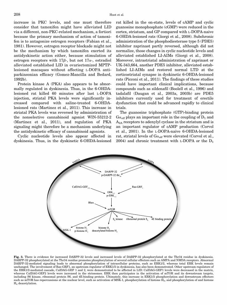
increase in PKC levels, and one must thereforeconsider that tamoxifen might have alleviated LIDvia a different, non-PKC-related mechanism, a fortioribecause the primary mechanism of action of tamoxi-fen is to antagonize estrogen receptors (Fabian et al.,1981). However, estrogen receptor blockade might notbe the mechanism by which tamoxifen exerted itsantidyskinetic action either, because stimulation ofestrogen receptors with 17b-, but not 17a-, estradiolalleviated established LID in ovariectomized MPTP-lesioned macaques without affecting L-DOPA anti-parkinsonian efficacy (Gomez-Mancilla and Bedard,1992b).Protein kinase A (PKA) also appears to be abnor-
mally regulated in dyskinesia. Thus, in the 6-OHDA-lesioned rat killed 60 minutes after last L-DOPAinjection, striatal PKA levels were significantly in-creased compared with saline-treated 6-OHDA-lesioned rats (Martinez et al., 2011). This increase instriatal PKA levels was reversed by administration ofthe nonselective cannabinoid agonist WIN-55212-2(Martinez et al., 2011), and regulation of PKAsignaling might therefore be a mechanism underlyingthe antidyskinetic efficacy of cannabinoid agonists.Cyclic nucleotide levels also appear affected in
dyskinesia. Thus, in the dyskinetic 6-OHDA-lesioned
rat killed in the on-state, levels of cAMP and cyclicguanosine monophosphate (cGMP) were reduced in thecortex, striatum, and GP compared with L-DOPA-naive6-OHDA-lesioned rats (Giorgi et al., 2008). Subchronicadministration of the phosphodiesterase type 5 (PDE5)inhibitor zaprinast partly reversed, although did notnormalize, those changes in cyclic nucleotide levels andalleviated established LI-AIMs (Giorgi et al., 2008).Moreover, intrastriatal administration of zaprinast orUK-343,664, another PDE5 inhibitor, alleviated estab-lished LI-AIMs and restored normal LTD at thecorticostriatal synapse in dyskinetic 6-OHDA-lesionedrats (Picconi et al., 2011). The findings of these studiescould have important clinical implications, becausecompounds such as sildenafil (Boolell et al., 1996) andtadalafil (Daugan et al., 2003a, 2003b) are PDE5inhibitors currently used for treatment of erectiledysfunction that could be advanced rapidly to clinicaltrials.
The guanosine triphosphate (GTP)-binding proteinGaolf plays an important role in the coupling of D1 andA2A receptors to adenylyl cyclase in the striatum and isan important regulator of cAMP production (Corvolet al., 2001). In the L-DOPA-naive 6-OHDA-lesionedrat, striatal levels of Gaolf were elevated (Corvol et al.,2004) and chronic treatment with L-DOPA or the D1
Fig. 3. There is evidence for increased DARPP-32 levels and increased levels of DARPP-32 phosphorylated at the Thr34 residue in dyskinesia.DARPP-32 phosphorylated at the Thr34 residue promotes phosphorylation of several cellular effectors such as AMPA and NMDA receptors. AbnormalDARPP-32-mediated signaling leads to abnormal phosphorylation of intracellular proteins, such as ERK1/2, whereas total ERK levels remainunchanged. The involvement of Ras-GRF1, an upstream regulator of ERK1/2 in dyskinesia, has also been demonstrated. Other upstream regulators ofthe ERK1/2-mediated cascade, CalDAG-GEF 1 and 2, were demonstrated to be affected in LID: CalDAG-GEF1 levels were decreased in the matrix,whereas CalDAG-GEF2 levels were increased in the striosomes. ERK then participates in the activation of mTOR and its downstream targets,including S6 kinase, ribosomal protein S6, and 4E-binding protein. Ultimately, this increase in ERK1/2 phosphorylation and downstream effectorssuch as mTOR has repercussions at the nuclear level, such as activation of MSK-1, phosphorylation of histone H3, and phosphorylation of and histoneH4 deacetylation.
208 Huot et al.
agonist SKF-81,297, but not ropinirole (last doseadministered 12 hours prior to the sacrifice, implyingthat the animals were killed in the off-state), normal-ized striatal levels of Gaolf (Corvol et al., 2004). Thefindings of that study are difficult to reconcile withthose mentioned in the previous paragraph,i.e., reductions in cAMP in the striatum of dyskinetic6-OHDA-lesioned rats. However, as mentioned above,increases in absolute levels of an enzyme, here Gaolf,may not necessarily reflect its activity, because otherfactors such as phosphorylation are also important.Moreover, perhaps the increase in Gaolf levels repre-sents a compensatory and not an etiological mecha-nism that aims at restoring normal cAMP levels indyskinesia. Lastly, reduced cAMP levels in dyskineticrats were demonstrated in the on-state, whereasincreased Gaolf were demonstrated in the off-state,perhaps accounting for the discrepancy betweenstudies.
XVI. Emerging Concepts in Dyskinesia
The field of LID is an area of intense and continuousresearch. Some emerging themes in the pathophysiol-ogy of dyskinesia will be briefly reviewed in thissection.
A. Angiogenesis and Blood-Brain Barrier
In the dyskinetic 6-OHDA-lesioned rat treated for 2weeks with L-DOPA and 5-bromo-29-deoxyuridine(BrdU) and killed 24 hours after last drug administra-tion, there was an increase in BrdU-positive endothe-lial cells within the denervated striatum, indicatingendothelial proliferation in LID (Westin et al., 2006).In a subsequent experiment, stimulation of D1 recep-tors and activation of ERK1/2 were demonstrated to bekey in the endothelial proliferation process (Lindgrenet al., 2009). In agreement with endothelial prolifera-tion, vascular endothelial growth factor (VEGF) levelswere found to be increased in the denervated striatumof dyskinetic 6-OHDA-lesioned rats killed 24 hoursafter last dose of L-DOPA, as well as in PD patientswith a history of dyskinesia (Ohlin et al., 2011). VEFGmRNA levels were particularly elevated within theastrocytes and their end-feet processes. These studies
have also suggested increased blood-brain barrierpermeability in the dyskinetic state. How angiogenesisand altered blood-brain barrier permeability contrib-ute to dyskinesia remain unknown at the moment.However, these findings tally very well with a PETstudy that demonstrated increased blood flow and flow/metabolism dissociation in the putamen/GP complex ofPD patients in the on-state, whether dyskinetic or not(Hirano et al., 2008).
B. Inflammation
In a study performed in the 6-OHDA-lesioned rat,systemic administration of the endogenous steroidcorticosterone and intrastriatal administration of aninterleukin-1 receptor antagonist both attenuated theexpression of established LI-AIMs (Barnum et al.,2008). In that study, de novo treatment with cortico-sterone also attenuated the development of LI-AIMs(Barnum et al., 2008). Striatal levels of interleukin-1bwere also found to be elevated in the dopamine-depleted striatum of dyskinetic rats killed 2 hoursafter last L-DOPA dose (Barnum et al., 2008). Themechanisms by which anti-inflammatory agents alle-viate LID are far from being elucidated. However,following corticosterone administration, levels of bothPPE-B and PPT mRNA were normalized, suggestingnormalization of the activity along the direct pathway.
C. Fatty Acids
Pharmacological studies performed in the MPTP-lesioned macaque have demonstrated that chronicdaily administration of the v-3 fatty acid docosohex-aenoic acid (DHA) alleviated established LID whileattenuating the priming process when administered denovo with L-DOPA (Samadi et al., 2006; Mahmoudiet al., 2009). Very little is known about fatty acids inPD and animal models of PD in regions known to beassociated with motor control. Thus, one postmortemstudy investigating levels of v-3 and v-6 fatty acids inthe brain of dyskinetic MPTP-lesioned NHP anddyskinetic PD patients measured v-3 and v-6 levelsin the temporal cortex (Julien et al., 2006), and,although interesting, the results of that study bringlittle information with respect to dyskinesia. Themechanism(s) underlying the antidyskinetic action of
TABLE 6DARPP-32, ERK1/2, and histone deacetylation in dyskinesia
• DARPP-32 levels are increased in the striatum.• DARPP-32 abnormally phosphorylated at the Thr34 residue are increased in the striatum.• ERK1/2 phosphorylation is increased in the striatum.• Genetic inactivation of DARPP-32 attenuates the development of LI-AIMs in the 6-OHDA-lesioned mouse.• Blockade of the mitogen-activated kinase/ERK kinase with SL327 attenuates the development of LI-AIMs in the 6-OHDA-lesioned mouse.• Development of LI-AIMs is reduced in Ras-GRF1-knockout mice.• Striatal delivery of a dominant-negative construct of Ras-GRF1 attenuates the severity of established LID in the MPTP-lesioned macaque.• CalDAG-GEF1/2 levels are altered in the striatum of the dyskinetic 6-OHDA-lesioned rat killed in the off-state.• Histone H3 is dephosphorylated in the striatum of the dyskinetic MPTP-lesioned NHP.• Histone H4 is deacetylated in the striatum of both dyskinetic MPTP-lesioned NHPs and dyskinetic 6-OHDA-lesioned mice killed at peak
LID.
L-DOPA-Induced Dyskinesia Pharmacology 209
DHA remains unknown as does the effect of DHA ondopamine agonist-induced dyskinesia. Moreover, italso remains unknown whether DHA is involved inthe pathophysiology of LID or if it simply alleviatesLID by modulating dopaminergic and glutamatergictransmissions.Despite the current lack of understanding of the
mechanisms whereby DHA alleviates LID, the poten-tial antidyskinetic efficacy of DHA is promising,because DHA could be advanced rapidly to clinicaltrials. Indeed, DHA is commercially available and hasbeen associated with beneficial effect on the cardiovas-cular system (Mozaffarian and Rimm, 2006). However,perhaps somewhat undermining the potential of DHAas an antidyskinetic agent is the fact that, becauseDHA is readily available over the counter, manydyskinetic PD patients are already taking it anda reduction in dyskinesia severity has not beenreported. On the other hand, high doses were requiredto alleviate LID in the parkinsonian NHP, 100–200mg/kg p.o., and perhaps the doses taken by PD patientsare too low to alleviate LID. Further studies areneeded to validate DHA as a clinically relevantantidyskinetic molecule and to determine which doses,if any, are effective and well tolerated.
D. Exocytosis
The synaptic vesicle glycoprotein 2A (SV2A), which isinvolved in exocytosis (Crowder et al., 1999), mightplay a role in the pathophysiology of dyskinesia,because it is currently the only known binding site ofthe anticonvulsant levetiracetam (Lynch et al., 2004).Levetiracetam effectively reduced the severity ofdyskinesia elicited by either L-DOPA or the combina-tion of L-DOPA and ropinirole in the MPTP-lesionedmarmoset (Hill et al., 2003). In the MPTP-lesionedmacaque, levetiracetam effectively alleviated L-DOPA-induced chorea but was ineffective against dystonia(Bezard et al., 2004). Concomitant administration oflevetiracetam with amantadine potentiated the anti-dyskinetic action of amantadine in the L-DOPA-treatedMPTP-lesioned marmoset (Hill et al., 2004b). Further-more, levetiracetam interfered with the priming pro-cess when administered de novo in combination withL-DOPA to the MPTP-lesioned marmoset (Hill et al.,2004a). Clinically, however, an antidyskinetic effect oflevetiracetam was not demonstrated (Wolz et al., 2010;Stathis et al., 2011; Wong et al., 2011).The mechanism(s) by which modulation of SV2A
activity might alleviate dyskinesia remains to beestablished. SV2A increases probability of vesicularrelease by priming vesicles in neurons in the restingstate (Custer et al., 2006), and it is possible thatmodulating SV2A leads to more physiologic release ofdopamine and/or glutamate in the striatum. How-ever, at this stage, it is important to emphasize thatthe antidyskinetic efficacy of modulating SV2A in
preclinical studies does not necessarily indicate per-turbed exocytosis or altered SV2A function in dyskine-sia, because modulating SV2A may simply, for instance,reduce pulsatility of raphestriatal dopamine release. Invivo microdialysis studies are needed to confirm thatpossibility, whereas studies measuring SV2A levels andphosphorylation status are needed to see whether itsfunction is perturbed in LID.
However, another argument indicating possiblealtered exocytosis in LID comes from a study per-formed in the 6-OHDA-lesioned rat. Thus, chronictherapy with SKF-38,393 and development of AIMs ledto an increase, whereas chronic therapy with quinpir-ole led to a decrease, in striatal mRNA levels of thetranscription factor zif-268 when rats were killed 60minutes after last treatment administration (Cartaet al., 2010). Zif-268, also known as EGR1 (earlygrowth response protein 1) or NGF1-A (nerve growthfactor-induced protein 1A), is involved in the transcrip-tional regulation of synaptobrevin II (Petersohn andThiel, 1996), an important protein in exocytosis (Bockand Scheller, 1999).
E. Thyrotropin Releasing Hormone
A study performed in the 6-OHDA-lesioned rat hasfound increased levels of thyrotropin releasing hor-mone (TRH) mRNA and an increase in pro-TRHimmunostaining in the dorsal striatum of animalsexpressing AIMs killed 12 hours after last L-DOPAdosing (Cantuti-Castelvetri et al., 2010). These eleva-tions were present in striatal medium spiny neuronscontacting both the GP and the entopeduncularnucleus. How enhanced TRH in striatal projectionneurons contribute to LID has yet to be demonstrated,but some hypotheses can be put forth. For instance,TRH increases GABA release in vivo in the hippocam-pus of anesthetized rats (Deng et al., 2006a). Accordingto the classic model of the organization of the basalganglia presented above, a TRH-mediated increase inGABA release at the striato-GPi/SNr complex wouldinhibit the output structures of the basal ganglia,thereby disinhibiting the glutamatergic thalamocort-ical projections with resulting enhanced cortical exci-tation. Systemic administration of TRH was alsoshown to increase striatal dopamine release in anes-thetized rats (Kreutz et al., 1990), but the increase inTRH being localized to striatofugal neurons somewhatdiminishes the likelihood of that possibility.
XVII. Concluding Remarks
Dyskinesia appears as a complex phenomenon, andseveral systems are involved in their pathophysiology.Although it is difficult to pinpoint a single factorleading to the development and expression of LID,nonphysiologic, pulsatile dopamine release and synap-tic pruning in the previously denervated striatum both
210 Huot et al.
seem to play critical roles. Of the other neurotrans-mitter systems described, glutamatergic neurotrans-mission has been most thoroughly studied and there isclear evidence that enhanced glutamatergic trans-mission is involved in the priming and expression ofLID. At the moment, our understanding of dyskinesia,priming and expression, revolves around dopamineand glutamate transmissions and, with a few excep-tions, is centered on the striatum. As such, althoughLID can be clearly modulated by a range of opioid,cannabinoid, and histaminergic drugs, the role playedby such neuromodulators in LID pathophysiology isdifficult to define. For instance, is enhanced cannabi-noid transmission a direct etiological factor in dyski-nesia or is it an epiphenomenon secondary tononphysiologic dopaminergic and glutamatergic trans-missions? The 5-HT system is a notable exception andis clearly responsible, in part at least, for the pulsatiledelivery of dopamine to the denervated striatum.Ultimately, cellular transcription systems becomeaffected, leading to changes in chromatin structure.The understanding of the intracellular mechanismsunderlying the dyskinetic phenotype have progressedover the last few years, and it is now known thatcascades involving DARPP-32 and ERK1/2, leading tohistone H4 deacetylation, are abnormal in dyskinesia.The challenges associated with drug development in
a scenario defined by a complexity of the changesaffecting many neuromodulator systems perhaps ex-plain why to date amantadine, which for historicalreasons was able to somewhat bypass the modern drugdevelopment process, is the only widely used anti-dyskinetic agent. Indeed, as discussed above, it is farfrom a perfect drug; amantadine effects may wane overtime, and if its action is to dampen NMDA-mediatedglutamatergic transmission, it does not address themultitude of other abnormal processes occurring indyskinesia. In view of these diffuse changes describedherein, perhaps an approach targeting several neuro-transmitter systems simultaneously might be moresuited to alleviate dyskinesia and to maintain anantidyskinetic efficacy over a long-term period. Alter-nately, the solution may lie in targeting theintracellular/intranuclear changes taking place indyskinesia; targeting downstream changes might per-haps allow countering more efficiently the changesmediated by abnormal transmission along severalneurotransmitter systems at a point where theyconverge. To date however, such an approach has beenlimited by difficulty for most drugs to enter theintracellular compartment and the concern of inducingnuclear changes and modifications in gene expression.Additionally, the search for long-lasting formulationsof L-DOPA that would provide constant dopaminelevels within the brain for extended periods of timemust continue, because this could possibly be the bestway to prevent the emergence and expression of LID
and, as a corollary, the best and safest antidyskinetictreatment.
These opportunities notwithstanding, the reviewpresented here does illustrate how far the field ofpharmacology of dyskinesia has progressed in recentyears. The challenge may not be solely one ofidentifying the appropriate targets but in transitioningtherapeutics from phase II to phase III success, a paththat amantadine did not have to tread.
Authorship contributions
Wrote or contributed to the writing of the manuscript: Huot,Johnston, Koprich, Fox, Brotchie.
ReferencesAbrahám G, Sólyom S, Csuzdi E, Berzsenyi P, Ling I, Tarnawa I, Hamori T, Pallagi I,Horvath K, and Andrasi F, et al. (2000) New non competitive AMPA antagonists.Bioorg Med Chem 8:2127–2143.
Adham N, Romanienko P, Hartig P, Weinshank RL, and Branchek T (1992) The rat5-hydroxytryptamine1B receptor is the species homologue of the human 5-hydroxytryptamine1D beta receptor. Mol Pharmacol 41:1–7.
Aghajanian GK and Marek GJ (1997) Serotonin induces excitatory postsynapticpotentials in apical dendrites of neocortical pyramidal cells. Neuropharmacology36:589–599.
Ahmed MR and Berthet ABychkov E, Porras G, Li Q, Bioulac BH, Carl YT, Bloch B,Kook S, and Aubert I, et al. (2010) Lentiviral overexpression of GRK6 alleviates L-dopa-induced dyskinesia in experimental Parkinson’s disease. Sci Transl Med 2:28ra28.
Aizman O, Brismar H, Uhlén P, Zettergren E, Levey AI, Forssberg H, GreengardP, and Aperia A (2000) Anatomical and physiological evidence for D1 and D2dopamine receptor colocalization in neostriatal neurons. Nat Neurosci 3:226–230.
Alachkar A, Brotchie J, and Jones OT (2006) alpha2-Adrenoceptor-mediated modu-lation of the release of GABA and noradrenaline in the rat substantia nigra parsreticulata. Neurosci Lett 395:138–142.
Alachkar A, Brotchie JM, and Jones OT (2010) Binding of dopamine and 3-methox-ytyramine as l-DOPA metabolites to human alpha(2)-adrenergic and dopaminergicreceptors. Neurosci Res 67:245–249.
Alachkar A, Brotchie JM, and Jones OT (2012) Changes in the mRNA levels of a2Aand a 2C adrenergic receptors in rat models of parkinson’s disease and L: -DOPA-induced dyskinesia. J Mol Neurosci 46:145–152.
Alagarsamy S, Marino MJ, Rouse ST, Gereau RW 4th, Heinemann SF, and Conn PJ(1999a) Activation of NMDA receptors reverses desensitization of mGluR5 in na-tive and recombinant systems. Nat Neurosci 2:234–240.
Alagarsamy S, Rouse ST, Gereau RW 4th, Heinemann SF, Smith Y, and Conn PJ(1999b) Activation of N-methyl-D-aspartate receptors reverses desensitization ofmetabotropic glutamate receptor, mGluR5, in native and recombinant systems.Ann N Y Acad Sci 868:526–530.
Alonso-Frech F, Zamarbide I, Alegre M, Rodríguez-Oroz MC, Guridi J, Manrique M,Valencia M, Artieda J, and Obeso JA (2006) Slow oscillatory activity and levodopa-induced dyskinesias in Parkinson’s disease. Brain 129:1748–1757.
Andersson M, Hilbertson A, and Cenci MA (1999) Striatal fosB expression is causallylinked with l-DOPA-induced abnormal involuntary movements and the associatedupregulation of striatal prodynorphin mRNA in a rat model of Parkinson’s disease.Neurobiol Dis 6:461–474.
Andersson M, Konradi C, and Cenci MA (2001) cAMP response element-bindingprotein is required for dopamine-dependent gene expression in the intact but notthe dopamine-denervated striatum. J Neurosci 21:9930–9943.
Anichtchik OV, Peitsaro N, Rinne JO, Kalimo H, and Panula P (2001) Distributionand modulation of histamine H(3) receptors in basal ganglia and frontal cortex ofhealthy controls and patients with Parkinson’s disease. Neurobiol Dis 8:707–716.
Anichtchik OV, Rinne JO, Kalimo H, and Panula P (2000) An altered histaminergicinnervation of the substantia nigra in Parkinson’s disease. Exp Neurol 163:20–30.
Anonymous (1974) Editorial: Dopa decarboxylase inhibitors. BMJ 4:250–251.Ansah TA, Ferguson MC, and Nayyar T (2011) The 5-HT(2A) receptor antagonistM100907 produces antiparkinsonian effects and decreases striatal glutamate.Front Syst Neurosci 5:48.
Arai A, Tomiyama M, Kannari K, Kimura T, Suzuki C, Watanabe M, KawarabayashiT, Shen H, and Shoji M (2008) Reuptake of L-DOPA-derived extracellular DA inthe striatum of a rodent model of Parkinson’s disease via norepinephrine trans-porter. Synapse 62:632–635.
Arai R, Karasawa N, Geffard M, and Nagatsu I (1995) L-DOPA is converted todopamine in serotonergic fibers of the striatum of the rat: a double-labeling im-munofluorescence study. Neurosci Lett 195:195–198.
Arai R, Karasawa N, Geffard M, Nagatsu T, and Nagatsu I (1994) Immunohisto-chemical evidence that central serotonin neurons produce dopamine from exoge-nous L-DOPA in the rat, with reference to the involvement of aromatic L-aminoacid decarboxylase. Brain Res 667:295–299.
Arai R, Karasawa N, and Nagatsu I (1996) Aromatic L-amino acid decarboxylase ispresent in serotonergic fibers of the striatum of the rat. A double-labeling immu-nofluorescence study. Brain Res 706:177–179.
L-DOPA-Induced Dyskinesia Pharmacology 211
Aubert I, Guigoni C, Håkansson K, Li Q, Dovero S, Barthe N, Bioulac BH, Gross CE,Fisone G, and Bloch B, et al. (2005) Increased D1 dopamine receptor signaling inlevodopa-induced dyskinesia. Ann Neurol 57:17–26.
Aubert I, Guigoni C, Li Q, Dovero S, Bioulac BH, Gross CE, Crossman AR, Bloch B,and Bezard E (2007) Enhanced preproenkephalin-B-derived opioid transmission instriatum and subthalamic nucleus converges upon globus pallidus internalis in L-3,4-dihydroxyphenylalanine-induced dyskinesia. Biol Psychiatry 61:836–844.
Ba M, Kong M, Yang H, Ma G, Lu G, Chen S, and Liu Z (2006) Changes in subcellulardistribution and phosphorylation of GluR1 in lesioned striatum of 6-hydroxydopamine-lesioned and l-dopa-treated rats. Neurochem Res 31:1337–1347.
Bara-Jimenez W, Dimitrova TD, Sherzai A, Aksu M, and Chase TN (2006) Glutamaterelease inhibition ineffective in levodopa-induced motor complications. Mov Disord21:1380–1383.
Bara-Jimenez W, Sherzai A, Dimitrova T, Favit A, Bibbiani F, Gillespie M, MorrisMJ, Mouradian MM, and Chase TN (2003) Adenosine A(2A) receptor antagonisttreatment of Parkinson’s disease. Neurology 61:293–296.
Barbeau A (1969a) L-dopa therapy in Parkinson’s disease: a critical review of nineyears’ experience. Can Med Assoc J 101:59–68.
Barbeau A (1969b) [Treatment of Parkinson’s disease by L-DOPA]. Union Med Can98:183–186.
Barbeau A (1971) Long-term side-effects of levodopa. Lancet 1:395.Barbeau A, Gillo-Joffroy L, and Mars H (1971) Treatment of Parkinson’s disease withlevodopa and Ro 4-4602. Clin Pharmacol Ther 12:353–359.
Barker AT, Jalinous R, and Freeston IL (1985) Non-invasive magnetic stimulation ofhuman motor cortex. Lancet 1:1106–1107.
Barnum CJ, Bhide N, Lindenbach D, Surrena MA, Goldenberg AA, Tignor S,Klioueva A, Walters H, and Bishop C (2012) Effects of noradrenergic denervationon L-DOPA-induced dyskinesia and its treatment by a- and b-adrenergic re-ceptor antagonists in hemiparkinsonian rats. Pharmacol Biochem Behav 100:607–615.
Barnum CJ, Eskow KL, Dupre K, Blandino P Jr, Deak T, and Bishop C (2008)Exogenous corticosterone reduces L-DOPA-induced dyskinesia in the hemi-parkinsonian rat: role for interleukin-1beta. Neuroscience 156:30–41.
Baronti F, Mouradian MM, Davis TL, Giuffra M, Brughitta G, Conant KE, and ChaseTN (1992) Continuous lisuride effects on central dopaminergic mechanisms inParkinson’s disease. Ann Neurol 32:776–781.
Bateup HS, Santini E, Shen W, Birnbaum S, Valjent E, Surmeier DJ, Fisone G,Nestler EJ, and Greengard P (2010) Distinct subclasses of medium spiny neuronsdifferentially regulate striatal motor behaviors. Proc Natl Acad Sci USA 107:14845–14850.
Beaulieu JM and Gainetdinov RR (2011) The physiology, signaling, and pharma-cology of dopamine receptors. Pharmacol Rev 63:182–217.
Berg D, Godau J, Trenkwalder C, and Eggert KCsoti IStorch AHuber HMorelli-Canelo MStamelou MRies V, et al. (2011) AFQ056 treatment of levodopa-induceddyskinesias: results of 2 randomized controlled trials. Mov Disord 26:1243–1250.
Berger B (1978) In vitro uptake of dopamine in serotoninergic nerve terminals:a fluorescence histochemical study on vibratome sections of the rat cerebral cortex.Adv Biochem Psychopharmacol 19:405–408.
Berger B and Glowinski J (1978) Dopamine uptake in serotoninergic terminals invitro: a valuable tool for the histochemical differentiation of catecholaminergic andserotoninergic terminals in rat cerebral structures. Brain Res 147:29–45.
Berthet A, Bezard E, Porras G, Fasano S, Barroso-Chinea P, Dehay B, Martinez A,Thiolat ML, Nosten-Bertrand M, and Giros B, et al. (2012) L-DOPA impairs pro-teasome activity in parkinsonism through D1 dopamine receptor. J Neurosci 32:681–691.
Berthet A, Porras G, Doudnikoff E, Stark H, Cador M, Bezard E, and Bloch B (2009)Pharmacological analysis demonstrates dramatic alteration of D1 dopamine re-ceptor neuronal distribution in the rat analog of L-DOPA-induced dyskinesia. JNeurosci 29:4829–4835.
Berton O, Guigoni C, Li Q, Bioulac BH, Aubert I, Gross CE, Dileone RJ, Nestler EJ,and Bezard E (2009) Striatal overexpression of DeltaJunD resets L-DOPA-induceddyskinesia in a primate model of Parkinson disease. Biol Psychiatry 66:554–561.
Bettler B, Kaupmann K, Mosbacher J, and Gassmann M (2004) Molecular structureand physiological functions of GABA(B) receptors. Physiol Rev 84:835–867.
Bézard E, Ferry S, Mach U, Stark H, Leriche L, Boraud T, Gross C, and Sokoloff P(2003) Attenuation of levodopa-induced dyskinesia by normalizing dopamine D3receptor function. Nat Med 9:762–767.
Bezard E, Hill MP, Crossman AR, Brotchie JM, Michel A, Grimée R, and Klitgaard H(2004) Levetiracetam improves choreic levodopa-induced dyskinesia in the MPTP-treated macaque. Eur J Pharmacol 485:159–164.
Bezard E, Ravenscroft P, Gross CE, Crossman AR, and Brotchie JM (2001) Upre-gulation of striatal preproenkephalin gene expression occurs before the appearanceof parkinsonian signs in 1-methyl-4-phenyl- 1,2,3,6-tetrahydropyridine monkeys.Neurobiol Dis 8:343–350.
Bhide NS, Lindenbach D, Barnum CJ, Surrena MA, and Bishop C (2010) The pan-B-adrenergic receptor antagonist (+/2)propranolol attenuates dyskinesia producedby l-DOPA, but not by D1 and D2 receptor agonists (Abstract 557.19/N7). SocNeurosci.
Bibbiani F, Costantini LC, Patel R, and Chase TN (2005a) Continuous dopaminergicstimulation reduces risk of motor complications in parkinsonian primates. ExpNeurol 192:73–78.
Bibbiani F, Oh JD, Kielaite A, Collins MA, Smith C, and Chase TN (2005b) Combinedblockade of AMPA and NMDA glutamate receptors reduces levodopa-inducedmotor complications in animal models of PD. Exp Neurol 196:422–429.
Bibbiani F, Oh JD, Petzer JP, Castagnoli N Jr, Chen JF, Schwarzschild MA,and Chase TN (2003) A2A antagonist prevents dopamine agonist-induced motorcomplications in animal models of Parkinson’s disease. Exp Neurol 184:285–294.
Bishop C, Krolewski DM, Eskow KL, Barnum CJ, Dupre KB, Deak T, and Walker PD(2009) Contribution of the striatum to the effects of 5-HT1A receptor stimulation inL-DOPA-treated hemiparkinsonian rats. J Neurosci Res 87:1645–1658.
Blanchet P, Bédard PJ, Britton DR, and Kebabian JW (1993) Differential effect ofselective D-1 and D-2 dopamine receptor agonists on levodopa-induced dyskinesiain 1-methyl-4-phenyl-1,2,3,6-tetrahydropyridine- exposed monkeys. J PharmacolExp Ther 267:275–279.
Blanchet PJ, Allard P, Grégoire L, Tardif F, and Bédard PJ (1996a) Risk factors forpeak dose dyskinesia in 100 levodopa-treated parkinsonian patients. Can J NeurolSci 23:189–193.
Blanchet PJ, Calon F, Martel JC, Bédard PJ, Di Paolo T, Walters RR, and PierceyMF (1995) Continuous administration decreases and pulsatile administrationincreases behavioral sensitivity to a novel dopamine D2 agonist (U-91356A) inMPTP-exposed monkeys. J Pharmacol Exp Ther 272:854–859.
Blanchet PJ, Fang J, Gillespie M, Sabounjian L, Locke KW, Gammans R, MouradianMM, and Chase TN (1998a) Effects of the full dopamine D1 receptor agonistdihydrexidine in Parkinson’s disease. Clin Neuropharmacol 21:339–343.
Blanchet PJ, Grondin R, and Bédard PJ (1996b) Dyskinesia and wearing-off followingdopamine D1 agonist treatment in drug-naive 1-methyl-4-phenyl-1,2,3,6-tetrahydropyridine-lesioned primates. Mov Disord 11:91–94.
Blanchet PJ, Konitsiotis S, and Chase TN (1997) Motor response to a dopamine D3receptor preferring agonist compared to apomorphine in levodopa-primed 1-methyl-4-phenyl-1,2,3,6-tetrahydropyridine monkeys. J Pharmacol Exp Ther 283:794–799.
Blanchet PJ, Konitsiotis S, and Chase TN (1998b) Amantadine reduces levodopa-induced dyskinesias in parkinsonian monkeys. Mov Disord 13:798–802.
Blanchet PJ, Konitsiotis S, Whittemore ER, Zhou ZL, Woodward RM, and Chase TN(1999) Differing effects of N-methyl-D-aspartate receptor subtype selective antag-onists on dyskinesias in levodopa-treated 1-methyl-4-phenyl-tetrahydropyridinemonkeys. J Pharmacol Exp Ther 290:1034–1040.
Blanchet PJ, Metman LV, Mouradian MM, and Chase TN (1996c) Acute pharma-cologic blockade of dyskinesias in Parkinson’s disease. Mov Disord 11:580–581.
Bock JB and Scheller RH (1999) SNARE proteins mediate lipid bilayer fusion. ProcNatl Acad Sci USA 96:12227–12229.
Boeve BF, Silber MH, and Ferman TJ (2004) REM sleep behavior disorder in Par-kinson’s disease and dementia with Lewy bodies. J Geriatr Psychiatry Neurol 17:146–157.
Bonsi P, Cuomo D, De Persis C, Centonze D, Bernardi G, Calabresi P, and Pisani A(2005) Modulatory action of metabotropic glutamate receptor (mGluR) 5 onmGluR1 function in striatal cholinergic interneurons. Neuropharmacology 49(Suppl 1):104–113.
Boolell M, Allen MJ, Ballard SA, Gepi-Attee S, Muirhead GJ, Naylor AM, OsterlohIH, and Gingell C (1996) Sildenafil: an orally active type 5 cyclic GMP-specificphosphodiesterase inhibitor for the treatment of penile erectile dysfunction. Int JImpot Res 8:47–52.
Boraud T, Bezard E, Bioulac B, and Gross CE (2001) Dopamine agonist-induceddyskinesias are correlated to both firing pattern and frequency alterations of pal-lidal neurones in the MPTP-treated monkey. Brain 124:546–557.
Bordet R, Ridray S, Carboni S, Diaz J, Sokoloff P, and Schwartz JC (1997) Inductionof dopamine D3 receptor expression as a mechanism of behavioral sensitization tolevodopa. Proc Natl Acad Sci USA 94:3363–3367.
Bordet R, Ridray S, Schwartz JC, and Sokoloff P (2000) Involvement of the directstriatonigral pathway in levodopa-induced sensitization in 6-hydroxydopamine-lesioned rats. Eur J Neurosci 12:2117–2123.
Bordia T, Campos C, Huang L, and Quik M (2008) Continuous and intermittentnicotine treatment reduces L-3,4-dihydroxyphenylalanine (L-DOPA)-induced dys-kinesias in a rat model of Parkinson’s disease. J Pharmacol Exp Ther 327:239–247.
Bordia T, Campos C, McIntosh JM, and Quik M (2010) Nicotinic receptor-mediatedreduction in L-DOPA-induced dyskinesias may occur via desensitization. J Phar-macol Exp Ther 333:929–938.
Boyce S, Rupniak NM, Steventon MJ, and Iversen SD (1990) Nigrostriatal damage isrequired for induction of dyskinesias by L-DOPA in squirrel monkeys. Clin Neu-ropharmacol 13:448–458.
Braak H, Del Tredici K, Rüb U, de Vos RA, Jansen Steur EN, and Braak E (2003)Staging of brain pathology related to sporadic Parkinson’s disease.Neurobiol Aging24:197–211.
Braak H, Ghebremedhin E, Rüb U, Bratzke H, and Del Tredici K (2004) Stages in thedevelopment of Parkinson’s disease-related pathology. Cell Tissue Res 318:121–134.
Breivogel CS, Sim LJ, and Childers SR (1997) Regional differences in cannabinoidreceptor/G-protein coupling in rat brain. J Pharmacol Exp Ther 282:1632–1642.
Brooks DJ, Piccini P, Turjanski N, and Samuel M (2000) Neuroimaging of dyskinesia.Ann Neurol 47:S154–158; discussion S158–S159.
Brown P and Eusebio A (2008) Paradoxes of functional neurosurgery: clues frombasal ganglia recordings. Mov Disord 23:12–20, quiz 158.
Brown TM, Brotchie JM, and Fitzjohn SM (2003) Cannabinoids decrease cortico-striatal synaptic transmission via an effect on glutamate uptake. J Neurosci 23:11073–11077.
Buck K and Ferger B (2009) Comparison of intrastriatal administration of nor-adrenaline and l-DOPA on dyskinetic movements: a bilateral reverse in vivomicrodialysis study in 6-hydroxydopamine-lesioned rats. Neuroscience 159:16–20.
Buck K and Ferger B (2010) The selective alpha1 adrenoceptor antagonist HEATreduces L-DOPA-induced dyskinesia in a rat model of Parkinson’s disease. Synapse64:117–126.
Buck K, Voehringer P, and Ferger B (2010) The alpha(2) adrenoceptor antagonistidazoxan alleviates L-DOPA-induced dyskinesia by reduction of striatal dopaminelevels: an in vivo microdialysis study in 6-hydroxydopamine-lesioned rats. J Neu-rochem 112:444–452.
Burnashev N, Villarroel A, and Sakmann B (1996) Dimensions and ion selectivity ofrecombinant AMPA and kainate receptor channels and their dependence on Q/Rsite residues. J Physiol 496:165–173.
212 Huot et al.

Calabresi P, Gubellini P, Centonze D, Sancesario G, Morello M, Giorgi M, Pisani A,and Bernardi G (1999) A critical role of the nitric oxide/cGMP pathway in corti-costriatal long-term depression. J Neurosci 19:2489–2499.
Callagham N, Mcllroy M, and O’Connor M (1974) Treatment of Parkinson’s Diseasewith levodopa and amantadine used as single drugs and in combined therapy. Ir JMed Sci 143:67–78.
Calne RY, Collier DS, Lim S, Pollard SG, Samaan A, White DJ, and Thiru S (1989)Rapamycin for immunosuppression in organ allografting. Lancet 2:227.
Calon F, Birdi S, Rajput AH, Hornykiewicz O, Bédard PJ, and Di Paolo T (2002a)Increase of preproenkephalin mRNA levels in the putamen of Parkinson diseasepatients with levodopa-induced dyskinesias. J Neuropathol Exp Neurol 61:186–196.
Calon F, Dridi M, Hornykiewicz O, Bédard PJ, Rajput AH, and Di Paolo T (2004)Increased adenosine A2A receptors in the brain of Parkinson’s disease patientswith dyskinesias. Brain 127:1075–1084.
Calon F, Goulet M, Blanchet PJ, Martel JC, Piercey MF, Bédard PJ, and Di Paolo T(1995) Levodopa or D2 agonist induced dyskinesia in MPTP monkeys: correlationwith changes in dopamine and GABAA receptors in the striatopallidal complex.Brain Res 680:43–52.
Calon F, Morissette M, Ghribi O, Goulet M, Grondin R, Blanchet PJ, Bédard PJ,and Di Paolo T (2002b) Alteration of glutamate receptors in the striatum of dys-kinetic 1-methyl-4-phenyl-1,2,3,6-tetrahydropyridine-treated monkeys followingdopamine agonist treatment. Prog Neuropsychopharmacol Biol Psychiatry 26:127–138.
Calon F, Morissette M, Goulet M, Grondin R, Blanchet PJ, Bédard PJ, and Di Paolo T(1999) Chronic D1 and D2 dopaminomimetic treatment of MPTP-denervatedmonkeys: effects on basal ganglia GABA(A)/benzodiazepine receptor complex andGABA content. Neurochem Int 35:81–91.
Calon F, Morissette M, Goulet M, Grondin R, Blanchet PJ, Bédard PJ, and Di Paolo T(2000) 125I-CGP 64213 binding to GABA(B) receptors in the brain of monkeys:effect of MPTP and dopaminomimetic treatments. Exp Neurol 163:191–199.
Calon F, Morissette M, Rajput AH, Hornykiewicz O, Bédard PJ, and Di Paolo T(2003a) Changes of GABA receptors and dopamine turnover in the postmortembrains of parkinsonians with levodopa-induced motor complications. Mov Disord18:241–253.
Calon F, Rajput AH, Hornykiewicz O, Bédard PJ, and Di Paolo T (2003b) Levodopa-induced motor complications are associated with alterations of glutamate receptorsin Parkinson’s disease. Neurobiol Dis 14:404–416.
Canals M, Marcellino D, Fanelli F, Ciruela F, de Benedetti P, Goldberg SR, NeveK, Fuxe K, Agnati LF, Woods AS, and , et al. (2003) Adenosine A2A-dopamineD2 receptor-receptor heteromerization: qualitative and quantitative assessmentby fluorescence and bioluminescence energy transfer. J Biol Chem 278:46741–46749.
Cantuti-Castelvetri I, Hernandez LF, Keller-McGandy CE, Kett LR, Landy A, Hol-lingsworth ZR, Saka E, Crittenden JR, Nillni EA, Young AB, and , et al. (2010)Levodopa-induced dyskinesia is associated with increased thyrotropin releasinghormone in the dorsal striatum of hemi-parkinsonian rats. PLoS ONE 5:e13861.
Cao X, Liang L, Hadcock JR, Iredale PA, Griffith DA, Menniti FS, Factor S,Greenamyre JT, and Papa SM (2007) Blockade of cannabinoid type 1 receptorsaugments the antiparkinsonian action of levodopa without affecting dyskinesias in1-methyl-4-phenyl-1,2,3,6-tetrahydropyridine-treated rhesus monkeys. J Phar-macol Exp Ther 323:318–326.
Cao X, Yasuda T, Uthayathas S, Watts RL, Mouradian MM, Mochizuki H, and PapaSM (2010) Striatal overexpression of DeltaFosB reproduces chronic levodopa-induced involuntary movements. J Neurosci 30:7335–7343.
Carey RJ (1991) Chronic L-dopa treatment in the unilateral 6-OHDA rat: evidencefor behavioral sensitization and biochemical tolerance. Brain Res 568:205–214.
Carlsson T, Carta M, Winkler C, Björklund A, and Kirik D (2007) Serotonin neurontransplants exacerbate L-DOPA-induced dyskinesias in a rat model of Parkinson’sdisease. J Neurosci 27:8011–8022.
Carpentier AF, Bonnet AM, Vidailhet M, and Agid Y (1996) Improvement oflevodopa-induced dyskinesia by propranolol in Parkinson’s disease. Neurology 46:1548–1551.
Carroll CB, Bain PG, Teare L, Liu X, Joint C, Wroath C, Parkin SG, Fox P, Wright D,and Hobart J, et al. (2004) Cannabis for dyskinesia in Parkinson disease: a ran-domized double-blind crossover study. Neurology 63:1245–1250.
Carta AR, Frau L, Pinna A, and Morelli M (2010) Dyskinetic potential of dopamineagonists is associated with different striatonigral/striatopallidal zif-268 expression.Exp Neurol 224:395–402.
Carta M, Carlsson T, Kirik D, and Björklund A (2007) Dopamine released from 5-HTterminals is the cause of L-DOPA-induced dyskinesia in parkinsonian rats. Brain130:1819–1833.
Carta M, Carlsson T, Muñoz A, Kirik D, and Björklund A (2008a) Involvement of theserotonin system in L-dopa-induced dyskinesias. Parkinsonism Relat Disord 14(Suppl 2):S154–S158.
Carta M, Carlsson T, Muñoz A, Kirik D, and Björklund A (2008b) Serotonin-dopamine interaction in the induction and maintenance of L-DOPA-induced dys-kinesias. Prog Brain Res 172:465–478.
Caulfield MP and Birdsall NJ (1998) International Union of Pharmacology.XVII. Classification of muscarinic acetylcholine receptors. Pharmacol Rev 50:279–290.
Cawello W, Braun M, and Boekens H (2009) Absorption, disposition, metabolic fate,and elimination of the dopamine agonist rotigotine in man: administration by in-travenous infusion or transdermal delivery. Drug Metab Dispos 37:2055–2060.
Cedarbaum JM (1987) Clinical pharmacokinetics of anti-parkinsonian drugs. ClinPharmacokinet 13:141–178.
Cenci MA, Lee CS, and Björklund A (1998) L-DOPA-induced dyskinesia in the rat isassociated with striatal overexpression of prodynorphin- and glutamic aciddecarboxylase mRNA. Eur J Neurosci 10:2694–2706.
Cenci MA and Lundblad M (2007) Ratings of L-DOPA-induced dyskinesia in theunilateral 6-OHDA lesion model of Parkinson’s disease in rats and mice. CurrProtoc Neurosci Chapter 9:Unit 9.25.
Cenci MA, Tranberg A, Andersson M, and Hilbertson A (1999) Changes in the re-gional and compartmental distribution of FosB- and JunB-like immunoreactivityinduced in the dopamine-denervated rat striatum by acute or chronic L-dopatreatment. Neuroscience 94:515–527.
Chahl LA (2006) Tachykinins and neuropsychiatric disorders. Curr Drug Targets 7:993–1003.
Chang B, Daniele CA, Gallagher K, Madonia M, Mitchum RD, Barrett L, Vezina P,and McGehee DS (2011) Nicotinic excitation of serotonergic projections from dorsalraphe to the nucleus accumbens. J Neurophysiol 106:801–808.
Charron G, Doudnikoff E, Laux A, Berthet A, Porras G, Canron MH, Barroso-ChineaP, Li Q, Qin C, and Nosten-Bertrand M, et al. (2011) Endogenous morphine-likecompound immunoreactivity increases in parkinsonism. Brain 134:2321–2338.
Chassain C, Eschalier A, and Durif F (2003) Antidyskinetic effect of magnesiumsulfate in MPTP-lesioned monkeys. Exp Neurol 182:490–496.
Chavkin C, James IF, and Goldstein A (1982) Dynorphin is a specific endogenousligand of the kappa opioid receptor. Science 215:413–415.
Chen L, Togasaki DM, Langston JW, Di Monte DA, and Quik M (2005) Enhancedstriatal opioid receptor-mediated G-protein activation in L-DOPA-treated dyski-netic monkeys. Neuroscience 132:409–420.
Chrétien M, Benjannet S, Gossard F, Gianoulakis C, Crine P, Lis M, and Seidah NG(1979) From beta-lipotropin to beta-endorphin and ‘pro-opio-melanocortin’. Can JBiochem 57:1111–1121.
Cicchetti F, Prensa L, Wu Y, and Parent A (2000) Chemical anatomy of striatalinterneurons in normal individuals and in patients with Huntington’s disease.Brain Res Brain Res Rev 34:80–101.
Civelli O, Bunzow JR, and Grandy DK (1993) Molecular diversity of the dopaminereceptors. Annu Rev Pharmacol Toxicol 33:281–307.
Clark EA and Hill SJ (1996) Sensitivity of histamine H3 receptor agonist-stimulated[35S]GTP gamma[S] binding to pertussis toxin. Eur J Pharmacol 296:223–225.
Conn PJ and Pin JP (1997) Pharmacology and functions of metabotropic glutamatereceptors. Annu Rev Pharmacol Toxicol 37:205–237.
Corbett AD, Henderson G, McKnight AT, and Paterson SJ (2006) 75 years of opioidresearch: the exciting but vain quest for the Holy Grail. Br J Pharmacol 147 (Suppl1):S153–S162.
Corvol JC, Muriel MP, Valjent E, Féger J, Hanoun N, Girault JA, Hirsch EC,and Hervé D (2004) Persistent increase in olfactory type G-protein alpha subunitlevels may underlie D1 receptor functional hypersensitivity in Parkinson disease. JNeurosci 24:7007–7014.
Corvol JC, Studler JM, Schonn JS, Girault JA, and Hervé D (2001) Galpha(olf) isnecessary for coupling D1 and A2a receptors to adenylyl cyclase in the striatum. JNeurochem 76:1585–1588.
Costa-Mattioli M, Sossin WS, Klann E, and Sonenberg N (2009) Translational controlof long-lasting synaptic plasticity and memory. Neuron 61:10–26.
Cotzias GC (1969) Parkinsoism and dopa. J Chronic Dis 22:297–301.Cotzias GC, Papavasiliou PS, and Gellene R (1969a) L-dopa in parkinson’s syndrome.N Engl J Med 281:272.
Cotzias GC, Papavasiliou PS, and Gellene R (1969b) Modification of Parkinsonism—
chronic treatment with L-DOPA. N Engl J Med 280:337–345.Cotzias GC, Van Woert MH, and Schiffer LM (1967) Aromatic amino acids andmodification of parkinsonism. N Engl J Med 276:374–379.
Court JA, Piggott MA, Lloyd S, Cookson N, Ballard CG, McKeith IG, Perry RH,and Perry EK (2000) Nicotine binding in human striatum: elevation in schizo-phrenia and reductions in dementia with Lewy bodies, Parkinson’s disease andAlzheimer’s disease and in relation to neuroleptic medication. Neuroscience 98:79–87.
Cox H, Togasaki DM, Chen L, Langston JW, Di Monte DA, and Quik M (2007) Theselective kappa-opioid receptor agonist U50,488 reduces L-dopa-induced dyski-nesias but worsens parkinsonism in MPTP-treated primates. Exp Neurol 205:101–107.
Cramer H, Jost S, Reuner C, Milios E, Geiger J, and Mundinger F (1991) Ventricularfluid neuropeptides in Parkinson’s disease. II. Levels of substance P-like immu-noreactivity. Neuropeptides 18:69–73.
Creese I, Burt DR, and Snyder SH (1977) Dopamine receptor binding enhancementaccompanies lesion-induced behavioral supersensitivity. Science 197:596–598.
Crittenden JR, Cantuti-Castelvetri I, Saka E, Keller-McGandy CE, Hernandez LF,Kett LR, Young AB, Standaert DG, and Graybiel AM (2009) Dysregulation ofCalDAG-GEFI and CalDAG-GEFII predicts the severity of motor side-effects in-duced by anti-parkinsonian therapy. Proc Natl Acad Sci USA 106:2892–2896.
Crowder KM, Gunther JM, Jones TA, Hale BD, Zhang HZ, Peterson MR, SchellerRH, Chavkin C, and Bajjalieh SM (1999) Abnormal neurotransmission in micelacking synaptic vesicle protein 2A (SV2A). Proc Natl Acad Sci USA 96:15268–15273.
Custer KL, Austin NS, Sullivan JM, and Bajjalieh SM (2006) Synaptic vesicle protein2 enhances release probability at quiescent synapses. J Neurosci 26:1303–1313.
Cutler JA (1998) Calcium-channel blockers for hypertension—uncertainty continues.N Engl J Med 338:679–681.
Damiano AM, McGrath MM, Willian MK, Snyder CF, LeWitt PA, Reyes PF, RichterRR, and Means ED (2000) Evaluation of a measurement strategy for Parkinson’sdisease: assessing patient health-related quality of life. Qual Life Res 9:87–100.
Daniele A, Albanese A, Gainotti G, Gregori B, and Bartolomeo P (1997) Zolpidem inParkinson’s disease. Lancet 349:1222–1223.
Daugan A, Grondin P, Ruault C, Le Monnier de Gouville AC, Coste H, Kirilovsky J,Hyafil F, and Labaudinière R (2003a) The discovery of tadalafil: a novel and highlyselective PDE5 inhibitor. 1: 5,6,11,11a-tetrahydro-1H-imidazo[19,59:1,6]pyrido[3,4-b]indole-1,3(2H)-dione analogues. J Med Chem 46:4525–4532.
Daugan A, Grondin P, Ruault C, Le Monnier de Gouville AC, Coste H, Linget JM,Kirilovsky J, Hyafil F, and Labaudinière R (2003b) The discovery of tadalafil:
L-DOPA-Induced Dyskinesia Pharmacology 213
a novel and highly selective PDE5 inhibitor. 2: 2,3,6,7,12,12a-hexahydropyrazino[19,29:1,6]pyrido[3,4-b]indole-1,4-dione analogues. J Med Chem 46:4533–4542.
de Esch IJ, Thurmond RL, Jongejan A, and Leurs R (2005) The histamine H4 re-ceptor as a new therapeutic target for inflammation. Trends Pharmacol Sci 26:462–469.
de la Fuente-Fernández R, Lu JQ, Sossi V, Jivan S, Schulzer M, Holden JE, Lee CS,Ruth TJ, Calne DB, and Stoessl AJ (2001) Biochemical variations in the synapticlevel of dopamine precede motor fluctuations in Parkinson’s disease: PET evidenceof increased dopamine turnover. Ann Neurol 49:298–303.
de la Fuente-Fernández R, Schulzer M, Mak E, Calne DB, and Stoessl AJ (2004a)Presynaptic mechanisms of motor fluctuations in Parkinson’s disease: a probabi-listic model. Brain 127:888–899.
de la Fuente-Fernández R, Sossi V, Huang Z, Furtado S, Lu JQ, Calne DB, Ruth TJ,and Stoessl AJ (2004b) Levodopa-induced changes in synaptic dopamine levelsincrease with progression of Parkinson’s disease: implications for dyskinesias.Brain 127:2747–2754.
Dekundy A, Gravius A, Hechenberger M, Pietraszek M, Nagel J, Tober C, van derElst M, Mela F, Parsons CG, and Danysz W (2011) Pharmacological character-ization of MRZ-8676, a novel negative allosteric modulator of subtype 5 metabo-tropic glutamate receptors (mGluR5): focus on L: -DOPA-induced dyskinesia. JNeural Transm 118:1703–1716.
Dekundy A, Lundblad M, Danysz W, and Cenci MA (2007) Modulation of L-DOPA-induced abnormal involuntary movements by clinically tested compounds: furthervalidation of the rat dyskinesia model. Behav Brain Res 179:76–89.
Dekundy A, Pietraszek M, Schaefer D, Cenci MA, and Danysz W (2006) Effects ofgroup I metabotropic glutamate receptors blockade in experimental models ofParkinson’s disease. Brain Res Bull 69:318–326.
Del Dotto P, Pavese N, Gambaccini G, Bernardini S, Metman LV, Chase TN,and Bonuccelli U (2001) Intravenous amantadine improves levadopa-induceddyskinesias: an acute double-blind placebo-controlled study. Mov Disord 16:515–520.
DeLong MR (1990) Primate models of movement disorders of basal ganglia origin.Trends Neurosci 13:281–285.
DeLong MR and Wichmann T (2007) Circuits and circuit disorders of the basalganglia. Arch Neurol 64:20–24.
Deng PY, Porter JE, Shin HS, and Lei S (2006a) Thyrotropin-releasing hormoneincreases GABA release in rat hippocampus. J Physiol 577:497–511.
Deng YP, Lei WL, and Reiner A (2006b) Differential perikaryal localization in rats ofD1 and D2 dopamine receptors on striatal projection neuron types identified byretrograde labeling. J Chem Neuroanat 32:101–116.
Di Monte DA, McCormack A, Petzinger G, Janson AM, Quik M, and Langston WJ(2000) Relationship among nigrostriatal denervation, parkinsonism, and dyski-nesias in the MPTP primate model. Mov Disord 15:459–466.
Dimitrova TD, Bara-Jimenez W, Savola JM, Encarnation E, Mouradian MM,and Chase TN (2009) Alpha-2 adrenergic antagonist effects in Parkinson’s disease.(Abstract). Mov Disord 24 (Suppl 1):S261.
Ding Y, Won L, Britt JP, Lim SA, McGehee DS, and Kang UJ (2011) Enhancedstriatal cholinergic neuronal activity mediates L-DOPA-induced dyskinesia inparkinsonian mice. Proc Natl Acad Sci USA 108:840–845.
Dingledine R, Borges K, Bowie D, and Traynelis SF (1999) The glutamate receptorion channels. Pharmacol Rev 51:7–61.
Dodel RC, Singer M, Köhne-Volland R, Szucs T, Rathay B, Scholz E, and Oertel WH(1998) The economic impact of Parkinson’s disease. An estimation based on a 3-month prospective analysis. Pharmacoeconomics 14:299–312.
Dunah AW and Standaert DG (2001) Dopamine D1 receptor-dependent trafficking ofstriatal NMDA glutamate receptors to the postsynaptic membrane. J Neurosci 21:5546–5558.
Dunah AW, Wang Y, Yasuda RP, Kameyama K, Huganir RL, Wolfe BB,and Standaert DG (2000) Alterations in subunit expression, composition, andphosphorylation of striatal N-methyl-D-aspartate glutamate receptors in a rat 6-hydroxydopamine model of Parkinson’s disease. Mol Pharmacol 57:342–352.
Dupre KB, Eskow KL, Negron G, and Bishop C (2007) The differential effects of 5-HT(1A) receptor stimulation on dopamine receptor-mediated abnormal involuntarymovements and rotations in the primed hemiparkinsonian rat. Brain Res 1158:135–143.
Dupre KB, Ostock CY, Eskow Jaunarajs KL, Button T, Savage LM, Wolf W,and Bishop C (2011) Local modulation of striatal glutamate efflux by serotonin 1Areceptor stimulation in dyskinetic, hemiparkinsonian rats. Exp Neurol 229:288–299.
Durif F, Debilly B, Galitzky M, Morand D, Viallet F, Borg M, Thobois S, Broussolle E,and Rascol O (2004) Clozapine improves dyskinesias in Parkinson disease: a dou-ble-blind, placebo-controlled study. Neurology 62:381–388.
Durif F, Vidailhet M, Bonnet AM, Blin J, and Agid Y (1995) Levodopa-induced dys-kinesias are improved by fluoxetine. Neurology 45:1855–1858.
Duvoisin RC, Heikkila RE, and Manzino L (1982) Pergolide-induced circling in ratswith 6-hydroxydopamine lesions in the nigrostriatal pathway. Neurology 32:1387–1391.
Eggert K, Squillacote D, Barone P, Dodel R, Katzenschlager R, Emre M, Lees AJ,Rascol O, Poewe W, and Tolosa E, et al.; German Competence Network on Par-kinson’s Disease (2010) Safety and efficacy of perampanel in advanced Parkinson’sdisease: a randomized, placebo-controlled study. Mov Disord 25:896–905.
Ehringer H and Hornykiewicz O (1960) [Distribution of noradrenaline and dopamine(3-hydroxytyramine) in the human brain and their behavior in diseases of theextrapyramidal system]. Klin Wochenschr 38:1236–1239.
Emre M, Rinne UK, Rascol A, Lees A, Agid Y, and Lataste X (1992) Effects of a se-lective partial D1 agonist, CY 208-243, in de novo patients with Parkinson disease.Mov Disord 7:239–243.
Eskow KL, Dupre KB, Barnum CJ, Dickinson SO, Park JY, and Bishop C (2009) Therole of the dorsal raphe nucleus in the development, expression, and treatment ofL-dopa-induced dyskinesia in hemiparkinsonian rats. Synapse 63:610–620.
Eusebio A and Brown P (2007) Oscillatory activity in the basal ganglia. ParkinsonismRelat Disord 13 (Suppl 3):S434–S436.
Evans JR, Mason SL, Williams-Gray CH, Foltynie T, Brayne C, Robbins TW,and Barker RA (2011) The natural history of treated Parkinson’s disease in anincident, community based cohort. J Neurol Neurosurg Psychiatry 82:1112–1118.
Fabian C, Tilzer L, and Sternson L (1981) Comparative binding affinities of tamox-ifen, 4-hydroxytamoxifen, and desmethyltamoxifen for estrogen receptors isolatedfrom human breast carcinoma: correlation with blood levels in patients withmetastatic breast cancer. Biopharm Drug Dispos 2:381–390.
Fahn S (2000) The spectrum of levodopa-induced dyskinesias. Ann Neurol 47:S2–9;discussion S9–11.
Fahn S, Libsch LR, and Cutler RW (1971) Monoamines in the human neostriatum:topographic distribution in normals and in Parkinson’s disease and their role inakinesia, rigidity, chorea, and tremor. J Neurol Sci 14:427–455.
Fahn S, Oakes D, Shoulson I, Kieburtz K, Rudolph A, Lang A, Olanow CW, Tanner C,and Marek K; Parkinson Study Group (2004) Levodopa and the progression ofParkinson’s disease. N Engl J Med 351:2498–2508.
Fariello RG (1998) Pharmacodynamic and pharmacokinetic features of cabergoline.Rationale for use in Parkinson’s disease. Drugs 55 (Suppl 1):10–16.
Fasano S, Bezard E, D’Antoni A, Francardo V, Indrigo M, Qin L, Dovero S, Cerovic M,Cenci MA, and Brambilla R (2010) Inhibition of Ras-guanine nucleotide-releasingfactor 1 (Ras-GRF1) signaling in the striatum reverts motor symptoms associatedwith L-dopa-induced dyskinesia. Proc Natl Acad Sci USA 107:21824–21829.
Fernandez A, de Ceballos ML, Jenner P, and Marsden CD (1992) Striatal neuro-peptide levels in Parkinson’s disease patients. Neurosci Lett 145:171–174.
Fernandez A, de Ceballos ML, Jenner P, and Marsden CD (1994) Neurotensin,substance P, delta and mu opioid receptors are decreased in basal ganglia ofParkinson’s disease patients. Neuroscience 61:73–79.
Ferré S, Karcz-Kubicha M, Hope BT, Popoli P, Burgueño J, Gutiérrez MA, Casadó V,Fuxe K, Goldberg SR, and Lluis C, et al. (2002) Synergistic interaction betweenadenosine A2A and glutamate mGlu5 receptors: implications for striatal neuronalfunction. Proc Natl Acad Sci USA 99:11940–11945.
Fichna J, Janecka A, Costentin J, and Do Rego JC (2007) The endomorphin systemand its evolving neurophysiological role. Pharmacol Rev 59:88–123.
Filion M (1979) Effects of interruption of the nigrostriatal pathway and of dopami-nergic agents on the spontaneous activity of globus pallidus neurons in the awakemonkey. Brain Res 178:425–441.
Fitzgerald PB, Fountain S, and Daskalakis ZJ (2006) A comprehensive review of theeffects of rTMS on motor cortical excitability and inhibition. Clin Neurophysiol 117:2584–2596.
Foord SM, Bonner TI, Neubig RR, Rosser EM, Pin JP, Davenport AP, Spedding M,and Harmar AJ (2005) International Union of Pharmacology. XLVI. G protein-coupled receptor list. Pharmacol Rev 57:279–288.
Fox S, Silverdale M, Kellett M, Davies R, Steiger M, Fletcher N, Crossman A,and Brotchie J (2004) Non-subtype-selective opioid receptor antagonism in treat-ment of levodopa-induced motor complications in Parkinson’s disease. Mov Disord19:554–560.
Fox SH, Henry B, Hill M, Crossman A, and Brotchie J (2002a) Stimulation of can-nabinoid receptors reduces levodopa-induced dyskinesia in the MPTP-lesionednonhuman primate model of Parkinson’s disease. Mov Disord 17:1180–1187.
Fox SH, Henry B, Hill MP, Peggs D, Crossman AR, and Brotchie JM (2001) Neuralmechanisms underlying peak-dose dyskinesia induced by levodopa and apomor-phine are distinct: evidence from the effects of the alpha(2) adrenoceptor antago-nist idazoxan. Mov Disord 16:642–650.
Fox SH, Katzenschlager R, Lim SY, Ravina B, Seppi K, Coelho M, Poewe W, RascolO, Goetz CG, and Sampaio C (2011) The Movement Disorder Society Evidence-Based Medicine Review Update: Treatments for the motor symptoms of Parkin-son’s disease. Mov Disord 26 (Suppl 3):S2–S41.
Fox SH, Kellett M, Moore AP, Crossman AR, and Brotchie JM (2002b) Randomised,double-blind, placebo-controlled trial to assess the potential of cannabinoid re-ceptor stimulation in the treatment of dystonia. Mov Disord 17:145–149.
Franceschi M, Cecchetto R, Panerai AE, Truci G, Smirne S, and Canal N (1986)Plasma beta-endorphin and beta-lipotropin in patients with Parkinson’s disease.Clin Neuropharmacol 9:549–555.
Fredholm BB, IJzerman AP, Jacobson KA, Klotz KN, and Linden J (2001) In-ternational Union of Pharmacology. XXV. Nomenclature and classification ofadenosine receptors. Pharmacol Rev 53:527–552.
Fujita M, Ichise M, Zoghbi SS, Liow JS, Ghose S, Vines DC, Sangare J, Lu JQ,Cropley VL, and Iida H, et al. (2006) Widespread decrease of nicotinic acetylcholinereceptors in Parkinson’s disease. Ann Neurol 59:174–177.
Fulceri F, Biagioni F, Ferrucci M, Lazzeri G, Bartalucci A, Galli V, Ruggieri S,Paparelli A, and Fornai F (2007) Abnormal involuntary movements (AIMs) fol-lowing pulsatile dopaminergic stimulation: severe deterioration and morphologicalcorrelates following the loss of locus coeruleus neurons. Brain Res 1135:219–229.
Gancher ST, Nutt JG, and Woodward WR (1987) Peripheral pharmacokinetics oflevodopa in untreated, stable, and fluctuating parkinsonian patients. Neurology37:940–944.
Gardoni F, Picconi B, Ghiglieri V, Polli F, Bagetta V, Bernardi G, Cattabeni F, DiLuca M, and Calabresi P (2006) A critical interaction between NR2B and MAGUKin L-DOPA induced dyskinesia. J Neurosci 26:2914–2922.
Gardoni F, Sgobio C, Pendolino V, Calabresi P, Di Luca M, and Picconi B (2011)Targeting NR2A-containing NMDA receptors reduces L-DOPA-induced dyski-nesias. Neurobiol Aging 33:2138–2144.
Gerfen CR, Engber TM, Mahan LC, Susel Z, Chase TN, Monsma FJ Jr, and SibleyDR (1990) D1 and D2 dopamine receptor-regulated gene expression of striatonigraland striatopallidal neurons. Science 250:1429–1432.
Gershanik OS, Heikkila RE, and Duvoisin RC (1978) The role of serotonin in theaction of L-dopa in an animal model of parkinsonism. Neurology 29 (Suppl):S553.
214 Huot et al.

Gingrich JA and Caron MG (1993) Recent advances in the molecular biology of do-pamine receptors. Annu Rev Neurosci 16:299–321.
Giorgi M, D’Angelo V, Esposito Z, Nuccetelli V, Sorge R, Martorana A, Stefani A,Bernardi G, and Sancesario G (2008) Lowered cAMP and cGMP signalling in thebrain during levodopa-induced dyskinesias in hemiparkinsonian rats: new aspectsin the pathogenetic mechanisms. Eur J Neurosci 28:941–950.
Giuffra ME, Sethy VH, Davis TL, Mouradian MM, and Chase TN (1993) Milacemidetherapy for Parkinson’s disease. Mov Disord 8:47–50.
Gladding CM, Fitzjohn SM, and Molnár E (2009) Metabotropic glutamate receptor-mediated long-term depression: molecular mechanisms. Pharmacol Rev 61:395–412.
Goetz CG, Damier P, Hicking C, Laska E, Müller T, Olanow CW, Rascol O, and RussH (2007) Sarizotan as a treatment for dyskinesias in Parkinson’s disease: a double-blind placebo-controlled trial. Mov Disord 22:179–186.
Goetz CG, Laska E, Hicking C, Damier P, Müller T, Nutt J, Warren Olanow C, RascolO, and Russ H (2008) Placebo influences on dyskinesia in Parkinson’s disease. MovDisord 23:700–707.
Gold SJ, Hoang CV, Potts BW, Porras G, Pioli E, Kim KW, Nadjar A, Qin C, LaHosteGJ, and Li Q, et al. (2007) RGS9-2 negatively modulates L-3,4-dihydroxyphenylalanine-induced dyskinesia in experimental Parkinson’s disease. JNeurosci 27:14338–14348.
Goldstein DS (2003) Dysautonomia in Parkinson’s disease: neurocardiological ab-normalities. Lancet Neurol 2:669–676.
Gomez-Mancilla B and Bédard PJ (1992a) Effect of chronic treatment with(+)-PHNO, a D2 agonist in MPTP-treated monkeys. Exp Neurol 117:185–188.
Gomez-Mancilla B and Bédard PJ (1992b) Effect of estrogen and progesterone on L-dopa induced dyskinesia in MPTP-treated monkeys. Neurosci Lett 135:129–132.
Gomez-Mancilla B and Bédard PJ (1993) Effect of nondopaminergic drugs on L-dopa-induced dyskinesias in MPTP-treated monkeys. Clin Neuropharmacol 16:418–427.
Gomez-Ramirez J, Johnston TH, Visanji NP, Fox SH, and Brotchie JM (2006) His-tamine H3 receptor agonists reduce L-dopa-induced chorea, but not dystonia, inthe MPTP-lesioned nonhuman primate model of Parkinson’s disease. Mov Disord21:839–846.
Gony M, Lapeyre-Mestre M, and Montastruc JL; French Network of RegionalPharmacovigilance Centers (2003) Risk of serious extrapyramidal symptoms inpatients with Parkinson’s disease receiving antidepressant drugs: a pharmacoepi-demiologic study comparing serotonin reuptake inhibitors and other antidepres-sant drugs. Clin Neuropharmacol 26:142–145.
Goto Y, Otani S, and Grace AA (2007) The Yin and Yang of dopamine release: a newperspective. Neuropharmacology 53:583–587.
Goulet M, Grondin R, Blanchet PJ, Bédard PJ, and Di Paolo T (1996) Dyskinesiasand tolerance induced by chronic treatment with a D1 agonist administered inpulsatile or continuous mode do not correlate with changes of putaminal D1receptors in drug-naive MPTP monkeys. Brain Res 719:129–137.
Grace AA (1995) The tonic/phasic model of dopamine system regulation: its relevancefor understanding how stimulant abuse can alter basal ganglia function. DrugAlcohol Depend 37:111–129.
Grafe MR, Forno LS, and Eng LF (1985) Immunocytochemical studies of substance Pand Met-enkephalin in the basal ganglia and substantia nigra in Huntington’s,Parkinson’s and Alzheimer’s diseases. J Neuropathol Exp Neurol 44:47–59.
Graham WC, Sambrook MA, and Crossman AR (1993) Differential effect of chronicdopaminergic treatment on dopamine D1 and D2 receptors in the monkey brain inMPTP-induced parkinsonism. Brain Res 602:290–303.
Graybiel AM (1990) Neurotransmitters and neuromodulators in the basal ganglia.Trends Neurosci 13:244–254.
Graybiel AM and Ragsdale CW Jr (1978) Histochemically distinct compartments inthe striatum of human, monkeys, and cat demonstrated by acetylthiocholinester-ase staining. Proc Natl Acad Sci USA 75:5723–5726.
Greengard P (2001) The neurobiology of slow synaptic transmission. Science 294:1024–1030.
Greger IH, Ziff EB, and Penn AC (2007) Molecular determinants of AMPA receptorsubunit assembly. Trends Neurosci 30:407–416.
Gregoire L, Morin N, Ouattara B, Gasparini F, Bilbe G, Johns D, Vranesic I,Sahasranaman S, Gomez-Mancilla B, and Di Paolo T (2011) The acute anti-parkinsonian and antidyskinetic effect of AFQ056, a novel metabotropic glutamatereceptor type 5 antagonist, in l-Dopa-treated parkinsonian monkeys. ParkinsonismRelat Disord 17:270–276.
Grégoire L, Rassoulpour A, Guidetti P, Samadi P, Bédard PJ, Izzo E, Schwarcz R,and Di Paolo T (2008) Prolonged kynurenine 3-hydroxylase inhibition reducesdevelopment of levodopa-induced dyskinesias in parkinsonian monkeys. BehavBrain Res 186:161–167.
Grégoire L, Samadi P, Graham J, Bédard PJ, Bartoszyk GD, and Di Paolo T (2009)Low doses of sarizotan reduce dyskinesias and maintain antiparkinsonian efficacyof L-Dopa in parkinsonian monkeys. Parkinsonism Relat Disord 15:445–452.
Grondin R, Bédard PJ, Britton DR, and Shiosaki K (1997) Potential therapeutic useof the selective dopamine D1 receptor agonist, A-86929: an acute study in par-kinsonian levodopa-primed monkeys. Neurology 49:421–426.
Gubellini P, Picconi B, Bari M, Battista N, Calabresi P, Centonze D, Bernardi G,Finazzi-Agrò A, and Maccarrone M (2002) Experimental parkinsonism altersendocannabinoid degradation: implications for striatal glutamatergic trans-mission. J Neurosci 22:6900–6907.
Guigoni C, Doudnikoff E, Li Q, Bloch B, and Bezard E (2007) Altered D(1) dopaminereceptor trafficking in parkinsonian and dyskinetic non-human primates. Neuro-biol Dis 26:452–463.
Guigoni C, Dovero S, Aubert I, Li Q, Bioulac BH, Bloch B, Gurevich EV, Gross CE,and Bezard E (2005a) Levodopa-induced dyskinesia in MPTP-treated macaques isnot dependent on the extent and pattern of nigrostrial lesioning. Eur J Neurosci22:283–287.
Guigoni C, Li Q, Aubert I, Dovero S, Bioulac BH, Bloch B, Crossman AR, Gross CE,and Bezard E (2005b) Involvement of sensorimotor, limbic, and associative basal
ganglia domains in L-3,4-dihydroxyphenylalanine-induced dyskinesia. J Neurosci25:2102–2107.
Haas HL, Sergeeva OA, and Selbach O (2008) Histamine in the nervous system.Physiol Rev 88:1183–1241.
Hadj Tahar A, Bélanger N, Bangassoro E, Grégoire L, and Bédard PJ (2000a)Antidyskinetic effect of JL-18, a clozapine analog, in parkinsonian monkeys. Eur JPharmacol 399:183–186.
Hadj Tahar A, Grégoire L, Bangassoro E, and Bédard PJ (2000b) Sustained caber-goline treatment reverses levodopa-induced dyskinesias in parkinsonian monkeys.Clin Neuropharmacol 23:195–202.
Hadj Tahar A, Grégoire L, Darré A, Bélanger N, Meltzer L, and Bédard PJ (2004)Effect of a selective glutamate antagonist on L-dopa-induced dyskinesias in drug-naive parkinsonian monkeys. Neurobiol Dis 15:171–176.
Hahnen E, Hauke J, Tränkle C, Eyüpoglu IY, Wirth B, and Blümcke I (2008) Histonedeacetylase inhibitors: possible implications for neurodegenerative disorders. Ex-pert Opin Investig Drugs 17:169–184.
Halabe Bucay A (2008) Activation of the proopiomelanocortin gene with ketoconazoleas a treatment for Parkinson’s disease: a new hypothesis. Ann N Y Acad Sci 1144:237–242.
Hallett PJ and Brotchie JM (2007) Striatal delta opioid receptor binding in experi-mental models of Parkinson’s disease and dyskinesia. Mov Disord 22:28–40.
Hallett PJ, Dunah AW, Ravenscroft P, Zhou S, Bezard E, Crossman AR, Brotchie JM,and Standaert DG (2005) Alterations of striatal NMDA receptor subunits associ-ated with the development of dyskinesia in the MPTP-lesioned primate model ofParkinson’s disease. Neuropharmacology 48:503–516.
Halliday GM, Li YW, Blumbergs PC, Joh TH, Cotton RG, Howe PR, Blessing WW,and Geffen LB (1990) Neuropathology of immunohistochemically identifiedbrainstem neurons in Parkinson’s disease. Ann Neurol 27:373–385.
Hammerstad JP, Carter J, Nutt JG, Casten GC, Shrotriya RC, Alms DR, and TempleD (1986) Buspirone in Parkinson’s disease. Clin Neuropharmacol 9:556–560.
Hammerstad JP, Woodward WR, Nutt JG, Gancher ST, Block GA, and Cyhan G(1994) Controlled release levodopa/carbidopa 25/100 (Sinemet CR 25/100): phar-macokinetics and clinical efficacy in untreated parkinsonian patients. Clin Neu-ropharmacol 17:429–434.
Hammond C, Bergman H, and Brown P (2007) Pathological synchronization inParkinson’s disease: networks, models and treatments. Trends Neurosci 30:357–364.
Hanrieder J, Ljungdahl A, Falth M, Mammo SE, Bergquist J and Andersson M(2011) L-DOPA-induced dyskinesia is associated with regional increase of striataldynorphin peptides as elucidated by imaging mass spectrometry. Mol Cell Pro-teomics 10:M111.009308.
Haskó G, Linden J, Cronstein B, and Pacher P (2008) Adenosine receptors: thera-peutic aspects for inflammatory and immune diseases. Nat Rev Drug Discov 7:759–770.
Hauser RA, McDermott MP, and Messing S (2006) Factors associated with the de-velopment of motor fluctuations and dyskinesias in Parkinson disease. Arch Neurol63:1756–1760.
Hauser RA and Olanow CW (1993) Orobuccal dyskinesia associated with trihex-yphenidyl therapy in a patient with Parkinson’s disease. Mov Disord 8:512–514.
Hauser RA, Panisset M, Abbruzzese G, Mancione L, Dronamraju N, and KakariekaA; FIRST-STEP Study Group (2009) Double-blind trial of levodopa/carbidopa/entacapone versus levodopa/carbidopa in early Parkinson’s disease.Mov Disord 24:541–550.
Hauser RA, Rascol O, Korczyn AD, Jon Stoessl A, Watts RL, Poewe W, De Deyn PP,and Lang AE (2007) Ten-year follow-up of Parkinson’s disease patients randomizedto initial therapy with ropinirole or levodopa. Mov Disord 22:2409–2417.
Hauser RA, Shulman LM, Trugman JM, Roberts JW, Mori A, Ballerini R,and Sussman NM; Istradefylline 6002-US-013 Study Group (2008) Study ofistradefylline in patients with Parkinson’s disease on levodopa with motor fluc-tuations. Mov Disord 23:2177–2185.
He L, Di Monte DA, Langston JW, and Quik M (2000) Autoradiographic analysis ofN-methyl-D-aspartate receptor binding in monkey brain: effects of 1-methyl-4-phenyl-1,2,3, 6-tetrahydropyridine and levodopa treatment. Neuroscience 99:697–704.
Heckers S, Geula C, and Mesulam MM (1992) Cholinergic innervation of the humanthalamus: dual origin and differential nuclear distribution. J Comp Neurol 325:68–82.
Hely MA, Morris JG, Reid WG, and Trafficante R (2005) Sydney Multicenter Study ofParkinson’s disease: non-L-dopa-responsive problems dominate at 15 years. MovDisord 20:190–199.
Henderson G and McKnight AT (1997) The orphan opioid receptor and its endoge-nous ligand—nociceptin/orphanin FQ. Trends Pharmacol Sci 18:293–300.
Henry B, Duty S, Fox SH, Crossman AR, and Brotchie JM (2003) Increased striatalpre-proenkephalin B expression is associated with dyskinesia in Parkinson’s dis-ease. Exp Neurol 183:458–468.
Henry B, Fox SH, Crossman AR, and Brotchie JM (2001) Mu- and delta-opioid re-ceptor antagonists reduce levodopa-induced dyskinesia in the MPTP-lesioned pri-mate model of Parkinson’s disease. Exp Neurol 171:139–146.
Henry B, Fox SH, Peggs D, Crossman AR, and Brotchie JM (1999) The alpha2-adrenergic receptor antagonist idazoxan reduces dyskinesia and enhances anti-parkinsonian actions of L-dopa in the MPTP-lesioned primate model of Parkinson’sdisease. Mov Disord 14:744–753.
Herrero MT, Augood SJ, Hirsch EC, Javoy-Agid F, Luquin MR, Agid Y, Obeso JA,and Emson PC (1995) Effects of L-DOPA on preproenkephalin and pre-protachykinin gene expression in the MPTP-treated monkey striatum. Neurosci-ence 68:1189–1198.
Herrero MT, Levy R, Ruberg M, Luquin MR, Villares J, Guillen J, Faucheux B,Javoy-Agid F, Guridi J, and Agid Y, et al. (1996) Consequence of nigrostriataldenervation and L-dopa therapy on the expression of glutamic acid decarboxylasemessenger RNA in the pallidum. Neurology 47:219–224.
L-DOPA-Induced Dyskinesia Pharmacology 215

Hiley CR and Kaup SS (2007) GPR55 and the vascular receptors for cannabinoids. BrJ Pharmacol 152:559–561.
Hill MP, Bezard E, McGuire SG, Crossman AR, Brotchie JM, Michel A, Grimée R,and Klitgaard H (2003) Novel antiepileptic drug levetiracetam decreases dyski-nesia elicited by L-dopa and ropinirole in the MPTP-lesioned marmoset. MovDisord 18:1301–1305.
Hill MP, Brotchie JM, Crossman AR, Bezard E, Michel A, Grimée R, and Klitgaard H(2004a) Levetiracetam Interferes With the L-dopa priming process in MPTP-lesioned drug-naive marmosets. Clin Neuropharmacol 27:171–177.
Hill MP, Girard F, Keywood C, Poli SM, Crossman AR, Ravenscroft P, Li Q, BezardE, and Mutel V (2010) ADX48621, a novel mGlu5 negative allosteric modulatoralleviates L-DOPA-induced chorea and dystonia in the MPTP-lesioned macaquemodel of Parkinson’s disease (Abstract). Mov Disord 25 (Suppl 2):S281–S282.
Hill MP, Ravenscroft P, Bezard E, Crossman AR, Brotchie JM, Michel A, Grimée R,and Klitgaard H (2004b) Levetiracetam potentiates the antidyskinetic action ofamantadine in the 1-methyl-4-phenyl-1,2,3,6-tetrahydropyridine (MPTP)-lesionedprimate model of Parkinson’s disease. J Pharmacol Exp Ther 310:386–394.
Hill SJ (1990) Distribution, properties, and functional characteristics of three classesof histamine receptor. Pharmacol Rev 42:45–83.
Hill SJ, Ganellin CR, Timmerman H, Schwartz JC, Shankley NP, Young JM,Schunack W, Levi R, and Haas HL (1997) International Union of Pharmacology.XIII. Classification of histamine receptors. Pharmacol Rev 49:253–278.
Hirano S, Asanuma K, Ma Y, Tang C, Feigin A, Dhawan V, Carbon M, and EidelbergD (2008) Dissociation of metabolic and neurovascular responses to levodopa in thetreatment of Parkinson’s disease. J Neurosci 28:4201–4209.
Hodgson RA, Bedard PJ, Varty GB, Kazdoba TM, Di Paolo T, Grzelak ME, Pond AJ,Hadjtahar A, Belanger N, and Gregoire L, et al. (2010) Preladenant, a selective A(2A) receptor antagonist, is active in primate models of movement disorders. ExpNeurol 225:384–390.
Horikawa S, Takai T, Toyosato M, Takahashi H, Noda M, Kakidani H, Kubo T,Hirose T, Inayama S, and Hayashida H, et al. (1983) Isolation and structuralorganization of the human preproenkephalin B gene. Nature 306:611–614.
Howlett AC, Barth F, Bonner TI, Cabral G, Casellas P, Devane WA, Felder CC,Herkenham M, Mackie K, and Martin BR, et al. (2002) International Union ofPharmacology. XXVII. Classification of cannabinoid receptors. Pharmacol Rev 54:161–202.
Hsu A, Togasaki DM, Bezard E, Sokoloff P, Langston JW, Di Monte DA, and Quik M(2004) Effect of the D3 dopamine receptor partial agonist BP897 [N-[4-(4-(2-methoxyphenyl)piperazinyl)butyl]-2-naphthamide] on L-3,4-dihydroxyphenylalanine-induced dyskinesias and parkinsonism in squirrel mon-keys. J Pharmacol Exp Ther 311:770–777.
Huang L, Grady SR, and Quik M (2011a) Nicotine reduces L-Dopa-induced dyski-nesias by acting at {beta}2 nicotinic receptors. J Pharmacol Exp Ther 338:932–941.
Huang LZ, Campos C, Ly J, Ivy Carroll F, and Quik M (2011b) Nicotinic receptoragonists decrease L-dopa-induced dyskinesias most effectively in partially lesionedparkinsonian rats. Neuropharmacology 60:861–868.
Hung SW, Adeli GM, Arenovich T, Fox SH, and Lang AE (2010) Patient perception ofdyskinesia in Parkinson’s disease. J Neurol Neurosurg Psychiatry 81:1112–1115.
Huot P and Brotchie JM (2011) 5-HT(1A) receptor stimulation and L-DOPA-induceddyskinesia in Parkinson’s disease: bridging the gap between serotonergic andglutamatergic mechanisms. Exp Neurol 231:195–198.
Huot P and Fox SH (2011) Nondopaminergic treatments for Parkinson’s disease.Neurodegen Dis Manag 1:491–512.
Huot P, Fox SH, and Brotchie JM (2011a) The serotonergic system in Parkinson’sdisease. Prog Neurobiol 95:163–212.
Huot P, Fox SH, Newman-Tancredi A, and Brotchie JM (2011b) Anatomically-selective5-HT1A and 5-HT2A therapies for Parkinson’s disease - an approach to reducingdyskinesia without exacerbating parkinsonism? J Pharmacol Exp Ther 339:1–7.
Huot P, Johnston TH, Koprich JB, Aman A, Fox SH, and Brotchie JM (2012a) L-745,870 reduces L-3,4-dihydroxyphenlylalanine-induced dyskinesia in the 1-methyl-4-phenyl-1,2,3,6-tetrahydropyridine-lesioned macaque model of Parkin-son’s disease. J Pharmacol Exp Ther 342:576–585.
Huot P, Johnston TH, Koprich JB, Winkelmolen L, Fox SH, and Brotchie JM (2012c)Regulation of cortical and striatal 5-HT(1A) receptors in the MPTP-lesioned ma-caque. Neurobiol Aging 33:e207, e209–e219.
Huot P, Johnston TH, Lewis KD, Koprich JB, Reyes MG, Fox SH, Piggott MJ,and Brotchie JM (2011c) Characterization of 3,4-methylenedioxymethamphet-amine (MDMA) enantiomers in vitro and in the MPTP-lesioned primate: R-MDMAreduces severity of dyskinesia, whereas S-MDMA extends duration of ON-time. JNeurosci 31:7190–7198.
Huot P, Johnston TH, Winkelmolen L, Fox SH, and Brotchie JM (2012b) 5-HT(2A)receptor levels increase in MPTP-lesioned macaques treated chronically with L-DOPA. Neurobiol Aging 33:194.e195-115.
Huot P and Parent A (2007) Dopaminergic neurons intrinsic to the striatum. JNeurochem 101:1441–1447.
Hurley MJ, Jackson MJ, Smith LA, Rose S, and Jenner P (2005) Immunoautor-adiographic analysis of NMDA receptor subunits and associated postsynapticdensity proteins in the brain of dyskinetic MPTP-treated common marmosets. EurJ Neurosci 21:3240–3250.
Hurley MJ, Jolkkonen J, Stubbs CM, Jenner P, and Marsden CD (1996a) DopamineD3 receptors in the basal ganglia of the common marmoset and following MPTPand L-DOPA treatment. Brain Res 709:259–264.
Hurley MJ, Mash DC, and Jenner P (2000) Adenosine A(2A) receptor mRNA ex-pression in Parkinson’s disease. Neurosci Lett 291:54–58.
Hurley MJ, Mash DC, and Jenner P (2003) Expression of cannabinoid CB1 receptormRNA in basal ganglia of normal and parkinsonian human brain. J NeuralTransm 110:1279–1288.
Hurley MJ, Stubbs CM, Jenner P, and Marsden CD (1996b) D3 receptor expressionwithin the basal ganglia is not affected by Parkinson’s disease.Neurosci Lett 214:75–78.
Hutchinson WD, Levy R, Dostrovsky JO, Lozano AM, and Lang AE (1997) Effects ofapomorphine on globus pallidus neurons in parkinsonian patients. Ann Neurol 42:767–775.
Iravani MM, Costa S, Al-Bargouthy G, Jackson MJ, Zeng BY, Kuoppamäki M, ObesoJA, and Jenner P (2005) Unilateral pallidotomy in 1-methyl-4-phenyl-1,2,3,6-tetrahydropyridine-treated common marmosets exhibiting levodopa-induced dys-kinesia. Eur J Neurosci 22:1305–1318.
Iravani MM, Costa S, Jackson MJ, Tel BC, Cannizzaro C, Pearce RK, and Jenner P(2001) GDNF reverses priming for dyskinesia in MPTP-treated, L-DOPA-primedcommon marmosets. Eur J Neurosci 13:597–608.
Iravani MM, Tayarani-Binazir K, Chu WB, Jackson MJ, and Jenner P (2006) In 1-methyl-4-phenyl-1,2,3,6-tetrahydropyridine-treated primates, the selective 5-hy-droxytryptamine 1a agonist (R)-(+)-8-OHDPAT inhibits levodopa-induced dyski-nesia but only withincreased motor disability. J Pharmacol Exp Ther 319:1225–1234.
Jackson MJ, Smith LA, Al-Barghouthy G, Rose S, and Jenner P (2007) Decreasedexpression of l-dopa-induced dyskinesia by switching to ropinirole in MPTP-treatedcommon marmosets. Exp Neurol 204:162–170.
Jaunarajs KL, Dupre KB, Steiniger A, Klioueva A, Moore A, Kelly C, and Bishop C(2009) Serotonin 1B receptor stimulation reduces D1 receptor agonist-induceddyskinesia. Neuroreport 20:1265–1269.
Jenner P (2002) Pharmacology of dopamine agonists in the treatment of Parkinson’sdisease. Neurology 58(4, Suppl 1)S1–S8.
Jenner P (2008) Molecular mechanisms of L-DOPA-induced dyskinesia. Nat RevNeurosci 9:665–677.
Johansson PA, Andersson M, Andersson KE, and Cenci MA (2001) Alterations incortical and basal ganglia levels of opioid receptor binding in a rat model of l-DOPA-induced dyskinesia. Neurobiol Dis 8:220–239.
Johnston TH, Damude S, Huot P, Fox SH, Jones SW, Rusche JR, and Brotchie JM(2010a) The histone deacetylase (HDAC) inhibitor RGFP109 reverses establishedmotor complications in L-DOPA-treated parkinsonian primates (Abstract). SocNeurosci.
Johnston TH, Fox SH, McIldowie MJ, Piggott MJ, and Brotchie JM (2010b) Re-duction of L-DOPA-induced dyskinesia by the selective metabotropic glutamatereceptor 5 antagonist 3-[(2-methyl-1,3-thiazol-4-yl)ethynyl]pyridine in the 1-methyl-4-phenyl-1,2,3,6-tetrahydropyridine-lesioned macaque model of Parkin-son’s disease. J Pharmacol Exp Ther 333:865–873.
Johnston TH, Fox SH, Piggott MJ, Savola JM, and Brotchie JM (2010c) The a₂adrenergic antagonist fipamezole improves quality of levodopa action in Parkin-sonian primates. Mov Disord 25:2084–2093.
Johnston TH, Huot P, Fox SH, Wakefield JD, Sykes KA, Bartolini WP, Milne GT,Pearson JP, and Brotchie JM (2011) Fatty acid amide hydrolase (FAAH) inhibitionreduces L-3,4-dihydroxyphenylalanine-induced hyperactivity in the 1-methyl-4-phenyl-1,2,3,6-tetrahydropyridine-lesioned non-human primate model of Parkin-son’s disease. J Pharmacol Exp Ther 336:423–430.
Johnston TH, van der Meij A, Brotchie JM, and Fox SH (2010d) Effect of histamineH2 receptor antagonism on levodopa-induced dyskinesia in the MPTP-macaquemodel of Parkinson’s disease. Mov Disord 25:1379–1390.
Jonkers N, Sarre S, Ebinger G, and Michotte Y (2002) MK801 suppresses the L-DOPA-induced increase of glutamate in striatum of hemi-Parkinson rats. BrainRes 926:149–155.
Julien C, Berthiaume L, Hadj-Tahar A, Rajput AH, Bédard PJ, Di Paolo T, Julien P,and Calon F (2006) Postmortem brain fatty acid profile of levodopa-treated Par-kinson disease patients and parkinsonian monkeys. Neurochem Int 48:404–414.
Juncos JL, Fabbrini G, Mouradian MM, and Chase TN (1987a) Controlled releaselevodopa-carbidopa (CR-5) in the management of parkinsonian motor fluctuations.Arch Neurol 44:1010–1012.
Juncos JL, Fabbrini G, Mouradian MM, Serrati C, Kask AM, and Chase TN (1987b)Controlled release levodopa treatment of motor fluctuations in Parkinson’s disease.J Neurol Neurosurg Psychiatry 50:194–198.
Juncos JL, Mouradian MM, Fabbrini G, Serrati C, and Chase TN (1987c) Levodopamethyl ester treatment of Parkinson’s disease. Neurology 37:1242–1245.
Kannari K, Kurahashi K, Tomiyama M, Maeda T, Arai A, Baba M, Suda T,and Matsunaga M (2002) [Tandospirone citrate, a selective 5-HT1A agonist, alle-viates L-DOPA-induced dyskinesia in patients with Parkinson’s disease]. No ToShinkei 54:133–137.
Kas A, Bottlaender M, Gallezot JD, Vidailhet M, Villafane G, Gregoire MC, Coulon C,Valette H, Dolle F, and Ribeiro MJ, et al. (2009) Decrease of nicotinic receptors inthe nigrostriatal system in Parkinson’s disease. J Cereb Blood Flow Metab 29:1601–1608.
Kaye CM and Nicholls B (2000) Clinical pharmacokinetics of ropinirole. Clin Phar-macokinet 39:243–254.
Kenakin T (2005) New concepts in drug discovery: collateral efficacy and permissiveantagonism. Nat Rev Drug Discov 4:919–927.
Kenakin T (2011) Functional selectivity and biased receptor signaling. J PharmacolExp Ther 336:296–302.
Kennedy MB, Beale HC, Carlisle HJ, and Washburn LR (2005) Integration of bio-chemical signalling in spines. Nat Rev Neurosci 6:423–434.
Kleedorfer B, Lees AJ, and Stern GM (1991) Buspirone in the treatment of levodopainduced dyskinesias. J Neurol Neurosurg Psychiatry 54:376–377.
Kobilka BK (2011) Structural insights into adrenergic receptor function and phar-macology. Trends Pharmacol Sci 32:213–218.
Kobylecki C, Cenci MA, Crossman AR, and Ravenscroft P (2010) Calcium-permeableAMPA receptors are involved in the induction and expression of l-DOPA-induceddyskinesia in Parkinson’s disease. J Neurochem 114:499–511.
Konitsiotis S, Blanchet PJ, Verhagen L, Lamers E, and Chase TN (2000) AMPAreceptor blockade improves levodopa-induced dyskinesia in MPTP monkeys.Neurology 54:1589–1595.
216 Huot et al.
Konitsiotis S and Tsironis C (2006) Levodopa-induced dyskinesia and rotationalbehavior in hemiparkinsonian rats: independent features or components of thesame phenomenon? Behav Brain Res 170:337–341.
Koprich JB, Fox SH, Johnston TH, Goodman A, Le Bourdonnec B, Dolle RE,DeHaven RN, DeHaven-Hudkins DL, Little PJ, and Brotchie JM (2011) The se-lective mu-opioid receptor antagonist ADL5510 reduces levodopa-induced dyski-nesia without affecting antiparkinsonian action in MPTP-lesioned macaque modelof Parkinson’s disease. Mov Disord 26:1225–1233.
Koprich JB, Johnston TH, Huot P, Fox SH, and Brotchie JM (2009) New insights intothe organization of the basal ganglia. Curr Neurol Neurosci Rep 9:298–304.
Kreutz MR, Acworth IN, Lehnert H, and Wurtman RJ (1990) Systemic administra-tion of thyrotropin-releasing hormone enhances striatal dopamine release in vivo.Brain Res 536:347–352.
Kuan WL, Zhao JW, and Barker RA (2008) The role of anxiety in the development oflevodopa-induced dyskinesias in an animal model of Parkinson’s disease, and theeffect of chronic treatment with the selective serotonin reuptake inhibitor cit-alopram. Psychopharmacology (Berl) 197:279–293.
Kumar N, Van Gerpen JA, Bower JH, and Ahlskog JE (2005) Levodopa-dyskinesiaincidence by age of Parkinson’s disease onset. Mov Disord 20:342–344.
Kunishima N, Shimada Y, Tsuji Y, Sato T, Yamamoto M, Kumasaka T, Nakanishi S,Jingami H, and Morikawa K (2000) Structural basis of glutamate recognition bya dimeric metabotropic glutamate receptor. Nature 407:971–977.
Lader M (2008) Effectiveness of benzodiazepines: do they work or not? Expert RevNeurother 8:1189–1191.
Lane EL, Cheetham SC, and Jenner P (2006) Does contraversive circling in the 6-OHDA-lesioned rat indicate an ability to induce motor complications as well astherapeutic effects in Parkinson’s disease? Exp Neurol 197:284–290.
Lastres-Becker I, Cebeira M, de Ceballos ML, Zeng BY, Jenner P, Ramos JA,and Fernández-Ruiz JJ (2001) Increased cannabinoid CB1 receptor binding andactivation of GTP-binding proteins in the basal ganglia of patients with Par-kinson’s syndrome and of MPTP-treated marmosets. Eur J Neurosci 14:1827–1832.
Lavoie B and Parent A (1990) Immunohistochemical study of the serotoninergicinnervation of the basal ganglia in the squirrel monkey. J Comp Neurol 299:1–16.
Lavoie B and Parent A (1991) Serotoninergic innervation of the thalamus in theprimate: an immunohistochemical study. J Comp Neurol 312:1–18.
Lavoie B, Parent A, and Bédard PJ (1991) Effects of dopamine denervation onstriatal peptide expression in parkinsonian monkeys. Can J Neurol Sci 18(3,Suppl)373–375.
Lee CS, Cenci MA, Schulzer M, and Björklund A (2000) Embryonic ventral mesen-cephalic grafts improve levodopa-induced dyskinesia in a rat model of Parkinson’sdisease. Brain 123:1365–1379.
Lee T, Seeman P, Rajput A, Farley IJ, and Hornykiewicz O (1978) Receptor basis fordopaminergic supersensitivity in Parkinson’s disease. Nature 273:59–61.
Lees AJ, Shaw KM, and Stern GM (1977) “Off period” dystonia and “on period”choreoathetosis in levodopa-treated patients with Parkinson’s disease. Lancet 2:1034.
Lemay S, Chouinard S, Blanchet P, Masson H, Soland V, Beuter A, and Bédard MA(2004) Lack of efficacy of a nicotine transdermal treatment on motor and cognitivedeficits in Parkinson’s disease. Prog Neuropsychopharmacol Biol Psychiatry 28:31–39.
Leppik IE (2004) Zonisamide: chemistry, mechanism of action, and pharmacokinet-ics. Seizure 13 (Suppl 1):S5–9.
Lévesque JC and Parent A (2005a) GABAergic interneurons in human subthalamicnucleus. Mov Disord 20:574–584.
Lévesque M and Parent A (2005b) The striatofugal fiber system in primates:a reevaluation of its organization based on single-axon tracing studies. Proc NatlAcad Sci USA 102:11888–11893.
Levy R, Herrero MT, Ruberg M, Villares J, Faucheux B, Guridi J, Guillen J, LuquinMR, Javoy-Agid F, and Obeso JA, et al. (1995a) Effects of nigrostriatal denervationand L-dopa therapy on the GABAergic neurons in the striatum in MPTP-treatedmonkeys and Parkinson’s disease: an in situ hybridization study of GAD67 mRNA.Eur J Neurosci 7:1199–1209.
Levy R, Vila M, Herrero MT, Faucheux B, Agid Y, and Hirsch EC (1995b) Striatalexpression of substance P and methionin-enkephalin in genes in patients withParkinson’s disease. Neurosci Lett 199:220–224.
LeWitt PA (1992) Clinical studies with and pharmacokinetic considerations ofsustained-release levodopa. Neurology 42(1, Suppl 1)29–32, discussion 57–60.
LeWitt PA, Guttman M, Tetrud JW, Tuite PJ, Mori A, Chaikin P, and Sussman NM;6002-US-005 Study Group (2008) Adenosine A2A receptor antagonist istradefylline(KW-6002) reduces “off” time in Parkinson’s disease: a double-blind, randomized,multicenter clinical trial (6002-US-005). Ann Neurol 63:295–302.
Lewitt PA, Hauser RA, Lu M, Nicholas AP, Weiner W, Coppard N, Leinonen M,and Savola JM (2012) Randomized clinical trial of fipamezole for dyskinesia inParkinson disease (FJORD study). Neurology 79:163–169.
Lhermitte F, Agid Y, and Signoret JL (1978) Onset and end-of-dose levodopa-induceddyskinesias. Possible treatment by increasing the daily doses of levodopa. ArchNeurol 35:261–263.
Liégeois JF, Bruhwyler J, Damas J, Rogister F, Masereel B, Geczy J, and Delarge J(1995) Modulation of the clozapine structure increases its selectivity for the do-pamine D4 receptor. Eur J Pharmacol 273:R1–R3.
Lindenbach D, Ostock CY, Eskow Jaunarajs KL, Dupre KB, Barnum CJ, Bhide N,and Bishop C (2011) Behavioral and cellular modulation of L-DOPA-induced dys-kinesia by beta-adrenoceptor blockade in the 6-hydroxydopamine-lesioned rat. JPharmacol Exp Ther 337:755–765.
Lindgren HS, Andersson DR, Lagerkvist S, Nissbrandt H, and Cenci MA (2010) L-DOPA-induced dopamine efflux in the striatum and the substantia nigra in a ratmodel of Parkinson’s disease: temporal and quantitative relationship to the ex-pression of dyskinesia. J Neurochem 112:1465–1476.
Lindgren HS, Ohlin KE, and Cenci MA (2009) Differential involvement of D1 and D2dopamine receptors in L-DOPA-induced angiogenic activity in a rat model ofParkinson’s disease. Neuropsychopharmacology 34:2477–2488.
Lu HC, Gonzalez E, and Crair MC (2001) Barrel cortex critical period plasticity isindependent of changes in NMDA receptor subunit composition. Neuron 32:619–634.
Lucas G and Spampinato U (2000) Role of striatal serotonin2A and serotonin2Creceptor subtypes in the control of in vivo dopamine outflow in the rat striatum. JNeurochem 74:693–701.
Luginger E, Wenning GK, Bösch S, and Poewe W (2000) Beneficial effects of aman-tadine on L-dopa-induced dyskinesias in Parkinson’s disease. Mov Disord 15:873–878.
Lukas RJ, Changeux JP, Le Novère N, Albuquerque EX, Balfour DJ, Berg DK,Bertrand D, Chiappinelli VA, Clarke PB, Collins AC, and , et al. (1999) In-ternational Union of Pharmacology. XX. Current status of the nomenclature fornicotinic acetylcholine receptors and their subunits. Pharmacol Rev 51:397–401.
Lundblad M, Andersson M, Winkler C, Kirik D, Wierup N, and Cenci MA (2002)Pharmacological validation of behavioural measures of akinesia and dyskinesia ina rat model of Parkinson’s disease. Eur J Neurosci 15:120–132.
Lundblad M, Usiello A, Carta M, Håkansson K, Fisone G, and Cenci MA (2005)Pharmacological validation of a mouse model of l-DOPA-induced dyskinesia. ExpNeurol 194:66–75.
Luquin MR, Laguna J, and Obeso JA (1992a) Selective D2 receptor stimulationinduces dyskinesia in parkinsonian monkeys. Ann Neurol 31:551–554.
Luquin MR, Scipioni O, Vaamonde J, Gershanik O, and Obeso JA (1992b) Levodopa-induced dyskinesias in Parkinson’s disease: clinical and pharmacological classifi-cation. Mov Disord 7:117–124.
Lynch BA, Lambeng N, Nocka K, Kensel-Hammes P, Bajjalieh SM, Matagne A,and Fuks B (2004) The synaptic vesicle protein SV2A is the binding site for theantiepileptic drug levetiracetam. Proc Natl Acad Sci USA 101:9861–9866.
Maccarrone M, Gubellini P, Bari M, Picconi B, Battista N, Centonze D, Bernardi G,Finazzi-Agrò A, and Calabresi P (2003) Levodopa treatment reverses endocanna-binoid system abnormalities in experimental parkinsonism. J Neurochem 85:1018–1025.
Maeda T, Nagata K, Yoshida Y, and Kannari K (2005) Serotonergic hyperinnervationinto the dopaminergic denervated striatum compensates for dopamine conversionfrom exogenously administered l-DOPA. Brain Res 1046:230–233.
Maertens de Noordhout A and Delwaide PJ (1986) Open pilot trial of ritanserin inparkinsonism. Clin Neuropharmacol 9:480–484.
Maggi CA (1995) The mammalian tachykinin receptors. Gen Pharmacol 26:911–944.Maggio JE (1988) Tachykinins. Annu Rev Neurosci 11:13–28.Mahmoudi S, Samadi P, Gilbert F, Ouattara B, Morissette M, Grégoire L, RouillardC, Di Paolo T, and Lévesque D (2009) Nur77 mRNA levels and L-Dopa-induceddyskinesias in MPTP monkeys treated with docosahexaenoic acid. Neurobiol Dis36:213–222.
Maneuf YP, Nash JE, Crossman AR, and Brotchie JM (1996) Activation of the can-nabinoid receptor by delta 9-tetrahydrocannabinol reduces gamma-aminobutyricacid uptake in the globus pallidus. Eur J Pharmacol 308:161–164.
Manson AJ, Iakovidou E, and Lees AJ (2000) Idazoxan is ineffective for levodopa-induced dyskinesias in Parkinson’s disease. Mov Disord 15:336–337.
Manson AJ, Katzenschlager R, Hobart J, and Lees AJ (2001) High dose naltrexonefor dyskinesias induced by levodopa. J Neurol Neurosurg Psychiatry 70:554–556.
Maratos EC, Jackson MJ, Pearce RK, Cannizzaro C, and Jenner P (2003) Both short-and long-acting D-1/D-2 dopamine agonists induce less dyskinesia than L-DOPA inthe MPTP-lesioned common marmoset (Callithrix jacchus). Exp Neurol 179:90–102.
Maratos EC, Jackson MJ, Pearce RK, and Jenner P (2001) Antiparkinsonian activityand dyskinesia risk of ropinirole and L-DOPA combination therapy in drugnaïve MPTP-lesioned common marmosets (Callithrix jacchus). Mov Disord 16:631–641.
Marin C, Aguilar E, Bonastre M, Tolosa E, and Obeso JA (2005) Early administrationof entacapone prevents levodopa-induced motor fluctuations in hemiparkinsonianrats. Exp Neurol 192:184–193.
Marin C, Aguilar E, Mengod G, Cortés R, and Obeso JA (2009a) Effects of early vs.late initiation of levodopa treatment in hemiparkinsonian rats. Eur J Neurosci 30:823–832.
Marin C, Aguilar E, and Obeso JA (2006a) Coadministration of entacapone withlevodopa attenuates the severity of dyskinesias in hemiparkinsonian rats. MovDisord 21:646–653.
Marin C, Aguilar E, Rodríguez-Oroz MC, Bartoszyk GD, and Obeso JA (2009b) Localadministration of sarizotan into the subthalamic nucleus attenuates levodopa-induced dyskinesias in 6-OHDA-lesioned rats. Psychopharmacology (Berl) 204:241–250.
Marin C, Rodriguez-Oroz MC, and Obeso JA (2006b) Motor complications in Par-kinson’s disease and the clinical significance of rotational behavior in the rat: havewe wasted our time? Exp Neurol 197:269–274.
Marsden CD, Barry PE, Parkes JD, and Zilkha KJ (1973a) Treatment of Parkinson’sdisease with levodopa combined with L-alpha-methyldopahydrazine, aninhibitor of extracerebral DOPA decarboxylase. J Neurol Neurosurg Psychiatry 36:10–14.
Marsden CD, Parkes JD, and Rees JE (1973b) A year’s comparison of treatment ofpatients with parkinson’s disease with levodopa combined with carbidopa versustreatment with levodopa alone. Lancet 2:1459–1462.
Martínez-Mir MI, Pollard H, Moreau J, Traiffort E, Ruat M, Schwartz JC,and Palacios JM (1993) Loss of striatal histamine H2 receptors in Huntington’schorea but not in Parkinson’s disease: comparison with animal models. Synapse 15:209–220.
Martinez A, Macheda T, Morgese MG, Trabace L, and Giuffrida A (2011) The can-nabinoid agonist WIN55212-2 decreases l-DOPA-induced PKA activation anddyskinetic behavior in 6-OHDA-treated rats. Neurosci Res 72:236–242.
L-DOPA-Induced Dyskinesia Pharmacology 217
Matsuda LA, Lolait SJ, Brownstein MJ, Young AC, and Bonner TI (1990) Structureof a cannabinoid receptor and functional expression of the cloned cDNA. Nature346:561–564.
Mauborgne A, Javoy-Agid F, Legrand JC, Agid Y, and Cesselin F (1983) Decrease ofsubstance P-like immunoreactivity in the substantia nigra and pallidum of par-kinsonian brains. Brain Res 268:167–170.
McEntee WJ and Crook TH (1993) Glutamate: its role in learning, memory, and theaging brain. Psychopharmacology (Berl) 111:391–401.
McHugh D, Hu SS, Rimmerman N, Juknat A, Vogel Z, Walker JM, and BradshawHB(2010) N-arachidonoyl glycine, an abundant endogenous lipid, potently drives di-rected cellular migration through GPR18, the putative abnormal cannabidiol re-ceptor. BMC Neurosci 11:44.
Mechoulam R, Peters M, Murillo-Rodriguez E, and Hanus LO (2007) Cannabidiol—recent advances. Chem Biodivers 4:1678–1692.
Meco G, Marini S, Linfante I, Modarelli F, and Agnoli A (1988) Controlled single-blind crossover study of ritanserin and placebo in L-dopa-induced dyskinesias inParkinson’s disease. Curr Ther Res 43:262–270.
Mehta AK and Ticku MK (1999) An update on GABAA receptors. Brain Res BrainRes Rev 29:196–217.
Meissner W, Ravenscroft P, Reese R, Harnack D, Morgenstern R, Kupsch A, Klit-gaard H, and Bioulac BGross CE and Bezard E, et al. (2006) Increased slow os-cillatory activity in substantia nigra pars reticulata triggers abnormal involuntarymovements in the 6-OHDA-lesioned rat in the presence of excessive extracellularstriatal dopamine. Neurobiol Dis 22:586–598.
Mela F, Marti M, Dekundy A, Danysz W, Morari M, and Cenci MA (2007) Antago-nism of metabotropic glutamate receptor type 5 attenuates l-DOPA-induced dys-kinesia and its molecular and neurochemical correlates in a rat model ofParkinson’s disease. J Neurochem 101:483–497.
Merims D, Ziv I, Djaldetti R, and Melamed E (1999) Riluzole for levodopa-induceddyskinesias in advanced Parkinson’s disease. Lancet 353:1764–1765.
Mesulam MM, Mash D, Hersh L, Bothwell M, and Geula C (1992) Cholinergic in-nervation of the human striatum, globus pallidus, subthalamic nucleus, substantianigra, and red nucleus. J Comp Neurol 323:252–268.
Metman LV, Del Dotto P, LePoole K, Konitsiotis S, Fang J, and Chase TN (1999)Amantadine for levodopa-induced dyskinesias: a 1-year follow-up study. ArchNeurol 56:1383–1386.
Metman LV, van den Munckhof P, Klaassen AA, Blanchet P, Mouradian MM,and Chase TN (1997) Effects of supra-threshold levodopa doses on dyskinesias inadvanced Parkinson’s disease. Neurology 49:711–713.
Miguelez C, Aristieta A, Cenci MA, and Ugedo L (2011) The locus coeruleus is di-rectly implicated in L-DOPA-induced dyskinesia in parkinsonian rats: an electro-physiological and behavioural study. PLoS ONE 6:e24679.
Millan MJ, Maiofiss L, Cussac D, Audinot V, Boutin JA, and Newman-Tancredi A(2002) Differential actions of antiparkinson agents at multiple classes of mono-aminergic receptor. I. A multivariate analysis of the binding profiles of 14 drugs at21 native and cloned human receptor subtypes. J Pharmacol Exp Ther 303:791–804.
Mimaki T, Suzuki Y, Tagawa T, Karasawa T, and Yabuuchi H (1990) Interaction ofzonisamide with benzodiazepine and GABA receptors in rat brain. Med J OsakaUniv 39:13–17.
Mishina M, Ishiwata K, Naganawa M, Kimura Y, Kitamura S, Suzuki M, HashimotoM, Ishibashi K, Oda K, and Sakata M, et al. (2011) Adenosine A(2A) receptorsmeasured with [C]TMSX PET in the striata of Parkinson’s disease patients. PLoSONE 6:e17338.
Missale C, Nash SR, Robinson SW, Jaber M, and Caron MG (1998) Dopaminereceptors: from structure to function. Physiol Rev 78:189–225.
Molinari SP, Kaminski R, Di Rocco A, and Yahr MD (1995) The use of famotidine inthe treatment of Parkinson’s disease: a pilot study. J Neural Transm Park DisDement Sect 9:243–247.
Mollereau C, Simons MJ, Soularue P, Liners F, Vassart G, Meunier JC,and Parmentier M (1996) Structure, tissue distribution, and chromosomal locali-zation of the prepronociceptin gene. Proc Natl Acad Sci USA 93:8666–8670.
Montastruc JL, Rascol O, Senard JM, and Rascol A (1994) A randomised controlledstudy comparing bromocriptine to which levodopa was later added, with levodopaalone in previously untreated patients with Parkinson’s disease: a five year followup. J Neurol Neurosurg Psychiatry 57:1034–1038.
Morelli M, Di Paolo T, Wardas J, Calon F, Xiao D, and Schwarzschild MA (2007) Roleof adenosine A2A receptors in parkinsonian motor impairment and l-DOPA-induced motor complications. Prog Neurobiol 83:293–309.
Morgante F, Espay AJ, Gunraj C, Lang AE, and Chen R (2006) Motor cortex plas-ticity in Parkinson’s disease and levodopa-induced dyskinesias. Brain 129:1059–1069.
Morgese MG, Cassano T, Cuomo V, and Giuffrida A (2007) Anti-dyskinetic effects ofcannabinoids in a rat model of Parkinson’s disease: role of CB(1) and TRPV1receptors. Exp Neurol 208:110–119.
Morin N, Grégoire L, Gomez-Mancilla B, Gasparini F, and Di Paolo T (2010) Effect ofthe metabotropic glutamate receptor type 5 antagonists MPEP and MTEP inparkinsonian monkeys. Neuropharmacology 58:981–986.
Morissette M, Dridi M, Calon F, Hadj Tahar A, Meltzer LT, Bédard PJ, and Di PaoloT (2006a) Prevention of dyskinesia by an NMDA receptor antagonist in MPTPmonkeys: effect on adenosine A2A receptors. Synapse 60:239–250.
Morissette M, Dridi M, Calon F, Hadj Tahar A, Meltzer LT, Bédard PJ, and Di PaoloT (2006b) Prevention of levodopa-induced dyskinesias by a selective NR1A/2B N-methyl-D-aspartate receptor antagonist in parkinsonian monkeys: implication ofpreproenkephalin. Mov Disord 21:9–17.
Morissette M, Samadi P, Hadj Tahar A, Bélanger N, and Di Paolo T (2010) StriatalAkt/GSK3 signaling pathway in the development of L-Dopa-induced dyskinesias inMPTP monkeys. Prog Neuropsychopharmacol Biol Psychiatry 34:446–454.
Mouradian MM, Heuser IJ, Baronti F, and Chase TN (1990) Modification of centraldopaminergic mechanisms by continuous levodopa therapy for advanced Parkin-son’s disease. Ann Neurol 27:18–23.
Mouradian MM, Heuser IJ, Baronti F, Fabbrini G, Juncos JL, and Chase TN (1989)Pathogenesis of dyskinesias in Parkinson’s disease. Ann Neurol 25:523–526.
Mozaffarian D and Rimm EB (2006) Fish intake, contaminants, and human health:evaluating the risks and the benefits. JAMA 296:1885–1899.
Muenter MD, Sharpless NS, Tyce GM, and Darley FL (1977) Patterns of dystonia (“I-D-I” and “D-I-D-“) in response to l-dopa therapy for Parkinson’s disease. Mayo ClinProc 52:163–174.
Muenter MD and Tyce GM (1971) L-dopa therapy of Parkinson’s disease: plasma L-dopa concentration, therapeutic response, and side effects. Mayo Clin Proc 46:231–239.
Muller T, Olanow CW, Nutt J, Hicking C, Laska E, and Russ H (2006) The PADDY-2study: the evaluation of sarizotan for treatment-associated dyskinesia in Parkin-son’s disease patients. Mov Disord 21 (Suppl. 15):S591.
Muñoz A, Li Q, Gardoni F, Marcello E, Qin C, Carlsson T, Kirik D, Di Luca M,Björklund A, and Bezard E, et al. (2008) Combined 5-HT1A and 5-HT1B receptoragonists for the treatment of L-DOPA-induced dyskinesia. Brain 131:3380–3394.
Munro S, Thomas KL, and Abu-Shaar M (1993) Molecular characterization of a pe-ripheral receptor for cannabinoids. Nature 365:61–65.
Murata M, Hasegawa K, and Kanazawa I; Japan Zonisamide on PD Study Group(2007) Zonisamide improves motor function in Parkinson disease: a randomized,double-blind study. Neurology 68:45–50.
Muriel MP, Bernard V, Levey AI, Laribi O, Abrous DN, Agid Y, Bloch B, and HirschEC (1999) Levodopa induces a cytoplasmic localization of D1 dopamine receptors instriatal neurons in Parkinson’s disease. Ann Neurol 46:103–111.
Muriel MP, Orieux G, and Hirsch EC (2002) Levodopa but not ropinirole induces aninternalization of D1 dopamine receptors in parkinsonian rats. Mov Disord 17:1174–1179.
Nadjar A, Brotchie JM, Guigoni C, Li Q, Zhou SB, Wang GJ, Ravenscroft P, Georges F,Crossman AR, and Bezard E (2006) Phenotype of striatofugal medium spiny neuronsin parkinsonian and dyskinetic nonhuman primates: a call for a reappraisal of thefunctional organization of the basal ganglia. J Neurosci 26:8653–8661.
Nadjar A, Gerfen CR, and Bezard E (2009) Priming for l-dopa-induced dyskinesia inParkinson’s disease: a feature inherent to the treatment or the disease? ProgNeurobiol 87:1–9.
Nambu A, Tokuno H, and Takada M (2002) Functional significance of the cortico-subthalamo-pallidal ‘hyperdirect’ pathway. Neurosci Res 43:111–117.
Napolitano M, Picconi B, Centonze D, Bernardi G, Calabresi P, and Gulino A (2006)L-DOPA treatment of parkinsonian rats changes the expression of Src, Lyn andPKC kinases. Neurosci Lett 398:211–214.
Nappi G, Petraglia F, Martignoni E, Facchinetti F, Bono G, and Genazzani AR (1985)beta-Endorphin cerebrospinal fluid decrease in untreated parkinsonian patients.Neurology 35:1371–1374.
Nash JE and Brotchie JM (2000) A common signaling pathway for striatal NMDAand adenosine A2a receptors: implications for the treatment of Parkinson’s dis-ease. J Neurosci 20:7782–7789.
Nash JE, Ravenscroft P, McGuire S, Crossman AR, Menniti FS, and Brotchie JM(2004) The NR2B-selective NMDA receptor antagonist CP-101,606 exacerbates L-DOPA-induced dyskinesia and provides mild potentiation of anti-parkinsonianeffects of L-DOPA in the MPTP-lesioned marmoset model of Parkinson’s disease.Exp Neurol 188:471–479.
Neuman RS and Rahman S (1996) Ca2+ mobilizing agents mimic serotonin 5-HT2Afacilitation of N-methyl-D-aspartate depolarization. Behav Brain Res 73:273–275.
Nevalainen N, Af Bjerken S, Lundblad M, Gerhardt GA, and Stromberg I (2011)Dopamine release from serotonergic nerve fibers is reduced in L-DOPA-induceddyskinesia. J Neurochem 118:12–23.
Newman-Tancredi A (2011) Biased agonism at serotonin 5-HT1A receptors: prefer-ential postsynaptic activity for improved therapy of CNS disorders. Neuropsychi-atry 1:149–164.
Newman-Tancredi A, Audinot V, Chaput C, Verrièle L, and Millan MJ (1997) [35S]Guanosine-59-O-(3-thio)triphosphate binding as a measure of efficacy at humanrecombinant dopamine D4.4 receptors: actions of antiparkinsonian and antipsy-chotic agents. J Pharmacol Exp Ther 282:181–191.
Newman-Tancredi A, Cussac D, Audinot V, Nicolas JP, De Ceuninck F, Boutin JA,and Millan MJ (2002) Differential actions of antiparkinson agents at multipleclasses of monoaminergic receptor. II. Agonist and antagonist properties at sub-types of dopamine D(2)-like receptor and alpha(1)/alpha(2)-adrenoceptor. J Phar-macol Exp Ther 303:805–814.
Nicholas AP, Lubin FD, Hallett PJ, Vattem P, Ravenscroft P, Bezard E, Zhou S, FoxSH, Brotchie JM, and Sweatt JD, et al. (2008) Striatal histone modifications inmodels of levodopa-induced dyskinesia. J Neurochem 106:486–494.
Nichols DE and Nichols CD (2008) Serotonin receptors. Chem Rev 108:1614–1641.Noda M, Teranishi Y, Takahashi H, Toyosato M, Notake M, Nakanishi S, and NumaS (1982) Isolation and structural organization of the human preproenkephalingene. Nature 297:431–434.
Nutt JG (1990) Levodopa-induced dyskinesia: review, observations, and speculations.Neurology 40:340–345.
Nutt JG, Gunzler SA, Kirchhoff T, Hogarth P, Weaver JL, Krams M, Jamerson B,Menniti FS, and Landen JW (2008) Effects of a NR2B selective NMDA glutamateantagonist, CP-101,606, on dyskinesia and Parkinsonism. Mov Disord 23:1860–1866.
Nutt JG, Obeso JA, and Stocchi F (2000) Continuous dopamine-receptor stimulationin advanced Parkinson’s disease. Trends Neurosci 23(10, Suppl)S109–S115.
Obeso JA and Lanciego JL (2011) Past, present, and future of the pathophysiologicalmodel of the Basal Ganglia. Front Neuroanat 5:39.
Oh JD, Bibbiani F, and Chase TN (2002) Quetiapine attenuates levodopa-inducedmotor complications in rodent and primate parkinsonian models. Exp Neurol 177:557–564.
218 Huot et al.

Oh JD, Russell DS, Vaughan CL, and Chase TN (1998) Enhanced tyrosine phos-phorylation of striatal NMDA receptor subunits: effect of dopaminergic de-nervation and L-DOPA administration. Brain Res 813:150–159.
Ohlin KE, Francardo V, Lindgren HS, Sillivan SE, O’Sullivan SS, Luksik AS,Vassoler FM, Lees AJ, Konradi C, and Cenci MA (2011) Vascular endothelialgrowth factor is upregulated by L-dopa in the parkinsonian brain: implications forthe development of dyskinesia. Brain 134:2339–2357.
Oishi N, Hashikawa K, Yoshida H, Ishizu K, Ueda M, Kawashima H, Saji H,and Fukuyama H (2007) Quantification of nicotinic acetylcholine receptors inParkinson’s disease with (123)I-5IA SPECT. J Neurol Sci 256:52–60.
Olah ME (1997) Identification of A2a adenosine receptor domains involved in se-lective coupling to Gs. Analysis of chimeric A1/A2a adenosine receptors. J BiolChem 272:337–344.
Olanow CW, Fahn S, Muenter M, Klawans H, Hurtig H, Stern M, Shoulson I, KurlanR, Grimes JD, and Jankovic J, et al. (1994) A multicenter double-blind placebo-controlled trial of pergolide as an adjunct to Sinemet in Parkinson’s disease. MovDisord 9:40–47.
Olanow W, Schapira AH, and Rascol O (2000) Continuous dopamine-receptor stim-ulation in early Parkinson’s disease. Trends Neurosci 23(10, Suppl)S117–S126.
Ostock CY, Dupre KB, Jaunarajs KL, Walters H, George J, Krolewski D, Walker PD,and Bishop C (2011) Role of the primary motor cortex in L-Dopa-induced dyskinesiaand its modulation by 5-HT1A receptor stimulation.Neuropharmacology 61:753–760.
Otsuka M and Yoshioka K (1993) Neurotransmitter functions of mammalian tachy-kinins. Physiol Rev 73:229–308.
Ouattara B, Gasparini F, Morissette M, Grégoire L, Samadi P, Gomez-Mancilla B,and Di Paolo T (2010a) Effect of L-Dopa on metabotropic glutamate receptor 5 inthe brain of parkinsonian monkeys. J Neurochem 113:715–724.
Ouattara B, Grégoire L, Morissette M, Gasparini F, Vranesic I, Bilbe G, Johns DR,Rajput A, Hornykiewicz O, Rajput AH, and , , et al. (2011) Metabotropic glutamatereceptor type 5 in levodopa-induced motor complications. Neurobiol Aging 32:1286–1295.
Ouattara B, Hoyer D, Grégoire L, Morissette M, Gasparini F, Gomez-Mancilla B,and Di Paolo T (2010b) Changes of AMPA receptors in MPTP monkeys withlevodopa-induced dyskinesias. Neuroscience 167:1160–1167.
Overton HA, Babbs AJ, Doel SM, Fyfe MC, Gardner LS, Griffin G, Jackson HC,Procter MJ, Rasamison CM, and Tang-Christensen M, et al. (2006) Deorphaniza-tion of a G protein-coupled receptor for oleoylethanolamide and its use in thediscovery of small-molecule hypophagic agents. Cell Metab 3:167–175.
Pacher P, Bátkai S, and Kunos G (2006) The endocannabinoid system as an emergingtarget of pharmacotherapy. Pharmacol Rev 58:389–462.
Padovan-Neto FE, Echeverry MB, Chiavegatto S, and Del-Bel E (2011) Nitric OxideSynthase Inhibitor Improves De Novo and Long-Term l-DOPA-Induced Dyskinesiain Hemiparkinsonian Rats. Front Syst Neurosci 5:40.
Pahwa R, Factor SA, Lyons KE, Ondo WG, Gronseth G, Bronte-Stewart H, Hallett M,Miyasaki J, Stevens J, and Weiner WJ; Quality Standards Subcommittee of theAmerican Academy of Neurology (2006) Practice Parameter: treatment of Par-kinson disease with motor fluctuations and dyskinesia (an evidence-based review):report of the Quality Standards Subcommittee of the American Academy of Neu-rology. Neurology 66:983–995.
Panula P, Pirvola U, Auvinen S, and Airaksinen MS (1989) Histamine-immunoreactive nerve fibers in the rat brain. Neuroscience 28:585–610.
Paoletti P and Neyton J (2007) NMDA receptor subunits: function and pharmacology.Curr Opin Pharmacol 7:39–47.
Papa SM and Chase TN (1996) Levodopa-induced dyskinesias improved by a gluta-mate antagonist in Parkinsonian monkeys. Ann Neurol 39:574–578.
Papathanou M, Rose S, McCreary A, and Jenner P (2011) Induction and expression ofabnormal involuntary movements is related to the duration of dopaminergicstimulation in 6-OHDA-lesioned rats. Eur J Neurosci 33:2247–2254.
Parent A (1990) Extrinsic connections of the basal ganglia. Trends Neurosci 13:254–258.
Parent A, Côté PY, and Lavoie B (1995) Chemical anatomy of primate basal ganglia.Prog Neurobiol 46:131–197.
Parent A and Hazrati LN (1995a) Functional anatomy of the basal ganglia. I.The cortico-basal ganglia-thalamo-cortical loop. Brain Res Brain Res Rev 20:91–127.
Parent A and Hazrati LN (1995b) Functional anatomy of the basal ganglia. II. Theplace of subthalamic nucleus and external pallidum in basal ganglia circuitry.Brain Res Brain Res Rev 20:128–154.
Parent A, Sato F, Wu Y, Gauthier J, Lévesque M, and Parent M (2000) Organizationof the basal ganglia: the importance of axonal collateralization. Trends Neurosci 23(10, Suppl)S20–S27.
Parkes JD, Baxter RC, Curzon G, Knill-Jones RP, Knott PJ, Marsden CD, TattersallR, and Vollum D (1971) Treatment of Parkinson’s disease with amantadine andlevodopa. A one-year study. Lancet 1:1083–1086.
Parkin SG, Gregory RP, Scott R, Bain P, Silburn P, Hall B, Boyle R, Joint C, and AzizTZ (2002) Unilateral and bilateral pallidotomy for idiopathic Parkinson’s disease:a case series of 115 patients. Mov Disord 17:682–692.
Parkinson Study Group (2000) Pramipexole vs levodopa as initial treatment forParkinson disease: A randomized controlled trial. JAMA 284:1931–1938.
Parkinson Study Group (2001) Evaluation of dyskinesias in a pilot, randomized,placebo-controlled trial of remacemide in advanced Parkinson disease. Arch Neurol58:1660–1668.
Parkinson Study Group (2006) Randomized placebo-controlled study of the nicotinicagonist SIB-1508Y in Parkinson disease. Neurology 66:408–410.
Parkinson Study Group (2009) Long-term effect of initiating pramipexole vs levodopain early Parkinson disease. Arch Neurol 66:563–570.
Passani MB and Blandina P (2011) Histamine receptors in the CNS as targets fortherapeutic intervention. Trends Pharmacol Sci 32:242–249.
Pavón N, Martín AB, Mendialdua A, and Moratalla R (2006) ERK phosphorylationand FosB expression are associated with L-DOPA-induced dyskinesia in hemi-parkinsonian mice. Biol Psychiatry 59:64–74.
Pearce RK, Heikkilä M, Lindén IB, and Jenner P (2001) L-dopa induces dyskinesia innormal monkeys: behavioural and pharmacokinetic observations. Psychopharma-cology (Berl) 156:402–409.
Pearce RK, Jackson M, Britton DR, Shiosaki K, Jenner P, and Marsden CD (1999)Actions of the D1 agonists A-77636 and A-86929 on locomotion and dyskinesia inMPTP-treated L-dopa-primed common marmosets. Psychopharmacology (Berl)142:51–60.
Pearce RK, Jackson M, Smith L, Jenner P, and Marsden CD (1995) Chronic L-DOPAadministration induces dyskinesias in the 1-methyl-4- phenyl-1,2,3,6-tetrahydropyridine-treated common marmoset (Callithrix Jacchus). Mov Disord10:731–740.
Péchevis M, Clarke CE, Vieregge P, Khoshnood B, Deschaseaux-Voinet C, BerdeauxG, and Ziegler M; Trial Study Group (2005) Effects of dyskinesias in Parkinson’sdisease on quality of life and health-related costs: a prospective European study.Eur J Neurol 12:956–963.
Pérez V, Marin C, Rubio A, Aguilar E, Barbanoj M, and Kulisevsky J (2009) Effect ofthe additional noradrenergic neurodegeneration to 6-OHDA-lesioned rats inlevodopa-induced dyskinesias and in cognitive disturbances. J Neural Transm 116:1257–1266.
Pertwee RG (2006) Cannabinoid pharmacology: the first 66 years. Br J Pharmacol147 (Suppl 1):S163–S171.
Petersohn D and Thiel G (1996) Role of zinc-finger proteins Sp1 and zif268/egr-1 intranscriptional regulation of the human synaptobrevin II gene. Eur J Biochem 239:827–834.
Pezzoli G, Panerai AE, Di Giulio A, Longo A, Passerini D, and Carenzi A (1984)Methionine-enkephalin, substance P, and homovanillic acid in the CSF of par-kinsonian patients. Neurology 34:516–519.
Piccini P, Weeks RA, and Brooks DJ (1997) Alterations in opioid receptor binding inParkinson’s disease patients with levodopa-induced dyskinesias. Ann Neurol 42:720–726.
Picconi B, Bagetta V, Ghiglieri V, Paillè V, Di Filippo M, Pendolino V, Tozzi A,Giampà C, Fusco FR, and Sgobio C, et al. (2011) Inhibition of phosphodiesterasesrescues striatal long-term depression and reduces levodopa-induced dyskinesia.Brain 134:375–387.
Picconi B, Centonze D, Håkansson K, Bernardi G, Greengard P, Fisone G, Cenci MA,and Calabresi P (2003) Loss of bidirectional striatal synaptic plasticity in L-DOPA-induced dyskinesia. Nat Neurosci 6:501–506.
Picconi B, Paillé V, Ghiglieri V, Bagetta V, Barone I, Lindgren HS, Bernardi G,Angela Cenci M, and Calabresi P (2008) l-DOPA dosage is critically involved indyskinesia via loss of synaptic depotentiation. Neurobiol Dis 29:327–335.
Picconi B, Pisani A, Centonze D, Battaglia G, Storto M, Nicoletti F, Bernardi G,and Calabresi P (2002) Striatal metabotropic glutamate receptor function followingexperimental parkinsonism and chronic levodopa treatment. Brain 125:2635–2645.
Pilla M, Perachon S, Sautel F, Garrido F, Mann A, Wermuth CG, Schwartz JC,Everitt BJ, and Sokoloff P (1999) Selective inhibition of cocaine-seeking behaviourby a partial dopamine D3 receptor agonist. Nature 400:371–375.
Pisani A, Bernardi G, Ding J, and Surmeier DJ (2007) Re-emergence of striatalcholinergic interneurons in movement disorders. Trends Neurosci 30:545–553.
Pisani V, Moschella V, Bari M, Fezza F, Galati S, Bernardi G, Stanzione P, Pisani A,and Maccarrone M (2010) Dynamic changes of anandamide in the cerebrospinalfluid of Parkinson’s disease patients. Mov Disord 25:920–924.
Porter RJ, Partiot A, Sachdeo R, Nohria V, and Alves WM; 205 Study Group (2007)Randomized, multicenter, dose-ranging trial of retigabine for partial-onset seiz-ures. Neurology 68:1197–1204.
Postma JU and Van Tilburg W (1975) Visual hallucinations and delirium duringtreatment with amantadine (Symmetrel). J Am Geriatr Soc 23:212–215.
Pourcher E, Bonnet AM, Kefalos J, Dubois B, and Agid Y (1989) Effects of ety-benzatropine and diazepam on levodopa-induced diphasic dyskinesias in Parkin-son’s disease. Mov Disord 4:195–201.
Price PA, Parkes JD, and Marsden CD (1978) Sodium valproate in the treatment oflevodopa-induced dyskinesia. J Neurol Neurosurg Psychiatry 41:702–706.
Prikhojan A, Brannan T, and Yahr MD (2000) Comparative effects of repeated ad-ministration of dopamine agonists on circling behavior in rats. J Neural Transm107:1159–1164.
Quik M, Cox H, Parameswaran N, O’Leary K, Langston JW, and Di Monte D (2007)Nicotine reduces levodopa-induced dyskinesias in lesioned monkeys. Ann Neurol62:588–596.
Quik M, Police S, Langston JW, and Di Monte DA (2002) Increases in striatal pre-proenkephalin gene expression are associated with nigrostriatal damage but not L-DOPA-induced dyskinesias in the squirrel monkey. Neuroscience 113:213–220.
Quock RM, Burkey TH, Varga E, Hosohata Y, Hosohata K, Cowell SM, Slate CA,Ehlert FJ, Roeske WR, and Yamamura HI (1999) The delta-opioid receptor: mo-lecular pharmacology, signal transduction, and the determination of drug efficacy.Pharmacol Rev 51:503–532.
Raiteri M (2006) Functional pharmacology in human brain. Pharmacol Rev 58:162–193.
Rajput AH, Fenton ME, Di Paolo T, Sitte H, Pifl C, and Hornykiewicz O (2004)Human brain dopamine metabolism in levodopa-induced dyskinesia and wearing-off. Parkinsonism Relat Disord 10:221–226.
Rascol O, Arnulf I, Peyro-Saint Paul H, Brefel-Courbon C, Vidailhet M, Thalamas C,Bonnet AM, Descombes S, Bejjani B, and Fabre N, et al. (2001a) Idazoxan, analpha-2 antagonist, and L-DOPA-induced dyskinesias in patients with Parkinson’sdisease. Mov Disord 16:708–713.
Rascol O, Blin O, and Thalamas C, et al. (1999) ABT-431, a D1 receptor agonistprodrug, has efficacy in Parkinson’s disease. Ann Neurol 45:736–741.
L-DOPA-Induced Dyskinesia Pharmacology 219
Rascol O, Brooks DJ, Korczyn AD, De Deyn PP, Clarke CE, and Lang AE (2000) Afive-year study of the incidence of dyskinesia in patients with early Parkinson’sdisease who were treated with ropinirole or levodopa. 056 Study Group. N Engl JMed 342:1484–1491.
Rascol O, Brooks DJ, Korczyn AD, De Deyn PP, Clarke CE, Lang AE, and Abdalla M;056 Study Group (2006a) Development of dyskinesias in a 5-year trial of ropiniroleand L-dopa. Mov Disord 21:1844–1850.
Rascol O, Brooks DJ, Melamed E, Oertel W, Poewe W, Stocchi F, and Tolosa E;LARGO study group (2005) Rasagiline as an adjunct to levodopa in patients withParkinson’s disease and motor fluctuations (LARGO, Lasting effect in Adjuncttherapy with Rasagiline Given Once daily, study): a randomised, double-blind,parallel-group trial. Lancet 365:947–954.
Rascol O, Damier P, Goetz CG, Hicking C, Hock K, Muller T, Olanow CW, and Russ H(2006b) A large phase III study to evaluate the safety and efficacy of sarizotan inthe treatment of L-dopa-induced dyskinesia associated with Parkinson’s disease:The Paddy-1 study. Mov Disord 21 (Suppl. 15):S492–S493.
Rascol O, Fabre N, Blin O, Poulik J, Sabatini U, Senard JM, Ané M, Montastruc JL,and Rascol A (1994a) Naltrexone, an opiate antagonist, fails to modify motorsymptoms in patients with Parkinson’s disease. Mov Disord 9:437–440.
Rascol O, Ferreira J, Negre-Pages L, Perez-Lloret S, Lacomblez L, Galitzky M,Lemarie JC, Corvol JC, Brotchie JM, and Bossi L (2011) A proof-of-concept, ran-domized, placebo-controlled, multiple cross-overs (n-of-1) study of naftazone inParkinson’s disease. Fundam Clin Pharmacol 26:557–564.
Rascol O, Nutt JG, Blin O, Goetz CG, Trugman JM, Soubrouillard C, Carter JH,Currie LJ, Fabre N, and Thalamas C, et al. (2001b) Induction by dopamine D1receptor agonist ABT-431 of dyskinesia similar to levodopa in patients with Par-kinson disease. Arch Neurol 58:249–254.
Rascol O, Sabatini U, Brefel C, Fabre N, Rai S, Senard JM, Celsis P, Viallard G,Montastruc JL, and Chollet F (1998) Cortical motor overactivation in parkinsonianpatients with L-dopa-induced peak-dose dyskinesia. Brain 121:527–533.
Rascol O, Sabatini U, Chollet F, Fabre N, Senard JM, Montastruc JL, Celsis P, Marc-Vergnes JP, and Rascol A (1994b) Normal activation of the supplementary motorarea in patients with Parkinson’s disease undergoing long-term treatment withlevodopa. J Neurol Neurosurg Psychiatry 57:567–571.
Ravenscroft P, Chalon S, Brotchie JM, and Crossman AR (2004) Ropinirole versus L-DOPA effects on striatal opioid peptide precursors in a rodent model of Parkinson’sdisease: implications for dyskinesia. Exp Neurol 185:36–46.
Reddy S, Factor SA, Molho ES, and Feustel PJ (2002) The effect of quetiapine onpsychosis and motor function in parkinsonian patients with and without dementia.Mov Disord 17:676–681.
Rinne JO, Anichtchik OV, Eriksson KS, Kaslin J, Tuomisto L, Kalimo H, Röyttä M,and Panula P (2002) Increased brain histamine levels in Parkinson’s disease butnot in multiple system atrophy. J Neurochem 81:954–960.
Riss J, Cloyd J, Gates J, and Collins S (2008) Benzodiazepines in epilepsy: phar-macology and pharmacokinetics. Acta Neurol Scand 118:69–86.
Robelet S, Melon C, Guillet B, Salin P, and Kerkerian-Le Goff L (2004) Chronic L-DOPA treatment increases extracellular glutamate levels and GLT1 expression inthe basal ganglia in a rat model of Parkinson’s disease. Eur J Neurosci 20:1255–1266.
Roberts C (2006) ACP-103, a 5-HT2A receptor inverse agonist. Curr Opin InvestigDrugs 7:653–660.
Roberts E and Frankel S (1950) gamma-Aminobutyric acid in brain: its formationfrom glutamic acid. J Biol Chem 187:55–63.
Roberts E and Frankel S (1951) Glutamic acid decarboxylase in brain. J Biol Chem188:789–795.
Rogawski MA (2011) Revisiting AMPA receptors as an antiepileptic drug target.Epilepsy Curr 11:56–63.
Rosenberg G (2007) The mechanisms of action of valproate in neuropsychiatric dis-orders: can we see the forest for the trees? Cell Mol Life Sci 64:2090–2103.
Ross RA (2003) Anandamide and vanilloid TRPV1 receptors. Br J Pharmacol 140:790–801.
R�uzicka E, Roth J, Jech R, and Busek P (2000) Subhypnotic doses of zolpidemoppose dopaminergic-induced dyskinesia in Parkinson’s disease. Mov Disord 15:734–735.
Rylander D, Iderberg H, Li Q, Dekundy A, Zhang J, Li H, Baishen R, Danysz W,Bezard E, and Cenci MA (2010a) A mGluR5 antagonist under clinical developmentimproves L-DOPA-induced dyskinesia in parkinsonian rats and monkeys. Neuro-biol Dis 39:352–361.
Rylander D, Parent M, O’Sullivan SS, Dovero S, Lees AJ, Bezard E, Descarries L,and Cenci MA (2010b) Maladaptive plasticity of serotonin axon terminals inlevodopa-induced dyskinesia. Ann Neurol 68:619–628.
Rylander D, Recchia A, Mela F, Dekundy A, Danysz W, and Cenci MA (2009)Pharmacological modulation of glutamate transmission in a rat model of L-DOPA-induced dyskinesia: effects on motor behavior and striatal nuclear signaling. JPharmacol Exp Ther 330:227–235.
Samadi P, Grégoire L, and Bédard PJ (2004) The opioid agonist morphine decreasesthe dyskinetic response to dopaminergic agents in parkinsonian monkeys. Neuro-biol Dis 16:246–253.
Samadi P, Grégoire L, Morissette M, Calon F, Hadj Tahar A, Bélanger N, Dridi M,Bédard PJ, and Di Paolo T (2008a) Basal ganglia group II metabotropic glutamatereceptors specific binding in non-human primate model of L-Dopa-induced dyski-nesias. Neuropharmacology 54:258–268.
Samadi P, Grégoire L, Morissette M, Calon F, Hadj Tahar A, Dridi M, Belanger N,Meltzer LT, Bédard PJ, and Di Paolo T (2008b) mGluR5 metabotropic glutamatereceptors and dyskinesias in MPTP monkeys. Neurobiol Aging 29:1040–1051.
Samadi P, Grégoire L, Rassoulpour A, Guidetti P, Izzo E, Schwarcz R, and Bédard PJ(2005) Effect of kynurenine 3-hydroxylase inhibition on the dyskinetic and anti-parkinsonian responses to levodopa in Parkinsonian monkeys. Mov Disord 20:792–802.
Samadi P, Grégoire L, Rouillard C, Bédard PJ, Di Paolo T, and Lévesque D (2006)Docosahexaenoic acid reduces levodopa-induced dyskinesias in 1-methyl-4-phenyl-1,2,3,6-tetrahydropyridine monkeys. Ann Neurol 59:282–288.
Samadi P, Morissette M, Calon F, Hadj Tahar A, Dridi M, Belanger N, Meltzer LT,Bédard PJ, and Di Paolo T (2008c) Normalization of GABAA receptor specificbinding in the substantia nigra reticulata and the prevention of L-dopa-induceddyskinesias in MPTP parkinsonian monkeys. Synapse 62:101–109.
Samadi P, Rajput A, Calon F, Grégoire L, Hornykiewicz O, Rajput AH, and Di PaoloT (2009) Metabotropic glutamate receptor II in the brains of Parkinsonian patients.J Neuropathol Exp Neurol 68:374–382.
Sammut S, Dec A, Mitchell D, Linardakis J, Ortiguela M, and West AR (2006) Phasicdopaminergic transmission increases NO efflux in the rat dorsal striatum viaa neuronal NOS and a dopamine D(1/5) receptor-dependent mechanism. Neuro-psychopharmacology 31:493–505.
Sander SE, Lemm C, Lange N, Hamann M, and Richter A (2012) Retigabine, a K(V)7(KCNQ) potassium channel opener, attenuates L-DOPA-induced dyskinesias in 6-OHDA-lesioned rats. Neuropharmacology 62:1052–1061.
Santini E, Heiman M, Greengard P, Valjent E, and Fisone G (2009) Inhibition ofmTOR signaling in Parkinson’s disease prevents L-DOPA-induced dyskinesia. SciSignal 2:ra36.
Santini E, Sgambato-Faure V, Li Q, Savasta M, Dovero S, Fisone G, and Bezard E(2010) Distinct changes in cAMP and extracellular signal-regulated protein kinasesignalling in L-DOPA-induced dyskinesia. PLoS ONE 5:e12322.
Santini E, Valjent E, Usiello A, Carta M, Borgkvist A, Girault JA, Hervé D, Green-gard P, and Fisone G (2007) Critical involvement of cAMP/DARPP-32 and extra-cellular signal-regulated protein kinase signaling in L-DOPA-induced dyskinesia.J Neurosci 27:6995–7005.
Sasaki YF, Rothe T, Premkumar LS, Das S, Cui J, Talantova MV, Wong HK, Gong X,Chan SF, and Zhang D, et al. (2002) Characterization and comparison of the NR3Asubunit of the NMDA receptor in recombinant systems and primary cortical neu-rons. J Neurophysiol 87:2052–2063.
Savola JM, Hill M, Engstrom M, Merivuori H, Wurster S, McGuire SG, Fox SH,Crossman AR, and Brotchie JM (2003) Fipamezole (JP-1730) is a potent alpha2adrenergic receptor antagonist that reduces levodopa-induced dyskinesia in theMPTP-lesioned primate model of Parkinson’s disease. Mov Disord 18:872–883.
Sawada H, Oeda T, Kuno S, Nomoto M, Yamamoto K, Yamamoto M, Hisanaga K,and Kawamura T; Amantadine Study Group (2010) Amantadine for dyskinesias inParkinson’s disease: a randomized controlled trial. PLoS ONE 5:e15298.
Schapira AH, Barone P, Hauser RA, Mizuno Y, Rascol O, Busse M, Salin L, Juhel N,and Poewe W; Pramipexole ER Studies Group (2011) Extended-release prami-pexole in advanced Parkinson disease: a randomized controlled trial.Neurology 77:767–774.
Scheller D, Ullmer C, Berkels R, Gwarek M, and Lübbert H (2009) The in vitroreceptor profile of rotigotine: a new agent for the treatment of Parkinson’s disease.Naunyn Schmiedebergs Arch Pharmacol 379:73–86.
Schiffmann SN, Fisone G, Moresco R, Cunha RA, and Ferré S (2007) Adenosine A2Areceptors and basal ganglia physiology. Prog Neurobiol 83:277–292.
Schmaljohann J, Gündisch D, Minnerop M, Bucerius J, Joe A, Reinhardt M, GuhlkeS, Biersack HJ, and Wüllner U (2006) In vitro evaluation of nicotinic acetylcholinereceptors with 2-[18F]F-A85380 in Parkinson’s disease. Nucl Med Biol 33:305–309.
Schrag A (2005) Entacapone in the treatment of Parkinson’s disease. Lancet Neurol4:366–370.
Schuster S, Doudnikoff E, Rylander D, Berthet A, Aubert I, Ittrich C, Bloch B, CenciMA, Surmeier DJ, Hengerer B, and , et al. (2009) Antagonizing L-type Ca2+channel reduces development of abnormal involuntary movement in the rat modelof L-3,4-dihydroxyphenylalanine-induced dyskinesia. Biol Psychiatry 65:518–526.
Schuster S, Nadjar A, Guo JT, Li Q, Ittrich C, Hengerer B, and Bezard E (2008) The3-hydroxy-3-methylglutaryl-CoA reductase inhibitor lovastatin reduces severity ofL-DOPA-induced abnormal involuntary movements in experimental Parkinson’sdisease. J Neurosci 28:4311–4316.
Seeman P and Van Tol HH (1994) Dopamine receptor pharmacology. Trends Phar-macol Sci 15:264–270.
Senard JM, Brefel-Courbon C, Rascol O, and Montastruc JL (2001) Orthostatic hy-potension in patients with Parkinson’s disease: pathophysiology and management.Drugs Aging 18:495–505.
Sharma JC, Ross IN, Rascol O, and Brooks D (2008) Relationship between weight,levodopa and dyskinesia: the significance of levodopa dose per kilogram bodyweight. Eur J Neurol 15:493–496.
Shinotoh H, Inoue O, Hirayama K, Aotsuka A, Asahina M, Suhara T, Yamazaki T,and Tateno Y (1993) Dopamine D1 receptors in Parkinson’s disease and striato-nigral degeneration: a positron emission tomography study. J Neurol NeurosurgPsychiatry 56:467–472.
Sieradzan KA, Fox SH, Hill M, Dick JP, Crossman AR, and Brotchie JM (2001)Cannabinoids reduce levodopa-induced dyskinesia in Parkinson’s disease: a pilotstudy. Neurology 57:2108–2111.
Silverdale MA, Crossman AR, and Brotchie JM (2002) Striatal AMPA receptorbinding is unaltered in the MPTP-lesioned macaque model of Parkinson’s diseaseand dyskinesia. Exp Neurol 174:21–28.
Silverdale MA, Kobylecki C, Hallett PJ, Li Q, Dunah AW, Ravenscroft P, Bezard E,and Brotchie JM (2010) Synaptic recruitment of AMPA glutamate receptor sub-units in levodopa-induced dyskinesia in the MPTP-lesioned nonhuman primate.Synapse 64:177–180.
Silverdale MA, Nicholson SL, Crossman AR, and Brotchie JM (2005) Topiramatereduces levodopa-induced dyskinesia in the MPTP-lesioned marmoset model ofParkinson’s disease. Mov Disord 20:403–409.
Silverdale MA, Nicholson SL, Ravenscroft P, Crossman AR, Millan MJ, and BrotchieJM (2004) Selective blockade of D(3) dopamine receptors enhances the anti-parkinsonian properties of ropinirole and levodopa in the MPTP-lesioned primate.Exp Neurol 188:128–138.
220 Huot et al.
Smith AJ, Alder L, Silk J, Adkins C, Fletcher AE, Scales T, Kerby J, Marshall G,Wafford KA, and McKernan RM, et al. (2001) Effect of alpha subunit on allostericmodulation of ion channel function in stably expressed human recombinantgamma-aminobutyric acid(A) receptors determined using (36)Cl ion flux. MolPharmacol 59:1108–1118.
Smith CP, Oh JD, Bibbiani F, Collins MA, Avila I, and Chase TN (2007) Tamoxifeneffect on L-DOPA induced response complications in parkinsonian rats and pri-mates. Neuropharmacology 52:515–526.
Smith LA, Jackson MJ, Al-Barghouthy G, Rose S, Kuoppamaki M, Olanow W,and Jenner P (2005) Multiple small doses of levodopa plus entacapone producecontinuous dopaminergic stimulation and reduce dyskinesia induction in MPTP-treated drug-naive primates. Mov Disord 20:306–314.
Smith LA, Jackson MJ, Hansard MJ, Maratos E, and Jenner P (2003) Effect ofpulsatile administration of levodopa on dyskinesia induction in drug-naïve MPTP-treated common marmosets: effect of dose, frequency of administration, and brainexposure. Mov Disord 18:487–495.
Soghomonian JJ, Pedneault S, Blanchet PJ, Goulet M, Di Paolo T, and Bédard PJ(1996) L-DOPA regulates glutamate decarboxylases mRNA levels in MPTP-treatedmonkeys. Brain Res Mol Brain Res 39:237–240.
Sohn YH, Metman LV, Bravi D, Linfante I, Aotsuka A, Mouradian MM, and ChaseTN (1994) Levodopa peak response time reflects severity of dopamine neuron lossin Parkinson’s disease. Neurology 44:755–757.
Sokoloff P, Giros B, Martres MP, Bouthenet ML, and Schwartz JC (1990) Molecularcloning and characterization of a novel dopamine receptor (D3) as a target forneuroleptics. Nature 347:146–151.
Sokoloff P and Schwartz JC (1995) Novel dopamine receptors half a decade later.Trends Pharmacol Sci 16:270–275.
Stathis P, Konitsiotis S, Tagaris G, and Peterson D; VALID-PD Study Group (2011)Levetiracetam for the management of levodopa-induced dyskinesias in Parkinson’sdisease. Mov Disord 26:264–270.
Steinbusch HW, Sauren Y, Groenewegen H, Watanabe T, and Mulder AH (1986) His-taminergic projections from the premammillary and posterior hypothalamic region tothe caudate-putamen complex in the rat. Brain Res 368:389–393.
Stephenson FA (2006) Structure and trafficking of NMDA and GABAA receptors.Biochem Soc Trans 34:877–881.
Stocchi F and Olanow CW (2004) Continuous dopaminergic stimulation in early andadvanced Parkinson’s disease. Neurology 62(1, Suppl 1)S56–S63.
Stocchi F, Rascol O, Kieburtz K, Poewe W, Jankovic J, Tolosa E, Barone P, Lang AE,and Olanow CW (2010) Initiating levodopa/carbidopa therapy with and withoutentacapone in early Parkinson disease: the STRIDE-PD study. Ann Neurol 68:18–27.
Stocchi F, Vacca L, Berardelli A, De Pandis F, and Ruggieri S (2001) Long-durationeffect and the postsynaptic compartment: study using a dopamine agonist witha short half-life. Mov Disord 16:301–305.
Stocchi F, Vacca L, Ruggieri S, and Olanow CW (2005) Intermittent vs continuouslevodopa administration in patients with advanced Parkinson disease: a clinicaland pharmacokinetic study. Arch Neurol 62:905–910.
Stockwell KA, Scheller D, Rose S, Jackson MJ, Tayarani-Binazir K, Iravani MM,Smith LA, Olanow CW, and Jenner P (2009) Continuous administration of roti-gotine to MPTP-treated common marmosets enhances anti-parkinsonian activityand reduces dyskinesia induction. Exp Neurol 219:533–542.
Stockwell KA, Scheller DK, Smith LA, Rose S, Iravani MM, Jackson MJ, and JennerP (2010) Continuous rotigotine administration reduces dyskinesia resulting frompulsatile treatment with rotigotine or L-DOPA in MPTP-treated common mar-mosets. Exp Neurol 221:79–85.
Stoessel C, Heberlein A, Hillemacher T, Bleich S, and Kornhuber J (2010) Positivereinforcing effects of flupirtine—two case reports. Prog NeuropsychopharmacolBiol Psychiatry 34:1120–1121.
Subramaniam S, Napolitano F, Mealer RG, Kim S, Errico F, Barrow R, Shahani N,Tyagi R, Snyder SH, and Usiello A (2011) Rhes, a striatal-enriched small G protein,mediates mTOR signaling and L-DOPA-induced dyskinesia. Nat Neurosci 15:191–193.
Sunahara RK, Guan HC, O’Dowd BF, Seeman P, Laurier LG, Ng G, George SR,Torchia J, Van Tol HH, and Niznik HB (1991) Cloning of the gene for a humandopamine D5 receptor with higher affinity for dopamine than D1. Nature 350:614–619.
Surmeier DJ, Song WJ, and Yan Z (1996) Coordinated expression of dopaminereceptors in neostriatal medium spiny neurons. J Neurosci 16:6579–6591.
Svenningsson P and Greengard P (2007) p11 (S100A10)—an inducible adaptor pro-tein that modulates neuronal functions. Curr Opin Pharmacol 7:27–32.
Svenningsson P, Nishi A, Fisone G, Girault JA, Nairn AC, and Greengard P (2004)DARPP-32: an integrator of neurotransmission. Annu Rev Pharmacol Toxicol 44:269–296.
Swanson CJ, Bures M, Johnson MP, Linden AM, Monn JA, and Schoepp DD (2005)Metabotropic glutamate receptors as novel targets for anxiety and stress disorders.Nat Rev Drug Discov 4:131–144.
Tamim MK, Samadi P, Morissette M, Grégoire L, Ouattara B, Lévesque D, RouillardC, and Di Paolo T (2010) Effect of non-dopaminergic drug treatment on Levodopainduced dyskinesias in MPTP monkeys: common implication of striatal neuro-peptides. Neuropharmacology 58:286–296.
Tanaka H, Kannari K, Maeda T, Tomiyama M, Suda T, and Matsunaga M (1999)Role of serotonergic neurons in L-DOPA-derived extracellular dopamine in thestriatum of 6-OHDA-lesioned rats. Neuroreport 10:631–634.
Taquet H, Nomoto M, Rose S, Jenner P, Javoy-Agid F, Mauborgne A, Benoliel JJ,Marsden CD, Legrand JC, and Agid Y, et al. (1988) Levels of Met-enkephalin, Leu-enkephalin, substance P and cholecystokinin in the brain of the common marmosetfollowing long term 1-methyl-4-phenyl-1,2,3,6,-tetrahydropyridine treatment.Neuropeptides 12:105–110.
Tayarani-Binazir KA, Jackson MJ, Rose S, Olanow CW, and Jenner P (2010) Pra-mipexole combined with levodopa improves motor function but reduces dyskinesiain MPTP-treated common marmosets. Mov Disord 25:377–384.
Taylor JL, Bishop C, Ullrich T, Rice KC, and Walker PD (2006) Serotonin 2A receptorantagonist treatment reduces dopamine D1 receptor-mediated rotational behaviorbut not L-DOPA-induced abnormal involuntary movements in the unilateraldopamine-depleted rat. Neuropharmacology 50:761–768.
Taylor KM and Laverty R (1969) The metabolism of tritiated dopamine in regions ofthe rat brain in vivo. II. The significance of the neutral metabolites of catechol-amines. J Neurochem 16:1367–1376.
Tel BC, Zeng BY, Cannizzaro C, Pearce RK, Rose S, and Jenner P (2002) Alterationsin striatal neuropeptide mRNA produced by repeated administration of L-DOPA,ropinirole or bromocriptine correlate with dyskinesia induction in MPTP-treatedcommon marmosets. Neuroscience 115:1047–1058.
Tenovuo O, Rinne UK, and Viljanen MK (1984) Substance P immunoreactivity in thepost-mortem parkinsonian brain. Brain Res 303:113–116.
Testa CM, Standaert DG, Landwehrmeyer GB, Penney JB Jr, and Young AB (1995)Differential expression of mGluR5 metabotropic glutamate receptor mRNA by ratstriatal neurons. J Comp Neurol 354:241–252.
Thomas A, Iacono D, Luciano AL, Armellino K, Di Iorio A, and Onofrj M (2004)Duration of amantadine benefit on dyskinesia of severe Parkinson’s disease. JNeurol Neurosurg Psychiatry 75:141–143.
Tison F, Mons N, Geffard M, and Henry P (1991) The metabolism of exogenous L-dopa in the brain: an immunohistochemical study of its conversion to dopamine innon-catecholaminergic cells of the rat brain. J Neural Transm Park Dis DementSect 3:27–39.
Togasaki DM, Protell P, Tan LC, Langston JW, Di Monte DA, and Quik M (2005)Dyskinesias in normal squirrel monkeys induced by nomifensine and levodopa.Neuropharmacology 48:398–405.
Togasaki DM, Tan L, Protell P, Di Monte DA, Quik M, and Langston JW (2001)Levodopa induces dyskinesias in normal squirrel monkeys. Ann Neurol 50:254–257.
Traub M, Wagner HR, Hassan M, Jackson-Lewis V, and Fahn S (1985) The effects ofchronic bromocriptine treatment on behaviour and dopamine receptor binding inthe rat striatum. Eur J Pharmacol 118:147–154.
Troiano AR, de la Fuente-Fernandez R, Sossi V, Schulzer M, Mak E, Ruth TJ,and Stoessl AJ (2009) PET demonstrates reduced dopamine transporter expressionin PD with dyskinesias. Neurology 72:1211–1216.
Tsironis C, Marselos M, Evangelou A, and Konitsiotis S (2008) The course of dyski-nesia induction by different treatment schedules of levodopa in Parkinsonian rats:is continuous dopaminergic stimulation necessary? Mov Disord 23:950–957.
Turjanski N, Lees AJ, and Brooks DJ (1997) In vivo studies on striatal dopamine D1and D2 site binding in L-dopa-treated Parkinson’s disease patients with andwithout dyskinesias. Neurology 49:717–723.
Uitti RJ and Ahlskog JE (1996) Comparative review of dopamine receptor agonists inParkinson’s disease. CNS Drugs 5:369–388.
Valastro B, Andersson M, Lindgren HS, and Cenci MA (2007) Expression pattern ofJunD after acute or chronic L-DOPA treatment: comparison with deltaFosB.Neuroscience 144:198–207.
van de Vijver DA, Roos RA, Jansen PA, Porsius AJ, and de Boer A (2002) Start ofa selective serotonin reuptake inhibitor (SSRI) and increase of antiparkinsoniandrug treatment in patients on levodopa. Br J Clin Pharmacol 54:168–170.
van der Stelt M, Fox SH, Hill M, Crossman AR, Petrosino S, Di Marzo V,and Brotchie JM (2005) A role for endocannabinoids in the generation of parkin-sonism and levodopa-induced dyskinesia in MPTP-lesioned non-human primatemodels of Parkinson’s disease. FASEB J 19:1140–1142.
van Kampen JM and Stoessl AJ (2003) Effects of oligonucleotide antisense to dopa-mine D3 receptor mRNA in a rodent model of behavioural sensitization to levo-dopa. Neuroscience 116:307–314.
Van Tol HH, Bunzow JR, Guan HC, Sunahara RK, Seeman P, Niznik HB, and CivelliO (1991) Cloning of the gene for a human dopamine D4 receptor with high affinityfor the antipsychotic clozapine. Nature 350:610–614.
Vanover KE, Betz AJ, Weber SM, Bibbiani F, Kielaite A, Weiner DM, Davis RE,Chase TN, and Salamone JD (2008) A 5-HT2A receptor inverse agonist, ACP-103,reduces tremor in a rat model and levodopa-induced dyskinesias in a monkeymodel. Pharmacol Biochem Behav 90:540–544.
Vassilatis DK, Hohmann JG, Zeng H, Li F, Ranchalis JE, Mortrud MT, Brown A,Rodriguez SS, Weller JR, and Wright AC, et al. (2003) The G protein-coupledreceptor repertoires of human and mouse. Proc Natl Acad Sci USA 100:4903–4908.
Venderova K, Brown TM, and Brotchie JM (2005) Differential effects of endocanna-binoids on [(3)H]-GABA uptake in the rat globus pallidus. Exp Neurol 194:284–287.
Verhagen Metman L, Blanchet PJ, van den Munckhof P, Del Dotto P, Natté R,and Chase TN (1998a) A trial of dextromethorphan in parkinsonian patients withmotor response complications. Mov Disord 13:414–417.
Verhagen Metman L, Del Dotto P, Blanchet PJ, van den Munckhof P, and Chase TN(1998b) Blockade of glutamatergic transmission as treatment for dyskinesias andmotor fluctuations in Parkinson’s disease. Amino Acids 14:75–82.
Verhagen Metman L, Del Dotto P, Natté R, van den Munckhof P, and Chase TN(1998c) Dextromethorphan improves levodopa-induced dyskinesias in Parkinson’sdisease. Neurology 51:203–206.
Verhagen Metman L, Del Dotto P, van den Munckhof P, Fang J, Mouradian MM,and Chase TN (1998d) Amantadine as treatment for dyskinesias and motor fluc-tuations in Parkinson’s disease. Neurology 50:1323–1326.
Verhagen Metman L, Locatelli ER, Bravi D, Mouradian MM, and Chase TN (1997)Apomorphine responses in Parkinson’s disease and the pathogenesis of motorcomplications. Neurology 48:369–372.
Vila M, Levy R, Herrero MT, Ruberg M, Faucheux B, Obeso JA, Agid Y, and HirschEC (1997) Consequences of nigrostriatal denervation on the functioning of thebasal ganglia in human and nonhuman primates: an in situ hybridization study ofcytochrome oxidase subunit I mRNA. J Neurosci 17:765–773.
Villafane G, Cesaro P, Rialland A, Baloul S, Azimi S, Bourdet C, Le Houezec J,Macquin-Mavier I, and Maison P (2007) Chronic high dose transdermal nicotine inParkinson’s disease: an open trial. Eur J Neurol 14:1313–1316.
L-DOPA-Induced Dyskinesia Pharmacology 221
Visanji NP, de Bie RM, Johnston TH, McCreary AC, Brotchie JM, and Fox SH (2008)The nociceptin/orphanin FQ (NOP) receptor antagonist J-113397 enhances theeffects of levodopa in the MPTP-lesioned nonhuman primate model of Parkinson’sdisease. Mov Disord 23:1922–1925.
Visanji NP, Fox SH, Johnston T, Reyes G, Millan MJ, and Brotchie JM (2009a)Dopamine D3 receptor stimulation underlies the development of L-DOPA-induceddyskinesia in animal models of Parkinson’s disease. Neurobiol Dis 35:184–192.
Visanji NP, Fox SH, Johnston TH, Millan MJ, and Brotchie JM (2009b) Alpha1-adrenoceptors mediate dihydroxyphenylalanine-induced activity in 1-methyl-4-phenyl-1,2,3,6-tetrahydropyridine-lesioned macaques. J Pharmacol Exp Ther 328:276–283.
Visanji NP, Gomez-Ramirez J, Johnston TH, Pires D, Voon V, Brotchie JM, and FoxSH (2006) Pharmacological characterization of psychosis-like behavior in theMPTP-lesioned nonhuman primate model of Parkinson’s disease. Mov Disord 21:1879–1891.
Wang JB, Imai Y, Eppler CM, Gregor P, Spivak CE, and Uhl GR (1993) mu opiatereceptor: cDNA cloning and expression. Proc Natl Acad Sci USA 90:10230–10234.
Watanabe M, Maemura K, Kanbara K, Tamayama T, and Hayasaki H (2002) GABAand GABA receptors in the central nervous system and other organs. Int Rev Cytol213:1–47.
Watts RL, Lyons KE, Pahwa R, Sethi K, Stern M, Hauser RA, Olanow W, Gray AM,Adams B, and Earl NL; 228 Study Investigators (2010) Onset of dyskinesia withadjunct ropinirole prolonged-release or additional levodopa in early Parkinson’sdisease. Mov Disord 25:858–866.
Weiss JL, Ng LK, and Chase TN (1971) Long-lasting dyskinesia induced by levodopa.Lancet 1:1016–1017.
Westin JE, Andersson M, Lundblad M, and Cenci MA (2001) Persistent changes instriatal gene expression induced by long-term L-DOPA treatment in a rat model ofParkinson’s disease. Eur J Neurosci 14:1171–1176.
Westin JE, Lindgren HS, Gardi J, Nyengaard JR, Brundin P, Mohapel P, and CenciMA (2006) Endothelial proliferation and increased blood-brain barrier permeabil-ity in the basal ganglia in a rat model of 3,4-dihydroxyphenyl-L-alanine-induceddyskinesia. J Neurosci 26:9448–9461.
Westin JE, Vercammen L, Strome EM, Konradi C, and Cenci MA (2007) Spatio-temporal pattern of striatal ERK1/2 phosphorylation in a rat model of L-DOPA-induced dyskinesia and the role of dopamine D1 receptors. Biol Psychiatry 62:800–810.
Whone AL, Rabiner EA, Arahata Y, Luthra SK, Hargreaves R, and Brooks DJ (2002)Reduced substance P binding in Parkinson’s disease complicated by dyskinesias:an 18F-L829165 PET study (Abstract). Neurology 58 (Suppl):A488–A489.
Wolters EC, Hurwitz TA, Mak E, Teal P, Peppard FR, Remick R, Calne S, and CalneDB (1990) Clozapine in the treatment of parkinsonian patients with dop-aminomimetic psychosis. Neurology 40:832–834.
Wolz M, Löhle M, Strecker K, Schwanebeck U, Schneider C, Reichmann H, GrählertX, Schwarz J, and Storch A (2010) Levetiracetam for levodopa-induced dyskinesiain Parkinson’s disease: a randomized, double-blind, placebo-controlled trial. JNeural Transm 117:1279–1286.
Wong KK, Alty JE, Goy AG, Raghav S, Reutens DC, and Kempster PA (2011) Arandomized, double-blind, placebo-controlled trial of levetiracetam for dyskinesiain Parkinson’s disease. Mov Disord 26:1552–1555.
Wright CE, Sisson TL, Ichhpurani AK, and Peters GR (1997) Steady-state pharma-cokinetic properties of pramipexole in healthy volunteers. J Clin Pharmacol 37:520–525.
Yamato H, Kannari K, Shen H, Suda T, and Matsunaga M (2001) Fluoxetine reducesL-DOPA-derived extracellular DA in the 6-OHDA-lesioned rat striatum. Neuro-report 12:1123–1126.
Yeh KC, August TF, Bush DF, Lasseter KC, Musson DG, Schwartz S, Smith ME,and Titus DC (1989) Pharmacokinetics and bioavailability of Sinemet CR: a sum-mary of human studies. Neurology 39(Suppl 2):25–38.
Yoshida S, Okada M, Zhu G, and Kaneko S (2005) Effects of zonisamide on neuro-transmitter exocytosis associated with ryanodine receptors. Epilepsy Res 67:153–162.
Zagon IS, Verderame MF, Allen SS, and McLaughlin PJ (2000) Cloning, sequencing,chromosomal location, and function of cDNAs encoding an opioid growth factorreceptor (OGFr) in humans. Brain Res 856:75–83.
Zeng BY, Dass B, Owen A, Rose S, Cannizzaro C, Tel BC, and Jenner P (1999)Chronic L-DOPA treatment increases striatal cannabinoid CB1 receptor mRNAexpression in 6-hydroxydopamine-lesioned rats. Neurosci Lett 276:71–74.
Zeng BY, Jolkkonen J, Jenner P, and Marsden CD (1995) Chronic L-DOPA treatmentdifferentially regulates gene expression of glutamate decarboxylase, pre-proenkephalin and preprotachykinin in the striatum of 6-hydroxydopamine-lesioned rat. Neuroscience 66:19–28.
Zeng BY, Pearce RK, MacKenzie GM, and Jenner P (2000) Alterations in pre-proenkephalin and adenosine-2a receptor mRNA, but not preprotachykinin mRNAcorrelate with occurrence of dyskinesia in normal monkeys chronically treated withL-DOPA. Eur J Neurosci 12:1096–1104.
Zhang X, Andren PE, Chergui K, and Svenningsson P (2008a) Neurokinin B/NK3receptors exert feedback inhibition on L-DOPA actions in the 6-OHDA lesion ratmodel of Parkinson’s disease. Neuropharmacology 54:1143–1152.
Zhang X, Andren PE, Greengard P, and Svenningsson P (2008b) Evidence for a role ofthe 5-HT1B receptor and its adaptor protein, p11, in L-DOPA treatment of ananimal model of Parkinsonism. Proc Natl Acad Sci USA 105:2163–2168.
Zhou FM, Liang Y, and Dani JA (2001) Endogenous nicotinic cholinergic activityregulates dopamine release in the striatum. Nat Neurosci 4:1224–1229.
Zoli M, Moretti M, Zanardi A, McIntosh JM, Clementi F, and Gotti C (2002) Iden-tification of the nicotinic receptor subtypes expressed on dopaminergic terminals inthe rat striatum. J Neurosci 22:8785–8789.
222 Huot et al.


![Index [pharmrev.aspetjournals.org]pharmrev.aspetjournals.org/content/pharmrev/36/4/local/back-matter… · 338 INDEX Benzamidines, inhibitor ofclassical pathway, 229 Bile acid-dependent](https://img.pdfslide.us/doc/110x75/5e9fe02aa70cf17206521135/index-338-index-benzamidines-inhibitor-ofclassical-pathway-229-bile-acid-dependent.jpg)
![INDEX [pharmrev.aspetjournals.org]pharmrev.aspetjournals.org/content/pharmrev/6/4/local/back-matter.… · 546 INDEX Drugallergy-Continued dosage ofdrugaffecting, 395 duration of,408](https://img.pdfslide.us/doc/110x75/5e9d1621ae88997f3e7425b1/index-546-index-drugallergy-continued-dosage-ofdrugaffecting-395-duration-of408.jpg)


![INDEX [pharmrev.aspetjournals.org]pharmrev.aspetjournals.org/content/pharmrev/5/4/local/back-matter.pdf · effectonmyasthenia gravis, 64 ... Anemia, effects ofantibiotics on, 406](https://img.pdfslide.us/doc/110x75/5d45af6088c993f1188d864c/index-effectonmyasthenia-gravis-64-anemia-effects-ofantibiotics-on.jpg)
![Index [pharmrev.aspetjournals.org]pharmrev.aspetjournals.org/content/pharmrev/30/4/local/... · 2005-07-26 · INDEX 469 intissues withremote involvement withnervous system, 80 inbloodcells,93](https://img.pdfslide.us/doc/110x75/5f7f8afccf762d253719f201/index-2005-07-26-index-469-intissues-withremote-involvement-withnervous-system.jpg)

![Index [pharmrev.aspetjournals.org]pharmrev.aspetjournals.org/content/pharmrev/43/4/local/back-matter.pdf · primary structure, kidney (fig.), 497 ... pathophysiology, ... schematic](https://img.pdfslide.us/doc/110x75/5b82cde67f8b9a315b8bcf1a/index-primary-structure-kidney-fig-497-pathophysiology-schematic.jpg)


![Index [pharmrev.aspetjournals.org]pharmrev.aspetjournals.org/content/pharmrev/42/4/local/back-matter.pdf · Benzomorphan aromatic substituents ofmolecules (fig.), 365 non-benzomorphan-binding](https://img.pdfslide.us/doc/110x75/605cd54e20643b2bbb76713d/index-benzomorphan-aromatic-substituents-ofmolecules-fig-365-non-benzomorphan-binding.jpg)