Embed Size (px)
Citation preview
Lee H. Monsein1
Petra J. Jeffery2
Barend B. van Heerden2
Zsolt Szabo2
John R. Schwartz2
Edwaldo E. Camargo2
Jacqueline Chazaly1
Received March 19, 1991; revision requested June 5, 1991; revision received June 20, 1991; accepted June 24, 1991 .
P. J. Jeffery is supported in part by the PEEL Medical Trust, United Kingdom; B. B. van Heerden is supported in part by the South African Medical Research Council.
1 Division of Neuroradiology, Russell H. Morgan Department of Radiology and Radiological Science, Johns Hopkins University School of Medicine, Meyer 8-140, 600 N. Wolfe St., Baltimore, MD 21205 . Address reprint requests to L. H. Monsein.
2 Division of Nuclear Medicine, Russell H. Morgan Department of Radiology and Radiological Science, Johns Hopkins University School of Medicine, Baltimore, MD 21205.
0195-6108/91/1206-1045 © American Society of Neuroradiology
1045
Assessing Adequacy of Collateral Circulation During Balloon Test Occlusion of the Internal Carotid Artery with 99mTc-HMPAO SPECT
A balloon test occlusion of the internal carotid artery was performed in 11 patients with internal carotid artery aneurysms. Tolerance by patients was assessed by a combination of clinical examination; angiography; electroencephalography; 99mTc-hexamethylpropyleneamine oxime (99mTc-HMPAO) single-photon emission computed tomography (SPECT) with relative quantification; and, in four patients, 99mTc-HMPAO SPECT with absolute quantification of cerebral blood flow. During test occlusion, angiography showed a patent circle of Willis in all patients. No patient developed new clinical findings or electroencephalographic changes. The SPECT studies of five patients in whom 99mTcHMPAO was injected during test occlusion demonstrated changes from their baseline SPECT studies. The internal carotid artery was permanently occluded in two of these patients, neither of whom became symptomatic because of the occlusion. Three patients who demonstrated no changes between baseline and test occlusion SPECT studies underwent permanent occlusion of the internal carotid artery without incident, and postoperative SPECT images were unchanged from baseline.
Our preliminary results suggest that patients who have no changes between baseline and test occlusion 99mTc-HMPAO SPECT studies should have adequate collateral circulation to sustain cerebral blood flow after occlusion of the internal carotid artery if no thromboembolic episodes occur. In contrast, a patient's tolerance of permanent occlusion cannot be consistently and reliably predicted if there are changes between baseline and test occlusion SPECT studies. In these patients, absolute quantitation of cerebral blood flow is important. Greater numbers of patients are required to confirm these initial results.
AJNR 12:1045-1051 November/December 1991
Permanent occlusion of the internal carotid artery (ICA) is a recognized treatment for certain intracranial aneurysms when there is adequate collateral cerebral circulation [1]. As early as 1911, Matas [2] recommended a temporary occlusion test to predict a patient's tolerance of this procedure.
Temporary occlusion can be performed by digital compression [2-6], ligature, clamp [7-12], or balloon catheter [13-24]. In addition to clinical examination, with or without provocative hypotension, several ways of assessing the adequacy of collateral circulation during the test have been recommended. These include angiography [25], electroencephalography (EEG) [26-32] , somatosensory evoked potentials (SEPs) [33], stump pressures [12 , 32, 34-37], transcranial Doppler [22], xenon-133 with external probes [32, 36, 38, 39], stable xenon with CT [17-19] , and 99mTc-hexamethylpropyleneamine oxime (99mTc-HMPAO) single-photon emission computed tomography (SPECT) [5, 24] (Monsein et al., Symposium Neuroradiologicum, June 1990). This report describes the use of 99mTc-HMPAO SPECT with relative and absolute quantification of cerebral blood flow (CBF) in addition to clinical examination, angiography, and EEG in patients undergoing test occlusion of the ICA with a balloon catheter.

1046 MONSEIN ET AL. AJNR :12, November/December 1991
Subjects and Methods
The study population comprised 11 patients with ICA aneurysms who were referred to our institution for ICA occlusion (Table 1). The diagnosis of aneurysm had already been established by CT or MR and angiography.
this period , hemodynamic parameters, EEG, limb power, and speech were monitored, and digital subtraction angiography was performed to assess collateral flow via the contralateral ICA and vertebrobasilar system.
SPECT Studies
Balloon Test Occlusion
Sheaths were placed in both common femoral arteries. Diagnostic angiography of the bilateral ICAs and vertebral arteries was performed . The catheter was left in the ICA contralateral to the aneurysm. A ?-French double-lumen balloon occlusion catheter (inflated size, 1 em) was placed in the ICA below the aneurysm. After baseline EEG recording, the balloon was inflated with 30% contrast medium to completely occlude the artery . After 15 min of inflation, 20 mCi (740 mBq) of 99mTc-HMPAO was injected IV. The balloon was left inflated for another 5 min, after which it was deflated and removed . During
SPECT imaging was performed in all patients after IV injection of 20 mCi (740 mBq) of 99mTc-HMPAO (Ceretec, Amersham, England) on three occasions: (1) a baseline study on the day before the balloon test occlusion; (2) after a 15-min balloon test occlusion of the ICA, with the occlusion maintained for an additional 5 min after injection of tracer; and (3) before discharge, if permanent occlusion of the ICA was performed. In two patients (cases 4 and 9), an additional SPECT study was performed the day after test occlusion. The baseline and follow-up injections were made under minimal stimulation conditions (eyes closed, dark quiet room). A thermoplastic face mask and laser alignment were used to ensure reproducible positioning for compari-
TABLE 1: Summary of Patients Undergoing Test Occlusion of Internal Carotid Artery Aneurysms
Aneurysm Site/Case Time of SPECT Findings Clinical Follow-up No. SPECT Study
L carotid-cavernous sinus
1 Baseline Normal Permanent occlusion not yet Balloon occlusion Unchanged performed
2 Baseline Normal Permanent occlusion not yet Balloon occlusion Unchanged performed
3 Baseline Bilateral frontal hypoperfusion Permanent occlusion not yet Balloon occlusion Minimal hypoperfusion, L performed
hemisphere 4 Baseline Normal Severe stroke in L middle
Balloon occlusion Unchanged cerebral artery resulted Day after test oc- Unchanged from premature balloon
elusion detachment Follow-up Stroke, L middle cerebral ar-
tery 5 Baseline Defect, L inferior frontal lobe Balloon occlusion per-
Balloon occlusion Unchanged formed ; no neurlogic Follow-up Unchanged sequelae
R carotid-cavernous sinus
6 Baseline Normal Balloon occlusion per-Balloon occlusion Unchanged formed; no neurologic Follow-up Unchanged sequelae
7 Baseline Normal Permanent occlusion not yet Balloon occlusion Moderate hypoperfusion, R performed
hemisphere 8 Baseline Normal Balloon occlusion per-
Balloon occlusion Moderate hypoperfusion, R formed; no neurologic hemisphere sequelae
Follow-up Moderate hypoperfusion, R hemisphere
L ophthalmic artery 9 Baseline Normal Failed clipping, surgical oc-
Balloon occlusion Hypoperfusion, L visual cor- elusion of L internal ca-tex rotid artery; small postop-
erative infarct , L temporal
Day after test oc- Normal tip; full functional recovery
elusion Follow-up Hyperperfusion, L frontotem-
poral region; infarct, L tem-
10 poral tip
Baseline Normal Balloon occlusion per-Balloon occlusion Unchanged formed; no neurologic se-Follow-up Unchanged quelae
L posterior communi-eating artery
11 Baseline Normal Successful clipping of aneu-Balloon occlusion Marked hypoperfusion, L rysm; permanent occlu-
hemisphere sion not performed
AJNR :12, November/December 1991 BALLOON OCCLUSION OF ICAs 1047
son of sequential studies. Imaging was commenced 1 0 min after injection for the baseline and follow-up studies, and within 2 hr after the balloon test occlusion. A Toshiba 90B rotating-head SPECT camera with a slant hole collimator (six patients) and a Hitachi Neurospect 2000 four-headed camera (five patients) were used. Data were acquired through 360°, and 64 projections were obtained. Each projection was acquired for 30 sec. Data were reconstructed by means of the filtered back projection technique by using a Butterworth filter with a frequency cutoff of 0.3 (Toshiba) or 0.2 (Hitachi) , order 14. Slices were obtained in the oblique (parallel to the orbitomeatal line), transverse, sagittal , and coronal planes with a thickness of 12 mm (Toshiba) or 8 mm (Hitachi).
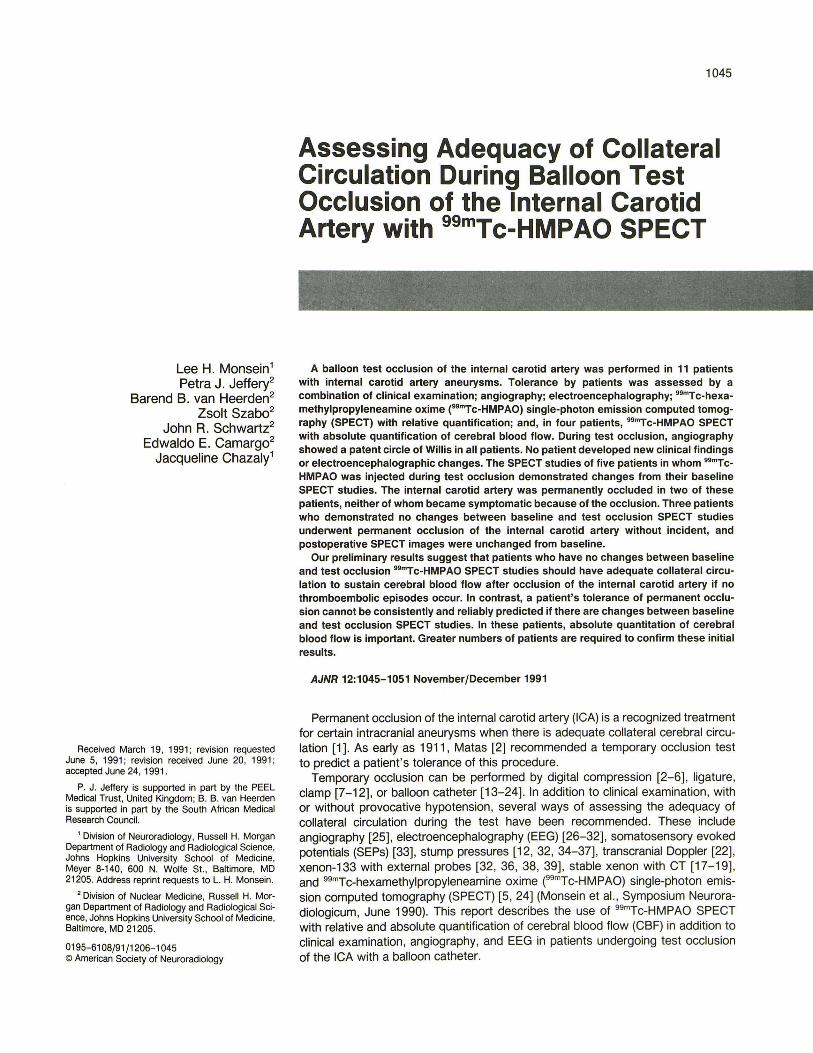
Relative quantification by means of region-of-interest (ROI) analysis was performed in all patients. A total of 14 ROis were placed on four transverse slices (Fig. 1 ). The slices were chosen by a single observer and were at the following levels: mid cerebellum, head of the caudate, thalamus, and mid parietal region . The same slices were used for each patient 's studies, and strenuous attempts were made to ensure comparative positioning of ROis . A ratio of counts per pixel within each ROI of the hemisphere on the side of occlusion to counts per pixel in the corresponding ROI of the contralateral hemisphere was calculated . The left/right hemisphere ratio in normal patients has been
Fig. 1.-Representative cerebral SPECT images illustrating four different levels of axial sections and regions of interest used in analysis.
A, Level of mid cerebellum. B, Level of head of caudate. C, Level of thalamus. D, Level of mid parietal lobe. CB = cerebellum, MCA2 = middle cerebral ar
tery territory (basal ganglia), ACA = anterior cerebral artery territory (medial frontal), MCA 1 = middle cerebral artery territory (lateral frontal), MCA3 = middle cerebral artery (high temporal), PCA = posterior cerebral artery territory (visual cortex), MCA4 = middle cerebral artery territory (parietal).
A
c
found to be approximately 1.00 ± 0.10 (2 SO) (Jeffery PJ , unpublished data). Hence, ratios different by more than 1 0% were considered to be important, particularly if all changes were in the same direction.
In four patients (cases 3 and 7-9), the CBF during test occlusion was calculated in ml ·1 00 g- 1
. min- ' by using a new method for absolute quantification of CBF with 99mTc-HMPAO SPECT data l40] . In one patient, the CBF during the baseline study was also calculated. In order to obtain absolute quantification, 99mTc-HMPAO was injected into an antecubital vein over 1 0 sec. A portable scintillation camera was positioned over the vertex of the head. Sixty frames (1 0 sec each) were acquired with a matrix size of 64 x 64.
An arterial catheter was used for collection of arterial blood. Arterial blood samples were withdrawn continuously at approximately 5-sec intervals for the first 2 min after the injection and every 1-2 min for the next 30 min. Blood samples in 0.5-ml volumes were immediately mixed with 2 ml of octanol and centri fuged for 3 min. Of the resulting supernatant, 0.5 ml was removed. The radioactivities of the total blood samples and of the samples extracted with octanol were measured in a well type scintillation counter.
The arterial input function was obtained by linear interpolation of the measured data points. A numeric integral of the input function was calculated for the first 5 min. The dynamic study was used to
CB
B ACA
MCA3 MCA4
PCA D
1048 MONSEIN ET AL. AJNR:12, November/December 1991
obtain activity curves from both hemispheres; these were then processed by deconvolutional analysis to obtain the impulse response function . The impulse response function value at 5 min was divided by the maximum of this function to obtain the retention fraction. The CBF was calculated as
where F = 1 mljmin, A9 , · = brain activity divided by the retention fraction, and A91 • = lipophilic arterial blood activity integrated over 5 min.
The brain activity was measured with the dedicated four-headed SPECT scanner (NeuroSpect 2000, Hitachi) 45 min after injection of 99mTc-HMPAO. The system was calibrated with a cylindrical phantom 22 em in diameter. Attenuation correction was performed after reconstruction with Chang's first-order method by using an attenuation coefficient of 0.11 em- ' . A representative slice including the basal ganglia and thalami was corrected for radioisotope decay and calibrated by using the calibration factor. This slice activity was then divided by the retention fraction , the 5-min integral of the input function, and the brain tissue density of 0.87 gjml to obtain the mean CBF slice. By using this mean CBF, all the individual pixel values of each brain slice were calculated and displayed in ml·1 00 g- ' ·min- ' tissue.
Permanent Occlusion
Two days to 6 weeks after the balloon test occlusion, permanent occlusion was performed in five patients, three of whom showed no change between baseline and test occlusion SPECT studies (cases 5, 6, and 10) and two of whom did show changes (cases 8 and 9). In four patients, a No. 9 Debrun latex balloon was introduced via the femoral artery and placed above and below the neck of the aneurysm. An additional balloon occluded the stump of the ICA. Balloons were filled with 30% contrast medium and detached from the catheter 15 min after inflation if there were no clinical or EEG changes with the inflated balloon in place. The patients were monitored in the intensive care unit for 24 hr after the procedure. In case 9, the ICA was ligated at surgery during an unsuccessful aneurysm clipping.
Six patients did not have occlusion of the ICA. One patient (case 4) sustained a stroke in the middle cerebral artery territory after premature balloon detachment during attempted endovascular treatment of the aneurysm. Another (case 11) underwent aneurysm clipping without carotid occlusion. In four patients (cases 1-3 and 7), permanent occlusion has not yet been attempted.
Results
Digital subtraction angiography performed during the balloon test occlusion revealed a patent circle of Willis in all patients. No significant changes in any patient were found in clinical examination, cardiovascular status, or EEG monitoring. There were no clinical complications.
The baseline SPECT studies were abnormal in two patients. One patient (case 3) had decreased perfusion in both frontal lobes of unknown origin. Another (case 5) had decreased perfusion in the left frontal lobe consequent to a previously attempted aneurysm surgery.
The SPECT studies of five patients (cases 3, 7-9, and 11), in whom 99mTc-HMPAO was injected during test occlusion, demonstrated changes from the baseline SPECT studies. In these patients, relatively decreased cerebral perfusion was
demonstrated on the side of the occlusion, and the most significant occlusionjnonocclusion cerebral hemisphere (ROI) ratios ranged from 0.81 to 0.86 (Table 2). In four of the patients, the hypoperfusion involved the anterior and middle cerebral artery territories on the side of the occlusion. One patient (case 9) had hypoperfusion in the territory of the left posterior cerebral artery during the balloon occlusion that was not seen on follow-up studies performed the next day or after final occlusion. The reason for this change is unclear. Perhaps it could have been caused by the vertebral artery injection that was done during the test occlusion procedure (i.e., vasospasm or embolus). The results of absolute quantification of CBF in four of these patients (cases 3 and 7-9) revealed the cerebral perfusion in the occluded hemisphere to be greater than 40 ml·1 00 g- 1
• min-' (normal, 40-60 ml·1 00 g- 1
• min- 1) in these patients (Table 3).
Follow-up studies were performed after the permanent occlusion of two patients in whom changes were seen between the baseline and test occlusion SPECT studies. One patient (case 9) developed an infarct in the left anterior temporal lobe after surgical manipulation, and the follow-up study revealed luxury perfusion surrounding the infarct. The followup study in case 8 had an appearance similar to that in the test occlusion study, and the patient remained asymptomatic.
Three patients (cases 5, 6, and 1 0) in whom no changes were demonstrated between baseline and test occlusion SPECT studies underwent permanent occlusion of the ICA and had an unremarkable course. Follow-up SPECT images in these patients were unchanged from baseline.
Discussion
The Cooperative Study of Intracranial Aneurysms and Subarachnoid Hemorrhage demonstrated that occlusion of the common carotid artery or ICA carries a 30% risk of ischemia of the ipsilateral cerebral hemisphere [1]. In 21% of these cases, onset of deficits is delayed for more than 48 hr after occlusion. Inadequate collateral circulation and thromboembolism are thought to be the two mechanisms causing ischemic complications [21]. Many authors have used temporary occlusion of the carotid artery in an attempt to identify those patients who will have inadequate cerebral circulation after permanent occlusion.
We use a balloon catheter for temporary occlusion of the ICA, a procedure well tolerated by patients. It is performed in the angiography suite, which allows imaging of the circle of Willis. That is more effective and reproducible than digital compression, which is recommended by some authors [2-6]. Surgical exposure of the carotid artery with application of a ligature or clamp has also been used but is more invasive. Theoretical advantages of gradual occlusion with ligatures or clamps vs abrupt occlusion with the balloon catheter have not been borne out [8, 41, 42]. CBF flow assessment with xenon-133 and external probes [32, 36, 38, 39] or stable xenon with CT scanning [17 -19] appears to improve the sensitivity to test occlusion over assessment with neurologic examination, stump pressure, EEG, SEP, or angiography alone and gives quantitative information. However, the equipment required for CBF determinations with these techniques

TABLE 2: Results of Relative Quantification of Cerebral Blood Flow in Patients Undergoing Test Occlusion of Internal Carotid Artery Aneurysms
Region of Interest/ Ratio by Case No.
Time of Measurement 2 3 4 5 6 7 8 9 10 11
Anterior cerebral artery territory {medial frontal)
Baseline 0.99 1.08 1.07 1.00 0.98 0.99 0.98 0.97 1.01 1.00 1.07 Balloon occlusion 1.00 1.08 0.92 1.00 0.97 1.00 0.93 0.89 1.00 0.97 0.99 Day after test occlusion 1.03 1.04 Follow-up 0.99 1.00 1.01 1.10 0.98
Middle cerebral artery territory {lateral frontal)
Baseline 1.01 1.00 1.13 1.05 0.93 1.05 0.94 1.00 1.02 1.00 0.96 Balloon occlusion 0.93 1.03 0.96 0.96 0.86 1.03 0.86 0.84 1.00 0.92 0.88 Day after test occlusion 0.95 1.10 Follow-up 0.86 1.02 0.78 1.26 1.04
Middle cerebral artery territory {basal ganglia)
Baseline 0.97 1.07 0.98 0.99 1.00 1.04 1.03 1.03 0.96 1.02 0.96 Balloon occlusion 1.05 0.99 1.09 0.94 0.95 0.97 0.93 0.96 0.98 0.96 0.86 Day after test occlusion 0.96 0.88 Follow-up 0.98 1.07 0.93 1.09 0.97
Middle cerebral artery territory {high temporal)
Baseline 0.96 0.97 1.06 0.96 0.95 1.06 0.98 1.03 1.06 0.92 0.97 Balloon occlusion 1.00 0.97 1.01 0.97 1.05 1.01 1.02 0.89 1.01 0.97 0.86 Day after test occlusion 0.97 1.04 Follow-up 0.91 1.10 0.96 1.16 0.99
Middle cerebral artery territory {parietal)
Baseline 0.96 1.11 1.03 0.96 0.99 0.96 0.97 0.96 0.95 0.94 0.99 Balloon occlusion 0.90 1.03 1.01 0.96 1.06 0.96 0.87 0.88 0.98 0.99 0.81 Day after test occlusion 1.00 1.01 Follow-up 1.04 1.04 0.87 1.05 0.98
Posterior cerebral artery {visual cortex)
Baseline 0.99 1.00 1.06 0.99 0.95 0.99 0.97 1.02 1.00 1.00 0.97 Balloon occlusion 0.99 0.98 1.08 1.01 1.01 1.03 0.88 0.84 0.82 1.08 0.93 Day after test occlusion 1.00 0.97 Follow-up 1.02 0.98 0.88 1.04 0.97
Note.-Ratios are of counts per pixel in region of interest on side of occlusion to counts per pixel in region of interest on contralateral side. Ratios in normal persons are about 1 .00 ± 0.1 0.
TABLE 3: Cerebral Blood Flow Values in Four Patients Whose SPECT Findings Changed Between Baseline and Test Occlusion
Cerebral Blood Flow {ml ·1 00 g-'. min- ')
Region of Interest/ Case 3 Case 7 Case 8 Case 9 Study Interval
Left Right Left Right Left Right Left Right
Anterior cerebral artery {medial frontal)
Baseline 46 45 Balloon occlusion 45 48 57 53 53 47 54 54
Middle cerebral artery territory {lat-eral frontal)
Baseline 43 43 Balloon occlusion 46 48 59 53 48 40 52 52
Middle cerebral artery territory {basal ganglia)
Baseline 48 50 Balloon occlusion 53 48 66 62 54 52 57 58
Middle cerebral artery territory {high temporal)
Baseline 46 44 Balloon occlusion 58 57 59 61 54 48 57 56
Middle cerebral artery territory (pa-rietal)
Baseline 42 41 Balloon occlusion 56 55 57 49 49 43 50 51
Posterior cerebral artery territory {visual cortex)
Baseline 44 44 Balloon occlusion 46 56 58 53 61 55 53 45
Cerebellum Baseline 54 54 Balloon occlusion 60 63 61 65 61 61 68 66
1050 MONSEIN ET AL. AJNR:12, November/December 1991
is not widely available, and there has been some concern about the effect of xenon itself on CBF [ 43]. External probe measurements with xenon-133 are easy to perform in the angiography suite and offer reproducible quantitative measurements but do not provide information about regional perfusion. Stable xenon with CT gives regional information but is cumbersome and requires transfer of the patient to another room with a carotid catheter in place [17 -19].
Matsuda et al. [5] have used 99mTc-HMPAO SPECT to assess cerebral perfusion during test occlusion of the carotid artery. Their study differed from our study in several aspects. They used manual compression of the common carotid for 5 min and injected 99mTc-HMPAO after only 30 sec of occlusion. The baseline study was obtained immediately prior to the occlusion, and then digitally subtracted from the second study. Brief occlusion of the carotid artery may not allow enough time for collateral circulation to be established. Compression of the common carotid artery may prevent collateral flow from the external carotid artery, which is of particular importance in elderly persons [37]. This may lead to false-positive results . Temporarily occluding the ICA with a balloon mimics the vascular effects of permanent occlusion, including leaving more time for establishment of collateral flow.
Our technique of assessing tolerance of ICA occlusion with 99mTc-HMPAO [24] (Monsein et al. , Symposium Neuroradiologicum, June 1990) has the advantages of being performed with readily available technology and offers regional information. The images in case 9 demonstrated the exquisite sensitivity of this technique along with its poor predictive value. Decreased perfusion was seen during the test occlusion in the territory of the left posterior cerebral artery, although the patient developed no symptoms and this abnormality was not seen again on subsequent studies.
The major disadvantage at the present time of using 99mTcHMPAO SPECT is that absolute quantification of CBF with this technique is difficult and not yet well validated. The
A B
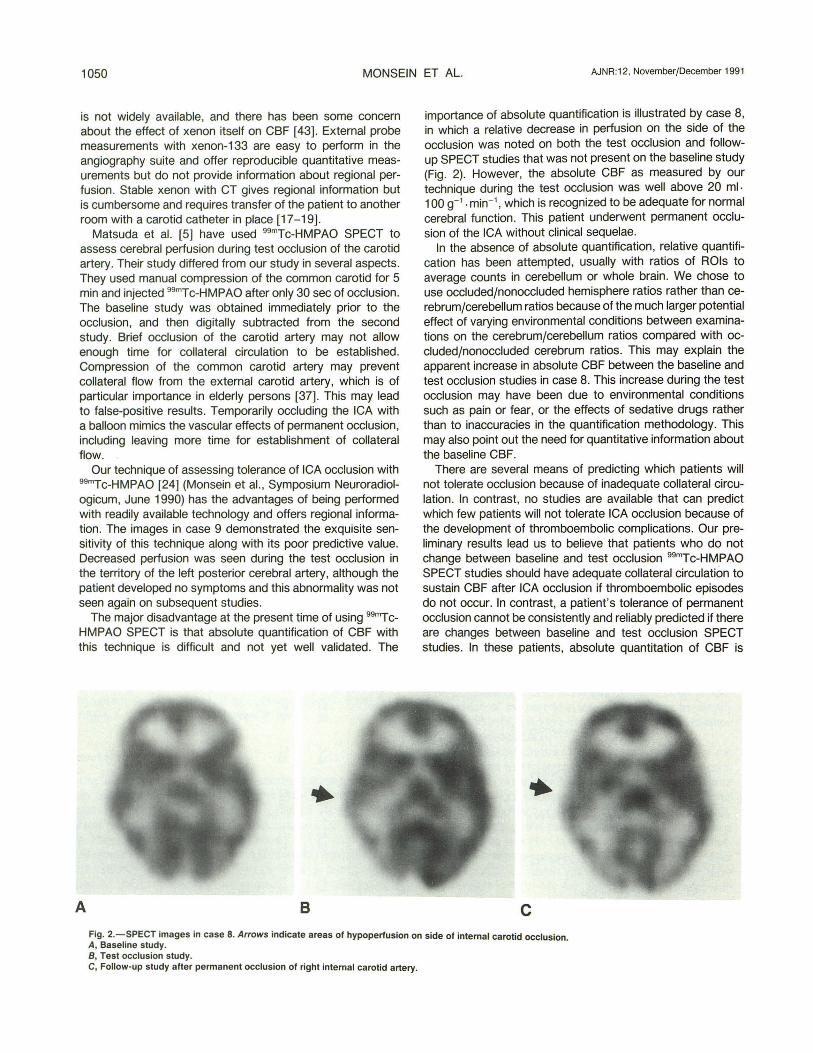
importance of absolute quantification is illustrated by case 8, in which a relative decrease in perfusion on the side of the occlusion was noted on both the test occlusion and followup SPECT studies that was not present on the baseline study (Fig. 2). However, the absolute CBF as measured by our technique during the test occlusion was well above 20 ml-1 00 g- 1 • min- 1 , which is recognized to be adequate for normal cerebral function . This patient underwent permanent occlusion of the ICA without clinical sequelae.
In the absence of absolute quantification, relative quantification has been attempted, usually with ratios of ROis to average counts in cerebellum or whole brain. We chose to use occludedjnonoccluded hemisphere ratios rather than cerebrum/cerebellum ratios because of the much larger potential effect of varying environmental conditions between examinations on the cerebrum/cerebellum ratios compared with occludedjnonoccluded cerebrum ratios. This may explain the apparent increase in absolute CBF between the baseline and test occlusion studies in case 8. This increase during the test occlusion may have been due to environmental conditions such as pain or fear, or the effects of sedative drugs rather than to inaccuracies in the quantification methodology. This may also point out the need for quantitative information about the baseline CBF.
There are several means of predicting which patients will not tolerate occlusion because of inadequate collateral circulation. In contrast, no studies are available that can predict which few patients will not tolerate ICA occlusion because of the development of thromboembolic complications. Our preliminary results lead us to believe that patients who do not change between baseline and test occlusion 99mTc-HMPAO SPECT studies should have adequate collateral circulation to sustain CBF after ICA occlusion if thromboembolic episodes do not occur. In contrast, a patient's tolerance of permanent occlusion cannot be consistently and reliably predicted if there are changes between baseline and test occlusion SPECT studies. In these patients, absolute quantitation of CBF is
c Fig. 2.- .SPECT images in case 8. Arrows indicate areas of hypoperfusion on side of internal carotid occlusion. A, Baselme study. 8 , Test occlusion study. C, Follow-up study after permanent occlusion of right internal carotid artery.

AJNR :12, November/December 1991 BALLOON OCCLUSION OF ICAs 1051
important. Greater numbers of patients are required to confirm these initial results .
ACKNOWLEDGMENT
We thank Glenda Davis for help in manuscript preparation.
REFERENCES
1. Nishioka H. Report on the cooperative study of intracranial aneurysms and subarachnoid hemorrhage. Section VIII , Part 1. Results of the treatment of intracranial aneurysms by occlusion of the carotid artery in the neck. J Neurosurg 1966;24:660-682
2. Matas R. Testing the efficiency of the collateral circulation as a preliminary to the occlusion of the great surgical arteries. Ann Surg 1911 ;53 : 1-43
3. Webster JE, Gurdjian ES. Carotid artery compression as employed both in the past and in the present. J Neurosurg 1958;15:372-383
4. Toole JF, Bevilacqua JE. The carotid compression test. Evaluation of the diagnostic reliability and prognostic significance. Neurology 1963;13: 601-606
5. Matsuda H, Higashi S, Asli IN , et al. Evaluation of cerebral collateral circulation by technetium-99m HM-PAO brain SPECT during Matas test: report of three cases. J Nucl Med 1988;29:1724-1729
6. McKissack W, Richardson A, Walsh L. Posterior communicating aneurysms. A controlled trial of the conservative and surgical treatment of ruptured aneurysms of the internal carotid artery at or near the point of origin of the posterior communicating artery. Lancet 1960;1: 1203-1206
7. Mount LA. Results of treatment of intracranial aneurysms using the Silverstone clamp. J Neurosurg 1959;16 :611-618
8. Heros RC . Thromboembolic complications after combined internal carotid ligation and extra- to intracranial bypass. Surg Neurol1984;21 :75-79
9. Drake CG. Giant intracranial aneurysms: experience with surgical treatment in 174 patients. Clin Neurosurg 1979;26: 12-95
10. Giannotta SL, McGillicuddy JE, Kindt GW. Gradual carotid artery occlusion in the treatment of inaccessible internal carotid artery aneurysm. Neurosurgery 1979;5:417-421
11 . Spetzler RF, Schuster H, Roski RA. Elective extracranial-intracranial arterial bypass in the treatment of inoperable giant aneurysms of the internal carotid artery. J Neurosurg 1980;53:22-27
12. Odom GL, Woodhall B, Tindall GT, Jackson JR. Changes in distal intravascular pressure and size of intracranial aneurysm following common carotid ligation . J Neurosurg 1962;19 :41-50
13. Berenstein A, Ransohoff J, Kupersmith M, Flamm E, Graeb D. Transvascular treatment of giant aneurysms of the cavernous carotid and vertebral arteries. Functional investigation and embolization. Surg Neural 1984;21 :3-12
14. Raymond J, Thron J. lntracavernous aneurysms: treatment by proximal balloon occlusion of the internal carotid artery. AJNR 1986;7 : 1087- 1092
15. Debrun G, Fox A, Drake F, Peerless S, Girvin J, Ferguson G. Giant unclippable aneurysms: treatment with detachable balloons. AJNR 1981;2:167-173
16. Serbinenko FA. Balloon catheterization and occlusion of major cerebral vessels. J Neurosurg 1974;41: 125-145
17. Erba SM, Horton JA, Latchaw RE, et al. Balloon test occlusion of the internal carotid artery with stable xenonfCT cerebral blood flow imaging. AJNR 1988;9:533-538
18. De Fries EJ, Sekhar LN , Horton JA, et al. A new method to predict safe resection of the internal carotid artery. Laryngoscope 1990;1 00 :85-88
19. Johnson DW, Stringer WA, Marks MP, Yonas H, Good WF, Gur D. Stable xenon CT cerebral blood flow imaging: rationale for and role in clinical decision making. AJNR 1991;12:201-21 3
20. Potts DG. Balloon test occlusion of the internal carotid artery with stable xenonfCT cerebral blood flow imaging. Invest Radiol 1989;24 :578-579
21. Fox AJ , Vinuela F, Pelz OM , et al. Use of detachable balloons for proximal artery occlusion in the treatment of unclippable cerebral aneurysms. J Neurosurg 1987;66: 40- 46
22. Braun IF, Battey PM, Fulenwider T, Per-Lee JH. Transcatheter carotid occlusion: an alternative to the surgical treatment of cervical carotid aneurysms. J Vase Surg 1986;4:299-302
23. Feaster SH , Powers A, Laws ER, Davis DO. Transcranial Doppler US as an alternative to angiography and balloon occlusion in estimating risk of carotid occlusion (abstr). Radiology 1990;177(P):281
24. Van Heerden BB, Monsein LH , Jeffery PJ , Debrun GM, Wagner HN, Camargo EE. Brain SPECT imaging to assess collateral circulation during trial balloon occlusion of internal carotid artery (ICA) (abstr). J Nucl Med 1990;31(P) :878
25. Jeffreys RV, Holmes AE . Common carotid ligation for the treatment of ruptured posterior communicating aneurysms. Neural Neurosurg Psychiatry 1971 ;34 :576-579
26. Trojaborg W, Boysen G. Relation between EEG, regional cerebral blood flow and internal carotid artery pressure during carotid endarterectomy. Electroencephalogr Clin Neurophysiol 1973;34: 61-69
27. Wells BA, Keats AS, Cooley DA. Increased tolerance to cerebral ischemia produced by general anesthesia during temporary carotid occlusion. Surgery 1963;54 :216-222
28. Perez-Borja C, Meyer JS. Electroencephalographic monitoring during reconstructive surgery of the neck vessels. Electroencephalogr Clin Neurophysiol1965;18 :1 61 - 169
29. Youmans JR, Kindt GW, Mitchell OC. Extended studies of direction of flow and pressure in the internal carotid artery following common carotid ligation. J Neurosurg 1967;27:250-254
30. Galbraith JG. Safeguards in carotid surgery. Surgery 1968;63 : 1010-1023 31 . Harris EJ , Brown WH , Pavy RN, Anderson W, Stone DW. Continuous
electroencephalographic monitoring during carotid artery endarterectomy. Surgery 1967;62 :441 - 447
32. Leech PJ , Miller JD, Fitch W, Barker J. Cerebral blood flow, internal carotid artery pressure, and the EEG as a guide to the safety of carotid ligation. J Neural Neurosurg Psychiatry 1974;37:854-862
33. Momma F, Wang AD, Symon L. Effects of temporary arterial occlusion on somatosensory evoked responses in aneurysm surgery. Surg Neural 1987;27:343- 352
34. Sweet WH , Bennett HS. Changes in internal carotid pressure during carotid and jugular occlusion and their clinical significance. J Neurosurg 1948;5: 178-195
35 . Bakay L, Sweet WH . Intra-arterial pressures in the neck and brain; late changes after carotid closure. Acute measurements after vertebral closure. J Neurosurg 1953;10:353-359
36 . Holmes AE , James IM, Wise CC. Observations on distal intravascular pressure changes and cerebral blood flow after common carotid artery ligation in man. J Neural Neurosurg Psychiatry 1971 ;34 :78-81
37. Heyman A, Tindall GT, Finney WHM, Woodhall B. Measurement of retinal artery and intracarotid pressures following carotid artery occlusion with the Crutchfield clamp. J Neurosurg 1960; 17 :297-305
38. Jennett WB , Harper AM, Gillespie FC. Measurement of regional cerebral blood-flow during carotid ligation. Lancet 1966;2 :1162-1163
39. Boysen , G. Cerebral blood flow measurements as a safeguard during carotid endarterectomy. Stroke 1971 ;2 : 1- 10
40. Szabo, Z, Monsein LH , Maruki Y, et al. Quantification of cerebral blood flow with Tc-99m HMPAO and SPECT (abstr) . Radiology 1990;177(P) :323
41 . Brice J, Dowsett D, Lower R. Some haemodynamic effects of carotid artery clamps. J Neural Neurosurg Psychiatry 1964;27 :580
42. Landolt M, Millikan CHL. Pathogenesis of cerebral infarction secondary to mechanical carotid artery occlusion. Stroke 1970;1 :52-62
43. Giller CA, Purdy P, Lindstrom WW. Effects of inhaled stable xenon on cerebral blood flow velocity. AJNR 1990;1 1: 177- 182
The reader's attention is directed to the commentary on this article, which appears on pages 1053-1 054.