Embed Size (px)
Citation preview

ASSESMENT OF KEY PARAMETERS ON THE PERFORMANCE OF THE
DELTOID MUSCLE IN REVERSE SHOULDER ARTHROPLASTY – A
MODELING AND SIMULATION BASED STUDY
NAVID ASLANI
Department of Design and Engineering, Faculty of Science and Technology, Bournemouth University, Talbot
Campus, Fern Barrow, Poole, Dorset, BH12 5BB, UK
SIAMAK NOROOZI
Department of Design and Engineering, Faculty of Science and Technology, Bournemouth University, Talbot
Campus, Fern Barrow, Poole, Dorset, BH12 5BB, UK
RICHARD HARTLEY
Bournemouth Royal Hospital, Bournemouth, UK
MIHAI DUPAC
Department of Design and Engineering, Faculty of Science and Technology, Bournemouth University, Talbot
Campus, Fern Barrow, Poole, Dorset, BH12 5BB, UK
PHILIP SEWELL
Department of Design and Engineering, Faculty of Science and Technology, Bournemouth University, Talbot
Campus, Fern Barrow, Poole, Dorset, BH12 5BB, UK
Reverse Shoulder Arthroplasty (RSA), in which anatomic concavities of glenohumeral joint
are inverted, is a popular treatment of arthritic shoulders with deficient rotator cuff. The
correct positioning of the glenohumeral centre of rotation and initial setting of the deltoid
length (Deltoid Tension) plays an important role in the outcome of the reverse shoulder
arthroplasty. A study of the key literature has shown that despite common use of RSA, its
biomechanical characteristics during motion are not fully understood. This study
investigates the influence of some of the key parameters on the intensity of the moment in a
shoulder after RSA during abduction in scapular plane. The kinematics after RSA are then
compared with the anatomic shoulder kinematics and differences are discussed.
Mathematical models of both the anatomical and reverse shoulder (RS) were developed in
MATLAB and in MSC ADAMS. The anatomical and RSA geometries were defined using
measurements obtained from previous X-Ray and MRI images of the shoulder girdle.
The results show that in RSA, the intensity of the moment generated in the glenohumeral
joint improves. However this improvement doesn’t show a constant trend and its intensity
can dramatically decrease in higher abduction.

Keywords: Rotator Cuff, Reverse Shoulder Arthroplasty (RSA), Deltoid, Shoulder
Kinematics, Simulation
Introduction
Reverse Shoulder Arthroplasty (RSA): A healthy shoulder has
specific characteristics in terms of range of motion, strength and
manoeuvrability it can provide. However, in an arthritic shoulder with rotator
cuff tear deficiency, characteristics are dramatically compromised. Rotator cuff
tear arthropathy (a condition that affects both shoulder strength and stability that
occurs when there is severe shoulder arthritis) can result in severe pain, and
difficulty in performing daily activities 1. There are many discussions about
shoulder implants 2,3
showing the Reverse Shoulder Arthroplasty (RSA) has
emerged as a an effective treatment of rotator cuff deficiencies in the shoulder.
Despite its success, this procedure has been associated with a relatively high
complication rate.
In RSA, as shown in Fig.1, the anatomic concavities of the
glenohumeral joint are inverted (by removing the humerus head and Scapula
fossa) to resolve the superior humeral head migration as a treatment of arthritis
in shoulders with rotator cuff deficiency, reducing pain and providing an
acceptable range of motion 4,5
.
The procedure shifts the centre of rotation medially relative to the
glenoid fossa to increase the effective lever arm and inferiorly Fig.1 to tension
the deltoid and improve its function 6,7,8
. Despite widespread use of RSA as
deficient rotator cuff treatment, a limited amount of data exists regarding the
functional outcome; especially with regards to the influence of biomechanical
and geometrical elements of the individual’s initial anatomic and post operation
prosthesis parameters. Currently there is no information on the importance of, or
the link between individuals’ initial, anatomical/geometry variations or
differences and the locating of the implant system during surgery on the
functional outcome of RSA.

Fig.1: Anatomic shoulder (left) Vs. Reverse shoulder (right)
Using a simulation it was possible to show the importance of the initial
geometrical differences in individuals and how it can inform the placement of
the implants. It also enables users to visualise the effect of lever arm beyond the
range of motion possible by the deltoid contraction. This will have an effect on
design of new implants glenoid to better control the lever arm length during
abduction.
Geometrical parameters of anatomic and prosthetic shoulder: X-Ray
and MRI images of the shoulder girdle shows a variety of morphology and
dimensional differences amongst individuals 9,10,11,12
.
Whilst no two individuals are the same, the normative range of motion
of the arm for all healthy individuals is practically the same. However, the
difference in anatomical sizes between individuals indicates there must exist an
optimised relationship between relative values of these key parameters in order
to obtain a defined abduction. All of these variables can play an important role
in the shoulder’s performance in terms of range of motion, strength and
manoeuvrability. After RSA the geometry and kinematics of the glenohumeral
joint will be totally changed. A standard RSA can result in different overall
geometry depending on the original size of the individual and also in terms of

the prosthesis size and positioning of prosthesis parts both on scapula and
humerus for each patient 13,14,15
.
Regarding information that can be extracted from X-Ray and MRI
images before and after surgery, it is possible to extract some key geometrical
parameters from such images as long as they are calibrated and are taken based
on a specific/standard protocol. These parameters can be used to define:
1) The origin of the deltoid on the acromion
2) The insertion points of the deltoid on the humerus
3) The centre of rotation of glenohumeral joint in 3D space
4) The available space and size of the glenoid sphere
all pre-operatively and post-operatively 16,17
.
The purposes of this study is to compare kinematic differences and to
determine the contributions of all the factors effecting the kinematics and
intensity of the total moment generated in the glenohumeral joint on the scapular
plane by the deltoid during abduction. This case study investigates and
compares both simulated normal anatomical and reverse shoulder in order to
evaluate the difference in their relative kinematics and the deltoid range of
possible active motion and their effect on the abduction levels. This study allows
the effect of change in the centre of rotation to be linked to the deltoid muscle’s
excess excursion where the deltoid is no longer able to generate the required
force to remain active beyond its normal operating range of contraction needed
to achieve full abduction in a normal shoulder.
This study demonstrated that all of the geometrical parameters, both in
normal shoulder and reverse shoulder (RS) either individually or in combination
can play an important role on the outcome of the surgery for each individual
11,18,16.
1. Methods
A musculoskeletal model of shoulder was developed in MSC ADAMS
software including glenohumeral joint, scapula, humerus and two segments of
deltoid (anterior and middle). Centre of rotation of GH joint was defined as
centre of humerus spherical head in anatomic shoulder and centre of prosthesis
glenoid in reverse shoulder. Both anterior and middle deltoids were modelled by

linear springs connected to the origin and insertion coordinates of deltoid on
scapula and humerus Fig.3. Springs deformation was considered as muscle
contraction and position and orientation of springs as deltoid force vector origin
and orientation 19,6
.
A mathematical model of shoulder was developed in MATLAB
software including all the geometrical dimensions of bones, GH joint, origin and
insertion coordinates of deltoid on humerus and scapula both for anatomic and
reverse shoulder. The distance between origin and insertion coordinates of each
muscle in 3D space was measured during arm abduction, as muscle length while
connecting points of these coordinates represent force vector origin and
direction.
Both models (musculoskeletal model of shoulder in MSC ADAMS and
mathematical model in MATLAB) revealed exactly same results.
These models were shown to be capable of creating a realistic
representation of the X-Ray and MRI image obtained from previous studies
9,10,11,12,16,17. The dimensions, coordinates, relative positions, perceived
displacements and centre of rotations, as well as trajectories and acceleration,
velocities and displacements can all be specified discretely and accurately
allowing for future parametric optimisation.
The shoulder is a very complex non-linear biomechanical system that
consists of three bones (the clavicle, humerus, and scapula) and four joints
(sternoclavicular, acromioclavicular, glenohumeral, and scapulothoracic) Fig.2.
Shoulder motion is generated by a combination of the motion of these four
joints.
The parameterised biomechanical model consists of the humerus, the
scapula, deltoid muscles, deltoid insertion points, position of Centre of Rotation
(COR) of Glenohumeral joint (GH) and deltoid tensioning before and after
surgery based on X-Ray and MRI images before and after RSA.
During arm abduction the Glenohumeral joint contributes 90o to 120
o
of abduction Fig.2. This leads to the assumption that after RSA the
glenohumeral joint must be able to achieve the same range of motion. This
outcome however, is not always guaranteed and the outcome varies between
individuals. This variation in the outcome after RSA is what prompted this

investigation. It is also assumed that after RSA the scapulothoracic joint still
provides 0o to 60
o degree of abduction which is independent of the deltoid
function 20,21,22
.
Fig.2: Shoulder Bones (left) Shoulder Joints (middle)
Scapulohumeral rhythm: in a full abduction of arm just around 120 degree of abduction is provided
by glenohumeral joint and the rest by scapulothoracic joint (right)
The origin (Centre of rotation), insertion coordinate and length of bones
and muscles were determined from anatomical and prosthetic measurements of
X-Ray and MRI images studies 9,10,11,12,16,17
.The wrapping of the muscle around
the bone was neglected due to previous studies which indicate wrapping takes
place in a limited range of motion (Low Abduction) 23,24,25,26
.
As shown in Fig.3 the fixed Oxyz coordinate system was used as a
centre of rotation of the glenohumeral joint on the scapula. The arm motion was
described in the scapular plane having θ as rotation of the glenohumeral joint 11
.
m, n, p = Distances between COR (Centre of Rotation) and origin of middle
deltoid on acromion along X,Y and Z axes, L = Distance between COR and
Insertion of deltoid on Humerus, β = angle between moment arm and force
vector of deltoid and F = Deltoid Force Vector.
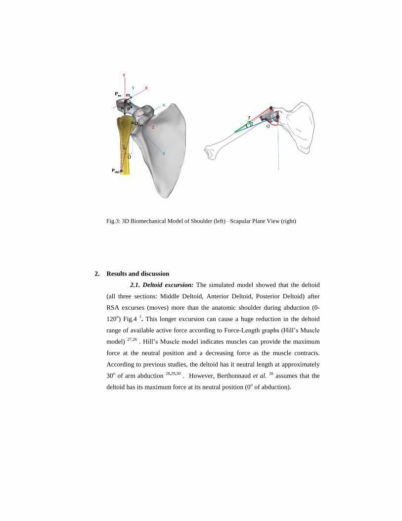
Fig.3: 3D Biomechanical Model of Shoulder (left) –Scapular Plane View (right)
2. Results and discussion
2.1. Deltoid excursion: The simulated model showed that the deltoid
(all three sections: Middle Deltoid, Anterior Deltoid, Posterior Deltoid) after
RSA excurses (moves) more than the anatomic shoulder during abduction (0-
120o) Fig.4
1. This longer excursion can cause a huge reduction in the deltoid
range of available active force according to Force-Length graphs (Hill’s Muscle
model) 27,26
. Hill’s Muscle model indicates muscles can provide the maximum
force at the neutral position and a decreasing force as the muscle contracts.
According to previous studies, the deltoid has it neutral length at approximately
30o of arm abduction
28,29,30 . However, Berthonnaud et al.
26 assumes that the
deltoid has its maximum force at its neutral position (0o of abduction).

Fig.4: Deltoid Length VS Abduction of GH joint (a) Middle Deltoid (b) Anterior Deltoid
red: anatomic / blue: reverse shoulder
This accelerating contraction of the deltoid in reverse shoulder causes
dramatic reduction in the available active force in it due to the muscle reaching
the end of its contraction range. In some cases the deltoid may exceed its
working range where it no longer can generate any force.
As shown in Fig.5, in Force-Length graph of middle deltoid, in
anatomic shoulder, when GH joint is in 0 degree of abduction, there exists little
passive force in the muscle having an available active force close to its
maximum. As the arm abducts more, the middle deltoid reaches its maximum
available active force at around 30o of abduction (where muscle reaches its
neutral length). After that the available active force decreases towards zero
(Maximum Abduction Angle). While in the reverse shoulder, the middle deltoid
starts its excursion approximately at the same muscle length of the anatomic
shoulder at 0o of abduction but it excurses more than the anatomic one during
abduction arriving almost at zero force 31
. Generally, the available maximum
active force of the Middle Deltoid in reverse shoulder is less than that of
anatomic shoulder during the same range of abduction angles. While for

Anterior Deltoid, the reverse shoulder can provide more force than the anatomic
one at the lower abduction angle. Effectively, the higher abduction angle follows
the same trend as that of the Middle Deltoid Fig.6.
Fig.5: (a) Available active force in middle deltoid VS muscle length (b) Available
active force in middle deltoid VS glenohumeral abduction angle
red: anatomic / blue: reverse shoulder / black: passive force in muscle
(Horizontal bars indicate deltoid excursion in anatomic and RS from 0o to 130o of
Glenohumeral joint abduction)
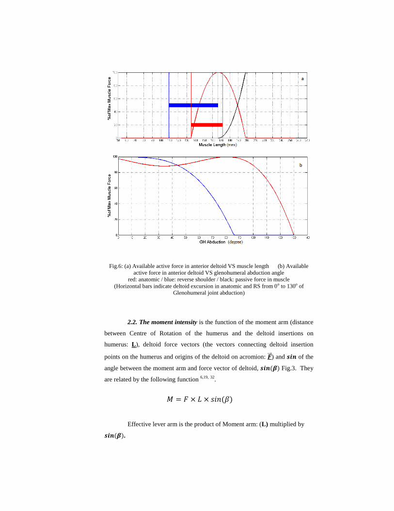
Fig.6: (a) Available active force in anterior deltoid VS muscle length (b) Available
active force in anterior deltoid VS glenohumeral abduction angle
red: anatomic / blue: reverse shoulder / black: passive force in muscle
(Horizontal bars indicate deltoid excursion in anatomic and RS from 0o to 130o of
Glenohumeral joint abduction)
2.2. The moment intensity is the function of the moment arm (distance
between Centre of Rotation of the humerus and the deltoid insertions on
humerus: L), deltoid force vectors (the vectors connecting deltoid insertion
points on the humerus and origins of the deltoid on acromion: ⃗⃗ ) and of the
angle between the moment arm and force vector of deltoid, Fig.3. They
are related by the following function 6,19,
32
.
Effective lever arm is the product of Moment arm: (L) multiplied by
.
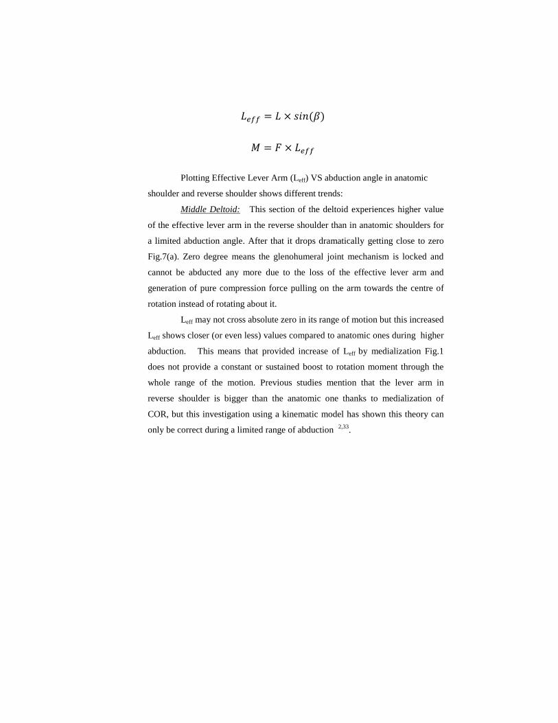
Plotting Effective Lever Arm (Leff) VS abduction angle in anatomic
shoulder and reverse shoulder shows different trends:
Middle Deltoid: This section of the deltoid experiences higher value
of the effective lever arm in the reverse shoulder than in anatomic shoulders for
a limited abduction angle. After that it drops dramatically getting close to zero
Fig.7(a). Zero degree means the glenohumeral joint mechanism is locked and
cannot be abducted any more due to the loss of the effective lever arm and
generation of pure compression force pulling on the arm towards the centre of
rotation instead of rotating about it.
Leff may not cross absolute zero in its range of motion but this increased
Leff shows closer (or even less) values compared to anatomic ones during higher
abduction. This means that provided increase of Leff by medialization Fig.1
does not provide a constant or sustained boost to rotation moment through the
whole range of the motion. Previous studies mention that the lever arm in
reverse shoulder is bigger than the anatomic one thanks to medialization of
COR, but this investigation using a kinematic model has shown this theory can
only be correct during a limited range of abduction 2,33
.

Fig.7: deltoid Effective Lever Arm VS Abduction of GH joint (a) middle deltoid (b)
anterior deltoid
red: anatomic / blue: reverse shoulder
For example, looking at Fig.7(a), at 10 degree of glenohumeral joint
abduction the effective lever arm in anatomic shoulder has a value equal to 20
mm while the prosthetic shoulder has an effective lever arm equal to 43 mm
which is more than twice that of the anatomic one of the same patient.
However, at 80 degrees of glenohumeral abduction the anatomic shoulder has an
effective lever arm equal to 40 mm while at this angle the prosthetic reverse
shoulder is 50mm. The results show that the rate of change of the lever arm
does not follow a linear trend and this medicalization Fig.1 in RS is only
advantageous during a limited range of abduction.
Anterior Deltoid: As shown in Fig.7(b), in reverse shoulder, Leff of the
Anterior Deltoid will increase at the beginning of abduction while its effect
decreases in higher abduction.
Fig.7 clearly shows the effect of the change in Lever arm length and its
dependency on the subtended angle (β).
In these graphs absolute values of Leff have been demonstrated. The
anatomic Leff graph has intersected zero effective lever arm at angle around 35o
of abduction and regarding absolute value before this angle Leff has had a

negative value which means it does not assist the arm to abduct in low abduction
while reverse shoulder has positive Leff during whole abduction which is useful.
2.3. Deltoid pre-tensioning as a solution? The Deltoid length can be
defined as the distance between origins of the deltoid on the acromion and its
insertion points on the humerus. In reverse shoulder arthroplasty the deltoid is
lengthened to increase its efficiency and it must be performed by increasing the
distance between the origin of the deltoid on the acromion and its insertion point
on the humerus 8,17,16,33
.
There are two solutions to increase this length which are:
(1) Increasing L Fig.3 (Distance between centre of rotation and insertion of
deltoid on humerus). L depends on the position of the socket of the
prosthesis on the humerus, diameter of the ball of the prosthesis and the size
of the spacers used. Increasing this value will result in middle deltoid
working range, a shift to the right on Force-Length graphs as shown in
Fig.8(a). As can be seen in Fig.8(b), increased L is not affecting Leff .The
same trend is observed for Anterior Deltoid as shown in Fig.8(c),(d) .
Fig
.8:
(a)
%o
f M
ax M
usc
le F
orc
e V
S M
usc
le L
ength
in
mid
dle
del
toid
.
Ho
rizo
nta
l b
ars
sho
w M
usc
le E
xcu
rsio
n.
Bla
ck g
rap
h r
evea
ls p
assi
ve
forc
e in
mu
scle
(b)
Eff
ecti
ve
Lev
er A
rm V
S G
H A
bd
uct
ion
(c)
%o
f M
ax M
usc
le F
orc
e V
S M
usc
le L
ength
in
ante
rio
r d
elto
id.
Ho
rizo
nta
l b
ars
sho
w M
usc
le E
xcu
rsio
n.
Bla
ck g
rap
h r
evea
ls p
assi
ve
forc
e in
mu
scle
(d)
Eff
ecti
ve
Lev
er A
rm V
S G
H A
bd
uct
ion
Red
: A
nat
om
ic /
Blu
e: R
ever
se S
ho
uld
er /
Gre
en:
Rev
erse
Sh
ou
lder
wit
h I
ncr
ease
d L

(2) Increasing n (distance between acromion and centre of rotation) Fig.3. This
requires placing the ball of prosthesis more inferiorly on scapula. As shown
in Error! Reference source not found.(a), when the COR is moved in the
reverse shoulder more inferiorly, initial middle deltoid length will be
increased while more excursion of deltoid occurs during abduction with a
shift in the working range of deltoid to right in the Force-Length graph. As
shown in Error! Reference source not found.(b) Leff trend will generally
improve still showing a drop in higher abduction. Excessive moving of
COR inferiorly can result over stressing that can result in stress fracture
34,35. Error! Reference source not found.(a),(b) show that deltoid
tensioning can optimise deltoid excursion in Force-Length graph with a
developed effect on the effective lever arm. The same trend is observed for
Anterior Deltoid as shown in Error! Reference source not found.(c),(d).

Fig
.9:
(a)
%o
f M
ax M
usc
le F
orc
e V
S M
usc
le L
ength
in
mid
dle
del
toid
.
Ho
rizo
nta
l b
ars
sho
w M
usc
le E
xcu
rsio
n.
Bla
ck g
rap
h r
evea
ls p
assi
ve
forc
e in
mu
scle
(b)
Eff
ecti
ve
Lev
er A
rm V
S G
H A
bd
uct
ion
(c)
%o
f M
ax M
usc
le F
orc
e V
S M
usc
le L
ength
in
an
teri
or
del
toid
.
Ho
rizo
nta
l b
ars
sho
w M
usc
le E
xcu
rsio
n.
Bla
ck g
rap
h r
evea
ls p
assi
ve
forc
e in
mu
scle
(d)
Eff
ecti
ve
Lev
er A
rm V
S G
H A
bd
uct
ion
Red
: A
nat
om
ic /
Blu
e: R
ever
se S
ho
uld
er /
Gre
en:
Rev
ers
e S
ho
uld
er w
ith
In
crea
sed
n
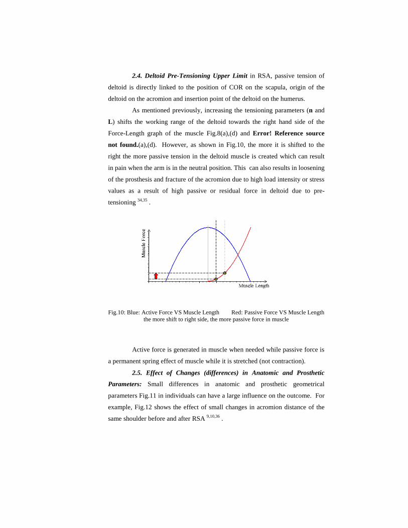
2.4. Deltoid Pre-Tensioning Upper Limit in RSA, passive tension of
deltoid is directly linked to the position of COR on the scapula, origin of the
deltoid on the acromion and insertion point of the deltoid on the humerus.
As mentioned previously, increasing the tensioning parameters (n and
L) shifts the working range of the deltoid towards the right hand side of the
Force-Length graph of the muscle Fig.8(a),(d) and Error! Reference source
not found.(a),(d). However, as shown in Fig.10, the more it is shifted to the
right the more passive tension in the deltoid muscle is created which can result
in pain when the arm is in the neutral position. This can also results in loosening
of the prosthesis and fracture of the acromion due to high load intensity or stress
values as a result of high passive or residual force in deltoid due to pre-
tensioning 34,35
.
Fig.10: Blue: Active Force VS Muscle Length Red: Passive Force VS Muscle Length
the more shift to right side, the more passive force in muscle
Active force is generated in muscle when needed while passive force is
a permanent spring effect of muscle while it is stretched (not contraction).
2.5. Effect of Changes (differences) in Anatomic and Prosthetic
Parameters: Small differences in anatomic and prosthetic geometrical
parameters Fig.11 in individuals can have a large influence on the outcome. For
example, Fig.12 shows the effect of small changes in acromion distance of the
same shoulder before and after RSA 9,10,36
.
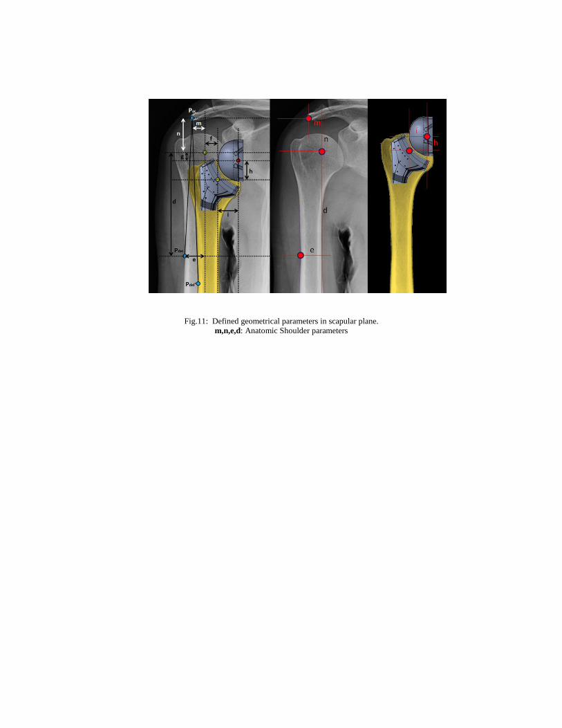
Fig.11: Defined geometrical parameters in scapular plane.
m,n,e,d: Anatomic Shoulder parameters

Fig
.12
: S
mal
l d
iffe
ren
ces
in n
(n
=1
5,1
7,1
9,2
1,2
3,2
5 m
m)
can
cau
se b
ig d
iffe
ren
ces
in m
usc
le e
xcu
rsio
n a
nd
Eff
ecti
ve
Lev
er A
rm d
uri
ng G
H a
bd
uct
ion
Lef
t: A
nat
om
ic S
ho
uld
er
R
igh
t: R
ever
se S
hou
lder
(Arr
ow
s sh
ow
in
crea
sin
g t
ren
d o
f n
fro
m1
5 t
o 2
5 m
m)

3. Discussion and results
A mathematical and 3D model of the anatomical and RSA were
developed using data from X-Ray and MRI images coming from previous
studies.
Different geometrical parameters were defined in each model (anatomic
and RS) Fig.11 and the effect of small changes in each one (in isolation) on the
overall kinematics and kinetics of the shoulder was investigated.
These parameters identify the Centre of rotation of glenohumeral joint
and the force vector of the deltoid knowing origin of the deltoid on the scapula
and its insertion point on the humerus both for the anatomic and RS shoulder
Fig.3.
The behaviours of the deltoid muscle was simulated and investigated
during glenohumeral joint full abduction both before and after RSA. The factors
considered for comparison of the functional outcome are classified as: 1) Deltoid
Excursion, 2) Effective Lever Arm, 3) Deltoid Tensioning and 4) Deltoid
Tensioning Upper Limit. Also, the differences these geometrical parameters
made on the outcome of the simulation were discussed.
In conclusion, regarding the fact that small differences in anatomic and
prosthetic parameters can affect dramatically the outcome of RSA, the
development of a structured approach/procedure for measurement is needed.
This would enable all measurement on all patients to be taken on similar or
identical planes to allow a more objective comparison of the Pre and post op
range of motion to be conducted. Also, to set the procedure for development of a
database and imaging techniques would allow better superposition of images
that allows RS to be located on the original image in order to take measurement
for various locations. Using a database of such images and optimisation of
kinematic graphs, the optimal decision could be made for individuals to have
possible maximum range of motion and least amount of pain.
4. REFERENCES
1. Meyer, D. C., Rahm, S., Farshad, M., Lajtai, G. & Wieser, K. Deltoid muscle
shape analysis with magnetic resonance imaging in patients with chronic rotator
cuff tears. BMC Musculoskelet Disord 14, 247 (2013).
2. Jazayeri, R. & Kwon, Y. W. Evolution of the reverse total shoulder prosthesis.
Bull. NYU Hosp. Jt. Dis. 69, 50–5 (2011).
3. Flatow, E. L. & Harrison, A. K. A history of reverse total shoulder arthroplasty.
Clin. Orthop. Relat. Res. 469, 2432–9 (2011).
4. Nam, D. et al. Reverse total shoulder arthroplasty: current concepts, results, and
component wear analysis. J. Bone Joint Surg. Am. 92 Suppl 2, 23–35 (2010).
5. Sanchez-Sotelo, J. Total shoulder arthroplasty. Open Orthop. J. 5, 106–14
(2011).
6. Terrier, a, Reist, A., Merlini, F. & Farron, A. Simulated joint and muscle forces
in reversed and anatomic shoulder prostheses. J. Bone Joint Surg. Br. 90, 751–6
(2008).
7. Kuechle, D. K. et al. The relevance of the moment arm of shoulder muscles with
respect to axial rotation of the glenohumeral joint in four positions. Clin.
Biomech. (Bristol, Avon) 15, 322–9 (2000).
8. De Wilde, L., Audenaert, E., Barbaix, E., Audenaert, A. & Soudan, K.
Consequences of deltoid muscle elongation on deltoid muscle performance: a
computerised study. Clin. Biomech. (Bristol, Avon) 17, 499–505 (2002).
9. Kircher, J. et al. Is there an association between a low acromion index and
osteoarthritis of the shoulder? Int. Orthop. 34, 1005–10 (2010).
10. Gu, G. & Yu, M. Y. Novel Physiotherapies Imaging Features and Clinical
Significance of the Acromion Morphological Variations. 2–5 (2013).
doi:10.4172/2165-7025.S2-003
11. Frankle, M. a, Teramoto, A., Luo, Z.-P., Levy, J. C. & Pupello, D. Glenoid
morphology in reverse shoulder arthroplasty: classification and surgical
implications. J. Shoulder Elbow Surg. 18, 874–85 (2009).
12. Werner, C. M. L. et al. Intermethod agreement and interobserver correlation of
radiologic acromiohumeral distance measurements. J. Shoulder Elbow Surg. 17,
237–40 (2008).

13. Gutiérrez, S., Comiskey, C. a, Luo, Z.-P., Pupello, D. R. & Frankle, M. a. Range
of impingement-free abduction and adduction deficit after reverse shoulder
arthroplasty. Hierarchy of surgical and implant-design-related factors. J. Bone
Joint Surg. Am. 90, 2606–15 (2008).
14. Gutiérrez, S., Keller, T. S., Levy, J. C., Lee, W. E. & Luo, Z.-P. Hierarchy of
stability factors in reverse shoulder arthroplasty. Clin. Orthop. Relat. Res. 466,
670–6 (2008).
15. Anglin, C., Wyss, U. P. & Pichora, D. R. Shoulder prosthesis subluxation:
Theory and experiment. J. Shoulder Elb. Surg. 9, 104–114 (2000).
16. Saltzman, M. D., Mercer, D. M., Warme, W. J., Bertelsen, A. L. & Matsen, F. a.
A method for documenting the change in center of rotation with reverse total
shoulder arthroplasty and its application to a consecutive series of 68 shoulders
having reconstruction with one of two different reverse prostheses. J. Shoulder
Elbow Surg. 19, 1028–33 (2010).
17. Lädermann, A. et al. Influence of arm lengthening in reverse shoulder
arthroplasty. J. Shoulder Elbow Surg. 21, 336–41 (2012).
18. Hoenecke, H. R., Tibor, L. M. & D’Lima, D. D. Glenoid morphology rather than
version predicts humeral subluxation: a different perspective on the glenoid in
total shoulder arthroplasty. J. Shoulder Elbow Surg. 21, 1136–41 (2012).
19. Kontaxis, a. & Johnson, G. R. The biomechanics of reverse anatomy shoulder
replacement – A modelling study. Clin. Biomech. 24, 254–260 (2009).
20. Lee, S. K., Yang, D. S., Kim, H. Y. & Choy, W. S. A comparison of 3D scapular
kinematics between dominant and nondominant shoulders during multiplanar arm
motion. Indian J. Orthop. 47, 135–42 (2013).
21. Ludewig, P. M., Hassett, D. R., Laprade, R. F., Camargo, P. R. & Braman, J. P.
Comparison of scapular local coordinate systems. Clin. Biomech. (Bristol, Avon)
25, 415–21 (2010).
22. Matsuki, K. et al. Dynamic in vivo glenohumeral kinematics during scapular
plane abduction in healthy shoulders. J. Orthop. Sports Phys. Ther. 42, 96–104
(2012).
23. Klepps, S. et al. A cadaveric study on the anatomy of the deltoid insertion and its
relationship to the deltopectoral approach to the proximal humerus. J. Shoulder
Elb. Surg. 13, 322–327 (2004).
24. Moser, T. et al. The deltoid, a forgotten muscle of the shoulder. Skeletal Radiol.
42, 1361–75 (2013).
25. Johnson, G. R., Spalding, D., Nowitzke, A. & Bogdukt, N. MODELLING THE
MUSCLES AND COORDINATE IMPLICATIONS OF THE SCAPULA DATA
AND FUNCTIONAL. J. Biomech. 29, (1996).
26. Berthonnaud, E., Morrow, M., Herzberg, G., An, K.-N. & Dimnet, J.
Biomechanical Model Predicting Values of Muscle Forces in the Shoulder Girdle
During Arm Elevation. J. Mech. Med. Biol. 10, 643–666 (2010).
27. Millard, M., Uchida, T., Seth, A. & Delp, S. L. Flexing computational muscle:
modeling and simulation of musculotendon dynamics. J. Biomech. Eng. 135,
021005 (2013).
28. FLAVIO ALMEIDA SALLES, A. Z. F. Isokinetic evaluation of eighteen male
patients submitted to surgical corretion of acute acromioclavicular luxation with a
minimum two-year follow-up. ACTA ORTOP BRAS 10, 19–24 (2002).
29. Terrier, A. et al. A musculoskeletal shoulder model based on pseudo-inverse and
null-space optimization. Med. Eng. Phys. 32, 1050–6 (2010).
30. Terrier, A., Reist, A., Vogel, A. & Farron, A. Effect of supraspinatus deficiency
on humerus translation and glenohumeral contact force during abduction. Clin.
Biomech. (Bristol, Avon) 22, 645–51 (2007).
31. Fridén, J. & Lieber, R. L. Quantitative evaluation of the posterior deltoid to
triceps tendon transfer based on muscle architectural properties. J. Hand Surg.
Am. 26, 147–55 (2001).
32. Schwartz, D. G. et al. The anterior deltoid’s importance in reverse shoulder
arthroplasty: a cadaveric biomechanical study. J. Shoulder Elbow Surg. 22, 357–
64 (2013).
33. Jobin, C. M. et al. Reverse total shoulder arthroplasty for cuff tear arthropathy:
the clinical effect of deltoid lengthening and center of rotation medialization. J.
Shoulder Elbow Surg. 21, 1269–77 (2012).
34. Schamblin, M. et al. In vitro quantitative assessment of total and bipolar shoulder
arthroplasties: a biomechanical study using human cadaver shoulders. Clin.
Biomech. (Bristol, Avon) 24, 626–31 (2009).
35. Boileau, P. et al. Revision surgery of reverse shoulder arthroplasty. J. Shoulder
Elbow Surg. 22, 1359–70 (2013).
36. Kappe, T., Cakir, B., Lippacher, S., Reichel, H. & Elsharkawi, M. Intraarticular
lesions in calcifying tendinitis: incidence and association with the acromion
index. Arch. Orthop. Trauma Surg. 131, 325–9 (2011).