Embed Size (px)
Citation preview
Seediscussions,stats,andauthorprofilesforthispublicationat:https://www.researchgate.net/publication/231897085
Assemblagesofmammalianhairandblood-feedingmidges(Insecta:Diptera:Psychodidae:Phlebotominae)inMioceneamber
ARTICLEinEARTHANDENVIRONMENTALSCIENCETRANSACTIONSOFTHEROYALSOCIETYOFEDINBURGH·MAY2005
ImpactFactor:0.94·DOI:10.1017/S0263593300001292
CITATIONS
11
READS
84
2AUTHORS:
EnriquePeñalver
InstitutoGeológicoyMinerodeEspaña
89PUBLICATIONS940CITATIONS
SEEPROFILE
DavidGrimaldi
AmericanMuseumofNaturalHistory
166PUBLICATIONS3,276CITATIONS
SEEPROFILE
Allin-textreferencesunderlinedinbluearelinkedtopublicationsonResearchGate,
lettingyouaccessandreadthemimmediately.
Availablefrom:EnriquePeñalver
Retrievedon:14January2016

Trunsuc~ions of rhe Rovrrl Socic.tj1 o f Edinb~rr~h: Earth Science.r, 96, 177-195, 2006 (for 2005)
Assemblages of mammalian hair and blood-feeding midges (Insecta: Diptera: Psychodidae: Phlebotominae) in Miocene amber
Enrique Pefialver and David Grimaldi
ABSTRACT: Five new fossil species of the Recent genus of blood-feeding sand flies Lut-omj~ia (Psychodidae: Phlebotominae) are described: L. Jilipalpis, L. miocena, L. paleopestis, L. scl~leei, and L. succini. All are preserved in Miocene amber from the Dominican Republic; today Hispaniola harbours only two known species of this genus. Recent Lutzomyia feed on a wide variety of terrestrial vertebrates, including reptiles, birds, and mammals. Three rare pieces of the amber are reported, two described in detail, which preserved assemblages of Lutzomyia swarms with strands of mammalian hair, indicating that a t least some of the fossil species were mammal feeders. Microstructure of the fossil hair offers little diagnostic evidence, but is very similar to that of insectivores in the Solenodontidae. Further preserved evidence indicates that the fossil midges swarmed about an arboreal nest or site of decayed wood that was worked by a mammal, but a t very specific times during formation of the amber. Other very rare Dominican amber pieces containing a flea and an ixodid tick also contain mammalian hairs of similar microstructure, together with Lutzonzyia sandflies, possibly reflecting the ectoparasite community of a Miocene mammal. This parasitic association has implications regarding the evolution of vectors of mammalian pathogens like Leishinanin and the study further reveals the extent of palaeobiological inference that is possible with amber.
KEY WORDS: Dominican Republic, fossil hair, Lirtzomjlia, mammalian hosts, microstructure, parasitic association, Solenodontidae, swarms.
Amber, or fossilised resin, is renowned for the fidelity with which it preserves delicate insects and other inclusions. This is most often appreciated as microscopic features like setae and sensilla on the external cuticle of terrestrial arthropods. While original reports of mkcromoIecuIar preservation in amber (DeSalle et nl. 1992: Cano e! (11. 1994) are now controversial (Austin et 01. 1997), amber does routinely preserve soft (non- chitinous) internal tissues, including cells, organelles like nuclei and mitochondria, and even the remains of endosymbiotic protists (Henwood 1992a, b; Grimaldi et al. 1994; Wier et al. 2002). Amber, thus, has arguably the finest ultrastructural preservation of any mode of fossilisation that is millions of years old. Another, less appreciated, aspect of amber's unique preservation concern the rare snapshots of behaviour, like copulating pairs of insects, females caught while ovipositing, insects snared in strands of spider webbing, and inquiline species captured with their hosts (Grimaldi 1996; Ross 1998; Weitschat & Wichard 2002). The most common inquilines in amber are ectoparasitic or phoretic mites clinging to their insect hosts (Weitschat & Wichard 2002), but there is also an occasional endoparasitic nematode caught bursting from an insect abdomen (Grimaldi et (11. 2002; Weitschat & Wichard 2002), o r ants captured with the mealybugs that they tended for the honeydew (Johnson et al. 2001). Such fossils provide minimum ages to some very intimate and obligate ecological associations.
Here, we report the exceptional preservation of swarms of blood-feeding (haematophagous) sand fly midges (Diptera: Psychodidae: Phlebotominae: Figs 1, 2a) preserved with very rare mammalian hair in Miocene amber from the Dominican Republic. Details of the preservation indicate that the midges
were feeding on the mammals whose hairs were preserved alongside them. We take this opportunity to describe five species of phlebotomines in this amber, in an effort to deter- mine the relationships and thus paleoecology of the midges. The midge-mammal taphonomic assemblage has significance regarding the history of association of phlebotomines with mammalian hosts, which is additionally significant since many phlebotomines transmit diseases of terrestrial tetrapods.
The Phlebotominae is a subfamily of approximately 500 described, primarily tropical and subtropical species in the family Psychodidae. Psychodidae are commonly called 'moth flies' for the fluffy appearance of the wings, which are covered with a thick vestiture of fine hairs. Phlebotomines themselves have the common name 'sand flies', which derives from the larval habit of breeding in soil, including sandy soil but almost always soils with significant organic content. Phlebotominae are sometimes classified in a separate family (e.g., Lewis 1971), but they are clearly closely related to all other psychodids so we are using the subfamily rank for them here. The only other psychodids besides phlebotomines that feed on blood are Sycoracinae and, like most other haematophagous Diptera, only the adult females feed on blood, which is required for the eggs to develop, though some species feed also on nectar. The main genera are Phlebotom~~s and Sergentomyia in the Old World and Lutzomyia in the New World. All three genera feed on amphibians, lizards, tortoises, birds, and mammals, with some host specificity shown by certain species (Lewis 1974). Species of Pl~lehotomus, for example, mostly feed on gerbils, hyraxes, murids, canids, and humans, and Sergentoinyia feeds mostly on reptiles and amphibians (Lewis 1974). L~tzomjirr has a broader palette than the other two genera. Phlebotomus
ENRIQUE PENALVER AND DAVID GRIMALDI
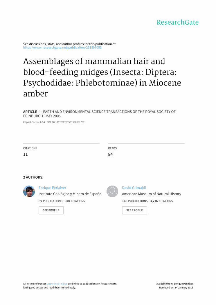
Figure 1 Photomicrograph of the holotype male Lufzomyia paleopestis sp. n. (AMNH DR6-146) in Miocene amber from the Dominican Republic.
BLOOD-FEEDING MIDGES AND HAIR IN AMBER 179
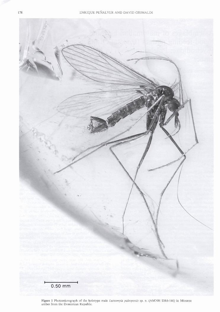
Figure 2 Photomicrographs of Miocene Lurzomyin in Dominican amber: (a) Female Lutzomyin sp. (AMNH DR14-567). The female is bloated, probably with a blood meal. No hairs o r other midges were preserved in the piece with the female; (b) Detail of female in (a), showing elements of the biting mouthparts, which are rarely displayed in the amber midges; (c) AMNH DR14-1268, which contains a small swarm of 6 males Lutzomyia filipalpis sp. n. and particles of wood (d), indicating the phlebotomines swarmed about areas of rotten wood on the amber tree.
and Lutzomyia further differ in that the former is typically found in open habitats such as savannas, scrub, and desert, while the latter is common in tropical forests. Also, of the species that transmit human diseases, Phlebotomus is notorious for the transmission of visceral leshmaniansis (or 'kala azar') in various regions of the Palearctic, while Lurzomyia transmits cutaneous leshmaniasis in large areas of the neotropics (reviewed by Lewis 1974; Killick-Kendrick 1999).
Leishmaniasis is caused by Leishmania, which is a trypano- somatid. Leishmania promastigotes develop in the fly in either the gizzard-like proventriculus, the mid gut (cardiac stomach), or the hind gut, depending on the species of parasite and fly, but they all migrate through the esophagous and fine mouth- parts of the fly into the secondary hosts' bloodstream when the fly is drawing blood. Approximately 30 species of phleboto- mines are believed to transmit 30 species of Leishmania in mammals, with some of the fly species transmitting several
species of these trypanosomes (Killick-Kendrick 1990). The most virulent Leishmanias in humans are L. donovuni in Asia, and L. mexicana and L. braziliensis in the American tropics. Phlebotomines transmit a host of other diseases as well, including bartonellosis (Oroya fever or Carrion's disease) in South America, which is caused by Burlonella bacillijormis (an a-proteobacterium) and transmitted by Lutzomyia. Certain species of Lulzomyia also transmit reptilian malaria parasites (Plasmodium), Trypanosoma, coccidians and hemogregarines (Ayala 1973; Warburg et al. 1991; Killick-Kendrick 1999), and both genera transmit arboviruses.
The oldest Phlebotominae are preserved in amber from the Early Cretaceous of Lebanon, which is approximately 135- 120 Ma, depending on the outcrops from where the amber derived. There are two species in the extinct genus Phleboto- miles in Lebanese amber, which Hennig (1972) proposed as a sister group to most of the Recent phlebotomines, and in fact

180 ENRIQUE PENALVER AND DAVID GRIMALDI
the species are very similar to the living genus Phlebotomus (Hennig 1972). Azar ef al. (1999) described two additional genera in this amber that were attributed to the Phleboto- minae. Given the diversity of the subfamily in the Early Cretaceous it is quite likely that this group originated in the Late Jurassic. Despite the name, Eophlebotomus Cockerell, 1917 in mid-Cretaceous amber from Myanmar is actually not closely related to phlebotomines (Hennig 1972; Duckhouse 2000; Azar et al., 2003), and this genus is also common in Albian amber from Alava, Spain (A. Arillo, pers. comm., 2005). There are, however, undescribed phlebotomines in Burmese amber. No phlebotomines occur in amber from the Albian of Spain (A. Arillo, pers. comm. 2005) o r France, nor in Turonian-aged New Jersey amber (Grimaldi et al. 2000), which is clearly a taphonomic effect. The genus Plzlebotomus occurs in amber from the Upper Eocene of the Baltic region and Lufzomyia (the genus we report on here) is preserved in Late Oligocene amber from southern Mexico (Quate 1963) and has previously been reported in Miocene Dominican amber (Poinar & Poinar 1999; Brazil & Filho 2002). Thus, the fossil record of phlebotomines is long but not very extensive, particularly for the Cenozoic. The earliest ones probably fed on early mammals as well as birds and other theropod dinosaurs, and when passerine birds and placental mammals radiated in the Late Cretaceous and early Cenozoic, they became the predominant hosts of these flies.
1. Materials and methods
Fossiliferous amber from the Dominican Republic on the Caribbean island of Hispaniola derives from outcrops in the mountains 10-20 km north and northeast of Santiago (reviewed in Grimaldi 1995). It is excavated by locals who sell the crude amber to dealers in Santiago and Santo Domingo, and so it is widely available commercially. The material for the current study was acquired through purchase, and as such its exact provenance within the outcrops of Dominican amber mines is unknown, but authenticity is certain based on UV fluorescence, a hot-needle test (which easily identifies plastics) and microsco~ic examination of the amber and its inclusions. Specifically, the manner of preservation, the types of syninclu- sions, and the fact that several pieces of the amber indepen- dently possessed similar inclusions and preservation all indicate that these pieces were not manufactured. Dominican amber is mid-Miocene, 15-20 Ma in age (Iturralde-Vinent & MacPhee 1996), though some popularised accounts of Dominican amber repeatedly and erroneously refer to some of this material as being Eocene (e.g., Poinar 1992; Poinar & Poinar 1999; see the discussion in Grimaldi 1995). This amber was formed by an extinct species of Hymenoea tree (Leguminosae: Caesalpinioidea), living species of which today exude copious resin when the bark is wounded by storms or boring insects (Langenheim 2003). Hymenaea today grows primarily in coastal habitats of western Central America, the Caribbean and throughout the Amazon Basin, with one species living in eastern Africa (Langenheim 2003).
Pieces of fossiliferous amber were prepared according to protocols in Nascimbene & Silverstein (2000). Smaller pieces containing single or only a few inclusions of phlebotomines were embedded in a high quality casting epoxy (Buehler, Inc.), which allowed the pieces to be sliced and polished very thin and close to the surface of the inclusions for optimal viewing. These specimens were applied to a glass microscope slide and studied under stereomicroscopy and up to 200 x magnifica- tion using a compound microscope. Pieces with hair and small
these could not be prepared. They were studied while immersed in mineral oil, which reduces the optical distortion caused by the curvature of the amber surface. Photomicrogra- phy used the Infinity"' K-2 long distance microscope and the M i c r O p t i c ~ ~ ? fibre optic flash unit (www.microptics.com). Two pieces with multiple midges and hairs in each were mapped by sketching the pieces, specimen by specimen, using a drawing tube attached to a Wild M-3 stereoscope. The system of wing venation generally follows the standard adopted for phlebotomine systematics (e.g., Young & Duncan, 1994), which itself is a slightly modified version of the Comstock-Needham system. Specimens are housed in the American Museum of Natural History in New York (AMNH), and in the Staatlichen Museum fiir Naturkunde in Stuttgart, Germany (SMNS).
2. Systematic paleontology
Class Insecta Linnaeus, 1758 Order Diptera Linnaeus, 1758
Family Psychodidae Newman, 1835 Subfamily Phlebotominae KertCsz, 1903
Genus Lutzomyia Franqa, 1924 Pintomyici Galati, 1995 (as genus). NEW COMBINATION.
Lutzomyia (Pintomyia) falcaorum (Brazil & Filho) Pinfomyici (Pifanomyia) falcaor~im Brazil & Filho, 2002: 502. NEW COMBINATION.
Diagnosis: Distinguished from the other fossil species described below by the following features of the male genitalia: presence of a spine on the paraphysis ("paramere" in Brazil & Filho 2002), and like L. poleopestis sp. n. (described below) there is a tuft of setae on the inner basal surface of the gonocoxite. Unlike paleopestis the paraphysis and lateral lobe are significantly shorter than the gonocoxite (paraphysis is equal in length and the lateral lobe longer in paleopestis).
Comments: We did not examine any material of this species, but relied on the original description of Brazil & Filho (2002). Though the illustration in that paper is extremely basic, the more detailed, written description clarifies some of the ambiguities. Brazil and Filho adopted the classification of Galati (1995) in placing this species, even though they admit that "the adoption of the genus Lutzomyicl [as in Young and Duncan's 1994 study] for the majority of the neotropical sand fly species can be justified" (Brazil & Filho 2002, p. 503). Indeed, the splitting of a monophyletic Lutzomyia into formal taxonomic units (i.e., eight genera, including Pintomyia, and nine subgenera) does not serve nomenclatural stability, particularly when DNA sequences indicate some ambiguity about species relationships (Beati et 01. 2004). Filho & Brazil (2003) mentioned additional, yet undescribed phlebotomine species in Dominican amber in the "genera" (sensu Galati 1995) Micropygomyia and Trichopygomyia.
Lutzomyia (Micropygomyia) Jilipalpis sp. n. (Figs 3a, f; 4a)
Derivation of name: Fili, meaning thread, and palpis, in reference to the long, fine apical palpomere.
Material: AMNH DR8-23 (1 d), DR8-169 (1 d), DR8-170 (1 d), DR8-171 (1 d), DRIO-276 (1 d), DR14-1268 (688) ; DR14-1338 (1 d), DR15-39 (7 dd) , DR12857 (2 dd) . Ho~o- type: AMNH DR8-167 (1 8).
Diagnosis: very long apical palpomere, nearly equal to swarms were larger and, since they were filled with inclusions, combined length of palpomeres 2-4, proboscis reaching to
BLOOD-FEEDING MIDGES AND HAIR IN AMBER 181
approximately middle of palpomere 3. Male genitalia: one apicallone subapical spine on gonostylus, two lateral spines; gonocoxite without tuft of setae on medial surface; lateral lobe with apex approximately at same level as apex of gonocoxite, with sparse setae; paraphysis with broad, setose base, narrow apically. Wing with base of vein R, and r-m crossvein very basal, nearly at same level as apex of Sc.
Description: Based mainly on the holotype specimen. Palpomeres measuring from I to 5: 0.04 mm; 0.14 mm; 0.13 mm; 0.09 mm and 0.3 1 mm (from AMNH DR14-1338); palpal formula 1.4.3.2.5. Antennae with ascoids simple, present a t least on flagellomeres I-V; ascoids short, reaching approximately to middle of home flagellomere. Newstead's spines not visible on palps. Abundant thoracic bristles present. Wings 1.92 mm length, 0.62mm width (L/W=3.10). Wings with abundant setae. Length of wing vein sections: a, 0.46 mm; p, 0.26 mm; 6, 0.10 mm; y, 0.29 mm. End of vein CuA, more distal than the end of R, and the bifurcation of R,+R,. Length of femur, tibia and basitarsus: foreleg (from AMNH DR8-23), 0.77 mm, 0.82 mm, 0.78 mm; midleg, 0.82 mm, 0.98 mm, 0.55 mm; hindleg, 0.82 mm, 1.12 mm, 0.51 mm. Distal spur on hind femur. Genitalia: gonocoxite 0.26 mm length and 0.06 mm width, gonostylus 0.14 mm length and 0.02 mm width, paraphysis simple (not divided), broad basally and 0.18 mm length, lateral lobe (not inflated) 0.27 mm length and 0.03 mm width.
Comments: This is the most common species in Dominican amber in terms of the number of identifiable (male) specimens, and it occurs in several pieces. Significant variation occurs in the wing of this species, with a LIW=3.10 in the holotype, to 3.63 in specimen AMNH DR8-171. Lutzonvia filipalpis has the distinctive characters of the subgenus L. (Micropygomyirr): antennal ascoids simple, those on flagellomere I1 not reaching to the distal end of that segment; palpomere 5 longer than palpomere 3; style with four spines and no subterminal seta; and the paraphysis simple. Lutzornyia (Micropygomyia) jili- palpis sp. n. differs from Phlebotomus paternus Quate, 1963 in Oligocene Mexican amber (which is actually a Lutzomyia) mainly on the basis of four spines on the gonostylus (five in L. pat ernus).
Lutzomyia succini sp. n. (Figs 3b, g; 4b)
Derivation of name from the Latin word succinurn (=amber).
Material: Holotype: AMNH DR-5-34 (1 8). Diagnosis: Proboscis reaching to apex of palpomere 3,
palpomere 5 very large (nearly equal to combined length of palpomeres 2 + 3 and half of palpomere 4). Male genitalia: gonostylus with single apical spine, single spine on mesal margin (on a tubercle near base of segment), one lateral spine; gonocoxite without basal tuft; lateral lobe with apex extended well beyond level of apex of gonocoxite, with numerous setae; paraphysis with sparse pubescence at apex.
Description: Palpomeres measuring from I to 5: 0.01 mm; 0.10 mm; 0. l l mm; 0.07 mm and 0.25 mm; palpal formula 1.4.2.3.5 (palpomere 2 a little shorter that 3). Newstead's spines not visible on palps. Three dorsal, long setae on the base of mouthparts. Antennal ascoids not visible. Thoracic bristles sparse. Wings 1.55 mm length and 0.45 mm width (LIW= 3.44). Wings with abundant setae. Length of wing vein sections: a, 0.31 mm; P, 0.19 mm; 6, 0.03 mm; y, 0.22 mm. Bifurcation of vein M I + , nearest to the level of bifurcation of R,,, Length of femora, tibiae and basitarsus: foreleg, 0.67 mm, 0.69 mm, 0.41 mm; midleg, 0.67 mm, ? mm, ? mm; hindleg, 0.68 mm, 1.01 mm, ? mm. Genitalia: gonocoxite
0.23 mm length and 0.06 rnrn width, gonostylus 0.10 mm length and 0.03 mm width, paraphysis simple (not divided) and 0.16 mm length and 0.04 mm width, lateral lobe 0.34 mm length and 0.03 mm width. Lateral lobe not inflated.
Comments: The holotype is a complete specimen, which is missing only a leg and part of three tarsi. This species seems to belong to the pilosa species group due to the combination of the following characters: palpomere 5 longer than palpomeres 3+4, gonostyle with three strong spines and small median setae, subterminal seta absent and paraphysis simple, but differs by the lateral lobe being long in comparison with the paraphysis (Young & Duncan 1994). Lutzomyia succini sp. n. has various characteristics similar o r identical to L. miocenu sp. n., mainly with respect to the genitalia, such as the shape of the gonocoxite, gonostylus, paraphysis and lateral'lobe, the presence of three spines on gonostylus, the setation of gono- coxite and gonostylus and the lateral lobe (Fig. 4b, c). The differences between these two new species are that L. succini has a larger body size, different palpal formula, palpomere 5 is longer, wing vein section 6 is 0.03 (vs. - 0.02 mm), the bifurcation of vein M I + , is nearest to the level of the bifurca- tion of R,,,, and the paraphysis has fewer setulae. Ltltzomyia succini has a palp very similar those of L. jilipalpis and L. paleopestis, except that palpomere 3 is smaller than palpomere 2 in L. jilipalpis and differs in the relative length of palpomere 5.
Lutsom~~ia n~iocena sp. n. (Figs 3c, h; 4c)
Derivation of name: In reference to the geological period of the amber.
Material: Holotype: AMNH DR6-147 (1 8 ) Diagnosis: Palp shorter, proboscis reaching nearly to apex
of palpomere 3, palpomere 4 very short (half the length of segment 3), length of palpomere 5 equal to combined length of palpomeres 3+4. Male genitalia: gonostylus with single apical spine, single spine on mesal margin (on a tubercle near base of segment), one lateral spine; gonocoxite without basal tuft; lateral lobe with apex extended well beyond level of apex of gonocoxite, with numerous setae; paraphysis with sparse pubescence at apex.
Description: Palpomeres measuring from 1 to 5: 0.02 mm; 0.09 mm; 0.09 mm; 0.05 mm and 0.14 mm; palpal formula 1.4.3.2.5 (palpomere 2 slightly longer than 3). Newstead's spines not visible on palps. Antennal ascoids visible on flagellomeres I-VI; ascoids short and simple. Thoracic bristles sparse. Wings 1.24 mm long, 0.35 mm wide (L/W=3.54). Wings with abundant setae. Length of wing vein sections: a, 0.22 mm; p, 0.14 mm; 6, - 0.02 mm; y, 0.22 mm. Bifurcation of vein M , , , nearest to level of separation of R,,, and R,. Length of femur, tibia and basitarsus: foreleg, 0.49 mm, 0.52 mm, 0.34 mm; midleg, 0.52 mm, 0.64 mm, 0.36 mm (any hindleg in articulation). Genitalia: gonocoxite 0.29 mm long, 0.06 mm wide; gonostylus 0.14 mm long, 0.03 mm wide; para- physis simple (not divided), 0.18 mm long, 0.04 mm wide; lateral lobe 0.34 mm long, 0.02 mm wide. Lateral lobe not inflated.
Comments: The holotype specimen is missing a leg and three legs are disarticulated. This species seems to belong to the pilosa species group due to the same combination of characters as in L. succini sp. n., as well as by the presence of simple antennal ascoids. Like L. szrccini sp. n., this species has a lateral lobe that is long in comparison to the paraphysis, which is a feature inconsistent with the pilosa species group (see Young & Duncan 1994).

ENRIQUE PENALVER AND DAVID GRlMALDl
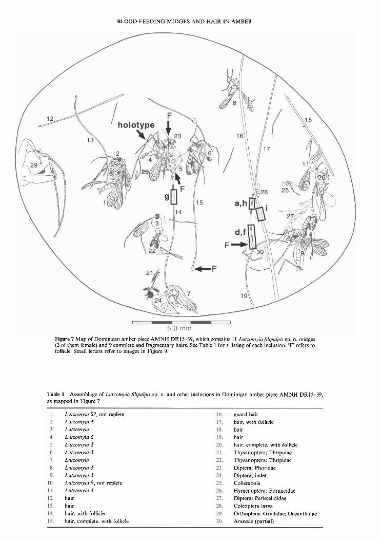
'a a. filipalpis b. succini C . miocena d . paleopestis e. schleei
i. paleopestis I Figure 3 Heads ( a x , lateral view) and wings (f-j) of new species of Miocene Lutrorn,vic~. Heads to same scale; wings not to same scale.
Lzrtzomvia paleopesris sp. n. (Figs 1; 3d, i; 4d)
Derivation of name: L., meaning "old pest." Material: AMNH DR14-554 (1 8). Holotype: AMNH
DR6-146 (1 d) Diagnosis: Apical palpomere about same length as pal-
pomeres 2+3, proboscis reaching to apex of palpomere 3. Male genitalia: gonostylus with single apical spine, mesa1 spine (blunt apices); gonocoxite with basomesal tuft of fine setae; paraphysis clavate (apex slightly clubbed, finely setulose), long, length equal to that of gonocoxite; lateral lobe very slender, setose, with dense fine setae at apex; aedeagus long, apically forked. Wing long and narrow, LIWz3.73.
Description: Based mainly on holotype. Palpomeres 1 to 5: 0.03 mm; 0.10 mm; 0.12 mm; 0.07 mm and 0.19 mm; palpal
formula 1.4.2.3.5. Newstead's spines not visible on palps. Antennae with simple, short ascoids, present at least on flagellomeres I-X. Thoracic bristles present. Wings 1.68 mm long and 0.45 mm wide. Wings with abundant setae. Length of wing vein sections: a, 0.32 mm; (3, 0.20 mm; 6, 0.00 mm; y, 0.24 mm. Length of femur, tibia, and basitarsus: foreleg, 0.65 mm, 0.73 mm, 0.42mm; midleg, 0.61 mm, 0.83 mm, 0.48 mm; hindleg, 0.70 mm, 0.86 mm, 0.52 mm. Genitalia: gonocoxite 0.23 mm long, 0.05 mm wide; gonostylus 0.10 mm long, 0.03 mm wide; paraphysis simple (not divided), 0.19 mm long; lateral lobe not inflated, 0.28 mm long, 0.02 mm wide.
Comments: The holotype of this species is a complete specimen. This species belongs to the verrucarurn species group due to a combination of the following characters: antenna1 ascoids simple, palpomere 5 longer than palpomere 3, coxite with a tuft of simple setae, style with two spines, paraphysis

BLOOD-FEEDING MIDGES AND HAIR IN AMBER
a. filipalpis ' , /
C . miocena
Figure 4 Male genitalia of new fossil species of Lulzomyia.
simple, without arms or extensions. Moreover, two strong Lutzomyia (Trichopygomyia) schleei sp. n. spines and one small subterminal seta on the gonostyle indicate (Figs 3e, j; 4e; 5 ) that this species belongs to the serrana series of the verrucarum species group. Many species in this group occur in montane Derivation of name: Patronym for Dr Dieter Schlee, forests (Young & Duncan 1994). formerly of the Stuttgart Museum, who had brought this

ENRIQUE PENALVER A N D DAVID GRIMALDI
Figure 5 Allotype (a) and holotype (b) of Lutzomyirr schlrri sp. n. (to the same scale), and details of the female head and genitalia, and male tibia1 spur. The location of the type specimens in the amber piece is indicated in Figure 8 and listed in Table 2.
exceptional piece of amber to the attention of Grimaldi and made the specimen available on loan.
Material: SMNS Do-5514-M (8 SS, 37 99 & 8 sex indet.). Holotype male (Fig. 5b), allotype female (Fig. 5a): location as given on map in Fig. 8.
Diagnosis: Apex of proboscis reaching to apex of pal- pomere 3; palpomeres 4 and 5 very short, combined length equal to palpomere 3. Male genitalia: gonocoxite with two tufts of setae on apical half; gonostylus with one apical and one subapical spine, two mesa1 spines on apical half at different levels; paraphysis short and thin, with sparse pubescence at apex; lateral lobe rod-like, with apex approximately at same level as apex of gonocoxite and with long setae apically. Wing: broad, LlW= 3.47.
Description: Based mainly on the holotype specimen. Pal- pomeres l to 5: 0.03 mm; 0.10 mm; 0.12 mm; 0.07 mm and 1.15 mm; palpal formula 1.4.2.3.5. Newstead's spines not visible on palps. Antennae with ascoids short and thin, obscure, present a t least on first flagellomere, lengths approxi- mately 112-113 that of flagellomere; ascoids apparently simple. Thorax with bristles abundant. Wings 1.63 mm long, 0.47 mm wide.
Wings with abundant setae. Length of wing vein sections: a, 0.31 mm; p, 0.22 mm; 6, 0.04 mm; y, 0.22 mm. Length of femur, tibia and basitarsus: foreleg, 0.65 mm, 0.92 mm, 0.52 mm; midleg, 0.66 mm, l .Ol mm, 0.60 mm; hindleg, 0.66 mm, 1.04 mm, 0.59 mm. Strong distal spur in hindtibia. Genitalia: gonocoxite 0.29 mm long, 0.06 mm wide; gonostylus
BLOOD-FEEDING MIDGES AND HAIR IN AMBER 185
0.16 mm long, 0.03 mm wide; paraphysis simple (not divided), 0.13 mm long; lateral lobe (not inflated) 0.27 mm long, 0.03 mm wide.
Female: based only on the allotype specimen, with greater body size and sparse setae on thorax, wings and legs; short and scarce ascoids on flagellomeres. Length of wing vein sections: a, 0.31 mm; p, 0.25 mm; 6, 0.07 mm; y, 0.26 mm. Genitalia (Fig. 5): subgenital plates, 0.10 mm long, 0.05 mm wide, with a rounded apex and dense setae; cerci rounded, 0.12 mm long, 0.05 mm wide, with fine, dense setae.
Comments: This piece contains an exceptional assemblage of 53 midges and 15 hairs or hair fragments (Fig. 8; Table 2), described below. All midges in the piece appear to belong to the same species, based on the distinctive palpomeres. Of the eight male midges, the best preserved one (the holotype: see Fig. 5b) has only a ventral view of the genitalia, so the male genitalia of this species shown in lateral view (Fig. 4e) is a reconstruction of four camera lucida drawings of three speci- mens, including the holotype. This species belongs to the subgenus L. (Trichopygomyia) due to a combination of the following characters: palpomere 5 short (less than the com- bined length of palpomeres 3+4), gonocoxite with groups of persistent setae, gonostyle with four spines inserted at different levels, subterminal seta absent, lateral lobe with largest dorsal setae at apex. This species has distinctive ascoids in that those of Recent species in the subgenus are longer.
2.1. Discussion of fossil species Phlebotomine taxonomy depends on various morphological characters, many of which are internal and could not be observed in the fossils (Lewis 1978; Martins et al. 1978; Young 1979; Young & Duncan 1994; Galati 1995). These include the number, shape, size, and positions of cibarial teeth, which are on a sclerotised sac within the head (the cibarium); armature on the pharynx; sperm pump in males (its length, width, degree of sclerotisation, shape of apices); spermathecae, which are sperm receptacles in females (their size, shape). Other obscure features are actually external, including Newstead's scales or sensilla on the maxillary palps and some of the ascoid sensilla on the flagellomeres (their distribution, shape, length). Both these types of structures are very fine and require observation using at least 200 x magnification and optimal preservation. Nonetheless, details of male genitalia, palps, and venation provided plenty of characters to reliably distinguish species, and so most morphological features used to study Recent phlebotomines were examined in the fossils. Only a few of the fossil species, though, can be reliably placed into modern species groups.
Lutzornyia jilipalpis sp. n. belongs to the subgenus L. (Micropygomyia), a group that was mentioned as being in Dominican amber and in Oligocene Mexican amber, but without any discussion or formal description (Filho & Brazil 2003). This group also occurs in the Recent biota of the Dominican Republic. LutsomyiaJilipalpis sp. n. is very similar to Lutzomyia (Micropygomyia) cayennensis. There are eight subspecies of this species occurring in Central America, north- ern South America, and the Greater Antilles in the West Indies (Dominican Republic, Haiti, Cuba, Jamaica, Cayman Brac Island and Puerto Rico). Lutzomyia paleopestis sp. n. belongs to the serrana series in the verrucarum species group, like the endemic species on Hispaniola L. christophei (the main differ- ence between the two is that the fossil species has three spines on the gonostylus, not two). The serrana series is distributed in the West Indies, Central and South America. With the excep- tion of one species, the species of L. (Trichophoromyia) inhabit lowland rain forests where many of them appear to have limited geographic ranges (Young & Duncan 1994), so L.
schleei sp. n. presumably had a similar habitat. This subgenus has also been mentioned as occurring in Dominican amber but without any description (Filho & Brazil 2003).
The assemblage of fossil Lutzomyia in Dominican amber is phylogenetically related to the extant fauna of the Greater Antilles and Central and South America, but the species diversity in this amber (six species) is far higher than expected for an island in this region. Considering the fact that amber will preserve a fraction of the complete species diversity in a fauna, it is very plausible that the extinct fauna of phleboto- mines on Hispaniola was two or three times the number in amber, perhaps a total of 12-18 species. The Recent fauna of Hispaniola, by contrast, harbours only two species/subspecies of phlebotomines: Lutzomyia [Micropygomyia] cayennensis hispaniolae and Lutzomyia [Pintomyia] christophei (Fairchild & Trapido 1950; Young & Duncan 1994). Cuba, which is the largest island in the Greater Antilles, harbours just five species (L. christophei; L. orestes; L. novoae; L. [Micropygomy~~) cubensis; L. [Micropygomyia] cayennensis); Jamaica has just two species (L. [Micropygomyia] duppyorum; L. [Micropygo- myiu] cayennensis), the Cayman Islands also has two species (L. orestes; L. [Micropygomyia] cayennensis), and Puerto Rico has just one species (L. [Micropygomyia] cayerznensis). Because phlebotomines are medically so important, and the Caribbean islands sustain large human populations, these midges have been well sampled there, so it is unlikely that few additional Recent species will be found in the region. Since insular faunas are generally depauperate, the fossil fauna in Dominican amber very likely reflects a time when the Greater Antilles were closer or even connected to the Central American mainland.
3. Midge-Mammal taphocenoses
3.1. Descriptions of amber pieces containing phlebotomines andlor mammalian hair and other ectoparasites
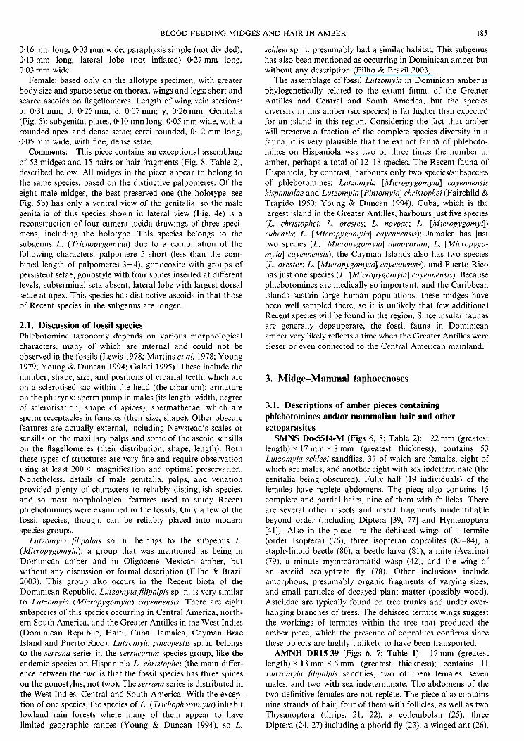
SMNS Do-5514-M (Figs 6, 8; Table 2): 22 mm (greatest length) x I7 mm x 8 rnrn (greatest thickness); contains 53 Lutzomyia schleei sandflies, 37 of which are females, eight of which are males, and another eight with sex indeterminate (the genitalia being obscured). Fully half (19 individuals) of the females have replete abdomens. The piece also contains 15 complete and partial hairs, nine of them with follicles. There are several other insects and insect fragments unidentifiable beyond order (including Diptera [39, 771 and Hymenoptera [41]). Also in the piece are the dehisced wings of a termite (order Isoptera) (76), three isopteran coprolites (82-84), a staphylinoid beetle (80), a beetle larva (81), a mite (Acarina) (79), a minute mymmaromatid wasp (42), and the wing of an asteiid acalyptrate fly (78). Other inclusions include amorphous, presumably organic fragments of varying sizes, and small particles of decayed plant matter (possibly wood). Asteiidae are typically found on tree trunks and under over- hanging branches of trees. The dehisced termite wings suggest the workings of termites within the tree that produced the amber piece, which the presence of coprolites confirms since these objects are highly unlikely to have been transported.
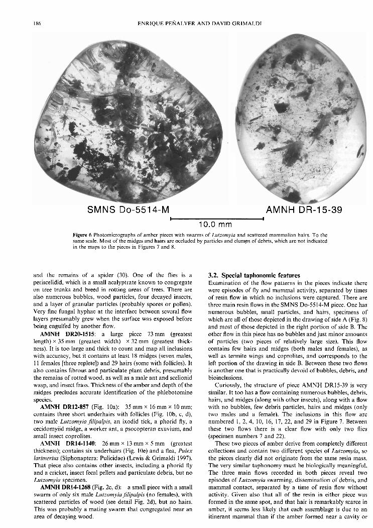
AMNH DR15-39 (Figs 6, 7; Table 1): 17 mm (greatest length) x I3 mm x 6 mm (greatest thickness); contains 11 Lurzomyia jilipalpis sandflies, two of them females, seven males, and two with sex indeterminate. The abdomens of the two definitive females are not replete. The piece also contains nine strands of hair, four of them with follicles, as well as two Thysanoptera (thrips: 21, 22), a collembolan (25), three Diptera (24, 27) including a phorid fly (23), a winged ant (26),
186 ENRIQUE PENALVER A N D DAVID GRIMALDI
SMNS DO-551 4-M AMNH DR-15-39 I I
10.0 mm Figure 6 Photomicrographs of amber pieces with swarms of Llrtzomyiu and scattered mammalian hairs. To the same scale. Most of the midges and hairs are occluded by particles and clumps of debris, which are not indicated in the maps to the pieces in Figures 7 and 8.
and the remains of a spider (30). One of the flies is a periscelidid, which is a small acalyptrate known to congregate on tree trunks and breed in rotting areas of trees. There are also numerous bubbles, wood particles, four decayed insects, and a layer of granular particles (probably spores or pollen). Very fine fungal hyphae at the interface between several flow layers presumably grew when the surface was exposed before being engulfed by another flow.
AMNH DR20-1515: a large piece 73mm (greatest length) x 35 mm (greatest width) x 32 mm (greatest thick- ness). It is too large and thick to count and map all inclusions with accuracy, but it contains at least 18 midges (seven males, 1 1 females [three replete]) and 29 hairs (some with follicles). It also contains fibrous and particulate plant debris, presumably the remains of rotted wood, as well as a male ant and scelionid wasp, and insect frass. Thickness of the amber and depth of the midges precludes accurate identification of the phlebotomine species.
AMNH DR12-857 (Fig. 10a): 35 mm x 16 mm x I0 mm; contains three short underhairs with follicles (Fig. lob, c, d), two male Lutzomyiajlipalpis, an ixodid tick, a phorid fly, a cecidomyiid midge, a worker ant, a psocopteran exuvium, and small insect coprolites.
AMNH DR14-1140: 26 mm x 13 mm x 5 mm (greatest thickness); contains six underhairs (Fig. 10e) and a flea, Pulex larimeritrs (Siphonaptera: Pulicidae) (Lewis & Grimaldi 1997). That piece also contains other insects, including a phorid fly and a cricket, insect fecal pellets and particulate debris, but no Lutzomyia specimen.
AMNH DR14-1268 (Fig. 2c, d): a small piece with a small swarm of only six male Lutzomyiajlipalpis (no females), with scattered particles of wood (see detail Fig. 2d), but no hairs. This was probably a mating swarm that congregated near an area of decaying wood.
3.2. Special taphonomic features ~xaminat ion of the flow patterns in the pieces indicate there were episodes of fly and mammal activity, separated by times of resin flow in which no inclusions were captured. There are three main resin flows in the SMNS Do-5514-M piece. One has numerous bubbles, small particles, and hairs, specimens of which are all of those depicted in the drawing of side A (Fig. 8) and most of those depicted in the right portion of side B. The other flow in this piece has no bubbles and just minor amounts of particles (two pieces of relatively large size). This flow contains few hairs and midges (both males and females), as well as termite wings and coprolites, and corresponds to the left portion of the drawing in side B. Between these two flows is another one that is practically devoid of bubbles, debris, and bioinclusions.
Curiously, the structure of piece AMNH DR15-39 is very similar. It too has a flow containing numerous bubbles, debris, hairs, and midges (along with other insects), along with a flow with no bubbles, few debris particles, hairs and midges (only two males and a female). The inclusions in this flow are numbered 1, 2, 4, 10, 16, 17, 22, and 29 in Figure 7. Between these two flows there is a clear flow with only two flies (specimen numbers 7 and 22).
These two pieces of amber derive from completely different collections and contain two different species of Lutzomyia, so the pieces clearly did not originate from the same resin mass. The very similar taphonomy must be biologically meaningful. The three main flows recorded in both pieces reveal two episodes of Lutzomyia swarming, dissemination of debris, and mammal contact, separated by a time of resin flow without activity. Given also that all of the resin in either piece was formed in the same spot, and that hair is remarkably scarce in amber, it seems less likely that each assemblage is due to an itinerant mammal than if the amber formed near a cavity or
BLOOD-FEEDING MIDGES A N D HAIR IN AMBER
Figure 7 Map of Dominican amber piece AMNH DR15-39, which contains 11 LutzomyiaJilipalpis sp. n. midges (2 of them female) and 9 complete and fragmentary hairs. See Table 1 for a listing of each inclusion. 'F' refers to follicle. Small letters refer to images in Figure 9.
Table 1 Assemblage of LutzomyiaJilipalpis sp. n. and other inclusions in Dominican amber piece AMNH DR15-39, as mapped in Figure 7
Lutzomyia P?, not replete Lutzomyia 6 Lutzomyia Lutzomyia 6 Lutzomyia 6 Lutzomyia 6 Llitzomyia Liitzomyia 6 Lutzomyia 6 Lul:omyia 9, not replete Lutzomyia 6 hair hair hair, with follicle hair, complete, with follicle
guard hair hair, with follicle hair hair hair, complete, with follicle Thysanoptera: Thripidae Thysanoptera: Thripidae Diptera: Phoridae Diptera, indet. Collembola Hymenoptera: Formicidae Diptera: Periscelididae Coleoptera larva Orthoptera: Gryllidae: Oecanthinae Araneae (partial)

ENRIQUE PENALVER AND DAVID GRIMALDI
BLOOD-FEEDING MIDGES A N D HAIR IN AMBER
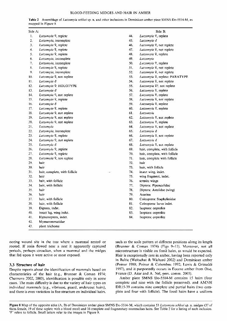
Table 2 Assemblage of Lutzomyiu schleei sp. n. and other inclusions in Dominican amber piece SMNS Do-5514-M, as mapped in Figure 8
Side I. 2. 3. 4. 5. 6. 7. 8. 9.
10. 11. 12. 13. 14. 15. 16. 17. 18. 19. 20. 21. 22. 23. 24. 25. 26. 27. 28. 29. 30. 31. 32. 33. 34. 35. 36. 37. 38. 39. 40. 41. 42. 43.
A: Lutzomyiu 9 , replete Lutzom~~ia, incomplete Lutzomyiu 9 , replete Lutzonl)liu 9, replete Lut:om)~iu 9, replete Llrtzomyiu. incomplete Lulzoni.viu, incomplete Lufzornyia 9, replete Lutzomyiu, incomplete L~rtzornviu 9, not replete Lufzom)~iu d Llrtzomyia 9: HOLOTYPE Lutzoniyiu d Ltrrzom)lia 9 , not replete Lur:omviu 9, replete Lutzomyiu d Lutzom!~iu 9, replete L~rtzorn~~iri 9, not replete Lutzornj1ia 9. not replete Lutzomyio 9 , not replete Lirt:omyicr Lutiomyin, incomplete Lutzornyici 9. replete Lutzom)lin 9 , not replete Lutzornyin d Lutzonfyicr 9, replete Lutzomviu 9, replete Lutzomyiu 9 , not replete hair hair hair, complete, with follicle hair hair, with follicle hair, with follicle hair hair hair, with follicle hair. with follicle Diptera. indet. insect leg, wing indet. Hymenoptera, indet. Mymarommatidae plant trichome
Side B: Llitzoniyiu 9, replete Lutzomyiu 6 Lutzomyiu 9, not replete Lulzomyiu P, not replete Lulzomyiu 9, replete Lut iomyi~ Llitzomyiu 9, replete Lutzomyiu 9, not replete Lurzomyia 9, not replete Lurzomyia 9, replete: PARATYPE Lutzornyi(i 9, not replete Lutzomjlia P?, not replete Lutzonlyiu 9, replete Lutzornyirr 9, replete Lurzomyicr P, not replete Lutzon~j~ict P, replete L11tzomvin 9, replete Lurzomyirr Lutzomyiu P, not replete Lurzom)iu 9, replete Llitzoni~~iu 9, not replete Lutzoniyiu d Lirtzonlyiu 9, not replete Lurzontjlio 6 Lutzon~jlirr 9, not replete hair, complete, with follicle hair, complete, with follicle hair, complete with follicle hair hair, with follicle insect wing. indet. wing fragment, indet. termite wings Diptera: Pipunculidae Diptera: Asteiidae (wing) Acarina Coleoptera: Staphylinidae Coleoptera: larva indet. Isoptera: coprolite Isoptera: coprolite Isoptera: coprolite
oozing wound site in the tree where a mammal nested or rooted. If resin flowed near a nest it apparently captured periods, perhaps circadian, when a mammal and the midges that fed upon it were active or most exposed.
3.3 Structure of hair Despite reports about the identification of mammals based on characteristics of the hair (e.g., Brunner & Coman 1974; Chernova 2002, 2003), identification is possible only in some cases. The main difficulty is due to the variety of hair types on individual mammals (e.g., vibrissae, guard, undercoat hairs), and there is even variation in fine structure on individual hairs,
such as the scale pattern at different positions along its length (Brunner & Coman 1974) (Figs 9-11). Moreover, not all microstructure is visible on fossil hairs, as would be expected. Hair is exceptionally rare in amber, having been reported only in Baltic (Weitschat & Wichard 2002) and Dominican amber (Poinar 1988; Poinar & Columbus 1992; Lewis & Grimaldi 1997), and it purportedly occurs in Eocene amber from Oise, France (D. Azar and A. Nel, pers. comm. 2005).
Amber piece SMNS Do-5514-M contains 15 hairs (four complete and nine with the follicle preserved), and AMNH DR15-39 contains nine complete and partial hairs (two com- plete and four with follicle). The fossil hairs have a uniform
Figure 8 Map of the opposite sides (A, B) of Dominican amber piece SMNS Do-5514M, which contains 53 Lirtzomyici sckleei sp. n. midges (37 of them female, 19 of these replete with a blood meal) and 16 complete and fragmentary mammalian hairs. See Table 2 for a listing of each inclusion. 'F' refers to follicle. Small letters refer to the images in Figure 9.

ENRIQUE PENALVER AND DAVID GRIMALDI
scale pattern and diameter along most of the length, tapering at the tip, and with a circular cross-section. These hairs are the guardlunder hair type. Those in the SMNS piece are more uniform in length and diameter, but the hairs in the AMNH piece have greater variation and the large hair (Fig. 7: speci- men 16) corresponds to a guard hair. Diameter of hairs in AMNH DR15-39 is approximately 44-51 pm (94pm in the largest hair), and approximately 63 pm for the hairs in SMNS Do-5514-M. Lengths of complete hairs are 10.7 mm in the AMNH piece (the incomplete guard hair is 13.0 mm) and 5.5 mm, 7.0 mm, 11.1 mm, and 13.3 mm in the SMNS piece (an incomplete hair is 14.8 mm). The medulla is preserved only in the short stems of some hairs and belongs to the simple type of Brunner & Coman's (1974) classification.
Three scale patterns have been observed on different hairs in the AMNH piece, but due to their proximity, similar propor- tions, and the fact that they have the same orientation, we assume these hairs are from the same individual mammal. All hairs in the SMNS piece have the same scale pattern and diameter, and thus must come from one individual mammal. Microscopic details of the hairs are as follows (terminology following Brunner & Coman 1974):
AMNH DR15-39 (see Figs 7, 9): Hair I (number 16): The larger, guard hair has a crenulate
scale margin, with a narrow distance between scale margins and a streaked-single chevron scale pattern (Fig. 9i).
Hair I1 (number 17): A thin hair with the follicle pre- served, having a smooth scale margin, with scale margins distant and arranged into a regular wave pattern. The posi- tions of the scales are strongly oblique with respect to the shaft of the hair (Fig. 9a, d, f, h).
Hair 111 (number 14): This hair is similar to the previous two but the positions of the scales are perpendicular to the hair's margin and each scale covers the entire diameter of the hair (Fig. 9g).
The scale pattern and size of the underhairs in this piece are very similar to that of three short underhairs (Fig. lob, c, d) in another piece of amber (AMNH DR12857; Fig. IOa), which contains two male LutzomyiaJilipalpis and an ixodid tick. The hair structure is also very similar to that of at least one of the six underhairs in piece AMNH DR14-1140 (Fig. IOe), which contains a pulicid flea.
SMNS Do-5514-M (Fig. 8): These hairs have scale mar- gins that are distant and smooth, and the scales are arranged into a regular wave pattern in positions that are perpendicular to the shaft of the hair (Fig. 9b, c, e, j).
Original comparisons were made between the fossil hairs and the hairs of various mammals, particularly Recent rela- tives of groups known to have inhabited the Antilles in the Miocene to the Holocene. The atlas by Brunner & Coman (1974) was also used. There were Early Oligocene (White & MacPhee 2001) to Holocene fossils of bats, sloths, rodents, insectivores, and monkeys in the Antilles. The main record is comprised of Quaternary cave fossils, but the record most germane to the present one comprises Miocene fossils of megalonychid sloths, capromyid rodents, platyrrhine primates, and a small insectivore (possibly Solendontidae) from Cuba and the Dominican Republic (MacPhee et al. 2003; MacPhee & Grimaldi 1996). Hair samples of Recent species derived from the AMNH mammal collection. In some cases the guard hairs were very dark, so in order to observe cuticle patterns under transmitted light with compound microscopy these hairs were bleached in either sodium hypochlorite or hydrogen peroxide. Living species examined were the following:
brasiliensis (from Jamaica), and Noctilio leporinus (from Venezuela). These species occasionally live in tree holes, and all species except T. brasiliensis occur on Hispaniola (Adrian Tejedor, pers. comm. 2004). The fossil record of West Indian bats is poor and only Quaternary in age, and origin of the endemic Antillean species is thought to be due to dispersal in the mid- to Late Cenozoic (Hedges 1996). Edentata: Bradypodidae (sloths): Choleopus didactylus (two-toed sloth, from Brazil). White & MacPhee (2001) reviewed the fossil record of sloths in the Antilles. Insectivora: Solenodon paradoxus, an endemic species of the family Solenodontidae, which has an isolated position among insectivores. Rodentia: Geocapromys bro~vnii (from Jamaica) and G. ingrahami (from the Bahamas). The former of these is endemic to Jamaica and also known from the Quaternary of that island (MacPhee & Flemming 2003). Primates: Callicebus torquatus (from Peru) and C. moloch (from Brazil). Two genera of Callicebinae occur in the Quaternary of Haiti, Jamaica, and the Dominican Republic (MacPhee et al. 1995; MacPhee & Horowitz 2004).
The microstructure of bat and sloth hair is very distinctive, and these orders can be readily dismissed regarding the identity of the fossil hairs. Bat hairs have distinctive dentate scale margins and simple coronal scale patterns. Sloth hairs have longitudinal grooves with pockets on the external surface of the hair shaft (Chernova 2000), and they are quite long (e.g., 70 mm in length or more). The hairs of Callicebus torquatus have a uniserial ladder-type of medulla in the guard hairs and very thin underhairs; the scale pattern of only the middle part of the underhair is similar to that of the area near the follicle in one of the fossil hairs (Fig. 90. Callicebus moloch has a biserial ladder-type of medulla and its guard hairs have a different scale pattern (regular wave, not streaked-single chevron). Guard hairs of Geocapromys brownii have a scale pattern like that in the fossil guard hair (Fig. 6, specimen 16), but the distance between scale margins is remarkably close by com- parison, and the hair diameter much greater. The scale pattern of the underhairs of Geocaprornys is similar to that of the fossil hairs, but Geocaprornys has a very different (uniserial ladder) type of medulla. Geocapromys ingrcrhami has a similar scale pattern to the hairs in the SMNS piece (Fig. 9j), but with a very thin diameter.
Based on size and microstructure, the fossil hairs are most consistent with those of solenodontid insectivores, though other insectivores and even rodents cannot be discounted as sources. Solenodontidae is a small family of shrew-like insec- tivores endemic to the Antilles, most species of which are extinct. The large guard hair in amber piece AMNH DR15-39 has a similar pattern and diameter to that of the Recent species Solenodon paradoxus (see Fig. I la), and the other hairs are very similar to the hairs of a juvenile individual of this species (Fig. 1 I b). Also, medulla structure of Solenodon and the fossil hairs is very similar, having the simple-interrupted type (Brun- ner & Coman 1974). Pleistocene bones of the extinct endemic species Solenodon rnarcanoi are known from Hispaniola, and it is unknown if the genus occurred in the Antilles prior to this.
Nesophontes is an extinct solenodontid genus known from throughout the Caribbean (MacPhee et al. 1999) (some au- thors place it in its own family, the Nesophontidae [Nowak 19911). The genus contains nine nominal and several other species, and the bones of four species are known from Hispani- ola. Nesophontes-like bones, without hair remains, occur in Dominican amber (MacPhee & Grimaldi 1996), and there is
Chiroptera (bats): Molosus molosus (from the Dominican evidence that the genus became extinct as late as the 19th-20th Republic), Artibeusjamaicensis (from Costa Rica), Tadarida centuries based on recently discovered material from central

BLOOD-FEEDING MIDGES AND HAIR IN AMBER 191
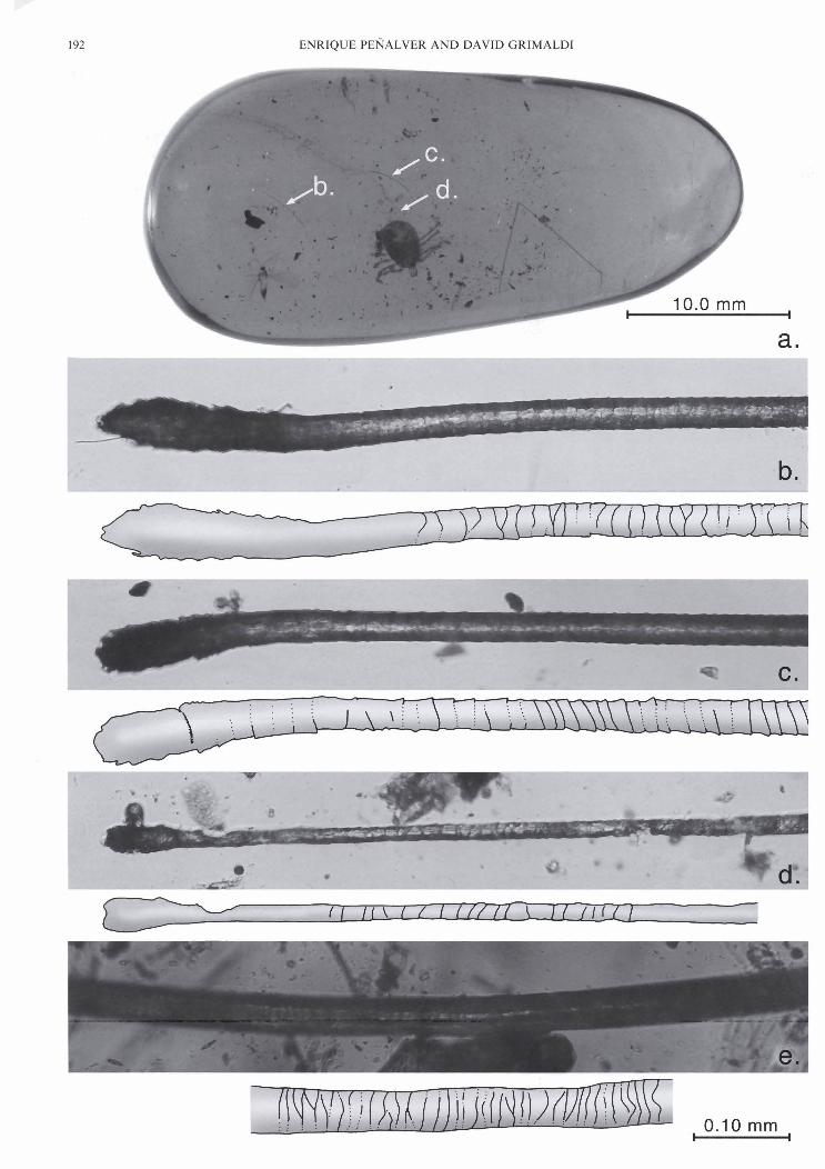
Figure 9 Detail of the mammalian hairs whose locations are mapped in Figures 7 and 8, (d) and (0 are the same specimen. Letters correspond to those hair sections mapped in Figures 7 and 8.

BLOOD-FEEDING MIDG iES AND HAIR IN AMBER 193
Figure 11 Scale pattern of two hairs of extant Solen(/on pcrrrrrlo.rus (Insectivora: Solenodontidae), endemic to Hispaniola: (a) Adult speci- men, AMNH 35331; (b) Juvenile, AMNH 90129. Compare the scale pattern with hairs in Figures 9 and 10.
Dominican Republic (MacPhee et al. 1999). The cranial cavity of a Nesopliontes skull preserved in a cave there contains four morphological types of hair, and though it is unknown which type of hair belongs to Nesophontes, one of the figured hair types (Fig. 4c in MacPhee et a/. 1999) is very similar to that of the hairs in Dominican amber we report here.
Discussion There is virtually no question that Lutzomyia were feeding on mammals in the Miocene of Hispaniola. Only three pieces are known to the senior author, among 100-200,000 screened over 20 years, which contain small swarms of phlebotomines and mammalian hair. Only two other pieces of Dominican amber in the AMNH collection of 6,000 pieces contain hair, one piece containing a tick (Ixodidae) and two male Lutzonzyiajlipalpis (AMNH DR12857), the other a flea (Lewis & Grimaldi 1997; AMNH DR14-1140). Hair structure in both pieces is very similar to that of the hairs in the phlebotomine pieces described here (Fig. 10). Hair is extremely rare in Dominican amber, and so the probability that it would be preserved along with blood-sucking and ectoparasitic arthropods based on chance alone is exceedingly remote, so there must be a biological association for this. Bloated female phlebotomines in two of the pieces in the present study always had a dark, granular material in the abdomen. Since only the females take a blood meal, and males were never bloated, presumably the bloated females contained a recent blood meal. This indicates the immediate proximity of their host, which must have been the mammal whose hairs were preserved alongside them.
Four of the amber pieces having multiple phlebotomines also contained particles of wood (Fig. 2d) as well as insect frass
and insects known to be associated with decaying tree trunks (e.g., termites, periscelidid and asteiid flies, and ants), which indicates the proximity of the mammal and midges to decaying wood. It is possible that the mammal was nesting in a decayed cavity within the Hyrnenaea amber tree, or was rooting into such an area in search of insects. There is little doubt that the mammals were arboreal. Lutzomyia is well known to feed on birds and mammals in forest canopies. In Neotropical forests Lutzomyia can be dramatically stratified between the canopy and the ground, with some species showing strong habitat preference (Dias-Lima et al. 2002). Many species rest on tree trunks, and some even have circadian migrations from the canopy to the ground and back.
It has been well documented here that two of the Miocene species of Lutzomyia fed from mammals, and it is known that species groups of phlebotomines have feeding preferences for either reptiles, birds, o r mammals (Lewis 1974; Killick- Kendrick 1990). However, it is unknown what the other four species fed upon, and there is direct evidence in Dominican amber of various vertebrates that could have been potential hosts. These include Ele~~therodactylus frogs (Poinar & Cannatella 1987), Anolis and Sphaerodactyltrs lizards (Rieppel 1980; Bohme 1984); several species of birds as based on the microstructure of feathers, including those of a woodpecker (Picidae) (Layborne et a/. 1994); and mammals. All evidence of mammals in Dominican amber is based on hairs, with the remarkable exception of a piece containing six vertebrae and four ribs of a small mammal (MacPhee & Grimaldi 1996). Structure of the vertebrae suggests it was an insectivore similar in size to endemic Nesophontes (Solenodontidae), and the structure of some hairs in the phlebotomine pieces is very similar to that of solenodontids. Living Solenodon are forest dwellers and mainly nocturnal, sheltering by day in hollow trees, tree cavities, caves, and burrows. They generally d o not build a nest except during the breeding season (MacFadden 1980; Nowak 1991), and are not considered to be arboreal. The attribution to Rodentia and Carnivora of other strands of hair in Dominican amber (Poinar 1988; Poinar & Columbus 1992) is based on little diagnostic evidence because of the limited information from hair that we discussed above. The known mammal hosts of Lutzomyici include armadillos, opossum, porcupine, kinkajou, sloths, anteaters, rabbits, various rodents, bats, and humans (D. J. Lewis 1975; Killick-Kendrick 1990).
Some modern species of the subgenus L. (Micropygomyia) feed on lizards. For example, L. (M.) cayennensis hispaniolae, endemic to the Dominican Republic, was commonly seen feeding on the backs of iguanid lizards in full sunlight, and females of L. (M.) cayennensis cayennensis have been found infected with trypanosomes in Venezuela and Colombia (Young & Duncan 1994). Species in the verrucarum species group, to which Lutzomyia paleopestis belongs, are suspected or proven vectors of Leishmania and Bartonella in Costa Rica and in northern South America (Young & Duncan 1994). The hosts of extant L. (Trichophoromyia) are unknown, but in wild-caught females of one species of the subgenus, L. ubiquit- olis, Leishmania lainsoni pathogens were detected (Young & Duncan 1994).
An independent approach to estimating feeding habits of phlebotomines (at least on a gross level, such as being lizard,
Figure 10 Photomicrograph (a) of a Dominican amber piece with two male L~rrzomyiufilipulpis sp. n., a tick (Ixodidae) and three scattered mammalian hairs (AMNH DR 12-857), and details of these hairs (W). Detail of a mammalian hair (e) in a Dominican amber piece with a flea, Pirlex lurimerius (AMNH DR14-1140). All hairs to the same scale.

194 ENRIQUE P E ~ ~ A L V E R AND DAVID GRIMALDI
bird, small o r large mammal feeders) is based o n the micro- scopic structure of the mouthparts (D. J. Lewis 1975). There is significant variation among species in the proportions of the labrum, mandibles, maxillary laciniae, and hypopharynx; the number and position of sensilla; and the number and size of serrations a t the distal ends of these structures (D. J. Lewis 1975; Brinson el al. 1993), which seems to have functional significance. Unfortunately, the mandibles, laciniae, and hypopharynx are rarely displayed in amber specimens (Fig. 2b); serrations on these elements being approximately 1 p m in length and width and thus barely visible using light microscopy under optimal preparations (let alone in amber). Thus, host preferences of the amber midges cannot be assessed based on mouthparts.
It may eventually be possible t o determine if these fossil phlebotomines harboured pathogenic microbes like Leishmania promastigotes, but determining that will require expendable specimens that have well-preserved internal tissues. Portions of the gut would need to be carefully extracted and embedded in epoxy, thin-sectioned with glass o r diamond blades, and examined a t 1,000 x and higher using transmis- sion electron microscopy (TEM). Light microscopy does not differentiate the organelles required to identify the promastig- otes (e.g., D. H. Lewis, 1975; Walters et a/. 1989), despite published reports of trypanosomatids in amber (Poinar and Poinar 2004, 2005; Poinar 2005). Since the remains of protists 2.5 p m in diameter were discerned in hind gut tissue of a primitive termite in Dominican amber using T E M (Wier et al. 2002), there is significant potential that if Leislzmania proma- stigotes o r other microbes were present in these fossil midges then unambiguous remains of them could possibly be recovered.
5. Acknowledgements
We deeply appreciate the assistance of Keith Luzzi, Steve Thurston, Paul Nascimbene, Zane Goehman, Dieter Schlee, Adrian Tejedor and Andre Nel, and the generous donations of Robert G. Goelet. Dany Azar, Michael S. Engel, and Susan Perkins provided helpful reviews. This research is a contribu- tion to the junior author's postdoctoral project "Taphonomy and palaeoecology of insects in Dominican amber" funded by a generous grant from the Spanish government.
6. References
Austin, J. J., Ross, A., Smith, A., Fortey, R. & Thomas, R. 1997. Problems of reproducibility: does geologically ancient DNA sur- vive in amber-preserved insects? Proceedings of the Royal Society, London B 264, 467-74.
Ayala, S. C. 1973. The phlebotomine sandfly-protozoan parasite community of central California grasslands. The American Midland Nururalist 89, 266-80.
Azar, D., Nel, A., Solignac, M., Paicheler, J.-C. & Bouchet, F. 1999. New genera and species of psychodid flies from the Lower Cretaceous amber of Lebanon. Paloeonrology 42, 1101-36.
Azar, D., Perrichot, V., Neradeau, D. & Nel, A. 2003. New psychodids from the Cretaceous ambers of Lebanon and France, with a discussion of Eophlehotomus connectans Cockerell, 1920 (Diptera, Psychodidae). Annals ofrhe Entomological Society of America 95: 1 17-26.
Beati, L., Ckeres, A. G., Lee, J. A. & Munstermann, L. E. 2004. Systematic relationships among Lutzomyia sand flies (Diptera: Psychodidae) of Peru and Colombia based on the analysis of 12s and 28s ribosomal DNA sequences. Inrernnrional Journal for Parasirology 34, 225-34.
Bohme, W. 1984. Erstfun eines fossilen kugelfingergeckos (Sauria: Gekkonidae: Sphaerodactylinae) aus Dominikanischem bernstein (Oligozln von Hispaniola, Antillen). Salamandra 20, 212-20.
Brazil, R. P. & Filho, J. D. A. 2002. Description of Pinto- myia (PiJanomyia) fulcaorum sp.n. (Diptera: Psychodidae:
Phlebotominae), a fossil sand fly from Dominican amber. Memdrias do Insrituto Osn~aldo Cruz 97, 501-3.
Brinson, F. J., McKeever, S. and Hagan, D. V. 1993. Comparative study of mouthparts of the phlebotomine sand flies Lutzomyio longipalpis, L. sl?annoni, and Phlebotomus paparasi (Diptera: Psychodidae). Annals of the E~~tomological Sociery of America 86, 470-83.
Brunner, H., & Coman, B. 1974. The Idenrification of Marnrnolian Huir. Melbourne, Australia: Inkata Press.
Cano, R. J., Borucki, M. K., Higby-Schweitzer, M., Poinar, H. N., Poinar, G. 0.. Jr. & Pollard, K. J. 1994. Bacillus DNA in fossil bees: an ancient symbiosis? Applied and Environmental Microbiology 60, 2 1647.
Chernova, 0. F. 2000. Unusual hair structure in sloths (Edentata: Bradypodidae). Doklady Biological Sciences 373, 400-4.
Chernova, 0. F. 2002. Architectonic and diagnostic significance of hair cuticle. Biology Bulletin (Moscow) 29, 23847.
Chernova, 0. F. 2003. Architectonic and diagnostic significance of hair cortex and medulla. Biolo~y Bulletin (Moscow) 30, 53-62.
Cockerell, T. D. A. 1917. Arthropods in Burmese amber. American Journal of Science 44, 360-8.
DeSalle, R., Gatesy, J., Wheeler, W., & Grimaldi, D. 1992. DNA sequences from a fossil termite in Oligo-Miocene amber and their phylogenetic implications. Science 257, 19334.
Dias-Lima, A,, Bermudez, E.C., Medeiros, J. F. & Sherlock, I. 2002. Vertical stratification of phlebotomine sandfly fauna (Diptera, Psychodidae) in a primary non-flooded forest of the Central Amazon, Amazonas State, Brazil. Cad Srrude Puhlico 18, 823-32.
Duckhouse, D. A. 2000. Redescription and re-evaluation of the Burmese amber psychodid Eoplllehoton7us connectens Cockerell and its phylogenetic position (Diptera: Psychodidae). Sysreniotic Entomology 25, 503-9.
Fairchild, G. B. & Trapido, H. 1950. The West Indian species of Phleboromus (Diptera Psychodidae). Annals of the En!omological Socierjl of America 43, 405-17.
Filho, J. D. A. & Brazil, R. P. 2003. Relationships of New World phlebotomine sand flies (Diptera: Psychodidae) based on fossil evidence. Memdrias do Institute Oslvnldo Cruz 98, 145-9.
Fran~a, C. 1924. Essai de classification des Phlebotomes. Insrirut Pasreur de I'Afrique dl1 Nord 1, 279-84.
Galati, E. A. B. 1995. Phylogenetic systematics of Phlebotominae (Diptera, Psychodidae) with emphasis on American groups. Boletin de lr Direccidn de Molririologia y Sanecinliento Amhiental 35 (Suppl. I), 13342.
Grimaldi, D. A. 1995. The age of Dominican amber. In Anderson K. B. & Crelling, J. C. (eds) Amher, Resinire, and Fossil Resins, 203-17. Washington D.C.: American Chemical Society Symposium Series 61 7.
Grimaldi, D. A. 1996. Amher: Window ro [he Pasr. New York: AMNHIAbrams.
Grimaldi, D., Bonwich, E., Delannoy, M. & Doberstein, S. 1994. Electron microscopic studies of mummified tissues in amber fossils. American Micseum Novitares 3097, 1-3 1.
Grimaldi, D. A,, Shedrinsky, A. & Wampler, T. P. 2000. A remarkable deposit of fossiliferous amber from the Upper Cretaceous (Turonian) of New Jersey. In Grimaldi, D. (ed.) Studies on Fossils in Amher, with Parricular Reference to the Cretaceous of New Jersey. Leiden: Backhuys.
Grimaldi, D. A,, Engel, M.S. & Nascimbene, P. C. 2002. Fossiliferous Cretaceous amber from Myanmar (Burma): its rediscovery, biotic diversity, and paleontological significance. American Museum Novirates 3361, 1-71.
Hedges, S. B. 1996. Historical biogeography of West Indian vertebrates. Annual Revie~ij of Ecology and Systematics 27, 163-96.
Hennig, W. 1972. Insektenfossilien aus der Unteren Kreide IV. Psychodidae (Phlebotominae), mit einer kritischen Ubersicht iiber das phylogenetische System der Familie und die bisher beschrie- benen Fossilien (Diptera). Sturrgurter BeitrZge zur Natitrklrnde 241, 149.
Henwood, A. 1992a. Exceptional preservation of dipteran flight muscle and the taphonomy of insects in amber. Polaios 7,203-12.
Henwood, A. 1992b. Soft-part preservation of beetles in Tertiary amber from the Dominican Republic. Palaeontology 35, 901-12.
Iturralde-Vinent, M. A. & MacPhee, R. D. E. 1996. Age and paleo- geographical origin of Dominican amber. Science 273, 1850-2.
Johnson, C., Agosti, D., Delabie, J. H., Dumpert, K., Williams, D. J., Tschirnhaus, M. von & Maschwitz, U. 2001. Acropyga and Azteca ants (Hymenoptera: Formicidae) with scale insects (Ster- norrhyncha: Coccoidea): 20 million years of intimate symbioses. American Museum Nosirates 3335, 1-1 8.

BLOOD-FEEDING MIDGES AND HAIR IN AMBER 195
Kertesz, K. 1903. Karalog der polaearktishen Dipteren. I. Orthorrhapha Nematocera. Budapest, vol. 1: 1-383.
Killick-Kendrick, R. 1990. Phlebotomine vectors of the leishmaniases: a review. Meclical and Veterinary Entomology 4, 1-24.
Killick-Kendrick, R. 1999. The biology and control of phlebotomine sand flies. Clinics in Dermatology 17, 279-89.
Langenheim, J. H. 2003. Plant Resins. Chemistry, Evolution, Ecology. and Ethnohotany. Portland, Oregon and Cambridge, UK: Timber Press.
Layborne, R. C., Deedrick, D. W. & Hueber, F. M. 1994. Feather in amber is earliest New World fossil of Picidae. The Wilson Bulletin 106, 18-25.
Lewis, D. H. 1975. Ultrastructural study of promastigotes of Leishmania from reptiles. Journal of Protozoology 22, 344-52.
Lewis, D. J . 1971. Phlebotomid sandflies. Bulletin o f the World Health Organization 44, 535-5 1 .
Lewis, D. J. 1974. The biology of Phlebotomidae in relation to leishmaniasis. Annrral re vie^^^ of Entomology 19, 363-84.
Lewis, D. J. 1975. Functional morphology of the mouthparts in New World phlebotomine sandflies (Diptera: Psychodidae). Transactions of the Royal Entomologiccrl Society, London 126, 497-532.
Lewis, D. J. 1978. The phlebotomine sandflies (Diptera: Psychodidae) of the Oriental Region. Bulletin of the British Museum (Nat~rrirl History), Entomology 37, 2 17-343.
Lewis, R. E. & Grimaldi, D. A. 1997. A pulicid flea in Miocene amber from the Dominican Republic (Insecta: Siphonaptera: Pulicidae). American Museum Novirates 3205, 1-9.
Linnaeus, C. 1758. System0 naturae per regna /ria nattrrae. Ed. X , vol. 1: 824 pp. Holmiae (=Stockholm).
McFadden, B. J. 1980. Rafting mammals or drifting islands?: Bioge- ography of the Greater Antillean insectivores Ncsophontes and Solenodon. Journal of Biogeography 7, 1 1-22.
MacPhee, R. D. E., Horovitz, I., Arredondo, 0 . & JimCnez Vazquez, 0. 1995. A new genus for the extinct Hispaniolan monkey Saimiri bernensis (Rimoli, 1977), with notes on its systematic position. American Museum Novitates 3134. 1-21.
MacPhee, R. D. E., Flemming, C., & Lunde, D . P. 1999. "Last occurrence" of the Antillean insectivoran Nesophontes: new radio- metric dates and their interpretation. American Museum Novitrrtes 3261, 1-20.
MacPhee, R. D. E., Iturralde-Vinent. M. A. & Gaffney, E. S. 2003. Domo de Zaza, an Early Miocene vertebrate locality in south- central Cuba, with notes on the tectonic evolution of Puerto Rico and the Mona Pasaje. American Museum Novitates 3394, 1 4 2 .
MacPhee, R. D. E. & Flemming, C. 2003. A possible heptaxodontine and other caviidan rodents from the Quaternary of Jamaica. American Museum Nositares 3422, 1 4 2 .
MacPhee, R. D. E. & Grimaldi, D. A. 1996. Mammal bones in Dominican amber. Nature 380, 489-90.
MacPhee, R. D. E. & Horovitz, I. 2004. New craniodental remains of the Quaternary Jamaican monkey Xenothrix mcgregori (Xeno- trichini, Callicebinae, Pitheciidae), with a reconsideration of the Aotus hypothesis. American Museum Novitates 3434, 1-51.
Martins, A. V., Williams, P., & Falczo, A. L. 1978. American sandflies (Diptera: Psychodidae, Plilehotominae). Rio de Janeiro: Academia Brasileira de Ciencias.
Nascimbene, P. & Silverstein, H. 2000. The preparation of fragile Cretaceous ambers for conservation and study of organismal inclusions. In Grimaldi, D. (ed.) Studies on Fossils in Amber, with Particular Reference to the Cretaceous of New Jersey. Leiden: Backhuys.
Newman, E. 1835. Attempted division of British insects into natural orders. Entomologist's Magazine 2, 279431.
Nowak, R. M. 1991. Walker's Mammals of the World, Vol. 1. Baltimore, Maryland: The Johns Hopkins University Press. Fifth Edition.
Poinar, G . O., Jr. 1988. Hair in Dominican amber: evidence for Tertiary land mammals in the Antilles. E-vperientio 44, 88-9.
Poinar, G. O., Jr. 1992. Lqe in Amber. Palo Alto, California: Stanford University Press.
Poinar, G., Jr. 2005. Triatomu dominicann sp. n. (Hemiptera: Reduviidae: Triatominae), and Trypanosoma antiyuus sp. n. (Stercoraria: Trypanosomatidae), the first fossil evidence of a triatomine-trypanosomatid vector association. Vector-Borne and Zoonotic Diseases 5, 72-8 1.
Poinar, G . 0 . . Jr. & Cannatella, D. C. 1987. An Upper Eocene frog from the Dominican Republic and its implications for Caribbean biogeography. Science 237, 12 15-6.
Poinar, G . O., Jr. &Columbus, J. T. 1992. Adhesive grass spikelet with mammalian hair in Dominican amber: first fossil evidence of epizoochory. Experientia 48, 9 0 6 8 .
Poinar, G.O., Jr. & Poinar, R. 1999. The Amber Forest. A Reconstruc- tion of a Vanished World. Princeton, New Jersey: Princeton University Press.
Poinar, G., Jr. & Poinar, R. 2004. Evidence of vector-borne disease of Early Cretaceous reptiles. Vector-Borne rmrl Zoonotic Diseases 4, 28 1 4 .
Poinar, G., Jr. & Poinar, R. 2005. Paleoleishmania prolerus n. gen., n. sp. (Trypanosomatidae: Kinetoplastida) from Cretaceous Burmese amber. Protist 155, 305-3 10.
Quate, L. W. 1963. Fossil Psychodidae in Mexican amber, part 2 (Diptera: Insecta). Journal of Paleontology 37, 1 10-8.
Rieppel, 0 . 1980. Green anole in Dominican amber. Nature 286, 4 8 6 7 .
Ross, A. J . 1998. Amber, The Natural Time Capsule. London: The Natural History Museum.
Walters, L. L., Chaplin, G . L., Modi, G . B. & Tesh. R. B. 1989. Ultrastructural biology of Leishmania (Viannia) panamensis (=Leishmania braziliensis panamensi.7) in Lutzomyia gomezi (Diptera: Psychodidae): a natural host-parasite association. Americrrn Journal of Tropical Medicine and Hygiene 41, 295-3 17.
Warburg, A., Ostovska, K. & Lawyer, P. G . 1991. Pathogens of phlebotomine sandflies: a review. Purossitologia 33, 519-26.
Weitschat, W., & Wichard, W. 2002. Atlas of Plants and Animals in Baltic Amber. Munich: Verlag Dr. Friedrich Pfeil.
White, J. L. & MacPhee, R. D. E. 2001. The sloths of the West Indies: a systematic and phylogenetic review. In Woods, C . A. & Sergile, F. E. (eds) Biogeography of the West Indies: Prrtterns and Perspectives, 201-35. C R C Press, second edition.
Wier, A,, Dolan, M., Grimaldi, D. A,, Guerrero, R., Wagensburg, J. & Margulis, L. 2002. Spirochete and protist symbionts of a termite (Mustotermes electrodominicus) in Miocene amber. Proceedings of the National Academy of Sciences, USA 99, 1410-3.
Young, D. G . 1979. A review of the bloodsucking psychodid flies of Colombia (Diptera: Phlebotominae and Sycoracinae). University o f Florida Agricultural E-vperiment Srotion Technical Bulletin 806, 266 pp.
Young, D. G . & Duncan, M. A. 1994. Guide to the identification and geographic distribution of Lutzomj~ia sandflies in Mexico, the West Indies, Central and South America (Diptera: Psychodidae). Memoirs of the Americr~n Entomological Institute 54, 881 pp.
ENRIQUE PENALVER and DAVID GRIMALDI, Division of Invertebrate Zoology, American Museum of Natural History, Central Park West a t 79th St., New York, New York 10024-5192 USA. e-mails: [email protected]; [email protected]
MS received 12 April 2005. Accepted for publication 10 October 2005.