Embed Size (px)
Citation preview
Department of Clinical Neuroscience Karolinska Institutet, Stockholm, Sweden
ASCORBATE IN THE OCULAR LENS
Vino C. Mody, Jr., M.D
Stockholm 2006
i
All previously published papers were reproduced with permission from the publisher. Published and printed by Karolinska University Press 2006 Box 200, SE-171 77 Stockholm, Sweden ©Vino C. Mody, Jr., M.D., ISBN 91-7140-256-X
ii
Abstract
Purpose: First, we intended to establish a method for sample preparation for measurement of ascorbate in whole rat and guinea pig lenses utilizing ultrafiltration and high performance liquid chromatography with ultraviolet radiation detection. Then, we aimed to investigate whether, in the albino rat, lens ascorbate concentration depends on solid dietary intake. Finally, we investigated if, in the pigmented guinea pig, lens ascorbate concentration may be elevated with drinking water supplementation.
Background: Ascorbate is an important dietary antioxidant. Ascorbate is an essential nutrient in the human and guinea pig, while the rat is capable of synthesizing ascorbate. In vitro and in vivo studies have demonstrated the protective effect of ascorbate against cataract development in the rat and guinea pig lens.
Methods: Albino Sprague Dawley rats were kept on solid diet supplemented with known amount of ascorbate for four weeks. In a second experiment, pigmented guinea pigs were kept on regular chow containing essential amount of ascorbate and drinking water supplemented with known amount of ascorbate. The animals were then sacrificed, the lenses extracted and homogenized in metaphosphoric acid. Ascorbate and other low molecular weight compounds were isolated with ultrafiltration and ascorbate was quantified with subsequent high performance liquid chromatography (HPLC) with ultraviolet radiation (UVR) detection at 254 nm. The UVR spectra for ascorbate and dehydroascorbate imply that 96% of the signal at 254 nm is ascorbate.
Results: We found that external and internal calibration provided similar results. Both methods had a linear absorbance response in the range used. All rat lenses were devoid of cataract. The baseline lens ascorbate content for rats receiving no ascorbate in the diet was significantly greater than zero. Lens ascorbate concentration increased linearly with dietary ascorbate intake with a statistically significant increase rate. All guinea pig lenses were devoid of cataract. All lenses contained a detectable concentration of ascorbate. Lens ascorbate concentration increased exponentially declining with drinking water supplementation concentration, up to a saturation level.
Conclusions: The method utilizing ultrafiltration and high performance liquid chromatography with ultraviolet radiation detection for measurement of whole lens ascorbate content in the rat and guinea pig lens is applicable. In the rat, lens ascorbate concentration linearly increases with solid dietary ascorbate intake without cataract development. In the guinea pig, lens ascorbate concentration increases exponentially declining to a saturation level with increasing drinking water ascorbate supplementation without cataract development.
iii

iv
Publications included in the thesis
I. Mody Jr. VC, Kakar M, Elfving Å, Söderberg PG, Löfgren S. Ascorbate In The Rat Lens, Dependence On Dietary Intake. Ophthalmic Research 2005; 37:142-149. Published by Karger Publishers.
II. Mody Jr. VC, Kakar M, Elfving Å, Söderberg PG, Löfgren S. Ascorbate In The Guinea Pig Lens, Dependence On Drinking Water Supplementation. Acta Ophthalmologica Scandinavica 2005; 83:228-233. Published by Blackwell Publishing.
v

vi
Table of contents
Abstract iii Publications included in the thesis v 1. Introduction ............................................................................................................................1
1.1 Ascorbate ........................................................................................................................1 1.2 Ascorbate requirement....................................................................................................1 1.3 Ascorbate function..........................................................................................................3 1.4 Ocular protection by ascorbate in vitro and in vivo........................................................3 1.5 Ascorbate in human epidemiological studies..................................................................3 1.6 Ascorbate pharmacodynamics ........................................................................................3 1.7 Free vs. bound ascorbate.................................................................................................4 1.8 Ascorbate measurement methods ...................................................................................5
2. Aims of the study....................................................................................................................7 3. Methods ..................................................................................................................................8
3.1 Experimental animals .....................................................................................................8 3.2 Experimental procedure..................................................................................................8
3.2.1 Ascorbate supplementation............................................................................................8 3.2.2 Lens sample preparation ................................................................................................9 3.2.3 Measurement of lens ascorbate concentration ...............................................................9
3.3 Statistical methods and parameters...............................................................................11 4. Results and discussion ..........................................................................................................12
4.1 Ascorbate in the rat lens, dependence on solid dietary intake ............................................12 4.2 Ascorbate in the guinea pig lens, dependence on drinking water supplementation............14
5. Conclusions ..............................................................................................................................16 6. Acknowledgments ....................................................................................................................17 7. References ................................................................................................................................18
vii

To My Parents Vino and Anita and Brother Beijoo
viii
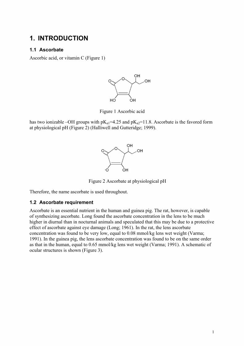
1. INTRODUCTION 1.1 Ascorbate Ascorbic acid, or vitamin C (Figure 1)
Figure 1 Ascorbic acid
has two ionizable –OH groups with pKa1=4.25 and pKa2=11.8. Ascorbate is the favored form at physiological pH (Figure 2) (Halliwell and Gutteridge; 1999).
Figure 2 Ascorbate at physiological pH
Therefore, the name ascorbate is used throughout.
1.2 Ascorbate requirement Ascorbate is an essential nutrient in the human and guinea pig. The rat, however, is capable of synthesizing ascorbate. Long found the ascorbate concentration in the lens to be much higher in diurnal than in nocturnal animals and speculated that this may be due to a protective effect of ascorbate against eye damage (Long; 1961). In the rat, the lens ascorbate concentration was found to be very low, equal to 0.08 mmol/kg lens wet weight (Varma; 1991). In the guinea pig, the lens ascorbate concentration was found to be on the same order as that in the human, equal to 0.65 mmol/kg lens wet weight (Varma; 1991). A schematic of ocular structures is shown (Figure 3).
1
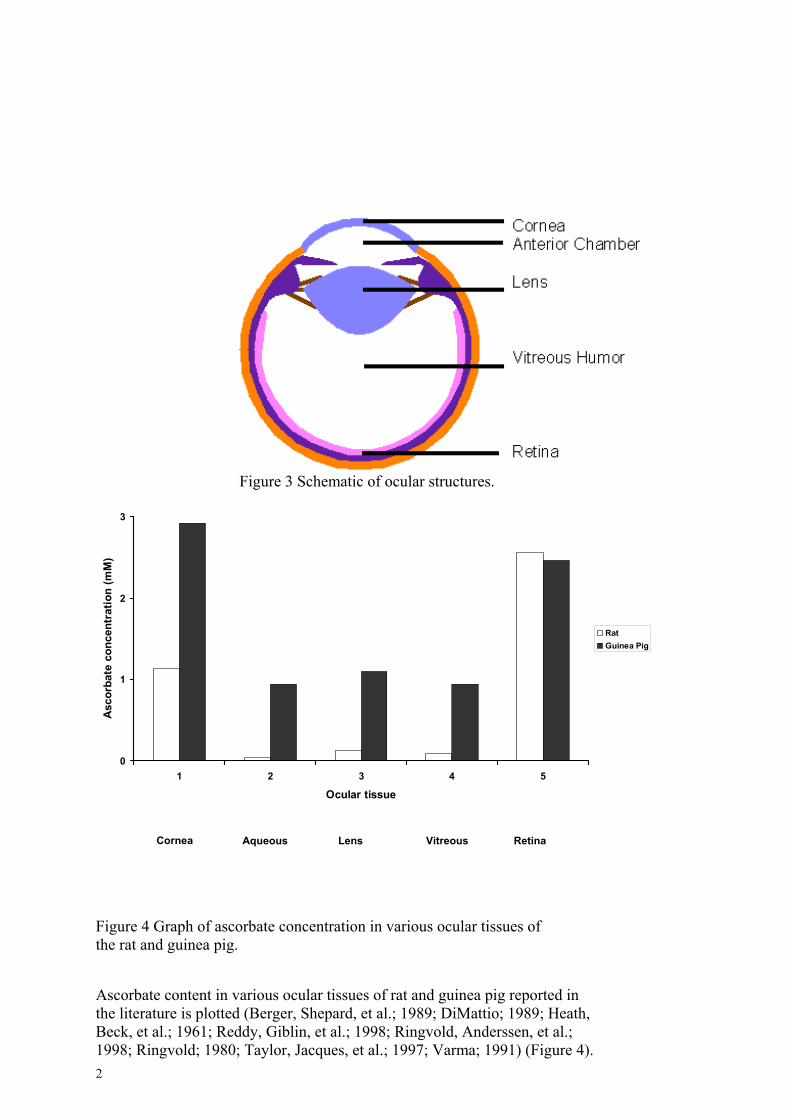
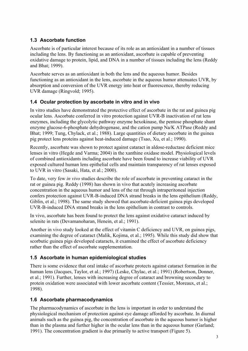
Figure 3 Schematic of ocular structures.
0
1
2
3
1 2 3 4
Ocular tissue
Asc
orba
te c
once
ntra
tion
(mM
)
RetVitreous Lens Aqueous Cornea
Figure 4 Graph of ascorbate concentration in various ocular tissues othe rat and guinea pig.
Ascorbate content in various ocular tissues of rat and guinea pig repthe literature is plotted (Berger, Shepard, et al.; 1989; DiMattio; 198Beck, et al.; 1961; Reddy, Giblin, et al.; 1998; Ringvold, Anderssen,1998; Ringvold; 1980; Taylor, Jacques, et al.; 1997; Varma; 1991) (2
5
ina
f
orted in 9; Heath, et al.; Figure 4).
RatGuinea Pig
1.3 Ascorbate function Ascorbate is of particular interest because of its role as an antioxidant in a number of tissues including the lens. By functioning as an antioxidant, ascorbate is capable of preventing oxidative damage to protein, lipid, and DNA in a number of tissues including the lens (Reddy and Bhat; 1999).
Ascorbate serves as an antioxidant in both the lens and the aqueous humor. Besides functioning as an antioxidant in the lens, ascorbate in the aqueous humor attenuates UVR, by absorption and conversion of the UVR energy into heat or fluorescence, thereby reducing UVR damage (Ringvold; 1995).
1.4 Ocular protection by ascorbate in vitro and in vivo In vitro studies have demonstrated the protective effect of ascorbate in the rat and guinea pig ocular lens. Ascorbate conferred in vitro protection against UVR-B inactivation of rat lens enzymes, including the glycolytic pathway enzyme hexokinase, the pentose phosphate shunt enzyme glucose-6-phosphate dehydrogenase, and the cation pump Na/K ATPase (Reddy and Bhat; 1999; Tung, Chylack, et al.; 1988). Large quantities of dietary ascorbate in the guinea pig protect lens proteins against heat-induced damage (Tsao, Xu, et al.; 1990).
Recently, ascorbate was shown to protect against cataract in aldose-reductase deficient mice lenses in vitro (Hegde and Varma; 2004) in the xanthine oxidase model. Physiological levels of combined antioxidants including ascorbate have been found to increase viability of UVR exposed cultured human lens epithelial cells and maintain transparency of rat lenses exposed to UVR in vitro (Sasaki, Hata, et al.; 2000).
To date, very few in vivo studies describe the role of ascorbate in preventing cataract in the rat or guinea pig. Reddy (1998) has shown in vivo that acutely increasing ascorbate concentration in the aqueous humor and lens of the rat through intraperitoneal injection confers protection against UVR-B-induced DNA strand breaks in the lens epithelium (Reddy, Giblin, et al.; 1998). The same study showed that ascorbate-deficient guinea pigs developed UVR-B-induced DNA strand breaks in the lens epithelium in contrast to controls.
In vivo, ascorbate has been found to protect the lens against oxidative cataract induced by selenite in rats (Devamanoharan, Henein, et al.; 1991).
Another in vivo study looked at the effect of vitamin C deficiency and UVR, on guinea pigs, examining the degree of cataract (Malik, Kojima, et al.; 1995). While this study did show that scorbutic guinea pigs developed cataracts, it examined the effect of ascorbate deficiency rather than the effect of ascorbate supplementation.
1.5 Ascorbate in human epidemiological studies There is some evidence that oral intake of ascorbate protects against cataract formation in the human lens (Jacques, Taylor, et al.; 1997) (Leske, Chylac, et al.; 1991) (Robertson, Donner, et al.; 1991). Further, lenses with increasing degree of cataract and browning secondary to protein oxidation were associated with lower ascorbate content (Tessier, Moreaux, et al.; 1998).
1.6 Ascorbate pharmacodynamics
3
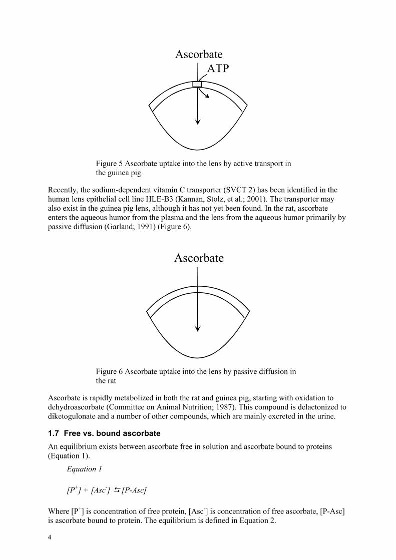
The pharmacodynamics of ascorbate in the lens is important in order to understand the physiological mechanism of protection against eye damage afforded by ascorbate. In diurnal animals such as the guinea pig, the concentration of ascorbate in the aqueous humor is higher than in the plasma and further higher in the ocular lens than in the aqueous humor (Garland; 1991). The concentration gradient is due primarily to active transport (Figure 5).
AscorbateATP
Figure 5 Ascorbate uptake into the lens by active transport in the guinea pig
Recently, the sodium-dependent vitamin C transporter (SVCT 2) has been identified in the human lens epithelial cell line HLE-B3 (Kannan, Stolz, et al.; 2001). The transporter may also exist in the guinea pig lens, although it has not yet been found. In the rat, ascorbate enters the aqueous humor from the plasma and the lens from the aqueous humor primarily by passive diffusion (Garland; 1991) (Figure 6).
Ascorbate
Figure 6 Ascorbate uptake into the lens by passive diffusion in the rat
Ascorbate is rapidly metabolized in both the rat and guinea pig, starting with oxidation to dehydroascorbate (Committee on Animal Nutrition; 1987). This compound is delactonized to diketogulonate and a number of other compounds, which are mainly excreted in the urine.
1.7 Free vs. bound ascorbate An equilibrium exists between ascorbate free in solution and ascorbate bound to proteins (Equation 1).
Equation 1
[P+] + [Asc-] [P-Asc]
Where [P+] is concentration of free protein, [Asc-] is concentration of free ascorbate, [P-Asc] is ascorbate bound to protein. The equilibrium is defined in Equation 2.
4
Equation 2
[ ][ ][ ]−+
−=
AscPAscPk
Here, k is the equilibrium coefficient.
Currently, it is difficult to measure true free ascorbate concentration in lens samples. Our method measures a fraction of the total ascorbate concentration, which is proportional to both the total ascorbate concentration and true free ascorbate concentration. Therefore, an increase in ascorbate concentration as measured with the utilized method corresponds to an increase in true free ascorbate concentration.
1.8 Ascorbate measurement methods Biochemists were able to measure ascorbate in lens extracts more than sixty years ago utilizing a color titration method with 2,6-dichlorophenolindophenol (Duke-Elder; 1968). However, the method was not specific for ascorbate since it also assayed for glutathione. In 1979 Omaye et al. determined ascorbate concentration in animal tissues using a spectrophotometric assay with 2,6-dichlorophenolindophenol (Omaye, Turnbull, et al.; 1979). The spectrophotometric assay is more specific for ascorbate than the color titration method. The technique has been applied to measure ascorbate in the rat lens (Reddy, Giblin, et al.; 1998).
Within the last fifteen years, a method of measuring intraocular ascorbate utilizing HPLC with either electrochemical (Blondin, Baragi, et al.; 1986; Taylor, Jahngen-Hodge, et al.; 1995) or UVR (Hallström, Carlsson, et al.; 1989) (Mody, Kakar, et al.; 2005) detection has been described. HPLC provides the advantage over previous methods of better sensitivity and specificity since molecules of interest can be separated from background molecules based on several molecular aspects such as size, solubility and electrical charge. Sensitivity in HPLC detection can be defined both qualitatively and quantitatively. Sensitivity in qualitative terms is the concentration of measured molecule that gives rise to an average signal twice that of the standard deviation of the signal from background noise, and in quantitative terms seven times.
HPLC with UVR detection is likely the simplest HPLC system for measuring ascorbate in biological samples. UVR detection is possible because of expressed absorption of UVR by ascorbate. The high molar extinction coefficient for ascorbate at 265 nm (neutral pH) or at 256 nm (acidic pH) allows for quantification of the ascorbic acid peak as it elutes from an HPLC column. The method of HPLC with UVR detection has been used for over a decade to analyze ascorbate from a number of tissues and plasma (Washko, Welch, et al.; 1992). HPLC with electrochemical detection has been applied to measure ascorbate in the lens tissue from Emory mice (Taylor, Jahngen-Hodge, et al.; 1995), guinea pigs, and humans (Taylor, Jacques, et al.; 1997).
Electrochemical detection must overcome two hurdles. First, the mobile phase must allow for separation of ascorbate from background molecules and be capable of carrying the electric charge. Second, it is difficult to maintain a constant, electrochemically reactive electrode surface (Washko, Welch, et al.; 1992). UVR detection may provide better specificity for a complex molecule since, when combined with ion-exchange chromatography, HPLC separates based on size, charge, and UVR absorption. Electrochemical detection separates based on only size and charge. Ultrafiltration for separation of very large molecules from smaller molecules and HPLC with UVR detection for separation of small molecules and for
5
measurement of ascorbate in rat lens samples were applied. The homogenization step used in processing the lenses is performed in metaphosphoric acid at low pH.
6
2. AIMS OF THE STUDY The aims of the study were:
I. Establish a method for sample preparation for measurement of ascorbate in whole lenses utilizing ultrafiltration and HPLC with UVR detection.
II. Investigate whether lens ascorbate concentration is dependent on solid dietary ascorbate intake in the albino rat.
III. Investigate whether lens ascorbate concentration may be elevated with drinking water supplementation in the guinea pig.
7
3. METHODS Albino Sprague Dawley rats and pigmented guinea pigs were supplemented with oral intake of ascorbate. The uptake in the lens was analyzed with HPLC.
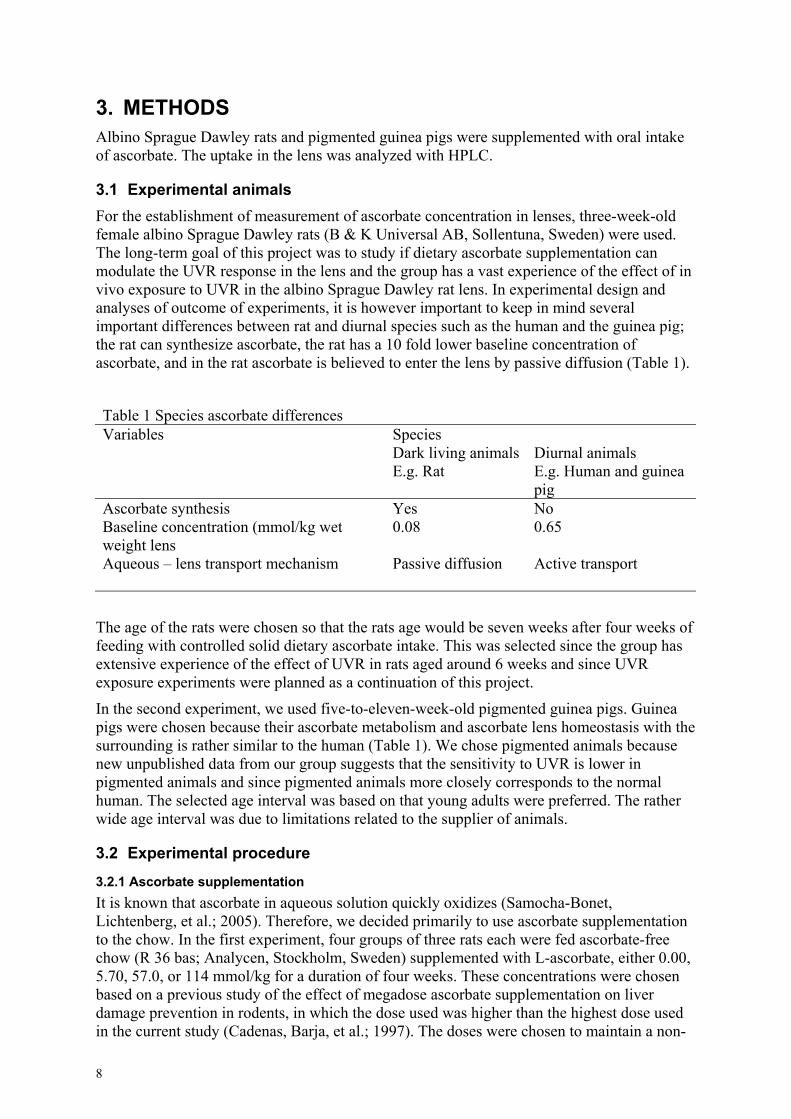
3.1 Experimental animals For the establishment of measurement of ascorbate concentration in lenses, three-week-old female albino Sprague Dawley rats (B & K Universal AB, Sollentuna, Sweden) were used. The long-term goal of this project was to study if dietary ascorbate supplementation can modulate the UVR response in the lens and the group has a vast experience of the effect of in vivo exposure to UVR in the albino Sprague Dawley rat lens. In experimental design and analyses of outcome of experiments, it is however important to keep in mind several important differences between rat and diurnal species such as the human and the guinea pig; the rat can synthesize ascorbate, the rat has a 10 fold lower baseline concentration of ascorbate, and in the rat ascorbate is believed to enter the lens by passive diffusion (Table 1).
Table 1 Species ascorbate differences Variables Species Dark living animals
E.g. Rat Diurnal animals E.g. Human and guinea pig
Ascorbate synthesis Yes No Baseline concentration (mmol/kg wet weight lens
0.08 0.65
Aqueous – lens transport mechanism Passive diffusion Active transport
The age of the rats were chosen so that the rats age would be seven weeks after four weeks of feeding with controlled solid dietary ascorbate intake. This was selected since the group has extensive experience of the effect of UVR in rats aged around 6 weeks and since UVR exposure experiments were planned as a continuation of this project.
In the second experiment, we used five-to-eleven-week-old pigmented guinea pigs. Guinea pigs were chosen because their ascorbate metabolism and ascorbate lens homeostasis with the surrounding is rather similar to the human (Table 1). We chose pigmented animals because new unpublished data from our group suggests that the sensitivity to UVR is lower in pigmented animals and since pigmented animals more closely corresponds to the normal human. The selected age interval was based on that young adults were preferred. The rather wide age interval was due to limitations related to the supplier of animals.
3.2 Experimental procedure
3.2.1 Ascorbate supplementation It is known that ascorbate in aqueous solution quickly oxidizes (Samocha-Bonet, Lichtenberg, et al.; 2005). Therefore, we decided primarily to use ascorbate supplementation to the chow. In the first experiment, four groups of three rats each were fed ascorbate-free chow (R 36 bas; Analycen, Stockholm, Sweden) supplemented with L-ascorbate, either 0.00, 5.70, 57.0, or 114 mmol/kg for a duration of four weeks. These concentrations were chosen based on a previous study of the effect of megadose ascorbate supplementation on liver damage prevention in rodents, in which the dose used was higher than the highest dose used in the current study (Cadenas, Barja, et al.; 1997). The doses were chosen to maintain a non-
8
toxic concentration. Four weeks of supplementation was chosen to achieve a steady state lens ascorbate concentration.
In the second experiment, we used drinking water supplementation with ascorbate, 0.00, 2.84, 5.68, or 8.52 mM, for a duration of four weeks. To minimize loss of ascorbate in the drinking water bottle, each bottle was covered in a black plastic bag and was changed with freshly prepared solution twice a day. The concentration of ascorbate in the drinking water was measured using HPLC with UVR detection. In addition, the chow fed to all animals contained 0.125 mol L-ascorbate/kg chow. This was because ascorbate is an essential compound for the guinea pig and the purpose of the experiment was to investigate the possibility to increase lenticular ascorbate with supplementation.
We used high amounts of ascorbate supplementation, up to ten-fold the amount used by Taylor et al. in a study on vitamin C in guinea pig eye tissues in relation to intake (Taylor, Jacques, et al.; 1997). The reason for not choosing to feed guinea pigs a low ascorbate or ascorbate-deficient diet was that ascorbate deprivation for two to three weeks in two studies caused guinea pigs to become ill and lose weight (Malik, Kojima, et al.; 1995; Reddy, Giblin, et al.; 1998). The theory was instead to see whether we could increase lens ascorbate levels through drinking water supplementation and whether the modulation could be used to study cataract models, including UVR. 3.2.2 Lens sample preparation At the end of the supplementation period, the rats were sacrificed with carbon dioxide asphyxiation. Each lens was extracted, photographed, wet weight measured, homogenized, and centrifuged in 1.0 ml of 0.25% metaphosphoric acid (Hallström, Carlsson, et al.; 1989).
When processing the lens samples for ascorbate measurement, it is important to add an agent to the sample to prevent metal catalyzed oxidation of ascorbate. The lens samples in the study were prepared in metaphosphoric acid. Besides protein denaturation, metaphosphoric acid prevents oxidation of ascorbate to dehydroascorbate because of high acidity and metal ion chelation (Koshiishi, Mamura, et al.; 1998; Washko, Welch, et al.; 1992).
The supernatant was ultrafiltered and the ultrafiltrate injected into an HPLC column.
3.2.3 Measurement of lens ascorbate concentration Our selection of method for ascorbate measurement among other methods published (Omaye, Turnbull, et al.; 1979) was based on that HPLC allows efficient separation of small molecules with higher sensitivity and better specificity than other methods (Hallström, Carlsson, et al.; 1989). The current HPLC method has the advantage over previously described methods of detecting even small concentrations of ascorbate in biological tissues, less than 0.06 µM, in the injected sample.
The column was an ion-exchange, reversed phase column. This column was chosen based on previous experience in other studies (Hallström, Carlsson, et al.; 1989). The mobile phase used in the HPLC system was 2 mM sulphuric acid, pH 2.4. This mobile phase was also selected based on experience from previous studies (Hallström, Carlsson, et al.; 1989) to avoid oxidation of ascorbate during the passage through the HPLC column.
Ascorbate was detected as UVR absorption at 254 nm at expected elution time checking for the pattern of surrounding peaks in the chromatogram. UVR provides specific detection with high sensitivity for ascorbate in biological tissues (Hallström, Carlsson, et al.; 1989) (Johnsen, Ringvold, et al.; 1985).
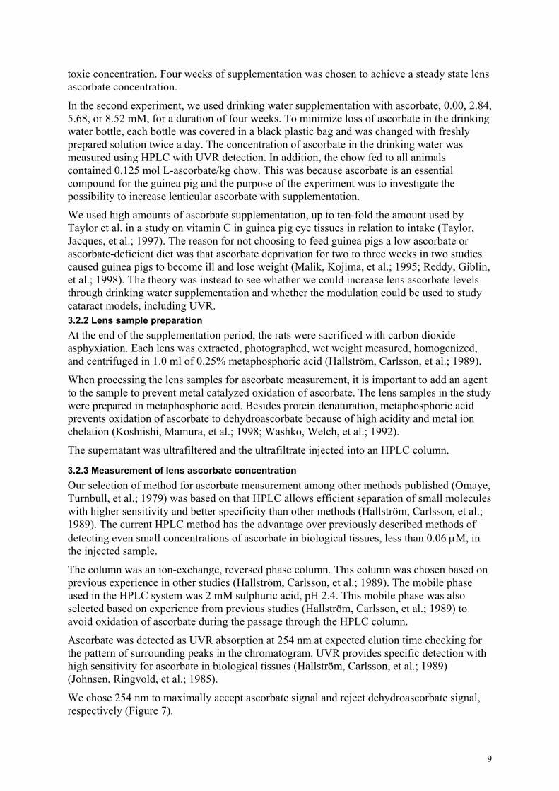
We chose 254 nm to maximally accept ascorbate signal and reject dehydroascorbate signal, respectively (Figure 7).
9
0
1
2
3
4
5
200 250Wav
Abs
orba
nce
Figure 7 Ultraviolet radiation spectra for 1dehydroascorbate in 2.5% metaphosphoricdenotes absorbance for the two spectra at 2
The absorbance ratio for the oxidized form of asfound to be 4.1 % at 254 nm (Figure 7). This imascorbate.
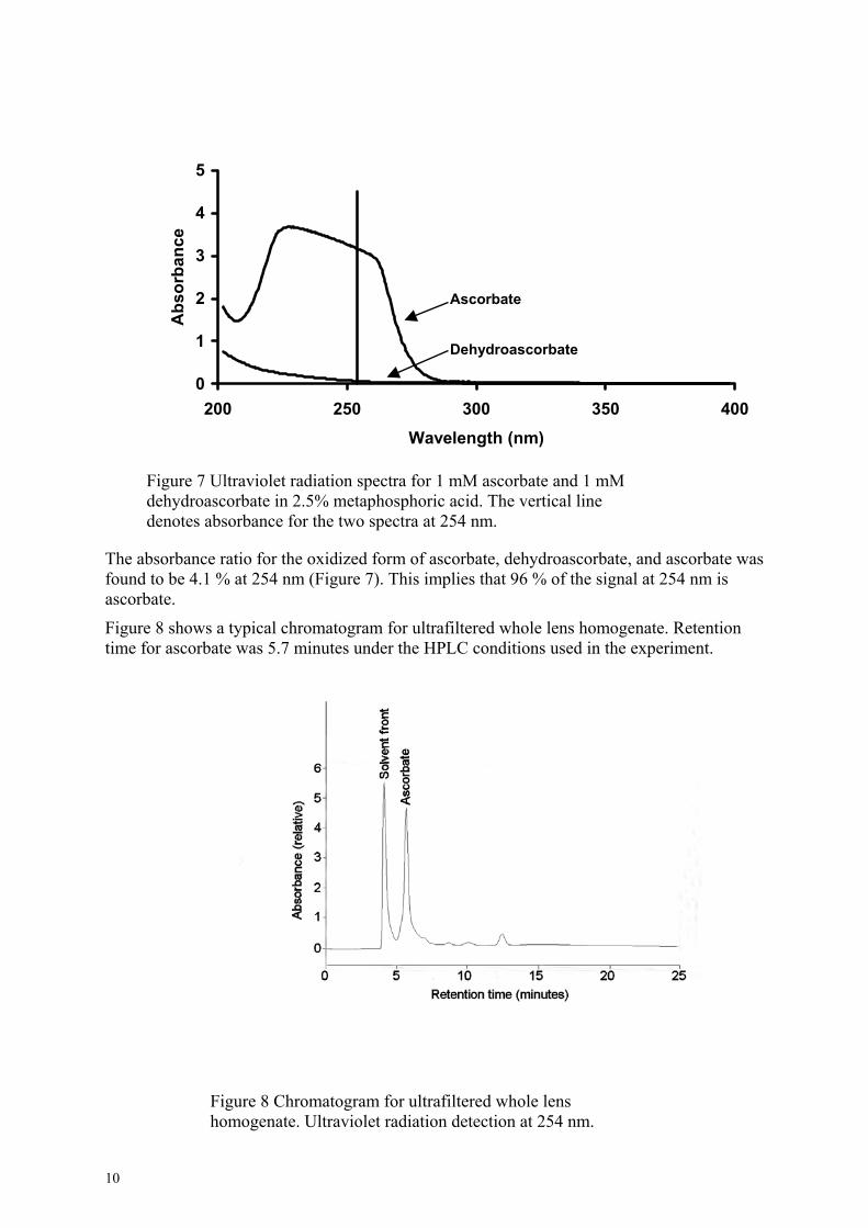
Figure 8 shows a typical chromatogram for ultratime for ascorbate was 5.7 minutes under the HP
Figure 8 Chromatogram for ultrafhomogenate. Ultraviolet radiation
10
Ascorbate Dehydroascorbate
300 350 400elength (nm)
mM ascorbate and 1 mM acid. The vertical line 54 nm.
corbate, dehydroascorbate, and ascorbate was plies that 96 % of the signal at 254 nm is
filtered whole lens homogenate. Retention LC conditions used in the experiment.
iltered whole lens detection at 254 nm.
The peak shape and symmetry allowed for resolution of the ascorbate peak. For analysis of sequential samples, we used chromatography software that was programmed to identify the ascorbate peak based on retention time and shape
The ascorbate concentration was calculated based on calibration against an external 10 µM L-ascorbate standard prepared from a commercially available ascorbate standard solution (Merck, Darmstadt, Germany). The low concentration standard was prepared by weighing a standard amount of commercially available ascorbate standard solution and adding water. The added amount of water was also weighed. The final concentration of ascorbate was calculated from the masses added of ascorbate standard solution and water, respectively.
There is always a risk that external calibration causes an error both in sensitivity and level of the calibration. We estimated this error by comparing external and internal calibration on a pooled ultrafiltrate of rat lenses.
External standard with ascorbate solutions ranging from 0 µM to 20 µM were prepared and measured on HPLC. Absorbance increased linearly as a function of concentration (r2>0.99; data not shown). Internal standard addition samples were created by adding known amounts of ascorbate ranging from 0 - 20 µM to samples of a portion of pooled, processed, whole rat lens ultrafiltrate solution. The samples were measured with HPLC. As for the external standard, the increase of absorbance as a function of concentration of standard added was linear (r2>0.98; data not shown). The ascorbate concentration in the rat lens ultrafiltrate was with the internal standard addition technique estimated to 1.87 mM and with external calibration technique to 1.95 mM. Considering the insignificant difference between the two methods, it was decided to only use the external calibration for future experiments.
The quantitative recovery of ascorbate in measurements was also determined. The supernatant was obtained after ultracentrifugation of grinded lens in metaphosphoric acid. The pellet after ultracentrifugation was re-extracted. The ascorbate concentration in the supernatant after re-extracting the pellet was 46 % of that in the supernatant of the lens homogenate (data not shown). This finding indicates that ascorbate in the pellet is released into solution upon re-extraction.
3.3 Statistical methods and parameters Throughout the thesis, the significance levels and confidence coefficients were set to 0.05 and 0.95, respectively.
11
4. RESULTS AND DISCUSSION 4.1 Ascorbate in the rat lens, dependence on solid dietary intake The current study aimed at evaluating a technique for sample preparation for determination of whole lens content of ascorbate. Further, it was intended to use the developed technique to investigate whether lens ascorbate concentration depends on dietary ascorbate intake.
The method for lens ascorbate measurement was described above.
The reason for trying to elevate lens ascorbate concentrations was to, in the future, study whether lens ascorbate modulates UVR-induced cataract in vivo.
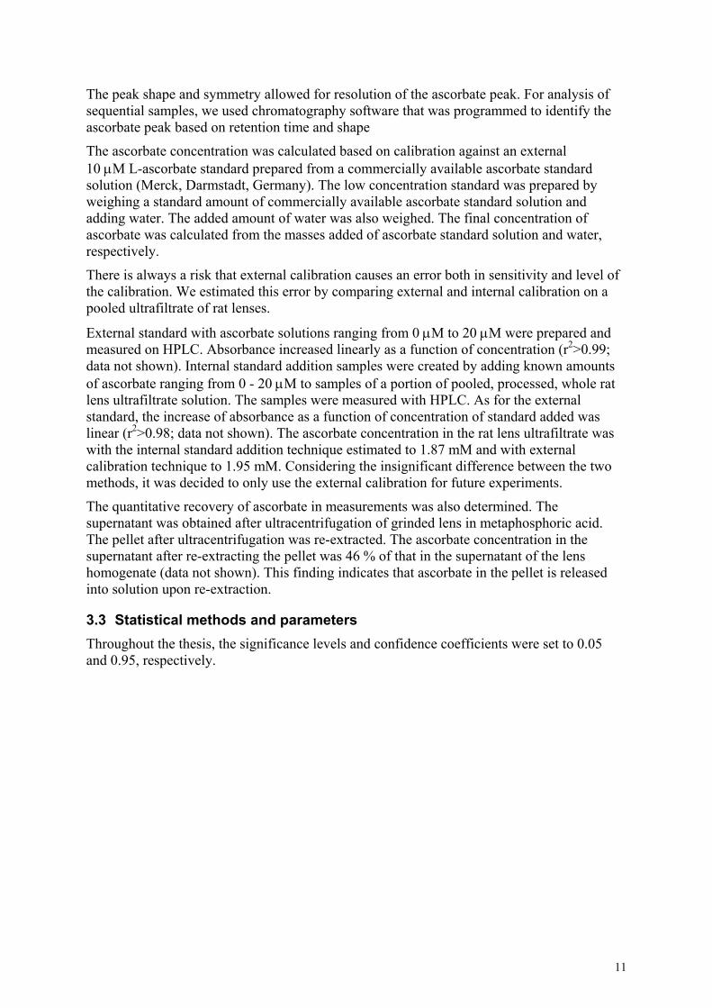
In order to exclude that the ascorbate supplementation per se modulates lens light scattering, all lenses were monitored macroscopically in incident light with a grid background. All lenses were devoid of cataract (Figure 9).
Figure 9 Lens photographs using bright field illumination for rats fed chow supplemented with varying amounts of ascorbate. A) 0 mmol/kg, B) 5.7 mmol/kg, C) 57 mmol/kg, and D) 114 mmol/kg. Grid square diameter is 0.79 mm.
Lens ascorbate concentration, expressed as µmol ascorbate/g wet weight lens, increased linearly with dietary ascorbate intake (mol/kg chow) (Figure 10).
12
Dietary ascorbate intake (mol/kg chow)
Lens
asc
orba
te c
once
ntra
tion
(mic
rom
ol/g
wet
wei
ght o
f len
s)
0.00
0.06
0.12
0.18
0.24
0.00 0.04 0.08 0.12
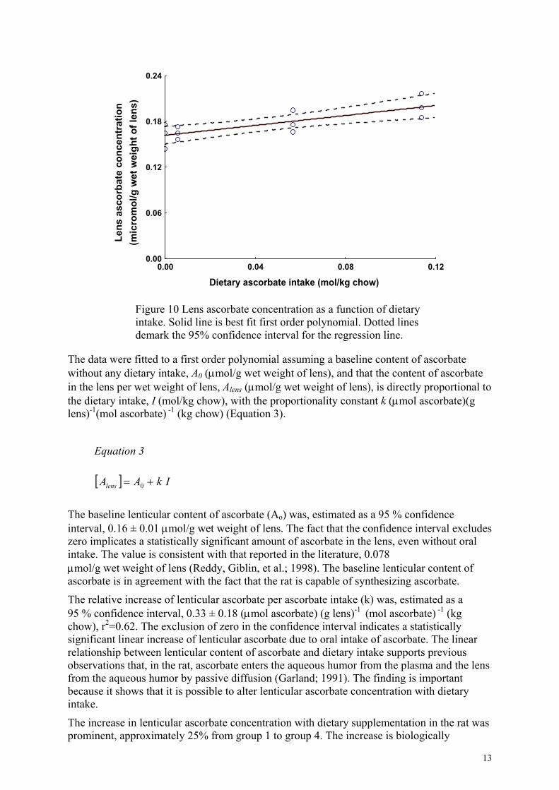
Figure 10 Lens ascorbate concentration as a function of dietary intake. Solid line is best fit first order polynomial. Dotted lines demark the 95% confidence interval for the regression line.
The data were fitted to a first order polynomial assuming a baseline content of ascorbate without any dietary intake, A0 (µmol/g wet weight of lens), and that the content of ascorbate in the lens per wet weight of lens, Alens (µmol/g wet weight of lens), is directly proportional to the dietary intake, I (mol/kg chow), with the proportionality constant k (µmol ascorbate)(g lens)-1(mol ascorbate) -1 (kg chow) (Equation 3).
Equation 3
[ ] IkAAlens += 0
The baseline lenticular content of ascorbate (Ao) was, estimated as a 95 % confidence interval, 0.16 ± 0.01 µmol/g wet weight of lens. The fact that the confidence interval excludes zero implicates a statistically significant amount of ascorbate in the lens, even without oral intake. The value is consistent with that reported in the literature, 0.078 µmol/g wet weight of lens (Reddy, Giblin, et al.; 1998). The baseline lenticular content of ascorbate is in agreement with the fact that the rat is capable of synthesizing ascorbate.
The relative increase of lenticular ascorbate per ascorbate intake (k) was, estimated as a 95 % confidence interval, 0.33 ± 0.18 (µmol ascorbate) (g lens)-1 (mol ascorbate) -1 (kg chow), r2=0.62. The exclusion of zero in the confidence interval indicates a statistically significant linear increase of lenticular ascorbate due to oral intake of ascorbate. The linear relationship between lenticular content of ascorbate and dietary intake supports previous observations that, in the rat, ascorbate enters the aqueous humor from the plasma and the lens from the aqueous humor by passive diffusion (Garland; 1991). The finding is important because it shows that it is possible to alter lenticular ascorbate concentration with dietary intake.
The increase in lenticular ascorbate concentration with dietary supplementation in the rat was prominent, approximately 25% from group 1 to group 4. The increase is biologically
13
important since ascorbate may serve as an antioxidant in the lens. The dependence of lens ascorbate concentration on dietary intake in the rat has not been studied previously. However, our data do correspond with an experiment analyzing the effect of a single intraperitoneal injection of ascorbate into the rat (Reddy, Giblin, et al.; 1998). The observed increase of lenticular ascorbate is important since one may use it to study the preventive effect of lenticular ascorbate against oxidatively- and photochemically-induced cataract in vivo using the rat model.
4.2 Ascorbate in the guinea pig lens, dependence on drinking water supplementation The study aimed at investigating whether lens ascorbate concentration can be increased by drinking water supplementation in the normal guinea pig fed standard chow diet containing ascorbate.
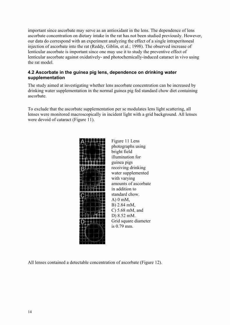
To exclude that the ascorbate supplementation per se modulates lens light scattering, all lenses were monitored macroscopically in incident light with a grid background. All lenses were devoid of cataract (Figure 11).
Figure 11 Lens photographs using bright field illumination for guinea pigs receiving drinking water supplemented with varying amounts of ascorbate in addition to standard chow. A) 0 mM, B) 2.84 mM, C) 5.68 mM, and D) 8.52 mM. Grid square diameter is 0.79 mm.
All lenses contained a detectable concentration of ascorbate (Figure 12).
14
0
0.25
0.5
0.75
1
0 3 6
Ascorbate supplementation in drinking water (mM)
Lens
asc
orba
te c
once
ntra
tion
(µm
ol/g
wet
wei
ght l
ens)
9
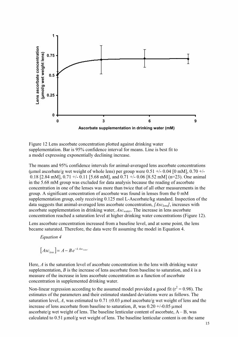
Figure 12 Lens ascorbate concentration plotted against drinking water supplementation. Bar is 95% confidence interval for means. Line is best fit to a model expressing exponentially declining increase.
The means and 95% confidence intervals for animal-averaged lens ascorbate concentrations (µmol ascorbate/g wet weight of whole lens) per group were 0.51 +/- 0.04 [0 mM], 0.70 +/- 0.18 [2.84 mM], 0.71 +/- 0.11 [5.68 mM], and 0.71 +/- 0.06 [8.52 mM] (n=23). One animal in the 5.68 mM group was excluded for data analysis because the reading of ascorbate concentration in one of the lenses was more than twice that of all other measurements in the group. A significant concentration of ascorbate was found in lenses from the 0 mM supplementation group, only receiving 0.125 mol L-Ascorbate/kg standard. Inspection of the data suggests that animal-averaged lens ascorbate concentration, [Asclens], increases with ascorbate supplementation in drinking water, Ascwater. The increase in lens ascorbate concentration reached a saturation level at higher drinking water concentrations (Figure 12).
Lens ascorbate concentration increased from a baseline level, and at some point, the lens became saturated. Therefore, the data were fit assuming the model in Equation 4.
Equation 4
[ ] waterAscklens eBAAsc −−=
Here, A is the saturation level of ascorbate concentration in the lens with drinking water supplementation, B is the increase of lens ascorbate from baseline to saturation, and k is a measure of the increase in lens ascorbate concentration as a function of ascorbate concentration in supplemented drinking water.
15
Non-linear regression according to the assumed model provided a good fit (r2 = 0.98). The estimates of the parameters and their estimated standard deviations were as follows. The saturation level, A, was estimated to 0.71 ±0.03 µmol ascorbate/g wet weight of lens and the increase of lens ascorbate from baseline to saturation, B, was 0.20 +/-0.05 µmol ascorbate/g wet weight of lens. The baseline lenticular content of ascorbate, A – B, was calculated to 0.51 µmol/g wet weight of lens. The baseline lenticular content is on the same

order as that reported in the literature for guinea pigs receiving a standard diet, 0.65 µmol/g wet weight of lens (Varma; 1991). The saturation rate, k , was 0.98 ±1.49 M-1 (1/k = 1.02 M).
In the guinea pig, uptake of ascorbate by the lens from the aqueous humor, and by the aqueous humor from the plasma, occurs by active transport (Garland; 1991). The currently found saturation of lenticular ascorbate secondary to drinking water supplementation (Figure 13) agrees with a previous finding in the guinea pig (Berger, Shepard, et al.; 1988). However, we used a higher amount of ascorbate supplementation. These two findings indicate that the active transport is concentration dependent and saturable.
The increase in lenticular ascorbate concentration with dietary supplementation in the guinea pig was significant, approximately 40 % from group 1 to group 4. The increase is important since it can be used to study the preventive effect of lenticular ascorbate against oxidatively- and photochemically-induced cataract in vivo. We plan to study the modulatory effect of ascorbate on UVR-induced cataract in vivo in the guinea pig.
5. CONCLUSIONS Grinding of the lens in metaphosphoric acid, ultrafiltration of the lens homogenate, and subsequent HPLC with UVR detection can be used to measure ascorbate in both the rat and the guinea pig lens.
The rat lens has a baseline ascorbate content even without any intake. In the rat, lens ascorbate concentration follows a linear dependence on dietary ascorbate intake without cataract development. The guinea pig lens has a detectable quantity of ascorbate with standard dietary ascorbate intake. In the guinea pig, lens ascorbate concentration increases with drinking water supplementation in an exponentially declining fashion, to a saturation level without cataract development.
16
6. ACKNOWLEDGMENTS The research was performed at the Research Department of St. Erik’s Eye Hospital and the Department of Physiology and Pharmacology of Karolinska Institutet, Stockholm Sweden. I would like to express sincere gratitude to all who have helped with completion of this thesis, and in particular to:
Dr. Stefan Löfgren, my main tutor. His outstanding support, enhancement of research skills and techniques, and critical analysis and discussion were beneficial in enabling completion of the experiments and the thesis.
Professor Per G. Söderberg, my co-tutor. He imparted training in essential research techniques, including experimental design and statistical analysis. His constructive criticism helped me to develop as a researcher.
Dr. Marcelo Ayala, my third tutor, for his support through discussion of research work.
Professor Emeritus Björn Tengroth for the outstanding support and guidance throughout the research project. His initial invitation to Karolinska Institutet enabled me to work at St. Erik’s Eye Hospital.
Professor Urban Ungerstedt for his outstanding collaborative support in enabling me to carry out experiments in the pharmacology laboratory.
Ms. Åse Elfving for her help with ascorbate analysis in the pharmacology laboratory.
Dr. Manoj Kakar for his support with experiments, beneficial discussions, and friendship.
Dr. Xiuqin Dong for fruitful discussions and encouragement.
Professor Bo Lindström for expanding my knowledge of statistics.
Ms. Maud Leindahl for her great help with photographic work.
Ms. Monica Aronsson and Ms. Therese Ottinger for their dedicated and hard work in maintaining the animals.
Associate professor Ingeborg Van Der Ploeg for generous use of equipment and discussions.
Ms. Jaana Johansson for her excellent secreterial assistance.
Mr. Mikael Östergren for outstanding computer support.
Dr. Jing Wang, Dr. Yihong Zhang, and Curry Bucht for support as colleagues.
The studies were supported financially by Swedish Society for Medical Research, Swedish Radiation Protection Institute, Karolinska Institutet Research Foundation, Gamla Tjänarinnor Foundation, Crown Princess Margareta Foundation, Karin Sandqvists Foundation, Swedish Medical Society, Swedish Council for Working Life and Social Research, project 2002-0598, Swedish Research Council, project K2004-74KX-15035-01A.
17

7. REFERENCES
Berger J, Shepard D, Morrow F, Sadowski J, Haire T, Taylor A (1988): Reduced and total ascorbate in guinea pig eye tissues in response to dietary intake. Curr Eye Res 7: 681-686. Berger J, Shepard D, Morrow F, Taylor A (1989): Relationship between dietary
intake and tissue levels of reduced and total vitamin C in the nonscorbutic guinea pig. J Nutr 119: 734-740. (5) Blondin J, Baragi V, Schwartz E, Sadowski JA, Taylor A (1986): Delay of UV-
induced eye lens protein damage in guinea pigs by dietary ascorbate. J Free Radic Biol Med 2: 275-281. (4) Cadenas S, Barja G, Poulsen HE, Loft S (1997): Oxidative DNA damage by
oxo8dG in the liver of guinea-pigs supplemented with graded dietary doses of ascorbic acid and alpha-tocopherol. Carcinogenesis 18: 2373-2377. Committee on Animal Nutrition NRC (1987): Ascorbic acid. In Vitamin
Tolerance of Animals. Anonymous. Washington, D.C.: National Academy Press 36-42. Devamanoharan PS, Henein M, Morris S, Ramachandran S, Richards RD, Varma
SD (1991): Prevention of selenite cataract by vitamin C. Exp Eye Res 52: 563-568. (5) DiMattio J (1989): A comparative study of ascorbic acid entry into aqueous and
vitreous humors of the rat and guinea pig. Invest Ophthalmol Vis Sci 30: 2320-2331. (11) Duke-Elder S (1968): The aqueous humor. In System of Ophthalmology: The
Physiology of the Eye and of Vision. IV:Anonymous. London: Henry Kimpton 169. Garland D (1991): Ascorbic acid and the eye. Am J Clin Nutr 54: 1198S-1202S. Halliwell B, Gutteridge JMC (1999): Ch 3 Antioxidant defenses. In Free radicals
in biology and medicine. Anonymous. New York: Oxford University Press 200-208. Hallström A, Carlsson A, Hillered L, Ungerstedt U (1989): Simultaneous
determination of lactate, pyruvate, and ascorbate in microdialysis samples from rat brain, blood, fat, and muscle using high-performance liquid chromatography. J Pharmacol Methods 22: 113-124. (2) Heath H, Beck TC, Rutter AC (1961): Biochemical changes in aphakia. Vision
Res 1: 274-286. Hegde KR, Varma SD (2004): Protective effect of ascorbate against oxidative
stress in the mouse lens. Biochim Biophys Acta 1670: 12-18. (1) Jacques PF, Taylor A, Hankinson SE, Willett WC, Mahnken B, Lee Y, Vaid K,
Lahav M (1997): Long-term vitamin C supplement use and prevalence of early age-related lens opacities. Am J Clin Nutr 66: 911-916. Johnsen H, Ringvold A, Blika S (1985): Ascorbic acid determination in serum
and aqueous humor by high-performance liquid chromatography. Acta Ophthalmol (Copenh) 63: 31-34. Kannan R, Stolz JQ, Prasad PD, Ganapathy V (2001): Vitamin C transport in
human lens epithelial cells: evidence for the presence of SVCT2. Exp Eye Res 73: 159-165. Koshiishi I, Mamura Y, Liu J, Imanari T (1998): Evaluation of an acidic
deproteinization for the measurement of ascorbate and dehydroascorbate in plasma samples. Clin Chem 44: 863-868. Leske MC, Chylac L, Suh-Yuh W (1991): The lens opacities case-control study:
risk factors for cataract. Arch Ophthalmol 109: 244-251.
18
Long C (1961): The Component Parts of the Eye. Biochemists´ Handbook. Princeton, N.J.: Van Nostrand Publishers 706-715.
19
Malik A, Kojima M, Sasaki K (1995): Morphological and biochemical changes in lenses of guinea pigs after vitamin-C-deficient diet and UV-B radiation. Ophthalmic Res 27: 189-196. Mody VC Jr, Kakar M, Elfving Å, Söderberg PG, Löfgren S (2005): Ascorbate In
The Rat Lens, Dependence On Dietary Intake. Ophthalmic Res 37: 142-149. Omaye ST, Turnbull JD, Sanberlica HE (1979): Selected methods for the
determination of ascorbic acid in animal cells, tissue and fluids. Methods Enzymol 62: 3-11. Reddy GB, Bhat KS (1999): Protection against UVB inactivation (in vitro) of rat
lens enzymes by natural antioxidants. Mol Cell Biochem 194: 41-45. Reddy VN, Giblin FJ, Lin LR, Chakrapani B (1998): The effect of aqueous
humor ascorbate on ultraviolet-B-induced DNA damage in lens epithelium. Invest Ophthalmol Vis Sci 39: 344-350. Ringvold A (1980): Aqueous humour and ultraviolet radiation. Acta Ophthalmol
Scand 58: 69-82. Ringvold A (1995): Quenching of UV-induced fluorescence by ascorbic acid in
the aqueous humor. Acta Ophthalmol Scand 73: 529-533. Ringvold A, Anderssen E, Kjonniksen I (1998): Ascorbate in the corneal
epithelium of diurnal and nocturnal species. Invest Ophthalmol Vis Sci 39: 2774-2777. Robertson J, Donner A, Trevithick J (1991): A possible role for vitamins C and E
in cataract prevention. Am J Clin Nutr 53: 346S-351S. Samocha-Bonet D, Lichtenberg D, Pinchuk I (2005): Kinetic studies of copper-
induced oxidation of urate, ascorbate and their mixtures. J Inorg Biochem 99: 1963-1972. (9) Sasaki H, Hata I, Shui YB, Kojima M, Sakamoto A, Kawakami Y, Takahashi N,
Sasaki K (2000): The effect of physiological levels of antioxidants on human lens epithelial cells and rat lenses damaged by UV irradiation [ARVO Abstract]. Invest Ophthalmol Vis Sci 41: 4. (4) Taylor A, Jacques PF, Nowell T, Perrone G, Blumberg J, Handelman G, Jozwiak
B, Nadler D (1997): Vitamin C in human and guinea pig aqueous, lens and plasma in relation to intake. Curr Eye Res 16: 857-864. Taylor A, Jahngen-Hodge J, Smith DE, Palmer VJ, Dallal GE, Lipman RD,
Padhye N, Frei B (1995): Dietary restriction delays cataract and reduces ascorbate levels in Emory mice. Exp Eye Res 61: 55-62. (1) Tessier F, Moreaux V, Birlouez-Aragon I, Junes P, Mondon H (1998): Decrease
in vitamin C concentration in human lenses during cataract progression. Int J Vitam Nutr Res 68: 309-315. Tsao C, Xu L, Young M (1990): Effect of dietary ascorbic acid on heat-induced
eye lens protein damage in guinea pigs. Ophthalmic Res 22: 106-110. Tung WH, Chylack LT, Andley UP (1988): Lens hexokinase deactivation by
near-UV irradiation. Curr Eye Res 7: 257-263. Varma SD (1991): Scientific basis for medical therapy of cataracts by anti-
oxidants. Am J Clin Nutr 53: 335S-345S. Washko PW, Welch RW, Dhariwal KR, Wang Y, Levine M (1992): Ascorbic
acid and dehydroascorbic acid analyses in biological samples. Anal Biochem 204: 1-14. (1)