Embed Size (px)
Citation preview
ARTICLE OPEN ACCESS
Assessment of disease progression in dysferlinopathyA 1-year cohort study
Ursula Moore, MBBchir, Marni Jacobs, PhD, Meredith K. James, PT, Anna G. Mayhew, PT, PhD, Roberto Fernandez-Torron, MD,
Jia Feng, MSc, Avital Cnaan, PhD, Michelle Eagle, PT, PhD, Karen Bettinson, MSc, Laura E. Rufibach, PhD, Robert Muni Lofra, PT,
Andrew M. Blamire, PhD, Pierre G. Carlier, MD, PhD, Plavi Mittal, PhD, Linda Pax Lowes, PT, PhD, Lindsay Alfano, DPT,
Kristy Rose, PT, PhD, Tina Duong, MPT, Katherine M. Berry, PT, Elena Montiel-Morillo, PT, Irene Pedrosa-Hernandez, PT,
Scott Holsten, PT, Mohammed Sanjak, PT, PhD, Ai Ashida, DPT, Chikako Sakamoto, PT, Takayuki Tateishi, PT, Hiroyuki Yajima, PT,
Aurelie Canal, PT, Gwenn Ollivier, PT, Valerie Decostre, PhD, Juan Bosco Mendez, MD, Nieves Sanchez-Aguilera Praxedes, PT,
Simone Thiele, PT, Catherine Siener, PT, MHS, Jeanine Shierbecker, PT, Julaine M. Florence, PT, MHS, DPT, Bruno Vandevelde,
Brittney DeWolf, PT, DPT, PCS, Meghan Hutchence, BSc, PT, Richard Gee, MPT, Juliana Prugel, PT, Elke Maron, PT,
Heather Hilsden, BA, Hanns Lochmuller, MD, Ulrike Grieben, MD, Simone Spuler, MD, Carolina Tesi Rocha, MD, John W. Day, MD,
Kristi J. Jones, MD, PhD, Diana X. Bharucha-Goebel, MD, Emmanuelle Salort-Campana, MD, Matthew Harms, MD,
Alan Pestronk, MD, Sabine Krause, MD, PhD, Olivia Schreiber-Katz, MD, Maggie C. Walter, MD, MA, Carmen Paradas, MD, PhD,
Jean-Yves Hogrel, PhD, Tanya Stojkovic, MD, Shin’ichi Takeda, MD, PhD, Madoka Mori-Yoshimura, MD, PhD, Elena Bravver, MD,
Susan Sparks, MD, Jordi Dıaz-Manera, MD, PhD, Luca Bello, MD, PhD, Claudio Semplicini, MD, PhD, Elena Pegoraro, MD, PhD,
Jerry R. Mendell, MD, Kate Bushby, MD, and Volker Straub, MD, for the Jain COS Consortium
Neurology® 2019;92:e461-e474. doi:10.1212/WNL.0000000000006858
Correspondence
Prof. Straub
volker.straub@
newcastle.ac.uk
AbstractObjectiveTo assess the ability of functional measures to detect disease progression in dysferlinopathy over 6 months and1 year.
MethodsOne hundred ninety-three patients with dysferlinopathy were recruited to the Jain Foundation’s InternationalClinical Outcome Study forDysferlinopathy. Baseline, 6-month, and 1-year assessments included adaptedNorth StarAmbulatory Assessment (a-NSAA), Motor Function Measure (MFM-20), timed function tests, 6-minute walk test(6MWT), Brooke scale, Jebsen test, manual muscle testing, and hand-held dynamometry. Patients also completedthe ACTIVLIM questionnaire. Change in each measure over 6 months and 1 year was calculated and comparedbetween disease severity (ambulant [mild, moderate, or severe based on a-NSAA score] or nonambulant [unable tocomplete a 10-meter walk]) and clinical diagnosis.
ResultsThe functional a-NSAA test was the most sensitive to deterioration for ambulant patients overall. The a-NSAA scorewas the most sensitive test in the mild andmoderate groups, while the 6MWTwas most sensitive in the severe group.The 10-meter walk test was the only test showing significant change across all ambulant severity groups. In non-ambulant patients, theMFMdomain 3, wrist flexion strength, and pinch gripweremost sensitive. Progression rates didnot differ by clinical diagnosis. Power calculations determined that 46 moderately affected patients are required todetermine clinical effectiveness for a hypothetical 1-year clinical trial based on the a-NSAA as a clinical endpoint.
ConclusionCertain functional outcome measures can detect changes over 6 months and 1 year in dysferlinopathy andpotentially be useful in monitoring progression in clinical trials.
ClinicalTrials.gov identifier:NCT01676077.
From the John Walton Muscular Dystrophy Research Centre (U.M., M.K.J., A.G.M., R.F.-T., M.E., K.B., R.M.L., H.H., H.L., K.B., V.S.), Newcastle University and Newcastle Hospitals NHS Foundation Trust, MRC Centre for NeuromuscularDiseases, Institute of Genetic Medicine, Central Parkway, Newcastle Upon Tyne, UK; Center for Translational Science (M.J., J.F., A. Cnaan), Division of Biostatistics and Study Methodology, Cooperative International NeuromuscularResearch Group (T.D., B.D.), and Department of Neurology (D.X.B.-G.), Children’s National Health System; Pediatrics, Epidemiology and Biostatistics (M.J., A. Cnaan), GeorgeWashington University, Washington, DC; Neuromuscular Area(R.F.-T.), Biodonostia Health Research Institute, Neurology Service, Donostia University Hospital, Donostia-San Sebastian, Spain; Jain Foundation (L.E.R., P.M.), Seattle,WA;Magnetic Resonance Centre (A.M.B.), Institute of CellularMedicine,Newcastle University, Newcastle Upon Tyne, UK; AIM &CEA NMR Laboratory (P.G.C.), Institute of Myology, Pitie-Salpetriere University Hospital, 47-83, Paris, France; Research Institute at Nationwide Children’s Hospital (L.P.L., L.A., K.M.B.,J.R.M.), TheOhio StateUniversity, Columbus; Institute forNeuroscience andMuscle Research (K.R.,M.Hutchence, K.J.J.), Children’sHospital atWestmead, University of Sydney, Australia; Lucile Salter PackardChildren’sHospital at Stanford(T.D.), 24349, Neurology, Palo Alto, CA; Physical Medicine and Rehabilitation (E.M.-M., I.P.-H.), Hospital de la Santa Creu i Sant Pau, Barcelona, Spain; Neuroscience Institute (S.H., M.S., E.B., S. Sparks), Carolinas Neuromuscular/ALS-MDACenter, CarolinasHealthCare System, Charlotte, NC; Department of Physical Rehabilitation (A.A., C. Sakamoto, T.T., H.Y.), National Center Hospital, National Center of Neurology and Psychiatry, Tokyo, Japan; Institut deMyologie (A. Canal,G.O., V.D., J.-Y.H., T.S.), AP-HP, GH Pitie-Salpetriere, Paris, France; Neurorehabilitation Unit (J.B.M.), Rehabilitation Hospital Universitario Virgen del Rocıo Sevilla; NeurophysiotherapyDepartment (N.S.-A.P.), Hospital Universitario Virgen delRocıo, Seville, Spain; Friedrich-Baur-Institute (S. Thiele, S.K., O.S.-K. M.C.W.), Department of Neurology, Ludwig-Maximilians-University of Munich, Germany; Department of Neurology (C. Siener, J.S., J.M.F., M. Harms, A.P.), WashingtonUniversity School of Medicine, St. Louis, MO; Centre de Reference des Maladies Neuromusculaires PACA Reunion Rhone Alpes (B.V., E.S.-C.), Hopital de la Timone, Aix-Marseille Universite, France; ELAN-PHYSIO (J.P., E.M.), Praxis furPhysiotherapieMaron; ChariteMuscle Research Unit (U.G., S. Spuler), Experimental and Clinical Research Center, a joint cooperation of the Charite Medical Faculty and theMax Delbruck Center forMolecularMedicine, Berlin, Germany;DepartmentofNeurology andNeurological Sciences (C.T.R., J.W.D.), StanfordUniversity School ofMedicine, CA;NIH (D.X.B.-G.), Bethesda,MD;Neuromuscular Unit (C.P.), Department ofNeurology,Hospital U. Virgendel Rocıo/Instituto deBiomedicinadeSevilla, Spain;DepartmentofNeurology (S. Takeda,M.M.-Y.),NationalCenterHospital,NationalCenterofNeurologyandPsychiatry, Tokyo, Japan;Centrode InvestigacionBiomedicaenRedenEnfermedadesRaras (J.D.-M.);Neuromuscular Disorders Unit (J.D.-M.), Neurology Department, Hospital de la Santa Creu i Sant Pau, Barcelona, Spain; and Department of Neuroscience (L.B., C. Semplicini, E.P.), University of Padova, Italy.
Go to Neurology.org/N for full disclosures. Funding information and disclosures deemed relevant by the authors, if any, are provided at the end of the article.
The Jain COS Consortium coinvestigators are listed in the Appendix at the end of the article.
The Article Processing Charge was funded by the Jain Foundation.
This is an open access article distributed under the terms of the Creative Commons Attribution-NonCommercial-NoDerivatives License 4.0 (CC BY-NC-ND), which permits downloading and sharing the work provided it isproperly cited. The work cannot be changed in any way or used commercially without permission from the journal.
Copyright © 2019 The Author(s). Published by Wolters Kluwer Health, Inc. on behalf of the American Academy of Neurology. e461
Dysferlinopathy is a rare, recessively inherited muscular dys-trophy caused by mutations in the DYSF1–4 gene. Potentialtherapies are in development, and some of them have enteredhuman clinical trials (NCT02579239, NCT01863004,NCT02710500). As the development of newly licensed andemerging treatments for Duchenne muscular dystrophy5,6
and spinal muscular atrophy type 17 has shown, proving ef-ficacy and satisfying regulators can be difficult in slowly pro-gressive and varied conditions.8
Appropriate powering of a clinical trial requires understandingof disease progression and responsiveness of various outcomemeasures over time.9 Selection of outcomemeasures for clinicaltrials in dysferlinopathy is particularly challenging. Dysferlin-opathy is characterized by a range of ages at onset and patternsof weakness and severity,4,10 variable rate of progression,4
modifying factors that are not clearly elucidated,10–13 and noappropriate measures for monitoring progression. While somemeasures such as the Motor Function Measure (MFM) havebeen shown to be reliable in dysferlinopathy,14 the variability ofprogression makes it difficult to demonstrate responsiveness insmall cohorts.
The Jain Clinical Outcome Study in Dysferlinopathy (COS)aims to address these difficulties by characterizing the clinical,biochemical, and radiologic parameters of 209 patients withdysferlinopathy over 3 years.
This article summarizes findings from a planned interimanalysis of baseline, 6-month, and year 1 data to determinewhether disease progression is detectable and which measuresare most able to detect this progress. We propose a powercalculation using the most sensitive measures to determinea potential clinical trial cohort required to demonstrate sig-nificant functional change.
MethodsThe COS studyThe COS study is an international collaborative study ofpatients with a genetic and/or protein assay–confirmed di-agnosis of dysferlinopathy.4 Screening, baseline, 6-month, and1-year visits took place between October 2012 and March2016. One hundred ninety-seven of the original 209 recruitedpatients completed a baseline visit. Of the original 209patients, 7 did not meet inclusion criteria, and 5 chose not tocontinue past the screening visit (figure 1). Full inclusion andexclusion criteria, the study protocol, and patient de-mographics have been described previously.4
Standard protocol approvals, registrations,and patient consentsThe study was initially approved by the following: NRESCommittee North East—Newcastle & North Tyneside onFebruary 2, 2012 (reference 211/NE/0360/R&D 5918). Thestudy was also approved by ethics review boards in eachcountry. All patients provided written informed consent. Thetrial was registered at ClinicalTrials.gov (NCT01676077).
Functional outcome measuresClinical assessments were standardized through trainings atinvestigator meetings, and all evaluations were performed bytrained clinicians. Assessments included the adapted NorthStar Ambulatory Assessment (a-NSAA),4 MFM-20, timedfunction tests (timed rise from floor, 10-m walk/run, 4-stairclimb and descend, Timed Up and Go, and the 6-minute walktest [6MWT]), Brooke Upper Extremity Scale, Jebsen HandFunction Test, manual muscle testing (MMT), and hand-helddynamometry (HHD). No assessments have been specificallyvalidated in a dysferlin-specific population. These assessmentswere chosen because of their widespread suitability for limbgirdle muscular dystrophy. The MFM-20 was selected overthe MFM-32 to reduce patient fatigue and duplication. Allassessments were attempted for ambulant patients. Non-ambulant patients did not complete the a-NSAA orambulation-based timed function tests.
Muscle strength was assessed with an 11-point scale forMMT.4 The best of 3 attempts for HHDwas used for analysis.
Functional ability was assessed with qualitative scales andtimed tests. Total scores were calculated for the a-NSAA15 inambulant patients, and the MFM-20 and the Brooke scalewere calculated in all patients. TheMFM-20 was also analyzedon the basis of its 3 components: D1 (standing and transfers),D2 (axial and proximal), and D3 (distal). Any patient with anincomplete score was excluded from analysis for that test. Toaccount for patients who became nonambulant or unable tocomplete timed function tests due to disease progression,timed test values were converted to velocity measures (eithermeter per second or task per second), with a 0-m/s velocityassigned for the first instance a patient was unable to completethe test.
Patient perception of progressionThe ACTIVLIM questionnaire, a Rasch-built patient-reported outcome measure,16 was used to capture patient-reported functional ability. The total score, out of a maximumof 36, was compared between visits. A higher score indicatesgreater functional ability.
Glossarya-NSAA = adapted North Star Ambulatory Assessment;COS = Jain Clinical Outcome Study in Dysferlinopathy;HHD = hand-held dynamometry; LGMD2B = limb girdle muscular dystrophy 2B;MFM =Motor FunctionMeasure;MMT =manual muscletesting; 6MWT = 6-minute walk test; SRM = standardized response mean.
e462 Neurology | Volume 92, Number 5 | January 29, 2019 Neurology.org/N

Statistical analysisStatistical analysis was completed with SAS 9.3 (SAS InstituteInc, Cary, NC). Mean and median change scores were cal-culated for each test between baseline and 6 months, 6months and year 1, and baseline and year 1. Data from somevisits were excluded if the visit occurred ±2 months outsidethe scheduled appointment date. A majority of functionaloutcome measures were not normally distributed; thus,comparisons between baseline and follow-up time pointswere conducted with the Wilcoxon signed rank-sum test andstandardized response mean (SRM).17 A comparison wasmade between the performance of patients with the mostcommon clinical diagnoses of limb girdle muscular dystrophy2B (LGMD2B) and Miyoshi myopathy with Kruskal-Wallistest with Steel-Dwass-Critchlow-Fligner correction for mul-tiple comparisons.18
Nonambulant patients (unable to walk 10 meters with usualorthotics and walking aids) were compared to 3 categories ofambulant patients: ambulation affected severely, moderately,or mildly. Using patients’ a-NSAA scores at baseline, we de-fined ambulant severity categories based on a clinical im-pression of disease severity and binomial distribution ofa-NSAA scores, with cut points of 0 to 10 (severe diseaseseverity), 11 to 40 (moderate disease severity), and 41+ (milddisease severity). The lower cut point was based on the end ofthe first a-NSAA peak, and the high cut point was placed at thestart of the second peak, with the wide spread in betweenrepresenting a plateau between peaks. Significance was de-fined as p ≤ 0.05. Results are expressed as median change(range) and p value.
Power analyses to support a hypothetical future clinical trialwere conducted to estimate the sample size required to detectsignificant statistical differences in progression over the courseof 1 year with PASS 14 software. Because change scores werenormally distributed, calculations used mean change scores
and SDs of the functional measure that demonstrated mostchange over time. For this analysis, the a-NSAA was used,which limited estimates to ambulant patients only. Samplesizes needed to evaluate a variety of expected drug effects werecalculated for reference, including halting of disease pro-gression (i.e., assuming mean progression estimates from thecurrent sample among untreated and no progression amongtreated patients), reflective of potential protein-restoring ge-netic therapies. An α level of 0.05, 80% power, and a treated-to-untreated ratio of 1:1 were assumed. For comparison witha widely used measure in muscular dystrophy, sample sizeswere also calculated on the basis of the 6MWT.
Data availability statementDeidentified cohort level data can be requested. All protocolassessments are in the public domain apart from the a-NSAA,which can be requested from the steering group (publicationin draft). The statistical plan is detailed above. All data for thisnatural history study will be stored for at least 5 years after theend of the study. Data can be requested by all interestedstakeholders for clinical research and trial planning. Datarequests are reviewed by the Jain COS Steering Committee,who can be contacted via the corresponding author.
ResultsOf the initial 197 eligible enrolled patients, 3 patients with-drew after baseline, and 1 withdrew after 6 months. This issummarized in figure 1. Patients included in this analysis werebetween 11 and 86 years of age at baseline and were at allstages of disease progression, from asymptomatic hyper-CKemia to nonambulant.
Functional outcome measuresFunctional outcome measure results are summarized in tables1 and 2, figure 2, and table e-1 (available from Dryad, doi.org/10.5061/dryad.tp08m60). Some of the assessments, by their
Figure 1 Flowchart of patient numbers at each visit
Number of patients who completed each assessment (baseline, 6 months, and 12 months) and how many of them were used for each analysis window.
Neurology.org/N Neurology | Volume 92, Number 5 | January 29, 2019 e463
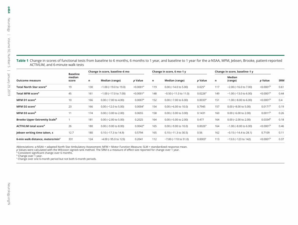
Table 1 Change in scores of functional tests from baseline to 6 months, 6 months to 1 year, and baseline to 1 year for the a-NSAA, MFM, Jebsen, Brooke, patient-reportedACTIVLIM, and 6-minute walk tests
Outcome measure
Baselinemedianscore
Change in score, baseline–6 mo Change in score, 6 mo–1 y Change in score, baseline–1 y
SRMn Median (range) p Value n Median (range) p Value nMedian(range) p Value
Total North Star scorea 19 130 −1.00 (−19.0 to 19.0) <0.0001a 119 0.00 (−14.0 to 5.00) 0.025a 117 −2.00 (−16.0 to 7.00) <0.0001b 0.61
Total MFM scorea 45 161 −1.00 (−17.0 to 7.00) <0.0001a 148 −0.50 (−11.0 to 11.0) 0.0226a 149 −1.00 (−13.0 to 6.00) <0.0001b 0.44
MFM D1 scorea 10 166 0.00 (−7.00 to 4.00) 0.0007a 152 0.00 (−7.00 to 6.00) 0.0033a 151 −1.00 (−8.00 to 6.00) <0.0001b 0.4
MFM D2 scorec 23 166 0.00 (−12.0 to 5.00) 0.0004c 154 0.00 (−6.00 to 10.0) 0.7945 157 0.00 (−8.00 to 5.00) 0.0171b 0.19
MFM D3 scoreb 11 174 0.00 (−3.00 to 2.00) 0.0655 158 0.00 (−3.00 to 3.00) 0.1431 160 0.00 (−6.00 to 2.00) 0.0011b 0.26
Brooke Upper Extremity Scaleb 1 181 0.00 (−2.00 to 5.00) 0.2025 164 0.00 (−5.00 to 2.00) 0.477 164 0.00 (−2.00 to 2.00) 0.0334b 0.18
ACTIVLIM total scorea 26 180 0.00 (−9.00 to 8.00) 0.0042a 165 0.00 (−9.00 to 10.0) 0.0026a 164 −1.00 (−8.00 to 6.00) <0.0001b 0.46
Jebsen writing time taken, s 12.7 180 0.10 (−17.3 to 14.9) 0.5794 165 0.10 (−11.3 to 30.5) 0.56 162 −0.15 (−14.4 to 28.1) 0.7109 0.11
6-min walk distance, meters/minc 331 124 −4.00 (−95.0 to 123) 0.2041 112 −7.00 (−110 to 51.0) 0.0003c 113 −13.0 (−123 to 142) <0.0001b 0.37
Abbreviations: a-NSAA = adapted North Star Ambulatory Assessment; MFM = Motor Function Measure; SLM = standardized response mean.p Values were calculated with the Wilcoxon signed-rank method. The SRM is a measure of effect size reported for change over 1 year.a Consistent significant change over 6 months.b Change over 1 year.c Change over one 6-month period but not both 6-month periods.
e464Neu
rology
|Vo
lume92,N
umber
5|
January
29,2019Neurology.org/N
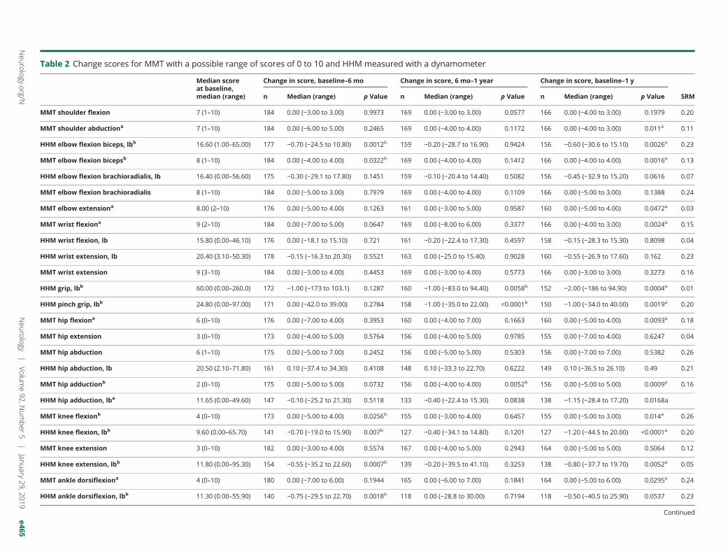
Table 2 Change scores for MMT with a possible range of scores of 0 to 10 and HHM measured with a dynamometer
Median scoreat baseline,median (range)
Change in score, baseline–6 mo Change in score, 6 mo–1 year Change in score, baseline–1 y
SRMn Median (range) p Value n Median (range) p Value n Median (range) p Value
MMT shoulder flexion 7 (1–10) 184 0.00 (−3.00 to 3.00) 0.9973 169 0.00 (−3.00 to 3.00) 0.0577 166 0.00 (−4.00 to 3.00) 0.1979 0.20
MMT shoulder abductiona 7 (1–10) 184 0.00 (−6.00 to 5.00) 0.2465 169 0.00 (−4.00 to 4.00) 0.1172 166 0.00 (−4.00 to 3.00) 0.011a 0.11
HHM elbow flexion biceps, lbb 16.60 (1.00–65.00) 177 −0.70 (−24.5 to 10.80) 0.0012b 159 −0.20 (−28.7 to 16.90) 0.9424 156 −0.60 (−30.6 to 15.10) 0.0026a 0.23
MMT elbow flexion bicepsb 8 (1–10) 184 0.00 (−4.00 to 4.00) 0.0322b 169 0.00 (−4.00 to 4.00) 0.1412 166 0.00 (−4.00 to 4.00) 0.0016a 0.13
HHM elbow flexion brachioradialis, lb 16.40 (0.00–56.60) 175 −0.30 (−29.1 to 17.80) 0.1451 159 −0.10 (−20.4 to 14.40) 0.5082 156 −0.45 (−32.9 to 15.20) 0.0616 0.07
MMT elbow flexion brachioradialis 8 (1–10) 184 0.00 (−5.00 to 3.00) 0.7979 169 0.00 (−4.00 to 4.00) 0.1109 166 0.00 (−5.00 to 3.00) 0.1388 0.24
MMT elbow extensiona 8.00 (2–10) 176 0.00 (−5.00 to 4.00) 0.1263 161 0.00 (−3.00 to 5.00) 0.9587 160 0.00 (−5.00 to 4.00) 0.0472a 0.03
MMT wrist flexiona 9 (2–10) 184 0.00 (−7.00 to 5.00) 0.0647 169 0.00 (−8.00 to 6.00) 0.3377 166 0.00 (−4.00 to 3.00) 0.0024a 0.15
HHM wrist flexion, lb 15.80 (0.00–46.10) 176 0.00 (−18.1 to 15.10) 0.721 161 −0.20 (−22.4 to 17.30) 0.4597 158 −0.15 (−28.3 to 15.30) 0.8098 0.04
HHM wrist extension, lb 20.40 (3.10–50.30) 178 −0.15 (−16.3 to 20.30) 0.5521 163 0.00 (−25.0 to 15.40) 0.9028 160 −0.55 (−26.9 to 17.60) 0.162 0.23
MMT wrist extension 9 (3–10) 184 0.00 (−3.00 to 4.00) 0.4453 169 0.00 (−3.00 to 4.00) 0.5773 166 0.00 (−3.00 to 3.00) 0.3273 0.16
HHM grip, lbb 60.00 (0.00–260.0) 172 −1.00 (−173 to 103.1) 0.1287 160 −1.00 (−83.0 to 94.40) 0.0058b 152 −2.00 (−186 to 94.90) 0.0004a 0.01
HHM pinch grip, lbb 24.80 (0.00–97.00) 171 0.00 (−42.0 to 39.00) 0.2784 158 −1.00 (−35.0 to 22.00) <0.0001b 150 −1.00 (−34.0 to 40.00) 0.0019a 0.20
MMT hip flexiona 6 (0–10) 176 0.00 (−7.00 to 4.00) 0.3953 160 0.00 (−4.00 to 7.00) 0.1663 160 0.00 (−5.00 to 4.00) 0.0093a 0.18
MMT hip extension 3 (0–10) 173 0.00 (−4.00 to 5.00) 0.5764 156 0.00 (−4.00 to 5.00) 0.9785 155 0.00 (−7.00 to 4.00) 0.6247 0.04
MMT hip abduction 6 (1–10) 175 0.00 (−5.00 to 7.00) 0.2452 156 0.00 (−5.00 to 5.00) 0.5303 156 0.00 (−7.00 to 7.00) 0.5382 0.26
HHM hip abduction, lb 20.50 (2.10–71.80) 161 0.10 (−37.4 to 34.30) 0.4108 148 0.10 (−33.3 to 22.70) 0.6222 149 0.10 (−36.5 to 26.10) 0.49 0.21
MMT hip adductionb 2 (0–10) 175 0.00 (−5.00 to 5.00) 0.0732 156 0.00 (−4.00 to 4.00) 0.0052b 156 0.00 (−5.00 to 5.00) 0.0009a 0.16
HHM hip adduction, lba 11.65 (0.00–49.60) 147 −0.10 (−25.2 to 21.30) 0.5118 133 −0.40 (−22.4 to 15.30) 0.0838 138 −1.15 (−28.4 to 17.20) 0.0168a
MMT knee flexionb 4 (0–10) 173 0.00 (−5.00 to 4.00) 0.0256b 155 0.00 (−3.00 to 4.00) 0.6457 155 0.00 (−5.00 to 3.00) 0.014a 0.26
HHM knee flexion, lbb 9.60 (0.00–65.70) 141 −0.70 (−19.0 to 15.90) 0.007b 127 −0.40 (−34.1 to 14.80) 0.1201 127 −1.20 (−44.5 to 20.00) <0.0001a 0.20
MMT knee extension 3 (0–10) 182 0.00 (−3.00 to 4.00) 0.5574 167 0.00 (−4.00 to 5.00) 0.2943 164 0.00 (−5.00 to 5.00) 0.5064 0.12
HHM knee extension, lbb 11.80 (0.00–95.30) 154 −0.55 (−35.2 to 22.60) 0.0007b 139 −0.20 (−39.5 to 41.10) 0.3253 138 −0.80 (−37.7 to 19.70) 0.0052a 0.05
MMT ankle dorsiflexiona 4 (0–10) 180 0.00 (−7.00 to 6.00) 0.1944 165 0.00 (−6.00 to 7.00) 0.1841 164 0.00 (−5.00 to 6.00) 0.0295a 0.24
HHM ankle dorsiflexion, lbb 11.30 (0.00–55.90) 140 −0.75 (−29.5 to 22.70) 0.0018b 118 0.00 (−28.8 to 30.00) 0.7194 118 −0.50 (−40.5 to 25.90) 0.0537 0.23
Continued
Neurolo
gy.org/N
Neurology
|Volum
e92,N
umber
5|
January29,2019
e465
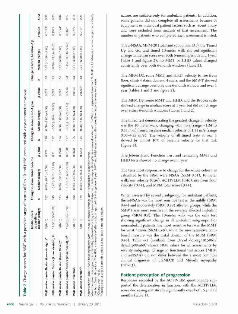
nature, are suitable only for ambulant patients. In addition,some patients did not complete all assessments because ofequipment or individual patient factors such as recent injuryand were excluded from analysis of that assessment. Thenumber of patients who completed each assessment is listed.
The a-NSAA, MFM-20 (total and subdomain D1), the TimedUp and Go, and timed 10-meter walk showed significantchange in median score over both 6-month periods and 1 year(table 1 and figure 2); no MMT or HHD values changedconsistently over both 6-month windows (table 2).
The MFM D2, some MMT and HHD, velocity to rise fromfloor, climb 4 stairs, descend 4 stairs, and the 6MWT showedsignificant change over only one 6-month window and over 1year (tables 1 and 2 and figure 2).
The MFM D3, some MMT and HHD, and the Brooke scaleshowed change in median score at 1 year but did not changeover either 6-month windows (tables 1 and 2).
The timed test demonstrating the greatest change in velocitywas the 10-meter walk, changing −0.1 m/s (range −1.24 to0.55 m/s) from a baseline median velocity of 1.11 m/s (range0.00–4.55 m/s). The velocity of all timed tests at year 1slowed by almost 10% of baseline velocity for that task(figure 2).
The Jebsen Hand Function Test and remaining MMT andHHD tests showed no change over 1 year.
The tests most responsive to change for the whole cohort, ascalculated by the SRM, were NSAA (SRM 0.61), 10-meterwalk/run velocity (0.56), ACTIVLIM (0.46), rise from floorvelocity (0.44), and MFM total score (0.44).
When assessed by severity subgroup, for ambulant patients,the a-NSAA was the most sensitive test in the mildly (SRM0.44) and moderately (SRM 0.89) affected groups, while the6MWT was most sensitive in the severely affected ambulantgroup (SRM 0.9). The 10-meter walk was the only testshowing significant change in all ambulant subgroups. Fornonambulant patients, the most sensitive test was the MMTfor wrist flexion (SRM 0.69), while the most sensitive com-bined measure was the distal domain of the MFM (SRM0.46). Table e-1 (available from Dryad doi.org/10.5061/dryad.tp08m60) shows SRM values for all assessments byseverity subgroup. Change in functional test scores (MFMand a-NSAA) did not differ between the 2 most commonclinical diagnoses of LGMD2B and Miyoshi myopathy(table 3).
Patient perception of progressionResponses recorded by the ACTIVLIM questionnaire sup-ported the deterioration in function, with the ACTIVLIMscore decreasing statistically significantly over both 6 and 12months (table 1).Ta
ble
2Chan
gescoresforMMTwithapossible
range
ofscoresof0to
10an
dHHM
mea
suredwithadyn
amometer
(con
tinue
d)
Medianscore
atbase
line,
median(range
)
Change
inscore
,base
line–6mo
Change
inscore
,6mo–1ye
ar
Change
inscore
,base
line–1y
SRM
nMedian(range
)pValue
nMedian(range
)pValue
nMedian(range
)pValue
MMTankle
planta
rflexion(kneestra
ight)
a3(0–10
)15
70.00
(−10
.0to
7.00
)0.27
313
90.00
(−6.00
to8.00
)0.21
213
70.00
(−10
.0to
8.00
)0.01
93a
0.23
HHM
ankle
planta
rflexo
rs(kneestra
ight),lb
12.60(0.00–
81.00)
140
−0.40
(−34
.2to
23.70)
0.21
127
−0.50
(−28
.6to
32.90)
0.22
912
3−0.70
(−33
.9to
30.20)
0.19
450.23
MMTankle
planta
rflexion(kneeflexe
d)a
3(0–10
)17
30.00
(−6.00
to7.00
)0.25
2815
40.00
(−5.00
to6.00
)0.24
215
30.00
(−6.00
to5.00
)0.01
74a
0.04
HHM
ankle
planta
rflexo
rs(kneeflexe
d),lb
b12
.20(0.00–
57.90)
144
−0.75
(−25
.6to
29.00)
0.01
38b
127
−0.30
(−34
.5to
22.10)
0.22
3412
4−1.15
(−39
.6to
22.60)
0.00
2a0.17
MMTankle
inve
rsion
7(0–10
)18
00.00
(−7.00
to6.00
)0.68
2816
50.00
(−5.00
to4.00
)0.81
5416
40.00
(−4.00
to5.00
)0.67
890.16
MMTankle
eve
rsionb
5(0–10
)17
90.00
(−6.00
to4.00
)0.82
3216
40.00
(−5.00
to4.00
)0.00
03b
164
0.00
(−6.00
to3.00
)0.01
3a0.21
Abbreviations:
HHM
=han
d-heldmyo
metry;M
MT=man
ual
muscle
testing;
SRM
=stan
dardized
resp
onse
mea
n.
When
thesa
memusclegroupsaretested
byboth
methods,they
aregrouped
toge
ther
toaidinterp
retation.p
Values
wereca
lculatedwiththeWilcoxo
nsign
ed-ran
kmethod.N
oMMTorHHM
asse
ssmen
tsch
ange
dco
nsisten
tly
ove
rboth
6-month
periods.
TheSR
M,a
mea
sure
ofeffect
size
,isreported
forch
ange
ove
r1ye
ar.M
MTan
dHHM
asse
ssmen
tsallshowed
low
resp
onsive
nes
sto
chan
ge.
aChan
geove
r1ye
ar.
bChan
geove
rasingle6-month
periodbutnotboth
6-month
periods.
e466 Neurology | Volume 92, Number 5 | January 29, 2019 Neurology.org/N
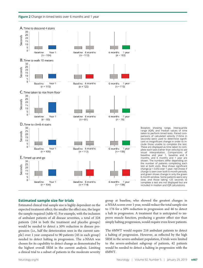
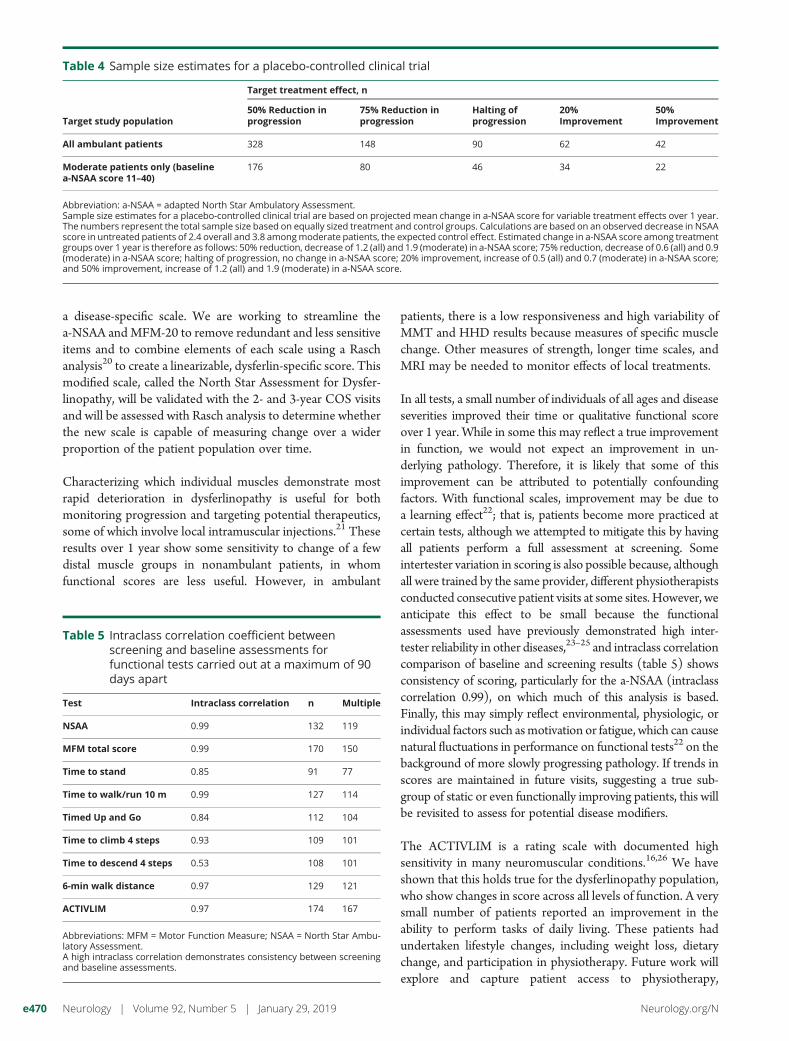
Estimated sample size for trialsEstimated clinical trial sample size is highly dependent on theexpected treatment effect; the smaller the effect size, the largerthe sample required (table 4). For example, with the inclusionof ambulant patients of all disease severities, a total of 328patients (164 in both the treatment and placebo groups)would be needed to detect a 50% reduction in disease pro-gression (i.e., half the deterioration seen in the current sam-ple) over 1 year compared to 90 patients (45 in each group)needed to detect halting in progression. The a-NSAA waschosen for its capability to detect change as demonstrated bythe highest overall SRM in the current analysis. Limitinga clinical trial to a subset of patients in the moderate severity
group at baseline, who showed the greatest changes ina-NSAA scores over 1 year, would reduce the total sample sizeto 176 for a 50% reduction in progression and 46 to detecta halt in progression. A treatment that is anticipated to im-prove muscle function, producing a greater effect size thansimply halting progression, would require even fewer patients.
The 6MWT would require 216 ambulant patients to detecta halting of progression. However, as reflected by the highSRM in the severe-ambulant population, if trials were limitedto the severe-ambulant subgroup of patients, 42 patientswould be needed to detect a halting in progression with the6MWT.
Figure 2 Change in timed tests over 6 months and 1 year
Boxplots showing range, interquartilerange (IQR), and median values of timetaken to perform timed tests. Paired com-parisons of calculated velocity (1/time inseconds) were used to determine signifi-cant vs insignificant change in order to in-clude those unable to complete the test.These are displayed as time taken to com-plete each task (rather than velocity) to aidvisual interpretation. Comparisons ofbaseline and year 1, baseline and 6months, and 6 months and 1 year areshown. The numbers differ depending onthe number of patients completing eachtest at both visits. Blue shows significantchange (p < 0.05) over 1 year; red shows ifchange is seen over both 6-month periods;and green shows change in only the green6-month window. Some patients were veryslow, and those taking >25 seconds tocomplete a test are not displayed but areincluded in median and IQR calculations.
Neurology.org/N Neurology | Volume 92, Number 5 | January 29, 2019 e467
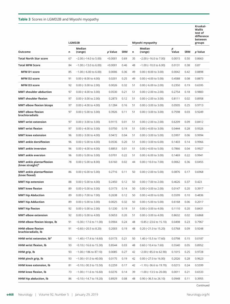
Table 3 Scores in LGMD2B and Miyoshi myopathy
Outcome
LGMD2B Miyoshi myopathy
Kruskal-Wallistest ofdifferencebetweengroups
nMedian(range) p Value SRM n
Median(range)
pValue SRM p Value
Total North Star score 67 −2.00 (−14.0 to 5.00) <0.0001 0.69 35 −2.00 (−16.0 to 7.00) 0.0015 0.50 0.8663
Total MFM Score 84 −1.00 (−13.0 to 6.00) <0.0001 0.46 48 −1.00 (−10.0 to 6.00) 0.0131 0.38 0.87
MFM D1 score 85 −1.00 (−6.00 to 6.00) 0.0006 0.36 49 0.00 (−8.00 to 3.00) 0.0042 0.42 0.8898
MFM D2 score 91 0.00 (−8.00 to 4.00) 0.0201 0.25 49 0.00 (−4.00 to 5.00) 0.4588 0.08 0.8873
MFM D3 score 92 0.00 (−3.00 to 2.00) 0.0026 0.32 51 0.00 (−6.00 to 2.00) 0.2350 0.19 0.6595
MMT shoulder abduction 97 0.00 (−4.00 to 3.00) 0.0530 0.21 51 0.00 (−2.00 to 2.00) 0.2754 0.18 0.9883
MMT shoulder flexion 97 0.00 (−3.00 to 2.00) 0.2873 0.12 51 0.00 (−2.00 to 3.00) 0.8111 0.02 0.8958
MMT elbow flexion biceps 97 0.00 (−4.00 to 4.00) 0.1284 0.16 51 0.00 (−3.00 to 3.00) 0.0505 0.25 0.9713
MMT elbow flexionbrachioradialis
97 0.00 (−5.00 to 3.00) 0.3926 0.11 51 0.00 (−3.00 to 3.00) 0.7598 0.03 0.9249
MMT wrist extension 97 0.00 (−3.00 to 3.00) 0.9115 0.01 51 0.00 (−2.00 to 2.00) 0.6209 0.09 0.8412
MMT wrist flexion 97 0.00 (−4.00 to 3.00) 0.0750 0.19 51 0.00 (−4.00 to 3.00) 0.0444 0.28 0.9326
MMT knee extension 96 0.00 (−3.00 to 4.00) 0.9472 0.04 51 0.00 (−3.00 to 5.00) 0.5997 0.06 0.9994
MMT ankle dorsiflexion 96 0.00 (−5.00 to 4.00) 0.0536 0.20 51 0.00 (−3.00 to 6.00) 0.1403 0.14 0.9966
MMT ankle inversion 96 0.00 (−4.00 to 4.00) 0.8853 0.01 51 0.00 (−4.00 to 5.00) 0.7866 0.04 0.9927
MMT ankle eversion 96 0.00 (−5.00 to 3.00) 0.0701 0.22 51 0.00 (−6.00 to 3.00) 0.1469 0.22 0.9941
MMT ankle plantarflexion(knee straight)a
78 0.00 (−5.00 to 8.00) 0.6160 0.02 44 0.00 (−10.0 to 7.00) 0.0062 0.36 0.0455
MMT ankle plantarflexion(knee flexed)
86 0.00 (−6.00 to 5.00) 0.2716 0.11 50 0.00 (−2.00 to 5.00) 0.0876 0.17 0.6968
MMT hip extension 88 0.00 (−5.00 to 4.00) 0.2450 0.12 50 0.00 (−7.00 to 2.00) 0.4626 0.37 0.423
MMT knee flexion 89 0.00 (−5.00 to 3.00) 0.3173 0.14 50 0.00 (−3.00 to 2.00) 0.0147 0.20 0.3917
MMT hip Abduction 89 0.00 (−7.00 to 7.00) 0.2638 0.12 50 0.00 (−4.00 to 6.00) 0.5599 0.10 0.4836
MMT hip Adduction 89 0.00 (−5.00 to 3.00) 0.0025 0.32 50 0.00 (−5.00 to 5.00) 0.6168 0.06 0.2017
MMT hip flexion 92 0.00 (−5.00 to 2.00) 0.1230 0.19 51 0.00 (−3.00 to 4.00) 0.1110 0.20 0.8431
MMT elbow extension 92 0.00 (−5.00 to 4.00) 0.0653 0.20 51 0.00 (−3.00 to 4.00) 0.8632 0.02 0.6868
HHM elbow flexion biceps, lb 91 −0.30 (−17.6 to 11.00) 0.0904 0.24 48 −0.85 (−23.6 to 15.10) 0.0498 0.23 0.7967
HHM elbow flexionbrachioradialis, lb
91 −0.60 (−20.5 to 8.20) 0.2003 0.19 48 0.20 (−21.0 to 15.20) 0.5768 0.09 0.9248
HHM wrist extension, lba 93 −1.40 (−17.4 to 14.60) 0.0173 0.21 50 1.40 (−15.5 to 17.60) 0.0798 0.15 0.0187
HHM wrist flexion, lb 93 −0.10 (−16.6 to 15.30) 0.8544 0.03 48 0.60 (−10.4 to 7.60) 0.5540 0.05 0.8952
HHM grip, lb 93 −1.00 (−186 to 87.10) 0.0085 0.27 42 −2.00 (−85.0 to 62.90) 0.1015 0.25 0.9728
HHM pinch grip, lb 93 −1.00 (−31.0 to 40.00) 0.0175 0.19 42 0.00 (−27.0 to 16.00) 0.2026 0.28 0.9623
HHM knee extension, lb 81 −0.10 (−30.3 to 15.50) 0.2259 0.17 42 −1.10 (−36.6 to 19.70) 0.0215 0.24 0.5599
HHM knee flexion, lb 73 −1.00 (−11.6 to 16.60) 0.0276 0.14 39 −1.00 (−13.5 to 20.00) 0.0011 0.21 0.6533
HHM hip abduction, lb 86 −0.10 (−14.7 to 19.20) 0.8929 0.08 48 0.90 (−36.5 to 26.10) 0.0948 0.11 0.3955
Continued
e468 Neurology | Volume 92, Number 5 | January 29, 2019 Neurology.org/N
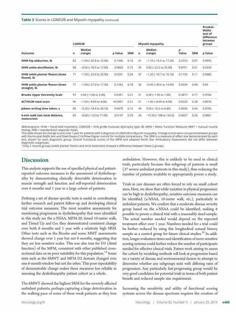
DiscussionThis analysis supports the use of specified physical and patient-reported outcome measures in the assessment of dysferlinop-athy by demonstrating clinically detectable deterioration inmuscle strength and function and self-reported deteriorationover 6 months and 1 year in a large cohort of patients.
Defining a set of disease specific tests is useful in coordinatingfurther research and patient follow-up and developing clinicaltrial outcome measures. The most sensitive assessments formonitoring progression in dysferlinopathy that were identifiedin this study are the a-NSAA, MFM-20, timed 10-meter walk,and Timed Up and Go; they demonstrated consistent changeover both 6 months and 1 year with a relatively high SRM.Other tests such as the Brooke and some MMT assessmentsshowed change over 1 year but not 6 months, suggesting thatthey are less sensitive scales. This was also true for D3 (distalfunction) of the MFM, consistent with other published cross-sectional data on its poor suitability for this population.14 Sometests such as the 6MWT and MFM D2 domain changed overone 6-month window but not the other. This poor repeatabilityof demonstrable change makes these measures less reliable inassessing the dysferlinopathy patient cohort as a whole.
The 6MWT showed the highest SRM for the severely affectedambulant patients, perhaps capturing a large deterioration inthe walking pace of some of these weak patients as they lose
ambulation. However, this is unlikely to be used in clinicaltrials, particularly because this subgroup of patients is small(37 severe-ambulant patients in this study), thus reducing thenumber of patients available to appropriately power a study.
Trials in rare diseases are often forced to rely on small cohortsizes. Here, we show that while variation in physical progressioncan be high in dysferlinopathy, sensitive outcome measures canbe identified (a-NSAA, 10-meter walk, etc.), particularly inambulant patients. We confirm that a moderate disease severitygroup based on the a-NSAA could be identified, making itpossible to power a clinical trial with a reasonably sized sample.The actual number needed would depend on the expectedtreatment effect over 1 year. Numbers needed for a trial couldbe further reduced by using this longitudinal natural historysample as a control group for future clinical studies.19 In addi-tion, longer evaluation times and identification ofmore sensitivescoring systems could further reduce the number of participantsneeded for effective clinical trials. Future work aiming to assessthe cohort by modeling methods will look at progression basedon a variety of disease and environmental factors to attempt todetermine whether any subgroups exist with differing rates ofprogression. Any particularly fast-progressing group would bevery good candidates for potential trials in terms of both patientbenefit and reduced sample size requirement.
Increasing the sensitivity and utility of functional scoringsystems across the disease spectrum requires the creation of
Table 3 Scores in LGMD2B and Miyoshi myopathy (continued)
Outcome
LGMD2B Miyoshi myopathy
Kruskal-Wallistest ofdifferencebetweengroups
nMedian(range) p Value SRM n
Median(range)
pValue SRM p Value
HHM hip adduction, lb 82 −1.00 (−20.8 to 13.40) 0.1396 0.16 41 −1.10 (−15.5 to 17.20) 0.2555 0.07 0.9905
HHM ankle dorsiflexion, lb 68 −0.65 (−18.5 to 17.00) 0.0833 0.15 35 0.00 (−22.2 to 25.90) 0.8751 0.01 0.5334
HHM ankle plantar flexors (kneeflexed), lb
71 −1.00 (−23.0 to 20.90) 0.0331 0.24 37 −1.20 (−10.7 to 16.10) 0.1155 0.11 0.9482
HHM ankle plantar flexors (kneestraight), lb
71 −1.00 (−27.0 to 17.30) 0.1283 0.18 36 −0.45 (−30.6 to 14.40) 0.9234 0.08 0.69
Brooke Upper Extremity Scale 97 0.00 (−1.00 to 2.00) 0.0301 0.21 51 0.00 (−1.00 to 1.00) 0.3877 0.17 0.9764
ACTIVLIM total score 95 −1.00 (−8.00 to 4.00) <0.0001 0.51 51 −1.00 (−8.00 to 6.00) 0.0520 0.28 0.8076
Jebsen writing time taken, s 95 −0.20 (−14.4 to 28.10) 0.6479 0.15 49 0.50 (−10.5 to 6.40) 0.5693 0.04 0.9795
6-min walk test total distance,meters/min
65 −8.00 (−123 to 71.00) 0.0137 0.33 34 −15.50 (−108 to 142.0) 0.0607 0.24 0.9881
Abbreviations: HHM = hand-held myometry; LGMD2B = limb girdle muscular dystrophy type 2B; MFM = Motor Function Measure; MMT = manual muscletesting; SRM = standardized response mean.This table shows the change scores over 1 year for patientswith a diagnosis of LGMD2BorMiyoshimyopathy. Change scoreswere comparedbetween groupswith the Kruskal-Wallis test and Steel-Dwass-Critchlow-Fligner control for multiple comparisons. The SRM is a measure of effect size demonstrated by eachtest, shown for each diagnostic group. Overall functional scores of the MFM and adapted North Star Ambulatory Assessment did not differ betweendiagnostic subgroups.a Only 2 muscle groups (ankle plantar flexion and wrist extension) showed a difference between these 2 groups.
Neurology.org/N Neurology | Volume 92, Number 5 | January 29, 2019 e469
a disease-specific scale. We are working to streamline thea-NSAA andMFM-20 to remove redundant and less sensitiveitems and to combine elements of each scale using a Raschanalysis20 to create a linearizable, dysferlin-specific score. Thismodified scale, called the North Star Assessment for Dysfer-linopathy, will be validated with the 2- and 3-year COS visitsand will be assessed with Rasch analysis to determine whetherthe new scale is capable of measuring change over a widerproportion of the patient population over time.
Characterizing which individual muscles demonstrate mostrapid deterioration in dysferlinopathy is useful for bothmonitoring progression and targeting potential therapeutics,some of which involve local intramuscular injections.21 Theseresults over 1 year show some sensitivity to change of a fewdistal muscle groups in nonambulant patients, in whomfunctional scores are less useful. However, in ambulant
patients, there is a low responsiveness and high variability ofMMT and HHD results because measures of specific musclechange. Other measures of strength, longer time scales, andMRI may be needed to monitor effects of local treatments.
In all tests, a small number of individuals of all ages and diseaseseverities improved their time or qualitative functional scoreover 1 year. While in some this may reflect a true improvementin function, we would not expect an improvement in un-derlying pathology. Therefore, it is likely that some of thisimprovement can be attributed to potentially confoundingfactors. With functional scales, improvement may be due toa learning effect22; that is, patients become more practiced atcertain tests, although we attempted to mitigate this by havingall patients perform a full assessment at screening. Someintertester variation in scoring is also possible because, althoughall were trained by the same provider, different physiotherapistsconducted consecutive patient visits at some sites. However, weanticipate this effect to be small because the functionalassessments used have previously demonstrated high inter-tester reliability in other diseases,23–25 and intraclass correlationcomparison of baseline and screening results (table 5) showsconsistency of scoring, particularly for the a-NSAA (intraclasscorrelation 0.99), on which much of this analysis is based.Finally, this may simply reflect environmental, physiologic, orindividual factors such asmotivation or fatigue, which can causenatural fluctuations in performance on functional tests22 on thebackground of more slowly progressing pathology. If trends inscores are maintained in future visits, suggesting a true sub-group of static or even functionally improving patients, this willbe revisited to assess for potential disease modifiers.
The ACTIVLIM is a rating scale with documented highsensitivity in many neuromuscular conditions.16,26 We haveshown that this holds true for the dysferlinopathy population,who show changes in score across all levels of function. A verysmall number of patients reported an improvement in theability to perform tasks of daily living. These patients hadundertaken lifestyle changes, including weight loss, dietarychange, and participation in physiotherapy. Future work willexplore and capture patient access to physiotherapy,
Table 4 Sample size estimates for a placebo-controlled clinical trial
Target study population
Target treatment effect, n
50% Reduction inprogression
75% Reduction inprogression
Halting ofprogression
20%Improvement
50%Improvement
All ambulant patients 328 148 90 62 42
Moderate patients only (baselinea-NSAA score 11–40)
176 80 46 34 22
Abbreviation: a-NSAA = adapted North Star Ambulatory Assessment.Sample size estimates for a placebo-controlled clinical trial are based on projected mean change in a-NSAA score for variable treatment effects over 1 year.The numbers represent the total sample size based on equally sized treatment and control groups. Calculations are based on an observed decrease in NSAAscore in untreated patients of 2.4 overall and 3.8 amongmoderate patients, the expected control effect. Estimated change in a-NSAA score among treatmentgroups over 1 year is therefore as follows: 50% reduction, decrease of 1.2 (all) and 1.9 (moderate) in a-NSAA score; 75% reduction, decrease of 0.6 (all) and 0.9(moderate) in a-NSAA score; halting of progression, no change in a-NSAA score; 20% improvement, increase of 0.5 (all) and 0.7 (moderate) in a-NSAA score;and 50% improvement, increase of 1.2 (all) and 1.9 (moderate) in a-NSAA score.
Table 5 Intraclass correlation coefficient betweenscreening and baseline assessments forfunctional tests carried out at a maximum of 90days apart
Test Intraclass correlation n Multiple
NSAA 0.99 132 119
MFM total score 0.99 170 150
Time to stand 0.85 91 77
Time to walk/run 10 m 0.99 127 114
Timed Up and Go 0.84 112 104
Time to climb 4 steps 0.93 109 101
Time to descend 4 steps 0.53 108 101
6-min walk distance 0.97 129 121
ACTIVLIM 0.97 174 167
Abbreviations: MFM = Motor Function Measure; NSAA = North Star Ambu-latory Assessment.A high intraclass correlation demonstrates consistency between screeningand baseline assessments.
e470 Neurology | Volume 92, Number 5 | January 29, 2019 Neurology.org/N
participation in exercise, and the use of aids and adaptations inthe home and workplace. These findings suggest that theACTIVLIM is a reliable patient-reported outcome measure indysferlinopathy that is a valuable complementary tool forclinical trials at any stage of the disease.
There are several limitations of this study. The first is therelatively short period of time over which change was ex-amined; this will be addressed in future analysis of this3-year study. Second, the use of ordinal, rather than con-tinuous, functional scales presented challenges in dataanalysis, necessitating the use of medians, making intuitiveinterpretation more difficult. However, these scales alsohave benefits because a single point change on a functionalchange necessarily represents a clinically, rather than sim-ply statistically, noticeable change. Future work will explorethe potential to linearize these Rasch-based measures,allowing us to keep the benefits of these scales while pro-viding the opportunity to subject them to more rigorousparametric statistics.
Although function is measured, the importance attributed bypatients to the loss of ability to perform 1 particular actionover another is not measured here. To clarify the clinicalsignificance of the statistically significant changes demon-strated here, further correlations are needed between thesefunctional and patient-reported measures, as well as consid-eration of patient valuation of the importance of the loss ofindividual functions.
Our current assessment of arm function is limited in thescales currently used in this study. Further work is requiredto develop or determine suitable measures for upper limbfunction.
This study demonstrates significant population-level changeover 6 months and 1 year in several functional and patient-reported measures. We anticipate that the planned combi-nation of the most sensitive elements of the a-NSAA andMFM to create a dysferlin-specific score will produce an im-portant measure that will become the basis for assessment ofprogression in dysferlinopathy regardless of ambulatory sta-tus. This measure combined with MRI data and further pa-tient subgroup analysis will add to the pathophysiologicunderstanding and trial readiness of the dysferlinopathypopulation and may further reduce the cohort size required topower potential clinical trials.
Author contributionsUrsula Moore: analysis or interpretation of the data, statisticalanalysis, drafting or revising the manuscript for intellectualcontent. Marni Jacobs: statistical analysis. Meredith James,analysis or interpretation of the data, drafting or revising themanuscript for intellectual content. Anna Mayhew and Rob-erto Fernandez Torron: analysis or interpretation of the data,drafting or revising the manuscript for intellectual content. JiaFeng: analysis or interpretation of the data, statistical analysis.
Avital Cnaan: design or conceptualization of the study,drafting or revising the manuscript for intellectual content.Karen Bettinson: major role in the acquisition of data, draftingor revising the manuscript for intellectual content. LauraRufibach: design or conceptualization of the study, drafting orrevising the manuscript for intellectual content. Robert MuniLofra, Andrew M. Blamire, and Pierre G. Carlier: design orconceptualization of the study, major role in the acquisition ofdata, drafting or revising the manuscript for intellectual con-tent. Plavi Mittal: design or conceptualization of the study,drafting or revising the manuscript for intellectual content.Michelle Eagle: design or conceptualization of the study,major role in the acquisition of data, drafting or revising themanuscript for intellectual content. Linda Lowes, LindsayAlfano, Kristy Rose, Tina Duong, Katherine Berry, ElenaMontiel-Morillo, Irene Pedrosa-Hernandez, Scott E. Holsten,Mohammed Sanjak, Ai Ashida, Chikako Sakamoto, TatyukiTateishi, Hiroyuki Yajima, Aurelie Canal, Gwenn Olliver,Valerie Decostre, Bosco Mendez, Nieves Sanchez-AguileraPraxedes, Simone Thiele, Catherine Siener, Jeannine Sheier-becker, Julaine Florence, Bruno Vandevelde, Brittney DeW-olf, Meghan Harman, Richard Gee, Juliane Prugel, ElkeMaron, and Heather Hilsden: major role in the acquisition ofdata, drafting or revising the manuscript for intellectual con-tent. Hanns Lochmuller: design or conceptualization of thestudy, drafting or revising the manuscript for intellectualcontent. Ulrike Grieben, Simone Spuler, Carolina Tesi Rocha,John W. Day, Kristi J. Jones, Diana X. Bharucha-Goebel,Emmanuelle Salort-Campana, Matthew Harms, Alan Pes-tronk, Sabine Krause, Olivia Schreiber-Katz, Maggie C. Wal-ter, Carmen Paradas, Jean-Yves Hogrel, Tanya Stojkovic,Shin’ich Takeda, Madoka Mori-Yoshimura, Elena Bravver,Susan Sparks, Jordi Dıaz-Manera, Luca Bello, Claudio Sem-plicini, Elena Pegoraro, Jerry R. Mendell, and Kate Bushby:design or conceptualization of the study, major role in theacquisition of data, drafting or revising the manuscript forintellectual content. Volker Straub: design or conceptualiza-tion of the study, major role in the acquisition of data, draftingor revising the manuscript for intellectual content, overallresponsibility for the content of this paper. For Study Group:the Jain Consortium: All members of the Jain Consortium arelisted in the Appendix. All members of the study group playeda role in acquisition of data.
AcknowledgmentThis study has been possible only thanks to the internationalcollaboration of several specialized centers promoted by theJain Foundation. The Jain COS Consortium thanks the studyparticipants and their families for their invaluable contribu-tions and acknowledges the ongoing support that the JainFoundation provides in the development, management, andanalysis of this study. The Jain Foundation, based in Seattle,WA, is focused entirely on LGMD2B/dysferlinopathy/Miyoshi myopathy. The foundation does not solicit fundingfrom patients but instead funds research and clinical studiesworldwide with the goal of finding treatments for dysferlin-opathy. Please visit jain-foundation.org for more information
Neurology.org/N Neurology | Volume 92, Number 5 | January 29, 2019 e471
about the foundation. If you are a patient with dysferlinopathy,please consider enrolling in their interactive dysferlinopathyregistry, which seeks to build a strong, engaged, and supportivecommunity ([email protected]).
Study fundingThe estimated US $4 million needed to fund this study isbeing provided by the Jain Foundation. The John WaltonCentre Muscular Dystrophy Research Centre is part of the
MRC Centre for Neuromuscular Diseases (grant MR/K000608/1).
DisclosureThe authors report no disclosures relevant to the manuscript.Go to Neurology.org/N for full disclosures.
Publication historyReceived by Neurology May 4, 2018. Accepted in final form October 1,2018.
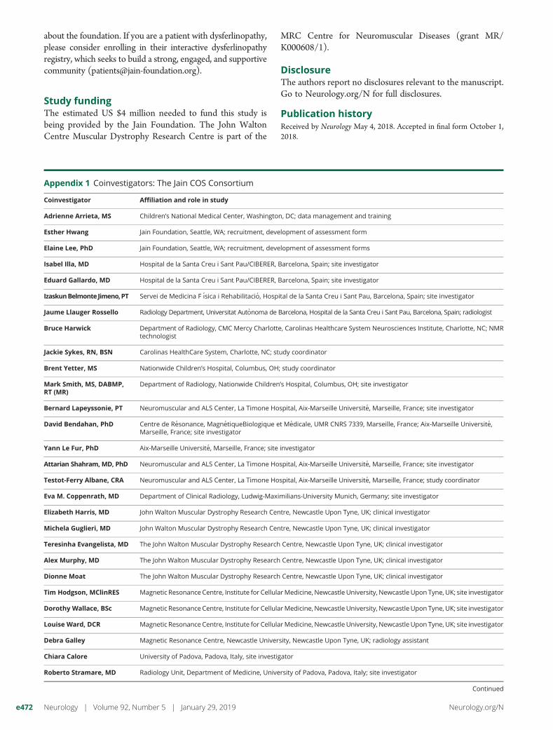
Appendix 1 Coinvestigators: The Jain COS Consortium
Coinvestigator Affiliation and role in study
Adrienne Arrieta, MS Children’s National Medical Center, Washington, DC; data management and training
Esther Hwang Jain Foundation, Seattle, WA; recruitment, development of assessment form
Elaine Lee, PhD Jain Foundation, Seattle, WA; recruitment, development of assessment forms
Isabel Illa, MD Hospital de la Santa Creu i Sant Pau/CIBERER, Barcelona, Spain; site investigator
Eduard Gallardo, MD Hospital de la Santa Creu i Sant Pau/CIBERER, Barcelona, Spain; site investigator
IzaskunBelmonteJimeno,PT Servei de Medicina F ısica i Rehabilitacio, Hospital de la Santa Creu i Sant Pau, Barcelona, Spain; site investigator
Jaume Llauger Rossello Radiology Department, Universitat Autonoma de Barcelona, Hospital de la Santa Creu i Sant Pau, Barcelona, Spain; radiologist
Bruce Harwick Department of Radiology, CMC Mercy Charlotte, Carolinas Healthcare System Neurosciences Institute, Charlotte, NC; NMRtechnologist
Jackie Sykes, RN, BSN Carolinas HealthCare System, Charlotte, NC; study coordinator
Brent Yetter, MS Nationwide Children’s Hospital, Columbus, OH; study coordinator
Mark Smith, MS, DABMP,RT (MR)
Department of Radiology, Nationwide Children’s Hospital, Columbus, OH; site investigator
Bernard Lapeyssonie, PT Neuromuscular and ALS Center, La Timone Hospital, Aix-Marseille Universite, Marseille, France; site investigator
David Bendahan, PhD Centre de Resonance, MagnetiqueBiologique et Medicale, UMR CNRS 7339, Marseille, France; Aix-Marseille Universite,Marseille, France; site investigator
Yann Le Fur, PhD Aix-Marseille Universite, Marseille, France; site investigator
Attarian Shahram, MD, PhD Neuromuscular and ALS Center, La Timone Hospital, Aix-Marseille Universite, Marseille, France; site investigator
Testot-Ferry Albane, CRA Neuromuscular and ALS Center, La Timone Hospital, Aix-Marseille Universite, Marseille, France; study coordinator
Eva M. Coppenrath, MD Department of Clinical Radiology, Ludwig-Maximilians-University Munich, Germany; site investigator
Elizabeth Harris, MD John Walton Muscular Dystrophy Research Centre, Newcastle Upon Tyne, UK; clinical investigator
Michela Guglieri, MD John Walton Muscular Dystrophy Research Centre, Newcastle Upon Tyne, UK; clinical investigator
Teresinha Evangelista, MD The John Walton Muscular Dystrophy Research Centre, Newcastle Upon Tyne, UK; clinical investigator
Alex Murphy, MD The John Walton Muscular Dystrophy Research Centre, Newcastle Upon Tyne, UK; clinical investigator
Dionne Moat The John Walton Muscular Dystrophy Research Centre, Newcastle Upon Tyne, UK; clinical investigator
Tim Hodgson, MClinRES Magnetic Resonance Centre, Institute for CellularMedicine, Newcastle University, Newcastle Upon Tyne, UK; site investigator
Dorothy Wallace, BSc Magnetic Resonance Centre, Institute for CellularMedicine, Newcastle University, Newcastle Upon Tyne, UK; site investigator
Louise Ward, DCR Magnetic Resonance Centre, Institute for CellularMedicine, Newcastle University, Newcastle Upon Tyne, UK; site investigator
Debra Galley Magnetic Resonance Centre, Newcastle University, Newcastle Upon Tyne, UK; radiology assistant
Chiara Calore University of Padova, Padova, Italy, site investigator
Roberto Stramare, MD Radiology Unit, Department of Medicine, University of Padova, Padova, Italy; site investigator
Continued
e472 Neurology | Volume 92, Number 5 | January 29, 2019 Neurology.org/N
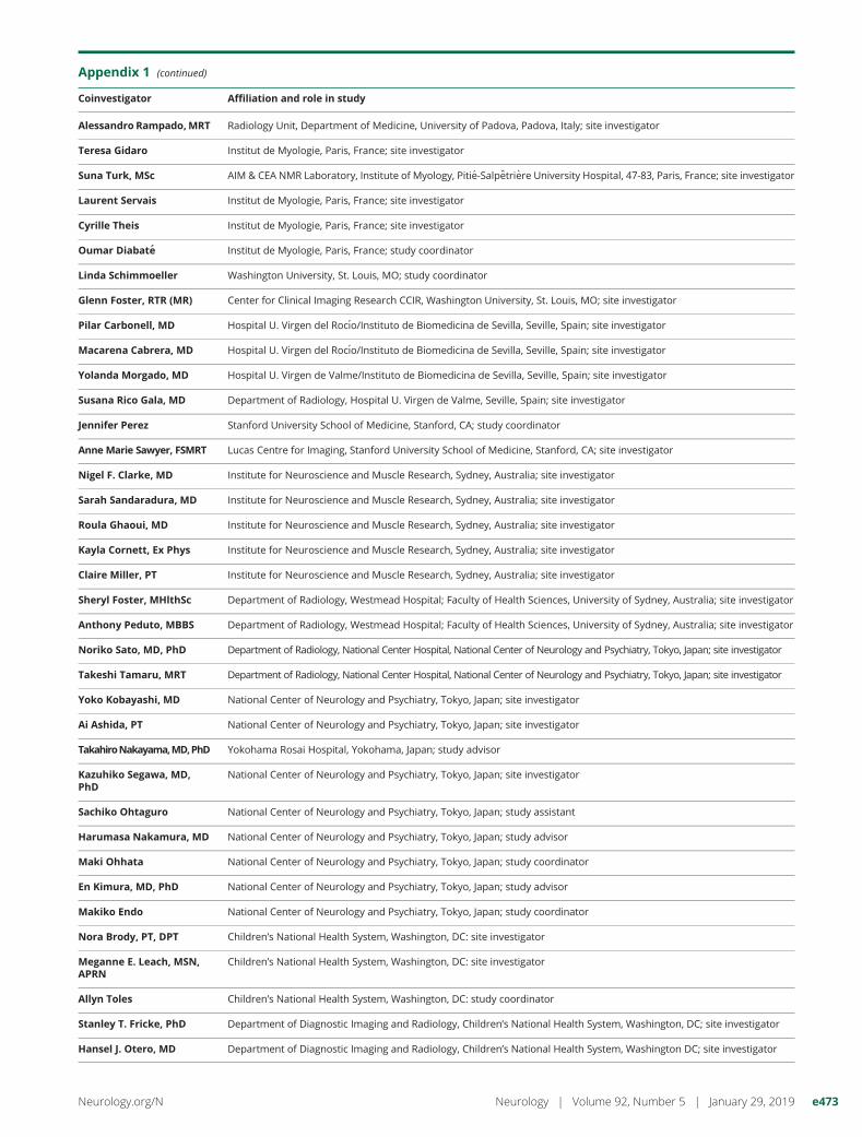
Appendix 1 (continued)
Coinvestigator Affiliation and role in study
Alessandro Rampado,MRT Radiology Unit, Department of Medicine, University of Padova, Padova, Italy; site investigator
Teresa Gidaro Institut de Myologie, Paris, France; site investigator
Suna Turk, MSc AIM & CEA NMR Laboratory, Institute of Myology, Pitie-Salpetriere University Hospital, 47-83, Paris, France; site investigator
Laurent Servais Institut de Myologie, Paris, France; site investigator
Cyrille Theis Institut de Myologie, Paris, France; site investigator
Oumar Diabate Institut de Myologie, Paris, France; study coordinator
Linda Schimmoeller Washington University, St. Louis, MO; study coordinator
Glenn Foster, RTR (MR) Center for Clinical Imaging Research CCIR, Washington University, St. Louis, MO; site investigator
Pilar Carbonell, MD Hospital U. Virgen del Rocıo/Instituto de Biomedicina de Sevilla, Seville, Spain; site investigator
Macarena Cabrera, MD Hospital U. Virgen del Rocıo/Instituto de Biomedicina de Sevilla, Seville, Spain; site investigator
Yolanda Morgado, MD Hospital U. Virgen de Valme/Instituto de Biomedicina de Sevilla, Seville, Spain; site investigator
Susana Rico Gala, MD Department of Radiology, Hospital U. Virgen de Valme, Seville, Spain; site investigator
Jennifer Perez Stanford University School of Medicine, Stanford, CA; study coordinator
Anne Marie Sawyer, FSMRT Lucas Centre for Imaging, Stanford University School of Medicine, Stanford, CA; site investigator
Nigel F. Clarke, MD Institute for Neuroscience and Muscle Research, Sydney, Australia; site investigator
Sarah Sandaradura, MD Institute for Neuroscience and Muscle Research, Sydney, Australia; site investigator
Roula Ghaoui, MD Institute for Neuroscience and Muscle Research, Sydney, Australia; site investigator
Kayla Cornett, Ex Phys Institute for Neuroscience and Muscle Research, Sydney, Australia; site investigator
Claire Miller, PT Institute for Neuroscience and Muscle Research, Sydney, Australia; site investigator
Sheryl Foster, MHlthSc Department of Radiology, Westmead Hospital; Faculty of Health Sciences, University of Sydney, Australia; site investigator
Anthony Peduto, MBBS Department of Radiology, Westmead Hospital; Faculty of Health Sciences, University of Sydney, Australia; site investigator
Noriko Sato, MD, PhD Department of Radiology, National Center Hospital, National Center of Neurology and Psychiatry, Tokyo, Japan; site investigator
Takeshi Tamaru, MRT Department of Radiology, National Center Hospital, National Center of Neurology and Psychiatry, Tokyo, Japan; site investigator
Yoko Kobayashi, MD National Center of Neurology and Psychiatry, Tokyo, Japan; site investigator
Ai Ashida, PT National Center of Neurology and Psychiatry, Tokyo, Japan; site investigator
TakahiroNakayama,MD,PhD Yokohama Rosai Hospital, Yokohama, Japan; study advisor
Kazuhiko Segawa, MD,PhD
National Center of Neurology and Psychiatry, Tokyo, Japan; site investigator
Sachiko Ohtaguro National Center of Neurology and Psychiatry, Tokyo, Japan; study assistant
Harumasa Nakamura, MD National Center of Neurology and Psychiatry, Tokyo, Japan; study advisor
Maki Ohhata National Center of Neurology and Psychiatry, Tokyo, Japan; study coordinator
En Kimura, MD, PhD National Center of Neurology and Psychiatry, Tokyo, Japan; study advisor
Makiko Endo National Center of Neurology and Psychiatry, Tokyo, Japan; study coordinator
Nora Brody, PT, DPT Children’s National Health System, Washington, DC: site investigator
Meganne E. Leach, MSN,APRN
Children’s National Health System, Washington, DC: site investigator
Allyn Toles Children’s National Health System, Washington, DC: study coordinator
Stanley T. Fricke, PhD Department of Diagnostic Imaging and Radiology, Children’s National Health System, Washington, DC; site investigator
Hansel J. Otero, MD Department of Diagnostic Imaging and Radiology, Children’s National Health System, Washington DC; site investigator
Neurology.org/N Neurology | Volume 92, Number 5 | January 29, 2019 e473
References1. Bushby K, Straub V. One gene, one or many diseases? Simplifying dysferlinopathy.
Neurology 2010;75:298–299.2. Nguyen K, Bassez G, Bernard R, et al. Dysferlin mutations in LGMD2B, Miyoshi
myopathy, and atypical dysferlinopathies. Hum Mutat 2005;26:165.3. Krahn M, Beroud C, Labelle V, et al. Analysis of the DYSF mutational spectrum in
a large cohort of patients. Hum Mutat 2009;30:E345–E375.4. Harris E, Bladen CL, Mayhew A, et al. The Clinical Outcome Study for Dysferlin-
opathy: an international multicenter study. Neurol Genet 2016;2:e89.5. Bushby K, Finkel R, Wong B, et al. Ataluren treatment of patients with nonsense
mutation dystrophinopathy. Muscle Nerve 2014;50:477–487.6. Ryan NJ. Ataluren: first global approval. Drugs 2014;74:1709–1714.7. Finkel RS, Mercuri E, Darras BT, et al. Nusinersen versus sham control in infantile-
onset spinal muscular atrophy. N Engl J Med 2017;377(18):1723–32.8. Mah JK. Current and emerging treatment strategies for Duchenne muscular dystro-
phy. Neuropsychiatr Dis Treat 2016;12:1795–1807.9. Shieh PB. Duchenne muscular dystrophy: clinical trials and emerging tribulations.
Curr Opin Neurol 2015;28:542–546.10. Fanin M, Angelini C. Progress and challenges in diagnosis of dysferlinopathy. Muscle
Nerve 2016;54:821–835.11. Klinge L, Aboumousa A, Eagle M, et al. New aspects on patients affected by dysferlin
deficient muscular dystrophy. J Neurol Neurosurg Psychiatry 2010;81:946–953.12. Klinge L, Dean AF, Kress W, et al. Late onset in dysferlinopathy widens the clinical
spectrum. Neuromuscul Disord 2008;18:288–290.13. Angelini C, Peterle E, Gaiani A, Bortolussi L, Borsato C. Dysferlinopathy course and
sportive activity: clues for possible treatment. Acta Myol 2011;30:127–132.14. Woudt L, Di Capua GA, Krahn M, et al. Toward an objective measure of functional
disability in dysferlinopathy. Muscle Nerve 2016;53:49–57.15. Tasca G, Iannaccone E,MonforteM, et al. MuscleMRI in Becker muscular dystrophy.
Neuromuscul Disord 2012;22(suppl 2):S100–S106.
16. Batcho CS, Van den Bergh PY, Van Damme P, Roy AJ, Thonnard JL, Penta M. Howrobust is ACTIVLIM for the follow-up of activity limitations in patients with neu-romuscular diseases? Neuromuscul Disord 2016;26:211–220.
17. Husted JA, Cook RJ, Farewell VT, Gladman DD.Methods for assessing responsiveness:a critical review and recommendations. J Clin Epidemiol 2000;53:459–468.
18. Douglas CE, Michael FA. On distribution-free multiple comparisons in the one-wayanalysis of variance. Commun Stat 1991;20:127–139.
19. Mendell JR, Goemans N, Lowes LP, et al. Longitudinal effect of eteplirsen versushistorical control on ambulation in Duchenne muscular dystrophy. Ann Neurol2016;79:257–271.
20. Mayhew A, Cano S, Scott E, Eagle M, Bushby K, Muntoni F. Moving towardsmeaningful measurement: Rasch analysis of the North Star Ambulatory As-sessment in Duchenne muscular dystrophy. Dev Med Child Neurol 2011;53:535–542.
21. ClinicalTrials.gov. rAAVrh74.MHCK7.DYSF.DV for Treatment of Dysferlinopathies[online]. Available at: clinicaltrials.gov/ct2/show/NCT02710500. Accessed August24, 2018.
22. Wadsworth CT, Krishnan R, Sear M, Harrold J, Nielsen DH. Intrarater reliability ofmanual muscle testing and hand-held dynametric muscle testing. Phys Ther 1987;67:1342–1347.
23. Scott E, Eagle M, Mayhew A, et al. Development of a functional assessment scalefor ambulatory boys with Duchenne muscular dystrophy. Physiother Res Int2012;17:101–109.
24. Berard C, Payan C, Hodgkinson I, Fermanian J. A motor function measure forneuromuscular diseases: construction and validation study. Neuromuscul Disord2005;15:463–470.
25. Cuthbert SC, Goodheart GJ Jr. On the reliability and validity of manual muscletesting: a literature review. Chiropr Osteopat 2007;15:4.
26. Vandervelde L, Van den Bergh PY, Goemans N, Thonnard JL. ACTIVLIM: a Rasch-built measure of activity limitations in children and adults with neuromuscular dis-orders. Neuromuscul Disord 2007;17:459–469.
e474 Neurology | Volume 92, Number 5 | January 29, 2019 Neurology.org/N
DOI 10.1212/WNL.00000000000068582019;92;e461-e474 Published Online before print January 9, 2019Neurology Ursula Moore, Marni Jacobs, Meredith K. James, et al.
Assessment of disease progression in dysferlinopathy: A 1-year cohort study
This information is current as of January 9, 2019
ServicesUpdated Information &
http://n.neurology.org/content/92/5/e461.fullincluding high resolution figures, can be found at:
References http://n.neurology.org/content/92/5/e461.full#ref-list-1
This article cites 25 articles, 4 of which you can access for free at:
Subspecialty Collections
http://n.neurology.org/cgi/collection/outcome_researchOutcome research
http://n.neurology.org/cgi/collection/all_neuromuscular_diseaseAll Neuromuscular Diseasefollowing collection(s): This article, along with others on similar topics, appears in the
Permissions & Licensing
http://www.neurology.org/about/about_the_journal#permissionsits entirety can be found online at:Information about reproducing this article in parts (figures,tables) or in
Reprints
http://n.neurology.org/subscribers/advertiseInformation about ordering reprints can be found online:
ISSN: 0028-3878. Online ISSN: 1526-632X.Wolters Kluwer Health, Inc. on behalf of the American Academy of Neurology.. All rights reserved. Print1951, it is now a weekly with 48 issues per year. Copyright Copyright © 2019 The Author(s). Published by
® is the official journal of the American Academy of Neurology. Published continuously sinceNeurology