Embed Size (px)
Citation preview
Article
Individual crypt genetic heterogeneity and the origin of metaplastic glandular epithelium in human Barrett's oesophagus
Leedham, S J, Preston, S L, McDonald, S A C, Elia, G, Bhandari, P, Poller, D, Harrison, R, Novelli, M R, Jankowski, Janusz and Wright, N A
Available at http://clok.uclan.ac.uk/16088/
Leedham, S J, Preston, S L, McDonald, S A C, Elia, G, Bhandari, P, Poller, D, Harrison, R, Novelli, M R, Jankowski, Janusz et al (2008) Individual crypt genetic heterogeneity and the origin of metaplastic glandular epithelium in human Barrett's oesophagus. Gut, 57 (8). pp. 10411048. ISSN 00175749
It is advisable to refer to the publisher’s version if you intend to cite from the work.http://dx.doi.org/10.1136/gut.2007.143339
For more information about UCLan’s research in this area go to http://www.uclan.ac.uk/researchgroups/ and search for <name of research Group>.
For information about Research generally at UCLan please go to http://www.uclan.ac.uk/research/
All outputs in CLoK are protected by Intellectual Property Rights law, includingCopyright law. Copyright, IPR and Moral Rights for the works on this site are retained by the individual authors and/or other copyright owners. Terms and conditions for use of this material are defined in the http://clok.uclan.ac.uk/policies/
CLoKCentral Lancashire online Knowledgewww.clok.uclan.ac.uk

Individual crypt genetic heterogeneity and the originof metaplastic glandular epithelium in humanBarrett’s oesophagus
S J Leedham,1 S L Preston,1,2 S A C McDonald,1,3 G Elia,1 P Bhandari,4 D Poller,5
R Harrison,6 M R Novelli,7 J A Jankowski,1,3,8 N A Wright1,2
See Commentary, p 1033
c Additional figures and tablesare published online only athttp://gut.bmj.com/content/vol57/issue8
1 Histopathology Unit, CancerResearch UK, London, UK;2 Institute of Cell and MolecularSciences, St Bartholomew’s andRoyal London School ofMedicine and Dentistry, QueenMary University, London, UK;3 Department of ClinicalPharmacology, University ofOxford, Oxford, UK;4 Department ofGastroenterology QueenAlexandra Hospital, Portsmouth,UK; 5 Pathology Department,Queen Alexandra Hospital,Portsmouth, UK; 6 PathologyDepartment, Leicester GeneralHospital, Leicester, UK;7 Histopathology Department,University College Hospital,London, UK; 8 Digestive DiseaseCentre, Leicester RoyalInfirmary, Leicester, UK
Correspondence to:Dr Simon J Leedham,Histopathology Unit, CancerResearch UK, 44 Lincoln’s InnFields, London WC2A 3PX, UK;[email protected]
Revised 23 January 2008Accepted 12 February 2008Published Online First27 February 2008
This paper is freely availableonline under the BMJ Journalsunlocked scheme, see http://gut.bmj.com/info/unlocked.dtl
ABSTRACTObjectives: Current models of clonal expansion in humanBarrett’s oesophagus are based upon heterogenous, flow-purified biopsy analysis taken at multiple segment levels.Detection of identical mutation fingerprints from thesebiopsy samples led to the proposal that a mutated clonewith a selective advantage can clonally expand to fill anentire Barrett’s segment at the expense of competingclones (selective sweep to fixation model). We aimed toassess clonality at a much higher resolution bymicrodissecting and genetically analysing individualcrypts. The histogenesis of Barrett’s metaplasia and neo-squamous islands has never been demonstrated. Weinvestigated the oesophageal gland squamous ducts asthe source of both epithelial sub-types.Methods: Individual crypts across Barrett’s biopsy andoesophagectomy blocks were dissected. Determination oftumour suppressor gene loss of heterozygosity patterns,p16 and p53 point mutations were carried out on a crypt-by-crypt basis. Cases of contiguous neo-squamous islandsand columnar metaplasia with oesophageal squamousducts were identified. Tissues were isolated by lasercapture microdissection and genetically analysed.Results: Individual crypt dissection revealed mutationpatterns that were masked in whole biopsy analysis.Dissection across oesophagectomy specimens demon-strated marked clonal heterogeneity, with multipleindependent clones present. We identified a p16 pointmutation arising in the squamous epithelium of theoesophageal gland duct, which was also present in acontiguous metaplastic crypt, whereas neo-squamousislands arising from squamous ducts were wild-type withrespect to surrounding Barrett’s dysplasia.Conclusions: By studying clonality at the crypt level wedemonstrate that Barrett’s heterogeneity arises frommultiple independent clones, in contrast to the selectivesweep to fixation model of clonal expansion previouslydescribed. We suggest that the squamous gland ductssituated throughout the oesophagus are the source of aprogenitor cell that may be susceptible to gene mutationresulting in conversion to Barrett’s metaplastic epithelium.Additionally, these data suggest that wild-type ducts maybe the source of neo-squamous islands.
Barrett’s oesophagus is the replacement of thenormal oesophageal stratified squamous epithe-lium with metaplastic glandular epithelium inresponse to inflammation and ulceration provokedby duodeno-gastroesophageal reflux.1 Oesophagealadenocarcinoma can arise from progression througha metaplasia–dysplasia–carcinoma sequence (MCS),and the presence of Barrett’s oesophagus increases
the risk of oesophageal adenocarcinoma by 30- to40-fold.2
Serial biopsies and molecular analysis of a cohortof patients with Barrett’s oesophagus has enabledresearchers to study the evolution of commontumour suppressor gene mutation patterns as theMCS progresses. These longitudinal clonal orderingstudies have shown that genetic and epigeneticinactivation of cyclin-dependent kinase N2 (p16) andgenetic inactivation of TP53 (p53) tumour suppres-sor genes occur early in the MCS3 with 88% of pre-dysplastic Barrett’s oesophagus tissue having adetectable p16 lesion.4 The demonstration of clonalp16 and p53 lesions throughout long lengths ofBarrett’s oesophagus4 5 suggests a common precursorlesion that undergoes clonal expansion, and has ledto the proposal that the MCS progresses as aconsequence of sequential tumour suppressor geneinactivation causing selective growth advantages.Growth advantages result in preferential expansionof a mutated clone and a mutation is said to have‘‘gone to fixation’’ when it expands throughout anentire field, extinguishing all competing clones. A‘‘selective sweep’’ is the process of natural selectiondriving a mutation to fixation.6 7 It has beensuggested that loss of each of the two p16 allelespredisposes to a selective sweep, and that p16mutation fixation occurs early in the progression ofBarrett’s oesophagus.6 The demonstration of similarp16 loss of heterozygosity (LOH), methylation andpoint mutation patterns in biopsy material takenfrom different levels of long Barrett’s oesophagussegments supports widespread clonal expansion andfixed mutations.4 However, phenotypic and geno-typic heterogeneity has also been described in someBarrett’s segments4 5 and clonal diversity in Barrett’ssegments has recently been shown to be associatedwith progression to adenocarcinoma.8
Despite years of active research the histogenesis ofBarrett’s oesophagus has never been demonstrated.Different theories have been proposed: proximalmigration of the gastric cardia; re-differentiation ofthe squamous epithelium and colonisation of cellsfrom the oesophageal gland ducts.9 10 Similar ques-tions remain regarding the origin of neo-squamousislands that can arise within fields of Barrett’s tissueafter acid suppression or endoscopic ablative ther-apy.11 12 Paulson et al13 demonstrated that thesesquamous islands were usually genetically wild-typedespite being surrounded by mutated Barrett’s tissue.They excluded encroachment of adjacent normalsquamous epithelium by only including patientswho developed isolated squamous islands, but were
Oesophagus
Gut 2008;57:1041–1048. doi:10.1136/gut.2007.143339 1041
group.bmj.com on November 1, 2016 - Published by http://gut.bmj.com/Downloaded from
unable to determine the source of the genetically normal tissuethat may have an important clinical role in re-epithelisation aftertreatment for Barrett’s oesophagus.
To date, most clonality studies have been carried out onheterogenous flow-purified whole biopsy samples. In this workour aims were to (1) study clonality at a crypt-by-crypt level,avoiding problems associated with contaminating normalstroma; and (2) examine the oesophageal gland squamous ducts
as the potential source of Barrett’s columnar epithelium andneo-squamous islands.
MATERIALS AND METHODS
Tissue and slidesParaffin-embedded biopsy (six biopsies from five patients) andoesophagectomy blocks (four blocks from patient 1, four blocks
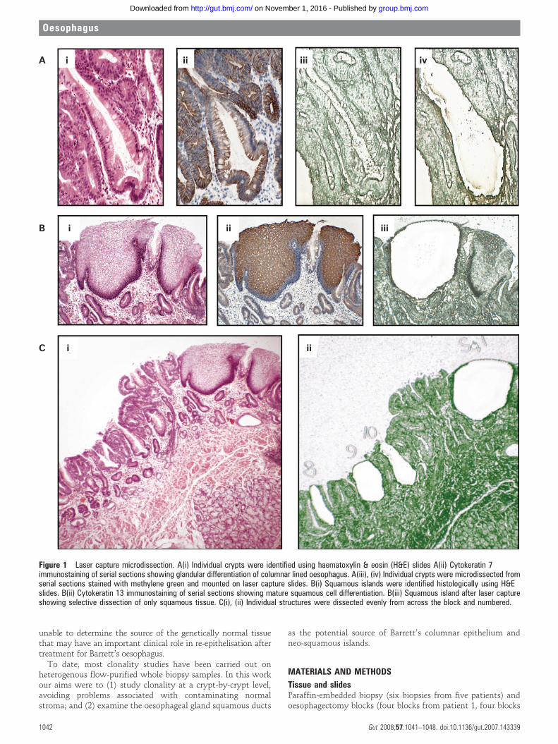
Figure 1 Laser capture microdissection. A(i) Individual crypts were identified using haematoxylin & eosin (H&E) slides A(ii) Cytokeratin 7immunostaining of serial sections showing glandular differentiation of columnar lined oesophagus. A(iii), (iv) Individual crypts were microdissected fromserial sections stained with methylene green and mounted on laser capture slides. B(i) Squamous islands were identified histologically using H&Eslides. B(ii) Cytokeratin 13 immunostaining of serial sections showing mature squamous cell differentiation. B(iii) Squamous island after laser captureshowing selective dissection of only squamous tissue. C(i), (ii) Individual structures were dissected evenly from across the block and numbered.
Oesophagus
1042 Gut 2008;57:1041–1048. doi:10.1136/gut.2007.143339
group.bmj.com on November 1, 2016 - Published by http://gut.bmj.com/Downloaded from
from patient 2 and two blocks from patient 3) were obtainedfrom the pathology archives of Leicester General Hospital.Tissue was independently assessed for Barrett’s metaplasia anddysplasia according to British Society of Gastroenterology 2005guidelines (www.bsg.org.uk), by at least two pathologists.Serial 5 mm sections were cut. Sections 1–3 and 5–7 weremounted onto P.A.L.M. membrane slides (P.A.L.M. MicrolaserTechnologies, Benried, Germany) and were stained withmethylene green. Section 4 was stained with haematoxylin &eosin (H&E).
Laser capture microdissection (fig 1)Suitable crypts for dissection were identified using the H&Eslide. The same crypts were identified on the slides stained withmethylene green. Individual crypt sections from the six serialslides were cut out from the laser capture slides and catapultedinto the adhesive caps of eppendorfs using the P.A.L.M. LaserMicrodissection system. Where constitutional DNA wasrequired for microsatellite analysis, serial areas of lamina propriawere microdissected. Catapulted sections on the cap wereimmersed in 12 ml of proteinase K solution (Arcturus Bioscience,Mt View, California, USA). After individual crypt dissectionresidual epithelial tissue was catapulted into a single tube andimmersed in 30 ml proteinase K for p53 gene screening. Negativecontrol tubes containing 12 ml proteinase K solution and nolaser capture material were included. Tubes were thencentrifuged at 4.5 g for 1 min and incubated at 65uC overnight.A 10 min incubation at 95uC denatured the proteinase K and thelysate was then stored at 220uC.
ImmunocytochemistryCytokeratin 7 and 13 staining was used to demonstrateglandular and squamous epithelial differentiation respec-tively.14 15 Serial sections of oesophagectomy blocks were cutat 4 mm and mounted on glass slides. Sections were de-waxedand rehydrated by standard methods. Endogenous peroxidasewas blocked with 3% H2O2 in methanol for 10 min. Antigenretrieval was achieved by 10 min microwaving in sodium citratebuffer at pH 6. Slides were incubated in 3% bovine serumalbumin in phosphate-buffered saline (PBS) for 15 min. Slidesunderwent primary antibody incubation with mouse mono-clonal antibodies against cytokeratin 7 (1:100 dilution of cloneOV-TL; Abcam, Cambridge, UK) or cytokeratin 13 (1:200dilution of clone AE8; Abcam). This was followed bybiotinylated rabbit anti-mouse secondary antibodies beforeapplication of a 1:500 dilution of the tertiary layer ofperoxidase-conjugated streptavidin (strep-HRP; Dako,Glostrup, Denmark). Each layer was applied for 45 min andthree 5 min PBS washes were performed between layers.Sections were then developed with 3,3-diaminobenzidinetetrahydrochloride solution (DAB; Sigma, Poole, UK) for2 min, followed by rinsing in tap water and light haematoxylincounterstaining. The positive control tissues used were duode-num (CK 7) and tonsil (CK 13). Negative controls underwent allsteps but were incubated with PBS instead of the primaryantibody solution.
Nested polymerase chain reaction and sequencingFirst and second round primers were designed to amplify exons5–9 of p53 and exon 2 of p16, using the primer 3 website (MIT,Cambridge, Massachusetts, USA). First round oligonucleotideprimer pairs were specifically designed to amplify a region thatincluded the amplicon covered by the primers used in the second
round of the polymerase chain reaction (PCR). Primer optimisa-tion determined the optimum reagent concentration andannealing temperature for each primer pair. Primer sequencesare tabulated in the supplementary information (supplementarytable 1A–C). Both PCR steps were carried out in an Omni PCRUV hood to minimise contamination (Bioquell, Andover,Berkshire, UK). Only products with an uncontaminatednegative control tube went forward for sequencing. PCRproduct was sequenced using BigDye terminator cycle sequen-cing on an ABI 3100 DNA sequencer (Applied Biosystems,Foster City, California, USA). The sequences obtained weredirectly compared to the revised Cambridge reference sequence,and any identified mutations were checked against theCatalogue of Somatic Mutations in Cancer (COSMIC) database(www.sanger.ac.uk) (Cambridge, UK). Sequencing wasrepeated twice from dissection lysate for the oesophageal glandsquamous duct work.
Microsatellite analysisThree microsatellite markers; D5S346 (APC gene), D9S932 (p16gene) and D17S786 (p53 gene), were used for LOH analysis.Constitutionally homozygous markers were scored as non-informative. Forward oligonucleotide primers were labelled atthe 59 end with the carboxy fluorescein (FAM) fluorescent marker(Sigma). PCR amplifications were performed using the LATAKARA kit (Takara Bio, Shiga, Japan). The PCR product wasanalysed on an ABI 3100 sequencer (Applied Biosystems) andgenotyper 2.5 software (Perkin-Elmer, Boston, Massachusetts,USA). At each marker, loss of heterozygosity was consideredpresent if the area under one allelic peak in the affected crypt wasless than 0.5 times or greater than 2 times that of the other allele,after correcting for the relative areas using constitutional DNA(microdissected areas of lamina propria tissue).
Statistical analysisThe associations between individual crypt point mutations andtumour suppressor gene allelic loss were analysed using the two-tailed Fisher’s exact test run on Prism 4.0 software (GraphpadSoftware, San Diego, California, USA).
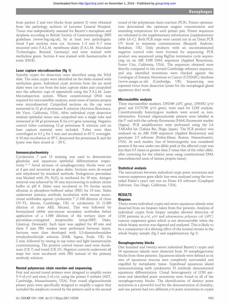
RESULTSBiopsiesThirty-seven individual crypts and seven squamous islands weredissected from six biopsies taken from five patients. Analysis ofindividual crypts from biopsy samples allowed detection ofLOH patterns in p16, p53 and adenomatous polyposis coli (APC)tumour suppressor genes which is not demonstrable when thewhole biopsy section was digested and analysed. This is likely tobe a consequence of a diluting effect of the normal stroma in thewhole biopsy sample (fig 2 and supplementary fig 1)
Oesophagectomy blocksOne hundred and twenty-seven individual Barrett’s crypts and14 squamous islands were dissected from 10 oesophagectomyblocks from three patients. Squamous islands were defined as anarea of squamous mucosa seen completely surrounded andengulfed by metaplastic tissue. Suprabasal squamous islandimmunostaining with cytokeratin 13 antibody demonstratedsquamous differentiation. Clonal heterogeneity of LOH pat-terns and identified point mutations were found in all of theoesophagectomy blocks. The identification of distinct pointmutations is a powerful tool for the demonstration of clonality,and one patient had two different p16 point mutations in crypts
Oesophagus
Gut 2008;57:1041–1048. doi:10.1136/gut.2007.143339 1043
group.bmj.com on November 1, 2016 - Published by http://gut.bmj.com/Downloaded from
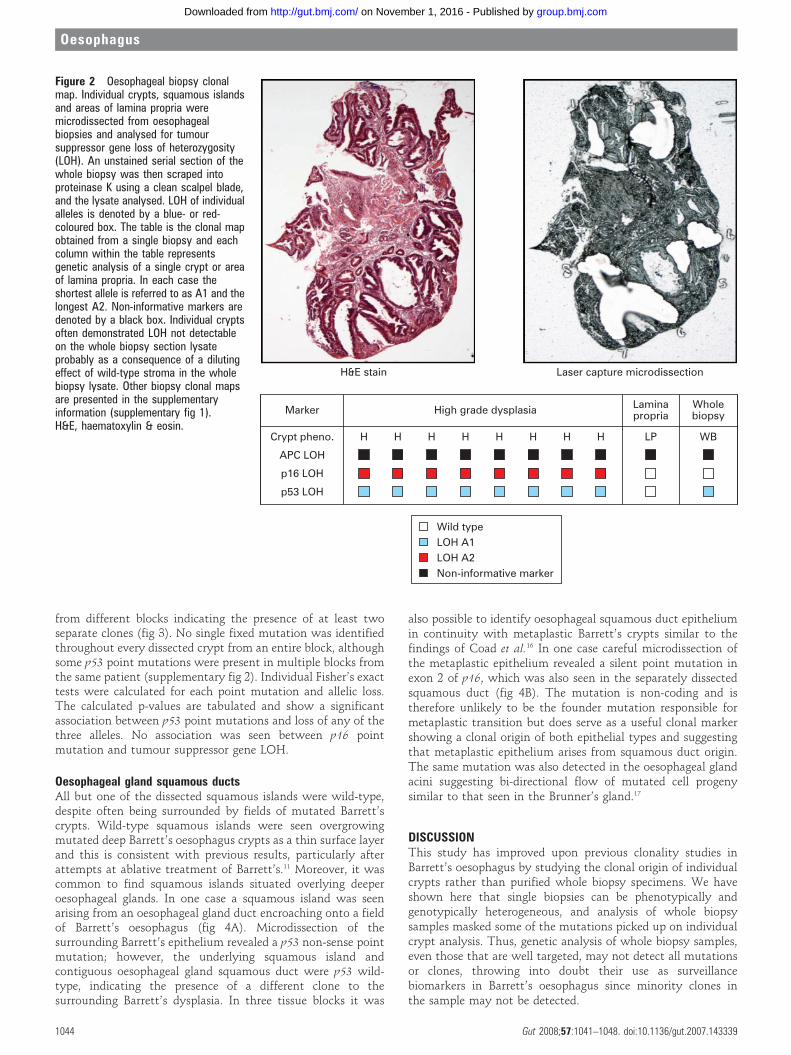
from different blocks indicating the presence of at least twoseparate clones (fig 3). No single fixed mutation was identifiedthroughout every dissected crypt from an entire block, althoughsome p53 point mutations were present in multiple blocks fromthe same patient (supplementary fig 2). Individual Fisher’s exacttests were calculated for each point mutation and allelic loss.The calculated p-values are tabulated and show a significantassociation between p53 point mutations and loss of any of thethree alleles. No association was seen between p16 pointmutation and tumour suppressor gene LOH.
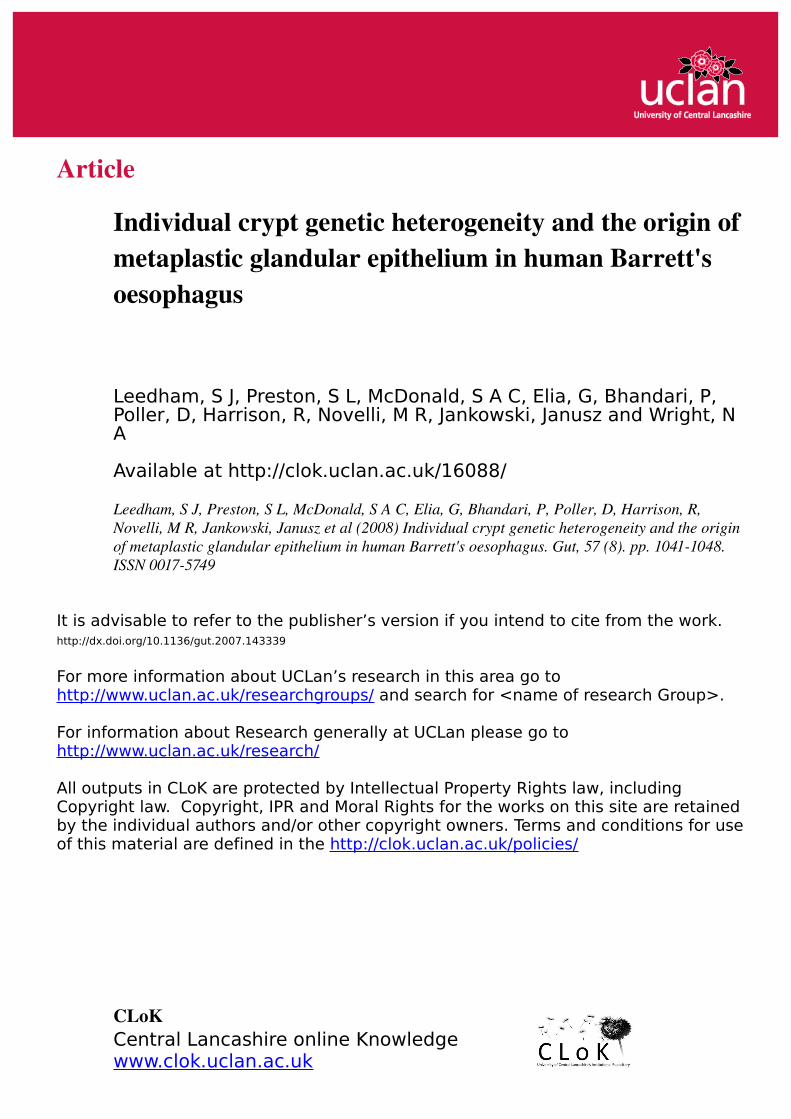
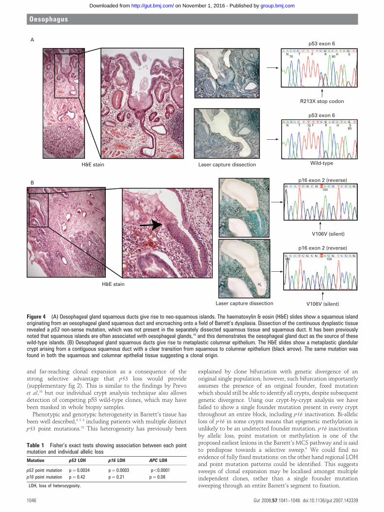
Oesophageal gland squamous ductsAll but one of the dissected squamous islands were wild-type,despite often being surrounded by fields of mutated Barrett’scrypts. Wild-type squamous islands were seen overgrowingmutated deep Barrett’s oesophagus crypts as a thin surface layerand this is consistent with previous results, particularly afterattempts at ablative treatment of Barrett’s.11 Moreover, it wascommon to find squamous islands situated overlying deeperoesophageal glands. In one case a squamous island was seenarising from an oesophageal gland duct encroaching onto a fieldof Barrett’s oesophagus (fig 4A). Microdissection of thesurrounding Barrett’s epithelium revealed a p53 non-sense pointmutation; however, the underlying squamous island andcontiguous oesophageal gland squamous duct were p53 wild-type, indicating the presence of a different clone to thesurrounding Barrett’s dysplasia. In three tissue blocks it was
also possible to identify oesophageal squamous duct epitheliumin continuity with metaplastic Barrett’s crypts similar to thefindings of Coad et al.16 In one case careful microdissection ofthe metaplastic epithelium revealed a silent point mutation inexon 2 of p16, which was also seen in the separately dissectedsquamous duct (fig 4B). The mutation is non-coding and istherefore unlikely to be the founder mutation responsible formetaplastic transition but does serve as a useful clonal markershowing a clonal origin of both epithelial types and suggestingthat metaplastic epithelium arises from squamous duct origin.The same mutation was also detected in the oesophageal glandacini suggesting bi-directional flow of mutated cell progenysimilar to that seen in the Brunner’s gland.17
DISCUSSIONThis study has improved upon previous clonality studies inBarrett’s oesophagus by studying the clonal origin of individualcrypts rather than purified whole biopsy specimens. We haveshown here that single biopsies can be phenotypically andgenotypically heterogeneous, and analysis of whole biopsysamples masked some of the mutations picked up on individualcrypt analysis. Thus, genetic analysis of whole biopsy samples,even those that are well targeted, may not detect all mutationsor clones, throwing into doubt their use as surveillancebiomarkers in Barrett’s oesophagus since minority clones inthe sample may not be detected.
Figure 2 Oesophageal biopsy clonalmap. Individual crypts, squamous islandsand areas of lamina propria weremicrodissected from oesophagealbiopsies and analysed for tumoursuppressor gene loss of heterozygosity(LOH). An unstained serial section of thewhole biopsy was then scraped intoproteinase K using a clean scalpel blade,and the lysate analysed. LOH of individualalleles is denoted by a blue- or red-coloured box. The table is the clonal mapobtained from a single biopsy and eachcolumn within the table representsgenetic analysis of a single crypt or areaof lamina propria. In each case theshortest allele is referred to as A1 and thelongest A2. Non-informative markers aredenoted by a black box. Individual cryptsoften demonstrated LOH not detectableon the whole biopsy section lysateprobably as a consequence of a dilutingeffect of wild-type stroma in the wholebiopsy lysate. Other biopsy clonal mapsare presented in the supplementaryinformation (supplementary fig 1).H&E, haematoxylin & eosin.
Oesophagus
1044 Gut 2008;57:1041–1048. doi:10.1136/gut.2007.143339
group.bmj.com on November 1, 2016 - Published by http://gut.bmj.com/Downloaded from
Dissection across large oesophagectomy blocks also revealedconsiderable phenotypic and genotypic heterogeneity in allcases. p16 point mutations were limited to single blocks andwere not significantly associated with loss of any alleles;however, there was a significant association (Fisher’s exact testp,0.004) between p53 point mutations and allelic loss of all
three tumour suppressor genes. This is consistent withfunctional loss of the cell cycle checkpoint activity of TP53protein, which is then permissive for widespread, large-scalegenetic changes. Additionally, p53 point mutations, althoughnot seen in every crypt, were often present in multiple blocksfrom a single oesophagectomy specimen, suggesting widespread
Figure 3 Clonal maps of two blocks from patient 1. Each table is the clonal map for the oesophagectomy specimen pictured and each column withinthe table represents the genetic analysis of a single crypt, squamous island or area of lamina propria. p16 point mutations are denoted by green- oryellow-coloured boxes as per the key. White boxes are wild-type. Clonal analysis revealed regional similarities in loss of heterozygosity (LOH) patternscorrelating with the observed phenotypic differences; however, there was no evidence of tissue-wide selective sweeps and no fixed founder mutationsindicating a common ancestral precursor. Two different, independent p16 point mutations were identified in the different blocks suggesting at least twodistinct clones. Only one squamous island contained a mutation and this is comparable with the results described by Paulson et al13 Clonal maps fromother patients are presented in the supplementary information (supplementary fig 2).
Oesophagus
Gut 2008;57:1041–1048. doi:10.1136/gut.2007.143339 1045
group.bmj.com on November 1, 2016 - Published by http://gut.bmj.com/Downloaded from
and far-reaching clonal expansion as a consequence of thestrong selective advantage that p53 loss would provide(supplementary fig 2). This is similar to the findings by Prevoet al,18 but our individual crypt analysis technique also allowsdetection of competing p53 wild-type clones, which may havebeen masked in whole biopsy samples.
Phenotypic and genotypic heterogeneity in Barrett’s tissue hasbeen well described,4 5 8 including patients with multiple distinctp53 point mutations.18 This heterogeneity has previously been
explained by clone bifurcation with genetic divergence of anoriginal single population; however, such bifurcation importantlyassumes the presence of an original founder, fixed mutationwhich should still be able to identify all crypts, despite subsequentgenetic divergence. Using our crypt-by-crypt analysis we havefailed to show a single founder mutation present in every cryptthroughout an entire block, including p16 inactivation. Bi-allelicloss of p16 in some crypts means that epigenetic methylation isunlikely to be an undetected founder mutation. p16 inactivationby allelic loss, point mutation or methylation is one of theproposed earliest lesions in the Barrett’s MCS pathway and is saidto predispose towards a selective sweep.6 We could find noevidence of fully fixed mutations: on the other hand regional LOHand point mutation patterns could be identified. This suggestssweeps of clonal expansion may be localised amongst multipleindependent clones, rather than a single founder mutationsweeping through an entire Barrett’s segment to fixation.
Figure 4 (A) Oesophageal gland squamous ducts give rise to neo-squamous islands. The haematoxylin & eosin (H&E) slides show a squamous islandoriginating from an oesophageal gland squamous duct and encroaching onto a field of Barrett’s dysplasia. Dissection of the continuous dysplastic tissuerevealed a p53 non-sense mutation, which was not present in the separately dissected squamous tissue and squamous duct. It has been previouslynoted that squamous islands are often associated with oesophageal glands,16 and this demonstrates the oesophageal gland duct as the source of thesewild-type islands. (B) Oesophageal gland squamous ducts give rise to metaplastic columnar epithelium. The H&E slides show a metaplastic glandularcrypt arising from a contiguous squamous duct with a clear transition from squamous to columnar epithelium (black arrow). The same mutation wasfound in both the squamous and columnar epithelial tissue suggesting a clonal origin.
Table 1 Fisher’s exact tests showing association between each pointmutation and individual allelic loss
Mutation p53 LOH p16 LOH APC LOH
p53 point mutation p = 0.0034 p = 0.0003 p,0.0001
p16 point mutation p = 0.42 p = 0.21 p = 0.08
LOH, loss of heterozygosity.
Oesophagus
1046 Gut 2008;57:1041–1048. doi:10.1136/gut.2007.143339
group.bmj.com on November 1, 2016 - Published by http://gut.bmj.com/Downloaded from
It has been proposed, but never proven, that submucosaloesophageal gland ducts may be the origin of metaplastic tissuein Barrett’s oesophagus.10 16 19 These results support thishypothesis by demonstrating a p16 point mutation originatingin microdissected squamous duct tissue that was also found inthe adjoining metaplastic crypt. The presence of an identicalmutation in the two different epithelial types is strong evidenceto suggest that the origin of the metaplastic tissue in humanBarrett’s oesophagus is a progenitor located in the oesophagealgland squamous ducts. Additionally, the presence of a wild-typesquamous island seen emerging from a wild-type squamousduct in the midst of, and completely surrounded by, a p53mutant field, strongly indicates a new clone development. Thissupports the hypothesis that neo-squamous islands can arise denovo from glandular tissue after Barrett’s ablation therapy,20 andextends the findings by Paulson et al13 by showing that these wild-type islands arise from non-mutated squamous duct tissue.
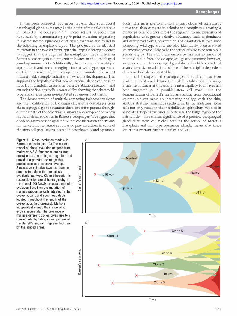
The demonstration of multiple competing independent clonesand the identification of the origin of Barrett’s oesophagus fromthe oesophageal gland squamous duct, structures present through-out the length of the oesophagus, allows the development of a newmodel of clonal evolution in Barrett’s oesophagus. We suggest thatduodeno-gastro-oesophageal reflux-induced ulceration and inflam-mation can induce tumour suppressor gene mutations in some ofthe stem cell populations located in oesophageal gland squamous
ducts. This gives rise to multiple distinct clones of metaplastictissue that then compete to colonise the oesophagus, creating amosaic pattern of clones across the segment. Clonal expansion ofpopulations with greater selective advantage leads to dominantand widespread clones; however, no single mutation is fixed sincecompeting wild-type clones are also identifiable. Non-mutatedsquamous ducts are likely to be the source of wild-type squamousislands (fig 5). These data are unable to rule out extension ofmutated tissue from the oesophageal–gastric junction; however,we propose that the oesophageal gland ducts should be consideredas an alternative or additional source of the multiple independentclones we have demonstrated here.
The cell biology of the oesophageal epithelium has beeninadequately studied despite the high mortality and increasingincidence of cancer at this site. The interpapillary basal layer hasbeen suggested as a possible stem cell zone21 but thedemonstration of Barrett’s metaplasia arising from oesophagealsquamous ducts raises an interesting analogy with the skin,another stratified squamous epithelium. In the epidermis, stemcells not only reside in the interfollicular epithelium but also inassociated deeper structures; specifically, the bulge region of thehair follicle.21 The clinical significance of a possible oesophagealgland duct stem cell niche, both as the source of Barrett’smetaplasia and wild-type squamous islands, means that thesestructures warrant further detailed analysis.
Figure 5 Clonal evolution models inBarrett’s oesophagus. (A) The currentmodel of clonal evolution adapted fromMaley et al.6 A founder mutation (redcross) occurs in a single progenitor andprovides a growth advantage thatpredisposes to a selective sweep.Successive selective sweeps result inprogression along the metaplasia–dysplasia pathway. Clone bifurcation isresponsible for clonal heterogeneity inthis model. (B) Newly proposed model ofevolution based on the mutation ofmultiple progenitor cells situated in theoesophageal gland squamous ductslocated throughout the length of theoesophagus (red crosses). Multipleindependent clones then arise whichevolve separately. The presence ofmultiple different clones gives rise to amosaic interdigitating clonal pattern ofthe Barrett’s segment represented hereby the striped areas.
Oesophagus
Gut 2008;57:1041–1048. doi:10.1136/gut.2007.143339 1047
group.bmj.com on November 1, 2016 - Published by http://gut.bmj.com/Downloaded from
In conclusion, we have demonstrated two important featuresof Barrett’s oesophagus. First, clonal heterogeneity arises frommultiple independent clones, previously undetectable by wholebiopsy analysis; and second, Barrett’s metaplasia and neo-squamous islands can arise from the oesophageal glandsquamous ducts.
Funding: SJL is funded by Medical Research Council; SLP, SACM and NAW arefunded by Cancer Research UK; and SACM and JAJ are also funded by OxfordUniversity.
Competing interests: None.
Ethics approval: Multicentre ethics approval was obtained from the OxfordshireResearch and Ethics Committee (MREC 07/Q1604/17).
REFERENCES1. Lagergren J, Bergstrom R, Lindgren A, et al. Symptomatic gastroesophageal reflux
as a risk factor for esophageal adenocarcinoma. N Engl J Med 1999;340:825–31.2. Jankowski JA, Wright NA, Meltzer SJ, et al. Molecular evolution of the metaplasia–
dysplasia–adenocarcinoma sequence in the esophagus. Am J Pathol 1999;154:965–73.3. Barrett MT, Sanchez CA, Prevo LJ, et al. Evolution of neoplastic cell lineages in
Barrett oesophagus. Nat Genet 1999;22:106–9.4. Wong DJ, Paulson TG, Prevo LJ, et al. p16(INK4a) lesions are common, early
abnormalities that undergo clonal expansion in Barrett’s metaplastic epithelium.Cancer Res 2001;61:8284–9.
5. Galipeau PC, Prevo LJ, Sanchez CA, et al. Clonal expansion and loss ofheterozygosity at chromosomes 9p and 17p in premalignant esophageal (Barrett’s)tissue. J Natl Cancer Inst 1999;91:2087–95.
6. Maley CC, Galipeau PC, Li X, et al. Selectively advantageous mutations and hitchhikers inneoplasms: p16 lesions are selected in Barrett’s esophagus. Cancer Res 2004;64:3414–27.
7. Maley CC, Reid BJ. Natural selection in neoplastic progression of Barrett’sesophagus. Semin Cancer Biol 2005;15:474–83.
8. Maley CC, Galipeau PC, Finley JC, et al. Genetic clonal diversity predicts progressionto esophageal adenocarcinoma. Nat Genet 2006;38:468–73.
9. Jankowski JA, Harrison RF, Perry I, et al. Barrett’s metaplasia. Lancet2000;356:2079–85.
10. Chang CL, Lao-Sirieix P, Save V, et al. Retinoic acid-induced glandular differentiationof the oesophagus. Gut 2007;56:906–17.
11. Biddlestone LR, Barham CP, Wilkinson SP, et al. The histopathology of treatedBarrett’s esophagus: squamous reepithelialization after acid suppression and laserand photodynamic therapy. Am J Surg Pathol 1998;22:239–45.
12. Wilkinson SP, Biddlestone L, Gore S, et al. Regression of columnar-lined (Barrett’s)oesophagus with omeprazole 40 mg daily: results of 5 years of continuous therapy.Aliment Pharmacol Ther 1999;13:1205–9.
13. Paulson TG, Xu L, Sanchez C, et al. Neosquamous epithelium does not typically arisefrom Barrett’s epithelium. Clin Cancer Res 2006;12:1701–6.
14. De Hertogh G, Van Eyken P, Ectors N, et al. On the origin of cardiac mucosa: ahistological and immunohistochemical study of cytokeratin expression patterns in thedeveloping esophagogastric junction region and stomach. World J Gastroenterol2005;11:4490–6.
15. Boch JA, Shields HM, Antonioli DA, et al. Distribution of cytokeratin markers inBarrett’s specialized columnar epithelium. Gastroenterology 1997;112:760–5.
16. Coad RA, Woodman AC, Warner PJ, et al. On the histogenesis of Barrett’soesophagus and its associated squamous islands: a three-dimensional study of theirmorphological relationship with native oesophageal gland ducts. J Pathol2005;206:388–94.
17. Ahnen DJ, Poulsom R, Stamp GW, et al. The ulceration-associated cell lineage(UACL) reiterates the Brunner’s gland differentiation programme but acquires theproliferative organization of the gastric gland. J Pathol 1994;173:317–26.
18. Prevo LJ, Sanchez CA, Galipeau PC, et al. p53-mutant clones and field effects inBarrett’s esophagus. Cancer Res 1999;59:4784–7.
19. Gillen P, Keeling P, Byrne PJ, et al. Experimental columnar metaplasia in the canineoesophagus. Br J Surg 1988;75:113–5.
20. Berenson MM, Johnson TD, Markowitz NR, et al. Restoration of squamous mucosaafter ablation of Barrett’s esophageal epithelium. Gastroenterology 1993;104:1686–91.
21. Seery JP. Stem cells of the oesophageal epithelium. J Cell Sci 2002;115:1783–9.
Robin Spiller, editor
A rare cause of ‘‘cellulitis’’
CLINICAL PRESENTATIONA 74-year-old man presented to the emergency department withlower back pain and left lower quadrant pain. His medicalhistory included Parkinson’s disease and left inguinal herniarepair. He was dyspnoeic and coughing.
On examination, he had fever (39.6uC), increased breathsounds and pain in left lower quadrant without rebound
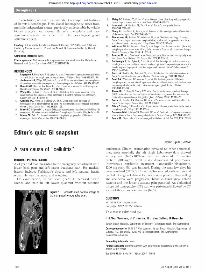
tenderness. Clinical examination revealed no other abnormal-ities, most especially the left thigh. Laboratory data showedleucocytosis (16.66109/litre) and an elevated C reactiveprotein (301 mg/l). Chest x ray demonstrated pneumonia.Intravenous antibiotic treatment (amoxicillin/clavulanate,1200 mg every 8h) was initiated. During the next few days hisfever remained (38.3uC). His left leg became red, oedematous andpainful. No signs of abscess formation were present. The swellingand erythema were progressive. Blood cultures grew entericbacteria and the lower quadrant pain persisted. An abdominalcomputed tomography (CT) scan was performed followed by CTscans of thorax and extremities (fig 1).
QUESTIONWhat is the diagnosis?See page 1089 for the answer
This case is submitted by:
R J Van Wensen, J P Ruurda, H J Van Geffen, K Bosscha
Jeroen Bosch Hospital, Department of Surgery, ’s-Hertogenbosch, The Netherlands
Correspondence to: Dr R J A Van Wensen, Jeroen Bosch Hospital, Department ofSurgery, P.O. Box 90153, 5200 ME ’s-Hertogenbosch, The Netherlands;[email protected]
Competing interests: None.
Patient consent: Informed consent was obtained for publication of the person’sdetails in this report.
Gut 2008;57:1048. doi:10.1136/gut.2007.141853
Figure 1 Reconstructed coronal image ofthe computed tomography scan.
Editor’s quiz: GI snapshot
Oesophagus
1048 Gut August 2008 Vol 57 No 8
group.bmj.com on November 1, 2016 - Published by http://gut.bmj.com/Downloaded from
in human Barrett's oesophagusepitheliumthe origin of metaplastic glandular
Individual crypt genetic heterogeneity and
Poller, R Harrison, M R Novelli, J A Jankowski and N A WrightS J Leedham, S L Preston, S A C McDonald, G Elia, P Bhandari, D
doi: 10.1136/gut.2007.1433392008 57: 1041-1048 originally published online February 27, 2008Gut
http://gut.bmj.com/content/57/8/1041Updated information and services can be found at:
These include:
MaterialSupplementary
http://gut.bmj.com/content/suppl/2008/07/03/57.8.1041.DC1.htmlSupplementary material can be found at:
References #BIBLhttp://gut.bmj.com/content/57/8/1041
This article cites 21 articles, 7 of which you can access for free at:
serviceEmail alerting
box at the top right corner of the online article. Receive free email alerts when new articles cite this article. Sign up in the
CollectionsTopic Articles on similar topics can be found in the following collections
(350)Oesophageal cancer
Notes
http://group.bmj.com/group/rights-licensing/permissionsTo request permissions go to:
http://journals.bmj.com/cgi/reprintformTo order reprints go to:
http://group.bmj.com/subscribe/To subscribe to BMJ go to:
group.bmj.com on November 1, 2016 - Published by http://gut.bmj.com/Downloaded from