Embed Size (px)
Citation preview
ACTA OPHTHALMOLOGICA VOL. 39 1961
From the Eye Department, Copenhagen City Hospital, Copenhagen (Head: Professor P o d Brcendstrzip, M . D.)
ARTERIOVENOUS ANEURYSM OF THE RETINA")
A Case of Spontaneous Thrombosis and >>Healing((
BY
Eilif GTegersen
By now the ophthalmological literature contains almost fifty cases of retinal arteriovenous aneurysms, also called racemose haemangioma. In Scandinavia the first cases were reported by Gertz (1916), Ehlers (1924), and Frandsen (1950). Recently, Bech & Jensen (1958) published two cases. Reviews of the literature may be found in the papers of Bech & Jensen and Rundles & Falls (1 95 1).
The case to be reported below is rather exceptional in having undergone spontaneous thrombosis and a kind of ,,healing<<. A brief description would, therefore, appear to be of interest.
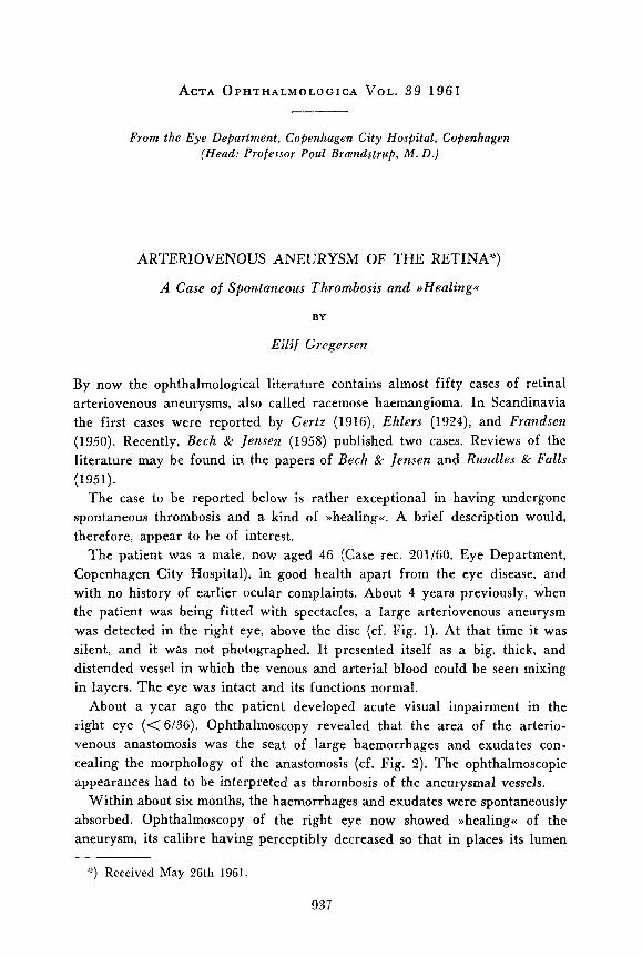
The patient was a male, now aged 46 (Case rec. 201/60, Eye Department, Copenhagen City Hospital), in good health apart from the eye disease, and with no history of earlier ocular complaints. About 4 years previously, when the patient was being fitted with spectacles, a large arteriovenous aneurysm was detected in the right eye, above the disc (cf. Fig. 1). At that time it was silent, and it was not photographed. I t presented itself as a big, thick, and distended vessel in which the venous and arterial blood could be seen mixing in layers. The eye was intact and its functions normal.
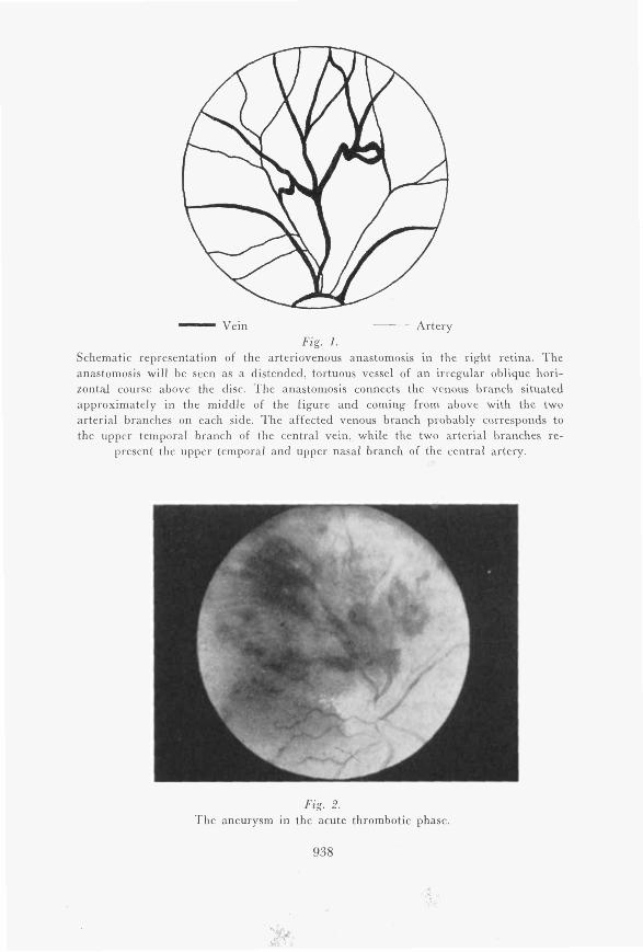
About a year ago the patient developed acute visual impairment in the right eye (<6/36). Ophthalmoscopy revealed that the area of the arterio- venous anastomosis was the seat of large haemorrhages and exudates con- cealing the morphology of the anastomosis (cf. Fig. 2 ) . The ophthalmoscopic appearances had to be interpreted as thrombosis of the aneurysmal vessels.
Within about six months, the haemorrhages and exudates were spontaneously absorbed. Ophthalmoscopy of the right eye now showed >>healing<< of the aneurysm, its calibre having perceptibly decreased so that in places its lumen
") Received May 26th 1961.
93 7
- Vein Artery Fig. I .
Schematic representation of the arteriovenous anastomosis in the right retina. The anastomosis will be seen as a distended, tortuous vessel of an irregular oblique hori- zontal course above the disc. The anastomosis connects the venous branch situated approximately in the middle of the figure and coming from above with the two arterial branches on each side. The affected venous branch probably corresponds to the upper temporal branch of the central vein, while the two arterial branches re-
present the upper temporal and upper nasal branch of the central artery.
Fig. 2. The aneurysm in the acute thrombotic phase.
938
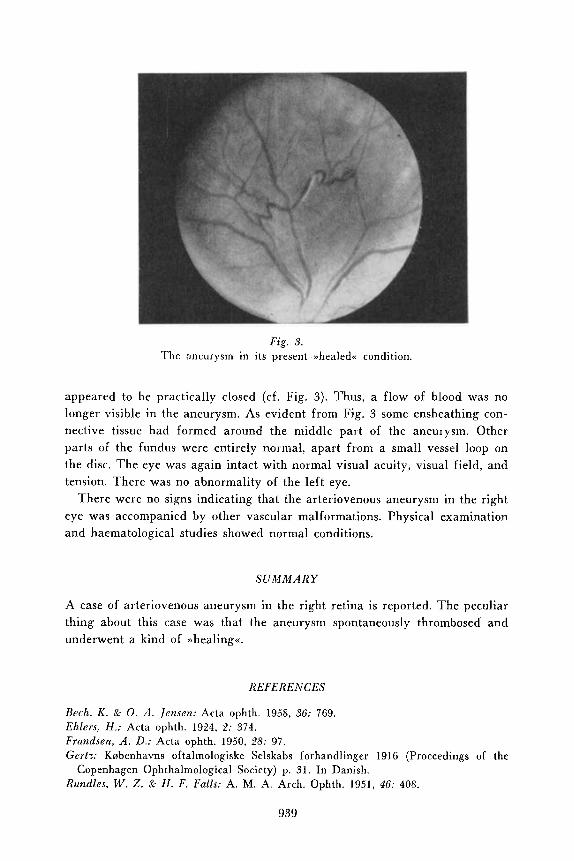
Fig. 3. The aneurysm in its present >>healed<< condition.
appeared to be practically closed (cf. Fig. 3). Thus, a flow of blood was no longer visible in the aneurysm. As evident from Fig. 3 some ensheathing con- nective tissue had formed around the middle part of the aneurysm. Other parts of the fundus were entirely normal, apart from a small vessel loop on the disc. The eye was again intact with normal visual acuity, visual field, and tension. There was no abnormality of the left eye.
There were no signs indicating that the arteriovenous aneurysm in the right eye was accompanied by other vascular malformations. Physical examination and haematological studies showed normal conditions,
SUMMARY
A case of arteriovenous aneurysm in the right retina is reported. The peculiar thing about this case was that the aneurysm spontaneously thrombosed and underwent a kind of >>healing<<.
R E F E R E N C E S
Bcch, K . & 0. A . Jensen: Acta ophth. 1958, 36: 769. Ehlers, H.: Acta ophth. 1924, 2: 374. Frandsen, A . D.: Acta ophth. 1950, 28: 97. Gertx Knbenhavns oftalmologiske Selskabs forhandlinger 1916 (Proceedings of the
Rundles, W. Z. & H . F. Falls: A. M. A. Arch. Ophth. 1951, 4F: 40s. Copenhagen Ophthalmological Society) p. 31. In Danish.
939


![Clinical Study …downloads.hindawi.com/journals/tswj/2012/386478.pdfembolism during deep vein aneurysm surgical repair [2, 18] or when thrombosis recurred in venous surgical area](https://img.pdfslide.us/doc/110x75/5f78114f0b793b21a578ee8f/clinical-study-embolism-during-deep-vein-aneurysm-surgical-repair-2-18-or-when.jpg)