Embed Size (px)
Citation preview

677
Braz J Med Biol Res 39(5) 2006
Arsenic trioxide alters nasopharyngeal cancer cellsBrazilian Journal of Medical and Biological Research (2006) 39: 677-685ISSN 0100-879X
Arsenic trioxide reduces the invasiveand metastatic properties ofnasopharyngeal carcinoma cellsin vitro
1Laboratory of Cancer Research, 2Department of Radiotherapy, Cancer Hospital,3Department of Cancer Molecular Biology, 4Department of Pathology,Shantou University Medical College, Shantou, P.R. China5Clinical Research Centre, Division of Dermatology,Department of Biomedicine and Surgery, Linkoping University, Linkoping,Sweden
C.W. Du1, B.G. Wen3,D.R. Li1, X. Peng2,
C.Q. Hong1, J.Y. Chen1,Z.Z. Lin2, X. Hong2,Y.C. Lin1, L.X. Xie2
M.Y. Wu4 and H. Zhang5
Abstract
Nasopharyngeal carcinoma (NPC) is notorious for the metastases,which are in close association with Epstein-Barr virus-encoded latentmembrane protein 1 (LMP1). Arsenic trioxide (As2O3) has beenshown to induce apoptosis and differentiation in NPC xenografts.Then, can it repress the cancer cells’ metastasis potential? To elucidatethis issue, the present study was performed. LMP1-negative cell lineHNE1 and LMP1-positive cell line HNE1-LMP1 were used as in vitromodel. Cells (1 x 10⁵/mL) were cultured with or without 3 µM As2O3
for 48 h. Then the survival cells were collected to investigate theirpotential of colony formation, attachment, invasion, and migration.Both confocal immunofluorescence staining and Western blot wereused to detect the changes of LMP1 expression. The changes of MMP-9 were examined by RT-PCR assay and Western blot. The results wereas follow: i) the colony formation inhibition rate (75.41 ± 3.9% inHNE1-LMP1 cells vs 37.89 ± 4.9% in HNE1 cells), the rate ofattachment (HNE1-LMP1 vs HNE1: 56.40 ± 3.5 vs 65.87 ± 5.9%), theinvasion inhibitory rate (HNE1-LMP1 vs HNE1: 56.50 ± 3.7 and27.91 ± 2.1%), and the migration inhibitory rate (HNE1-LMP1 vsHNE1: 48.70 ± 3.9 vs 29.19 ± 6.27%) were all significantly differentbetween the two cell lines (P < 0.01). ii) LMP1 was down-regulated inAs2O3-treated HNE1-LMP1 cells. iii) The reduction of MMP-9 wasfound in As2O3-treated groups, more evident in HNE1-LMP1 cells.Thus, we conclude that As2O3 can reduce metastasis potential of NPCcells, involving inhibition of MMP-9 expression. LMP1 were alsoreduced in this process and seemed to enhance anti-metastasis activityof As2O3.
CorrespondenceD.R. Li
Laboratory of Cancer Research
Cancer Hospital
Shantou University Medical College
No. 7 Raoping Road
Shantou
Guang Dong 515031
P.R. China
Fax: +86-754-856-0352
E-mail: [email protected]
Research supported by the Shantou
University Research and Development
Fund (L03002), Medical Science
Foundation of Guangdong Province
(B2004089) and the Traditional
Chinese Medicine Research Fund of
Guangdong Province (102053).
This study is also part of a major
research project of the Shantou
Science Technology Committee
([2003]119).
Received June 14, 2005
Accepted November 18, 2005
Key words• Arsenic trioxide (As2O3)• Nasopharyngeal carcinoma• Metastases• Latent membrane protein 1• Metalloproteinase 9

678
Braz J Med Biol Res 39(5) 2006
C.W. Du et al.
Introduction
Nasopharyngeal carcinoma (NPC) is acommon cancer in southern China. The dis-ease is notorious for its high invasivenessand metastatic activity. Clinically, tumor cellsoften disseminate to regional lymph nodesand to distant sites before forming a mass atthe primary site. Thus far, radiotherapy orradiochemotherapy is the treatment of choicefor this malignancy, but side effects as gas-trointestinal toxicity and myelosuppressionoften lead to the interruption of chemo-therapy. In addition, tumor metastasis re-mains a critical obstacle in clinical radio-therapy or combined radiochemotherapy.When metastatic disease develops after cura-tive radiotherapy, the prognosis is poor.Therefore, a novel therapeutic approach toNPC is strongly desired.
We have reported that arsenic trioxide(As2O3), at the dose of 5 mg/kg, suppressedthe growth of NPC xenografts by inducingpartial differentiation and apoptosis (1). Inour earlier work, the drug was shown toinhibit telomerase activity and to enhanceradiation-induced apoptosis (2,3). However,the effects of As2O3 on NPC cell invasionand metastasis are unclear. In the presentstudy, we investigated the antimetastatic ef-fects of As2O3 on human NPC cell linesusing the Matrigel invasion assay. In view ofthe important role of latent membrane pro-tein 1 (LMP1) in NPC metastasis, we deter-mined the influence of LMP1 on As2O3-mediated anti-metastatic activity.
Material and Methods
Cell lines
The HNE1-LMP1 cell line, which is con-stantly expressing LMP1 by artificially trans-fecting LMP1 cDNA into HNE1 cells, wasinvestigated as an in vitro model in ourstudy. The parental cell line HNE1, whichdoes not express LMP1, derived from poorly
differentiated NPC, was used as control. Thetwo cell lines were established at the CancerResearch Institute of Hunan Medical Uni-versity (4) and were kindly provided byProfessor Y. Cao. Cells were cultured inRPMI 1640 medium with 10% fetal bovineserum, kept in a humidified atmosphere of95% air and 5% CO2 at 37ºC.
Experimental design
Both cell lines, at the density of 1 x 10⁵/mL, were exposed to 3 µM As2O3 for 48 h.Floating cells were then discarded and theresidual cells were allowed to grow further.When the residual cells reached confluencethey were collected for study. Cells not treatedwith As2O3 were used as controls.
Colony formation assay
About 100 living cells were added to a60-mm culture dish containing 5 mL of cul-ture medium. The dishes were then placed ina humidified incubator containing 5% CO2
and incubated at 37ºC for 14 days. The num-ber of colonies containing more than 50 cellsin each dish was counted under a micro-scope. The inhibitory rate (IR) was calcu-lated as follows: IR (%) = (number of colo-nies formed in the control group - number ofcolonies formed in the test group)/number ofcolonies formed in the control group.
In vitro adhesion assay
Each well of the 96-well microplates wascoated with reconstituted basement mem-brane Matrigel (Becton Dickinson Labware,Franklin Lakes, NJ, USA), 2 µg per well.The coated wells were allowed to dry andappropriate serum-free RPMI 1640 mediumwas added to each well and incubated for 1h. The wells were then washed with PBS.Each well containing 4 x 10⁴ cells was incu-bated for 2 h at 37ºC in the presence of 5%CO2. Wells were washed 3 times with 200

679
Braz J Med Biol Res 39(5) 2006
Arsenic trioxide alters nasopharyngeal cancer cells
µL PBS, 40 µg MTT was added to each welland the plates were incubated for 4 h at 37ºCin the presence of 5% CO2. The liquid wasthen removed and 200 µL DMSO was addedto each well. The absorbance of each wellwas read with a microplate reader (Bio-Rad450, Hercules, CA, USA) at 492 nm. Dataare reported as the percentage of total cells,assuming that the adhesion of cells in thecontrol treatment represented 100%.
In vitro invasion assay
Assays were performed with the use ofFalcon cell culture inserts (pore size, 8.0 µm;Becton Dickinson). The chambers were setin a 6-well plate. The upper layer of theculture insert was then coated with 750 µgMatrigel, a reconstituted extracellular ma-trix (Becton Dickinson). Cells were seededat a density of 2 x 10⁴ cells/well into theupper layer of the culture insert and culturedwith serum-free DMEM. Then, 3 mL ofculture medium supplemented with 0.1%BSA and 250 µg solubilized Matrigel wasplaced into the lower layer of the cultureinsert as a chemoattractant. After the cellswere incubated for 24 h, the remaining cellsin the upper layer were swabbed with cottonand penetrating cells in the lower layer werefixed with 95% ethanol and removed forhematoxylin staining. Cells passing throughthe Matrigel matrix and each 8-µm pore ofthe culture insert were counted using lightmicroscopy. Ten fields per well werecounted. The IR was calculated as follows:IR (%) = (number of penetrating cells in thenegative control group - number of penetrat-ing cells in the test group)/number of cells inthe penetrating negative control group.
In vitro mobility assay
The assay was the same as for the inva-sive procedure described above except thatthe upper side of the polycarbonate mem-branes was not coated with Matrigel.
LMP1 expression assay
To determine whether LMP1 plays a rolein the metastatic potential of the cells wefirst used confocal immunofluorescencestaining to detect the changes in LMP1 ex-pression in HNE1-LMP1 cells after As2O3
treatment. Cells were cultured overnight ona glass coverslip, washed with PBS andfixed in 4% paraformaldehyde. To displayLMP1, the cells were first incubated withanti-LMP1 antibody (DAKO, Glostrup, Den-mark) and then reacted with their corre-sponding FITC-conjugated anti-IgG anti-body (DAKO) as secondary antibody. Tovisualize the nuclei, the cells were stainedwith 50 µg/mL propidium iodine containing100 µg DNase-free RNase A per mL and thefluorescent image was observed under a la-ser-scanning confocal microscope (Ultima312, Meridian Instruments Inc., Kent, WA,USA) using the following parameters: ex-cited light 488 nm, emission light 530 nmand pinhole 10-40 nm. Western blotting wasthen used to analyze the changes in LMP1expression. The procedure was similar tothat used for matrix metalloproteinase 9(MMP-9) examination, which is describedbelow.
MMP-9 expression assay
We first determined MMP-9 mRNA lev-els using semi-quantitative RT-PCR. TotalRNA was extracted with Trizol reagent(Gibco BRL, Gaithersburg, MD, USA) ac-cording to manufacturer instructions. Re-verse transcription was performed with theCASsuper two-step semi-quantitative RT-PCR kit (Casarray, Shanghai, China). Theprimers used for MMP-9 mRNA amplifica-tion were (F) 5'-GACTCGGTCTTTGAGGAGCC-3' (R) and 5'-GAACTCACGCGCCAGTAGAA-3' (350 bp) (5). PCR condi-tions were as follows: 94ºC for 5 min, fol-lowed by 32 cycles at 94ºC for 30 s, 56ºC for30 s, and 72ºC for 45 s, with a final extension
680
Braz J Med Biol Res 39(5) 2006
C.W. Du et al.
at 72ºC for 7 min. Electrophoresis of the RT-PCR products was performed on 1% agarosegel with a size marker under standard condi-tions. Simultaneously, transcripts encodingß-actin were detected in all samples andserved as internal controls using the primers5'-CCT CTA TGC CAA CAC AGT GC-3'(left) and 5'-GTA CTC CTG CTT GCT GATCC-3' (right).
In addition, SDS-PAGE and Western blotwere used to determine whether MMP-9expression was affected. Cells were collectedwith a cell scraper and placed in lysis buffer(1 mM Na3PO4, 20 mM HEPES, pH 7.4, 1mM PMSF, 10 µg/mL leupeptin, and 10 µg/mL aprotinin) for at least 1 h at 4ºC. The cellswere transferred to a clean tube and centri-fuged for 30 s at 1,500 g to eliminate unbro-ken cells. The supernatant was incubated onice for 1 h and then centrifuged at 10,000 g at4ºC for 5 min and the supernatant was col-lected and placed in a new microcentrifugetube. The protein was stored at -70ºC. Equalamounts of protein from each cell line wereadded to 12.5% polyacrylamide gel. Afterelectrophoresis, proteins were blotted ontoPVDF sheets (Amersham Pharmacia BiotechInc., Piscataway, NJ, USA) at 350 mA for 1h. The same antibodies used for immuno-fluorescence staining were used to detect
MMP-9 in each tumor cell line. Goat anti-mouse antibody conjugated with horserad-ish peroxidase was used as the secondaryantibody. The protein bands were visualizedwith ECL plus Western blotting detectionreagents (Amersham).
Statistical analysis
Each assay was performed in triplicateand repeated at least three times. Statisticalanalysis was performed using the SPSS 10.0for Windows package. Data are reported asmeans ± SD. Statistical differences wereevaluated by a one-way analysis of variance(ANOVA) followed by the Bonferroni posthoc test, with the level of significance set atP < 0.05.
Results
Effect of As2O3 on colony formation
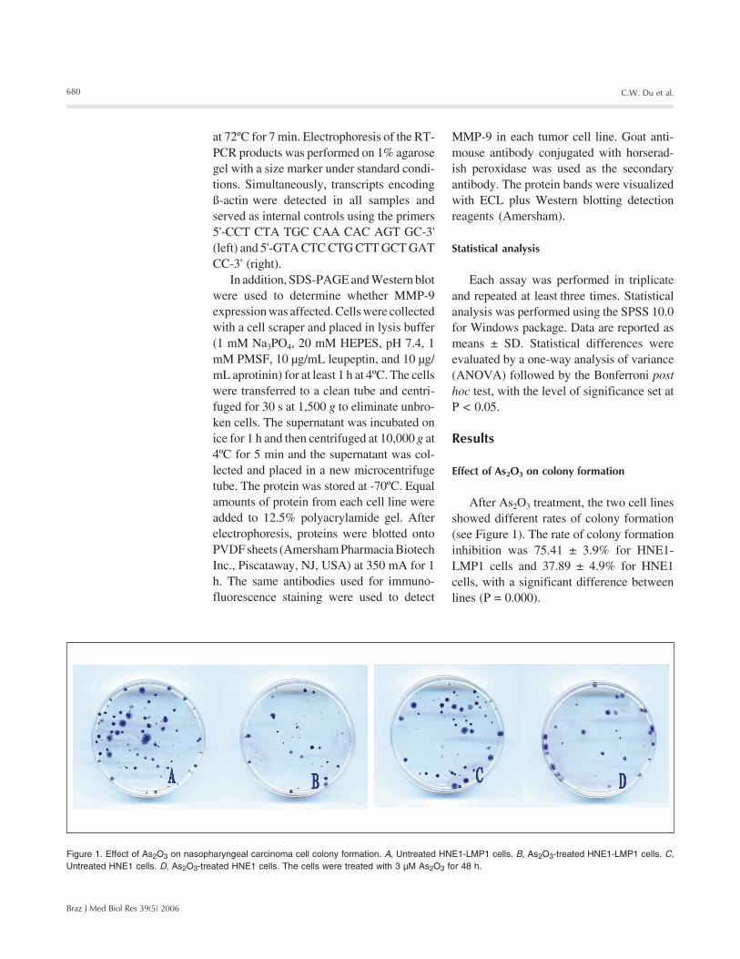
After As2O3 treatment, the two cell linesshowed different rates of colony formation(see Figure 1). The rate of colony formationinhibition was 75.41 ± 3.9% for HNE1-LMP1 cells and 37.89 ± 4.9% for HNE1cells, with a significant difference betweenlines (P = 0.000).
Figure 1. Effect of As2O3 on nasopharyngeal carcinoma cell colony formation. A, Untreated HNE1-LMP1 cells. B, As2O3-treated HNE1-LMP1 cells. C,Untreated HNE1 cells. D, As2O3-treated HNE1 cells. The cells were treated with 3 µM As2O3 for 48 h.

681
Braz J Med Biol Res 39(5) 2006
Arsenic trioxide alters nasopharyngeal cancer cells
Effect of As2O3 on the invasive ability of the cells
As shown in Figure 3, there were fewerinvading HNE1-LMP1 and HNE1 cells inthe As2O3 treatment group compared to the
Effect of As2O3 on adhesion
The influence of As2O3 treatment on theadhesion of both cell lines is shown in Fig-ure 2. Both lines showed significantly de-creased attachment to Matrigel, with valuesof 56.40 ± 3.5% for HNE1-LMP1 cells and65.87 ± 5.9% for HNE1 cells (P = 0.001).
negative control group (HNE1-LMP1: P =0.000; HNE1: P = 0.000). However, theinvasive ability of HNE1-LMP1 cells wassignificantly lower than that of HNE1 cellswhen submitted to treatment with the samedose of As2O3 (P = 0.000), with respectiveinhibitory rates of 56.50 ± 3.7 and 27.91%(P = 0.000).
Effect of As2O3 on cell chemotactic migrationability
The chemotactic ability of both cell linesafter treatment with As2O3 was lower than
Figure 2. Effect of As2O3 (3 µM) for 48 h on adhesion inboth cell lines. Absorbance of each well was obtainedat 492 nm. A, Absorbance for HNE1-LMP1 cells with-out As2O3 treatment. B, Absorbance for HNE1-LMP1cells after As2O3 treatment. C, Absorbance for HNE1cells without As2O3 treatment. D, Absorbance for HNE1cells after As2O3 treatment.
Abs
orba
nce
at 4
92 n
m
2.5
2.0
1.5
1.0
0.5
0A B C D
Figure 3. Effect of 3 µM As2O3 for 48 h on the invasive potential ofcells. The assay was performed using Matrigel as reconstitutedextracellular matrix in Falcon cell culture inserts in vitro. The smallrings are 8-µm membrane pores of the Falcon cell culture insertsindicated by a solid arrow. The invasive cells indicated by an openarrow were counted at least in 10 fields per insert. A, PenetratingHNE1-LMP1 cells in the negative control. B, Penetrating HNE1-LMP1 cells in the As2O3 group. C, Penetrating HNE1 cells in thenegative control. D, Penetrating HNE1 cells in the As2O3 group.E, Number of penetrating cells per insert (y-axis) in each group.Data are reported as the mean ± SD for three independent experi-ments carried out in triplicate, where A, B, C, and D in the x-axisstand for each experimental group described as above. H&Estaining. P < 0.05 compared to control (ANOVA).
Cel
l inv
asio
n (c
ells
per
inse
rt) 90
80706050
0
4030
1020
A B C D
E

682
Braz J Med Biol Res 39(5) 2006
C.W. Du et al.
that of their negative control group (HNE1-LMP1: P = 0.000; HNE1: P = 0.000), but theinhibitory rate of HNE1-LMP1 (48.70 ±3.9%) was significantly higher than that ofthe HNE1 group (29.19 ± 6.27%; P = 0.000).The effects of As2O3 on the chemotacticmigration ability of both cell lines are shownin Figure 4.
Effect of As2O3 on LMP1 expression
As shown in Figure 5, the positive stain-ing intensity of HNE1-LMP1 cells markedlydecreased after As2O3 treatment, as demon-strated by confocal microscopy. In parallel,Western blot showed that the level of LMP1was also reduced.
Figure 5. Effect of 3 µM As2O3 for 48 h on LMP1 expressed inHNE1-LMP1 cells. Top panel, Demonstration of LMP1 ex-pression by confocal immunofluorescence staining. The posi-tive signals were green-stained and located in the cytoplasmand membrane (400X). A, Strong green fluorescent intensityfor LMP1 signals in untreated cells. B, Reduction of greenfluorescent intensity for LMP1 signals in As2O3-treated cells.Bottom panel, Western blott analysis of LMP1 proteins. A,Strong positive staining of LMP1 expression in the controlgroup. B, Weak LMP1 expression in the As2O3 group.
63 kDa
Figure 4. Effect of 3 µM As2O3 for 48 h on the migrationpotential of cells. The small rings are 8-µm membrane poresof the Falcon cell culture inserts indicated by a solid arrow.The migration cells indicated by an open arrow were countedat least in 10 fields per insert. A, Penetrating HNE1-LMP1cells in the negative control. B, Penetrating HNE1-LMP1 cellsin the As2O3 group. C, Penetrating HNE1 cells in the negativecontrol. D, Penetrating HNE1 cells in the As2O3 group. E,Number of penetrating cells per insert (y-axis) in each group.Data are reported as the mean ± SD for three independentexperiments carried out in triplicate, where A, B, C, and D inthe x-axis stand for each experimental group described asabove. H&E staining. P < 0.05 compared to control (ANOVA).
Cel
l mig
ratio
n (c
ells
per
inse
rt) 90
80
706050
0
40
30
1020
A B C D
E

683
Braz J Med Biol Res 39(5) 2006
Arsenic trioxide alters nasopharyngeal cancer cells
Effect of As2O3 on MMP-9 expression
The level of MMP-9 mRNA was exam-ined by semi-quantitative RT-PCR in theHNE1-LMP1 and HNE1 cell lines. As shownin Figure 6, MMP-9 mRNA was expressedin both HNE1-LMP1 and HNE1 cells. How-ever, the extent of expression became veryfaint in HNE1-LMP1 cells after As2O3 treat-ment, whereas As2O3-treated HNE1 cellsshowed only moderate reduction of MMP-9mRNA expression. In addition, tumor cellsshowed remarkable alteration in the expres-sion of MMP-9 protein. In both cell lines,the expression of MMP-9 protein (92 kDa)decreased after treatment with As2O3. Thedown-regulated protein expression of MMP-9 was observed particularly in As2O3-treatedHNE1-LMP1 cells (Figure 7).
Discussion
NPC has highly invasive and metastaticproperties and is more metastatic than otherhead and neck carcinomas. Approximately90% of patients show cervical lymph nodemetastases as the most frequent finding ofNPC. At present, radiotherapy remains astandard treatment for this disease. Althoughintensity modulation radiotherapy delivers ahigher conformal radiation dose to the targetarea and may spare normal organs such asthe parotid glands in patients with early stagedisease, distant metastases still represent thepredominant mode of treatment failure (6,7).Thus, there is a great need for effective anti-metastasis treatment for patients with NPC.
As2O3, a component of Chinese medi-cine, has been known as a poison for manyyears. The drug has started drawing moreattention since the discovery of its clinicalefficacy in acute promyelocytic leukemiareported by Chinese investigators. Clinicaltrials administering As2O3 against other bloodmalignancies and even solid tumors havebeen conducted in recent years (8). FDAapproval of As2O3 against acute promyelo-
Figure 6. Effect of 3 µM As2O3for 48 h on MMP-9 mRNA ex-pression in HNE1-LMP1 andHNE1 cells. M, DNA markers.A, Untreated HNE1-LMP1 cells.B, As2O3-treated HNE1-LMP1cells. C, Untreated HNE1 cells.D, As2O3-treated HNE1 cells.The vertical scale at the left isbp. Experiments were per-formed on three different occa-sions with similar results.
Figure 7. Western blot showingthe effect of 3 µM As2O3 for 48 hon MMP-9 protein expression inHNE1-LMP1 and HNE1 cells.A, Untreated HNE1-LMP1 cells.B, As2O3-treated HNE1-LMP1cells. C, Untreated HNE1 cells.D, As2O3-treated HNE1 cells.
cytic leukemia without major clinical com-plications has quickly led to the testing ofAs2O3 against several types of cancers (9),even though the complete mechanism ofaction against solid or blood-borne tumors isunclear. At present, the drug is indicated as abroad-spectrum anticancer medicine for avariety of cancers. We have reported thatsingle doses of 5 mg/kg As2O3 cause apopto-sis and differentiation in NPC xenografts(1,2). Moreover, recent data indicated thatAs2O3 can induce vascular shutdown andnecrosis in esophageal carcinoma (10). Inaddition, As2O3 has also been reported toinhibit radiation-induced cell invasion (11).Considering these data as a whole, it wasreasonable to investigate whether As2O3 isable to inhibit the invasive and metastaticactivity of NPC cells.
Because we know that not all NPC cellscan be induced to apoptosis or differentia-tion after As2O3 treatment, the objective ofthe present study was to determine whether
726
553
500417311249200
151
MMP-9 (350 bp)
ß-actin (210 bp)
M A B C D
92 kDa
A B C D

684
Braz J Med Biol Res 39(5) 2006
C.W. Du et al.
the malignant biological behavior of residualcells can be changed. Therefore, the presentexperiment was designed to further extendour previous studies of the inhibitory effectsof As2O3 on cancer cells in NPC, with em-phasis on the following points: i) the poten-tial metastatic activity of residual NPC cellsafter As2O3 treatment and its possible mech-anism, and ii) the role of LMP1 in the anti-cancer effect of As2O3.
It is well known that tumor cell metasta-sis is a complex cascade of events. Essentialsteps include the degradation of extracellu-lar matrix and basement membrane. Theprocess involves multiple steps such as prolif-eration, adhesion and migration of tumorcells (11). In the present study, we found thatAs2O3 inhibited the cell potential for prolif-eration, attachment, invasion, and migra-tion. Previous studies have emphasized thatthe critical step for the control of metastasesis to retard local proliferation (12) and toblock cell attachment, invasion and motility.Current data indicate that 3 µM As2O3 for 48h can suppress clonogenic survival, espe-cially in HNE1-LMP1 cells. It has beenshown that cellular interactions with extra-cellular matrix will promote adhesion andmigration, which are thought to be requiredfor tumor metastases. Agents inhibiting cellattachment in vitro may decrease the inva-sion and metastatic potential of tumor cellsin vivo (13). In vitro invasion, attachment,proteolytic dissolution of the matrix, andmovement of tumor cells through Matrigeland polycarbonate are required. Thus, thereconstituted basement membrane invasionassay could reflect the invasive ability of thetumor. We reported here that, compared withuntreated cells, the number of residual HNE1-LMP1 and HNE1 cells after the action ofAs2O3 treatment through the membrane de-creased by up to 1- to 2-fold. This is the firstevidence that As2O3 can reduce the invasivepotential of NPC tumor cells in vitro.
NPC cells have a unique environment,because most carcinoma cells contain hu-
man Epstein-Barr virus as a major etiologicagent for carcinogenesis. Epstein-Barr vi-rus-encoded viral oncoprotein, LMP1, co-operatively induces cellular immortalizationand transformation by a series of signal trans-ductions (12). The LMP1 up-regulates vas-cular endothelial growth factor, cyclooxy-genase-2, and interleukin-8 (13-15) that isactively involved in the promotion of angio-genesis. We know that massive formation ofblood vessels at the tumor site will increasethe opportunity for tumor cells to enter thecirculation. Our data showed that As2O3 in-hibited LMP1 expression in HNE1-LMP1cells. As2O3-induced metastasis suppressionoccurred more easily in LMP1-positive NPCcells (HNE1-LMP1) than in the parental cells(HNE1) with no LMP1 expression. Whethermicrovessel synthesis was inhibited accord-ingly remains to be determined.
In the present study, we examined thealteration of MMP-9 mRNA levels by RT-PCR methods and its protein expression byWestern blot, because MMP-9 belongs to agene family of zinc-containing endopepti-dases which can degrade the extracellularmatrix and basement membrane, playing anessential role in the metastatic process. Onthe other hand, many studies have been pub-lished about the association of LMP1 andMMP-9 with the invasive and metastaticpotential of NPC (13,16). Our data indicatedthat As2O3 can down-regulate MMP-9 at themRNA and protein level in both cell lines tosome extent, but with strong inhibition in theHNE1-LMP1 cell line. Based on these find-ings, we suggest that LMP1 participates inthe enhancement of MMP-9 suppression in-duced by As2O3. Further studies are neededto clarify how As2O3 regulates the expres-sion of MMP-9 and how LMP1 increases thesensitivity of As2O3 in altering the invasiveand metastatic properties of NPC cells.
We analyzed the antiproliferative andantimetastatic effects of As2O3 in NPC celllines. The down-regulation of MMP-9 byAs2O3 may result in lower invasiveness by

685
Braz J Med Biol Res 39(5) 2006
Arsenic trioxide alters nasopharyngeal cancer cells
NPC cells. Our data also demonstrate thatLMP1 expression enhances the responsive-ness of tumor cells to As2O3. Based on theseresults, we present a new point of view about
References
1. Li DR, Du CW, Lin YC et al. (2002). Inhibition of growth of humannasopharyngeal cancer xenografts in SCID mice by arsenic trioxide.Tumori, 88: 522-526.
2. Du CW, Li DR, Lin YC et al. (2004). Differentiation of human na-sopharyngeal carcinoma xenografts and repression of telomeraseactivity induced by As2O3. National Medical Journal of India, 17: 67-70.
3. Li DR, Lin YC, Xie LX et al. (2003). Arsenic trioxide enhancesradiosensitivity in vitro of nasopharyngeal carcinoma. ExperimentalOncology, 25: 248-251.
4. Zhu HC, Yao KT, Li GY et al. (1992). Establishment and characteris-tic analysis of four epithelial tumor cell lines derived from NPC.Journal of Human Medical University (in Chinese), 17: 103-107.
5. Jiang Y, Xu W, Lu J et al. (2001). Invasiveness of hepatocellularcarcinoma cell lines: contribution of hepatocyte growth factor, c-met,and transcription factor Ets-1. Biochemical and Biophysical Re-search Communications, 286: 1123-1130.
6. Kam MK, Teo PM, Chau RM et al. (2004). Treatment of nasopharyn-geal carcinoma with intensity-modulated radiotherapy: the HongKong experience. International Journal of Radiation Oncology, Biol-ogy, Physics, 60: 1440-1450.
7. Lee N, Xia P, Quivey JM et al. (2002). Intensity-modulated radio-therapy in the treatment of nasopharyngeal carcinoma: an update ofthe UCSF experience. International Journal of Radiation Oncology,Biology, Physics, 53: 12-22.
8. Vuky J, Yu R, Schwartz L et al. (2002). Phase II trial of arsenictrioxide in patients with metastatic renal cell carcinoma. Investiga-tional New Drugs, 20: 327-330.
9. Antman KH (2001). Introduction: The history of arsenic trioxide in
cancer therapy. Oncologist, 6 (Suppl 2): 1-2.10. Shen ZY, Shen J, Chen JY et al. (2003). The inhibition of growth and
angiogenesis in heterotransplanted esophageal carcinoma viaintratumoral injection of arsenic trioxide. Oncology Reports, 10:1869-1874.
11. Wei LH, Lai KP, Chen CA et al. (2005). Arsenic trioxide preventsradiation-enhanced tumor invasiveness and inhibits matrix metallo-proteinase-9 through downregulation of nuclear factor kappa B.Oncogene, 24: 390-398.
12. Li HP & Chang YS (2003). Epstein-Barr virus latent membraneprotein 1: structure and functions. Journal of Biomedical Science,10: 490-504.
13. Lo AK, Huang DP, Lo KW et al. (2004). Phenotypic alterationsinduced by the Hong Kong-prevalent Epstein-Barr virus-encodedLMP1 variant (2117-LMP1) in nasopharyngeal epithelial cells. Inter-national Journal of Cancer, 109: 919-925.
14. Murono S, Inoue H, Tanabe T et al. (2001). Induction of cyclooxy-genase-2 by Epstein-Barr virus latent membrane protein 1 is in-volved in vascular endothelial growth factor production in nasopha-ryngeal carcinoma cells. Proceedings of the National Academy ofSciences, USA, 98: 6905-6910.
15. Ren Q, Sato H, Murono S et al. (2004). Epstein-Barr virus (EBV)latent membrane protein 1 induces interleukin-8 through the nuclearfactor-kappa B signaling pathway in EBV-infected nasopharyngealcarcinoma cell line. Laryngoscope, 114: 855-859.
16. Horikawa T, Yoshizaki T, Sheen TS et al. (2000). Association oflatent membrane protein 1 and matrix metalloproteinase 9 withmetastasis in nasopharyngeal carcinoma. Cancer, 89: 715-723.
the mechanism of the anti-NPC activity ofAs2O3 and provide a logical basis for theapplication of As2O3 to the treatment ofEpstein-Barr virus-associated NPC.

http://www.scielo.br
Scientific Electronic Library Online
FAPESP - Fundação de Amparo à
Pesquisa do Estado de São Paulo
BIREME - Centro Latino-Americano e do
Caribe de Informação em Ciências da Saúde
...............
......
SciELO is a virtual library of Brazilianscientific journals in electronic format.It organizes and publishes in theInternet/Web full texts of scientificjournals as well as indicators of usageand impact.
The general objective of SciELO is tocontribute to the advancement ofBrazilian scientific research bywidening and improving the process ofpublication, dissemination andevaluation of scientific literature.
Providing universal access to scientificjournals, SciELO will promote aremarkable increase in the visibilityand accessibility of nationalscientific literature.
The development and operation ofSciELO are sponsored by FAPESPand carried out in partnership withBIREME. SciELO had its pilot operationduring 1997 and the first semesterof 1998 with a collection of 10 journaltitles. Starting in July 1998, SciELO willprogressively include new titles.


![Lipid encapsulation of arsenic trioxide attenuates cervical cancer properties by inhibiting expression of folate receptor-[alpha] and human papillomavirus-E6 oncogene to induce apoptosis](https://img.pdfslide.us/doc/110x75/58853cb91a28ab26518b70d7/lipid-encapsulation-of-arsenic-trioxide-attenuates-cervical-cancer-properties.jpg)