Embed Size (px)
Citation preview
NANO EXPRESS Open Access
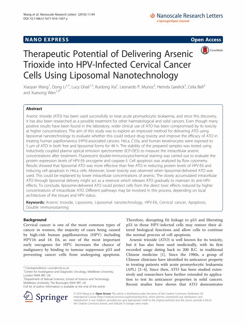
Therapeutic Potential of Delivering ArsenicTrioxide into HPV-Infected Cervical CancerCells Using Liposomal NanotechnologyXiaoyan Wang1, Dong Li1,3, Lucy Ghali1,3, Ruidong Xia2, Leonardo P. Munoz3, Hemda Garelick3, Celia Bell3
and Xuesong Wen1,3*
Abstract
Arsenic trioxide (ATO) has been used successfully to treat acute promyelocytic leukaemia, and since this discovery,it has also been researched as a possible treatment for other haematological and solid cancers. Even though manypositive results have been found in the laboratory, wider clinical use of ATO has been compromised by its toxicityat higher concentrations. The aim of this study was to explore an improved method for delivering ATO usingliposomal nanotechnology to evaluate whether this could reduce drug toxicity and improve the efficacy of ATO intreating human papillomavirus (HPV)-associated cancers. HeLa, C33a, and human keratinocytes were exposed to5 μm of ATO in both free and liposomal forms for 48 h. The stability of the prepared samples was tested usinginductively coupled plasma optical emission spectrometer (ICP-OES) to measure the intracellular arsenicconcentrations after treatment. Fluorescent double-immunocytochemical staining was carried out to evaluate theprotein expression levels of HPV-E6 oncogene and caspase-3. Cell apoptosis was analysed by flow cytometry.Results showed that liposomal ATO was more effective than free ATO in reducing protein levels of HPV-E6 andinducing cell apoptosis in HeLa cells. Moreover, lower toxicity was observed when liposomal-delivered ATO wasused. This could be explained by lower intracellular concentrations of arsenic. The slowly accumulated intracellularATO through liposomal delivery might act as a reservoir which releases ATO gradually to maintain its anti-HPVeffects. To conclude, liposome-delivered ATO could protect cells from the direct toxic effects induced by higherconcentrations of intracellular ATO. Different pathways may be involved in this process, depending on localarchitecture of the tissues and HPV status.
Keywords: Arsenic trioxide, Liposome, Liposomal nanotechnology, HPV-E6, Cervical cancer, Apoptosis,Double immunostaining
BackgroundCervical cancer is one of the most common types ofcancer in women, the majority of cases being causedby high-risk human papillomavirus (HPV) includingHPV16 and 18. E6, as one of the most importantearly oncogenes for HPV, increases the chance ofmalignancy by binding to tumour suppressor p53 andpreventing cancer cells from undergoing apoptosis.
Therefore, disrupting E6 linkage to p53 and liberatingp53 in those HPV-infected cells may restore their al-tered biological functions and allow cells to continuethe normal process of cell apoptosis.Arsenic trioxide (ATO) is well known for its toxicity,
but it has also been used medicinally, with its firstrecorded usage dating back to 200 B.C. in traditionalChinese medicine [1]. Since the 1980s, a group ofChinese clinicians have identified its anticancer propertyin treating patients with acute promyelocytic leukaemia(APL) [2–4]. Since then, ATO has been studied exten-sively and researchers have further extended its applica-tion to test its anticancer properties in solid cancers.Recent studies have shown that ATO demonstrates
* Correspondence: [email protected] for Investigative and Diagnostic Oncology, Middlesex University,London NW4 4BT, UK3Department of Natural Sciences, School of Science and Technology,Middlesex University, The Burroughs NW4 4BT, UKFull list of author information is available at the end of the article
© 2016 Wang et al. Open Access This article is distributed under the terms of the Creative Commons Attribution 4.0International License (http://creativecommons.org/licenses/by/4.0/), which permits unrestricted use, distribution, andreproduction in any medium, provided you give appropriate credit to the original author(s) and the source, provide a link tothe Creative Commons license, and indicate if changes were made.
Wang et al. Nanoscale Research Letters (2016) 11:94 DOI 10.1186/s11671-016-1307-y
anticancer activity against a variety of solid tumourmodels and cancer cell lines, including lung, liver,ovarian, cervical, breast and prostate cancers [5–10].The mechanisms of action of ATO in treating thesecancers are still not fully understood, although celldifferentiation, apoptosis induction, angiogenesis in-hibition and reactive oxygen species generation havebeen implicated as being involved [11]. Furthermore,much higher dosages of ATO are required for treat-ing malignancies rising from solid cancers [12] incomparison to haematopoietic ones due to their dis-tinct differences in tissue architecture. Therefore,some severe side effects from ATO may occur dur-ing the treatment including peripheral neuropathies,liver failure and cardiac toxicity, which could limitits clinical utility [8, 13].Our earlier work has shown that ATO can be used as
an agent to specifically target HPV-infected cervicalcancer cells [14]. However, it can only be used at a lowconcentration (up to 2 μM) as most of cells were killedafter their exposure to high concentrations of ATO.Therefore, an effective delivery system is needed toimprove the therapeutic index of the drug and to expandits clinical utility to treat solid tumours.The liposomal delivery system has been used for the
delivery of both lipophilic and hydrophilic drugs [15]. Itstarted from the use of conventional liposomes and hassince evolved to the development of stealth liposomesand now targeting liposomes. Most recently, it has beensuccessfully used in different applications includingtreating gliomas [16, 17] and breast cancers [18, 19]and managing infectious and inflammatory disorders[20] and heart diseases [21, 22]. In addition, othernanocarrier-delivering systems including using polymer-drug conjugates, dendrimers, micro/nano-particles andmicelles also emerged and have been used in variousapplications along with liposomal-delivering systems[22, 23]. Current nanotechnology provides us a superband promising opportunity to guide the targetingligands directly to the target sites with minimal dis-turbance to the surrounding cells and tissues, whichholds our hope for defeating cancers including HPV-associated cancers one day.Chen and co-workers have shown that targeted li-
posomal delivery of ATO potentiates its efficacy inrelatively insensitive solid tumours [24, 25]. To ourknowledge, no work has been done to investigate theeffects of ATO delivered by liposomes on the tre-atment of HPV-associated cancers. Therefore, an invitro study was carried out to investigate the antican-cer effects of liposomal-encapsulated ATO incomparison to free form ATO through the evaluationof protein expression levels of HPV-E6 oncogene andcaspase-3.
MethodsMaterialsSoy phosphatidylcholine (PC) was purchased from AvantiPolar Lipids (AL, USA). Methoxypolyethyleneglycol-di-stearoyl-phosphatidylethanolamine (DSPE-PEG2000; withmPEG MW2000Da) was obtained from Genzyme (UK).Cholesterol (Chol), PBS, Triton-100, ATO, nickel acetateand dialysis tubing were purchased from Sigma (UK).Methanol and dichloromethane were from Thermofisher(UK). RPMI1640, L-glutamine, penicillin-streptomycinand foetal bovine serum (FBS) were from Invitrogen LifeTechnologies (UK).
Liposome Preparation and CharacterizationLiposomes were composed of soy PC, cholesterol, andmethoxypolyethyleneglycol-di-stearoyl-phosphatidyletha-nolamine (DSPE-PEG2000; Genzyme). Liposomes wereprepared as described elsewhere [24]. Briefly, the lipidswere dissolved in methanol: dichloromethane1:2 (v/v) at aPC/cholesterol/DSPE-PEG2000 molar ratio of 54.7:45:0.3at room temperature. The lipid mixtures were depositedon the side wall of the rotary glass vial by removing thesolvent with nitrogen. The dried lipid films were hydratedin 730 mM nickel acetate (Ni(OAc)2) aqueous solutions.This process led to the spontaneous formation of pegy-lated liposomes. The liposome suspension was subse-quently subjected to 10 freeze-and-thaw cycles (freezingin liquid nitrogen for 3 min and thawing in 37 °C waterbath for 3 min). The liposomes were then downsized bypassing through 0.1-μm Anotop 10 filters (Whatman,UK). Extruded liposomes were dialysed against 10 mMsodium phosphate buffer at pH 7 to get rid of excessNi(OAc)2. The Ni(OAc)2-encapsulated liposomes werethen incubated with an ATO solution at room temperaturefor 2.5 h. After removal of extra unencapsulated ATO bydialysis, the concentrations of phospholipids (P), encapsu-lated ATO and nickel (Ni) in the liposomes were deter-mined by inductively coupled plasma optical emissionspectrometer (ICP-OES; Thermo-Scientific iCap 6500ICP, UK). The molar ratios of ATO/lipid were calcu-lated and used to assess loading efficiency and liposomestability. The mean liposome sizes were determinedby dynamic light scattering on a Zetasizer-Nano ZS(Malvern Instruments, UK).
Cell CultureTwo cervical cancer cell lines, HeLa and C33a (ATCC,USA), and a control cell line, human keratinocytes (HK)(Life Technologies, UK), were used in this study. HeLacells (HPV18 positive = 10 copies per cell) and C33a(HPV negative) were cultured in RPMI1640 media con-taining 10 % foetal calf serum, 100 U/ml of penicillinand 100 mg/ml streptomycin in 75-cm2 flasks. The cellswere grown in a humidified incubator containing 5 %
Wang et al. Nanoscale Research Letters (2016) 11:94 Page 2 of 8
CO2 and 95 % air at 37 °C until they reached 90 %confluence. The following experiments were then set upfor further studies following 48-h ATO exposure: quanti-fication of cellular arsenic uptake, fluorescent double im-munocytochemistry staining (HPV18 E6 and Caspase-3)and flow cytometry analysis for cell apoptosis.
Quantitative Analysis of Cellular Uptake of Arsenic byICP-OESCells were seeded into four 25-cm2 flasks at 2 × 10⁵
cells/flask. Following a 24-h cell attachment, cells weretreated as described below: control (cells in RPMImedia), ATO 5 μM (ATO5), liposomes only (Lip) andlipo-ATO 5 μM (ATO5 + Lip). After 48 h, the cells werewashed by PBS then trypsinized before they werecollected into Falcon tubes for further analysis. Cellswere extracted by adding 3–7 ml of nitric acid andtransferred to Teflon tubes to be digested in a MarsX-press microwave (Method EPA 3051A, 2007). The cellextracts were then transferred to centrifuge tubes, andarsenic concentration was analysed using ICP-OES. Theconcentration of arsenic was corrected by cell numberand total volume accordingly.
HPV-E6 and Caspase-3 Expression Levels Analysed byConfocal MicroscopyThe cells were counted and seeded at a density of5 × 104/ml on sterile cover slips placed in six-well plates.The cells were grown for overnight attachment beforeexposing to the drug treatment. After 48 h of drug treat-ment, the cells grown on cover slips were washed andfixed with 4 % paraformaldehyde in PBS for 10 min beforeimmunocytochemical staining.Fixed cells on coverslips were immunostained using a
double fluorescent staining method by labelling HPV-E6protein with fluomore cyanine 5 (in red) and activecaspase-3 protein (Abcam, Cambridge, UK) using fluor-escein isothiocyanate (FITC; in green) as describedpreviously [14]. Briefly, cells were permeablized by 0.2 %Triton-100, followed by 50 % horse serum for blocking.The first primary antibody, monoclonal anti-HPV16 E6/HPV18 E6 (Santa Cruz Biotechnology, Heidelberg,Germany) at a 1 in 150 dilution in PBS, was applied for90 min at room temperature, followed by 30 and 20 minof secondary and tertiary antibodies, using an ABCuniversal kit (Vector Lab, Peterborough, UK). Thetyramide signal amplification reagent conjugated bycyanine 5 (TSA-Cy5, PerkinElmer, Waltham, MA, USA)was then applied to detect any horseradish peroxidase-conjugated antibody bound to HPV-E6 protein.Next, goat serum was applied before polyclonal rabbit
antihuman caspase-3 antibody (Abcam, UK) at a 1 in100 dilution and was incubated with cells for 60 min.FITC-labelled HRP-conjugated anti-rabbit IgG (Sigma,
Dorset, UK) was added afterwards, followed by mount-ing using 6-diamidino-2-phenylindole (DAPI; stainingnuclei in blue) containing anti-fade ProLong Gold reagent(Life Technologies Ltd, Paisley, UK). The fluorescenceemitted from each slide was observed via a confocalmicroscope (Leica Microsystems, Wetzlar, Germany) andimages were recorded accordingly.
Analysis of Cell Apoptosis by Flow CytometryCells were seeded at 5 × 10⁵/ml in six-well culture platesand grown overnight before exposing to the drug. After48 h of drug treatment, the cells were trypsinized, washedtwice by PBS and then collected into 15-ml centrifugetubes for further staining. The cells were re-suspended inAnnexin-V binding buffer (BD, Oxford, UK) before incu-bating with monoclonal antibody Annexin-V conjugatedwith Alexa 488 (10 μg/ml) (Sigma, UK) for 30 min avoid-ing light. Propidium iodide (PI; 1 μg/ml) (Sigma, UK)was added afterwards, and all samples were analysedwithin 1 hr of PI staining using FACSCalibur (BD,Oxford, UK).
Statistical AnalysisStatistical analysis was carried out automatically follow-ing flow cytometry analysis through BD Calibur softwareprovided. Mean and coefficient of variation (CV) werecalculated accordingly.For immunostaining results from confocal microscopy,
an average number from positively stained cells in a totalof six fields of each sample were calculated and averagepercentages were recorded.
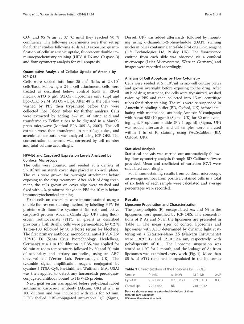
ResultsLiposome Preparation and CharacterizationThe phospholipids (P), encapsulated As, and Ni in theliposomes were quantified by ICP-OES. The concentra-tions of P, As and Ni in the liposomes are presented inTable 1. The mean sizes of control liposomes andliposomes with ATO determined by dynamic light scat-tering on a Zetasizer-Nano ZS (Malvern Instruments)were 118.9 ± 0.7 and 121.0 ± 2.4 nm, respectively, withpolydispersity of 0.1. The liposome suspension wasstored at 4 °C for 1 month, and the leakage of As fromliposomes was examined every week (Fig. 1). More than85 % of ATO remained encapsulated in the liposomes
Table 1 Characterization of the liposomes by ICP-OES
Sample P (mM) As (mM) Ni (mM) As/P
Lipo-ATO 2.37 ± 0.03 0.78 ± 0.23 2.71 ± 1.63 0.33
Control lipo 2.22 ± 0.04 ND 2.81 ± 0.12
Data are shown as means ± standard deviations of threereplicate measurementsND lower than detection limit
Wang et al. Nanoscale Research Letters (2016) 11:94 Page 3 of 8
after 1 month. No significant change in size or chargewas observed (Fig. 2).
Quantitative Analysis of Arsenic UptakeCells were incubated with the four samples: control(cells in RPMI media), ATO 5 μM (ATO5), liposomesonly (Lip) and lipo-ATO 5 μM (ATO5 + Lip) for 48 h.The cellular concentration of arsenic was determined byICP-OES (Table 2). Results showed that arsenic uptakewas significantly decreased in all three cell lines whenATO was delivered by liposomes (ATO5 + LIP) incomparison to free ATO (p < 0.001). In addition, thehighest arsenic uptake was observed in HeLa cells, andno arsenic was detected in any of the cell lines whentreated with cell media or control liposomes (Table 2).
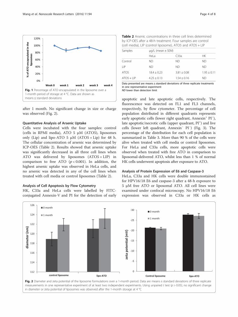
Analysis of Cell Apoptosis by Flow CytometryHK, C33a and HeLa cells were labelled by FITC-conjugated Annexin-V and PI for the detection of early
apoptotic and late apoptotic cells, respectively. Thefluorescence was detected on FL1 and FL3 channels,respectively, by flow cytometer. The percentage of cellpopulation distributed in different quadrants representsearly apoptotic cells (lower right quadrant, Annexin+ PI−),late apoptotic/necrotic cells (upper quadrant, PI+) and livecells (lower left quadrant, Annexin− PI−) (Fig. 3). Thepercentage of the distribution for each cell population issummarized in Table 3. More than 90 % of the cells werealive when treated with cell media or control liposomes.For HeLa and C33a cells, more apoptotic cells wereobserved when treated with free ATO in comparison toliposomal-delivered ATO, whilst less than 1 % of normalHK cells underwent apoptosis after exposure to ATO.
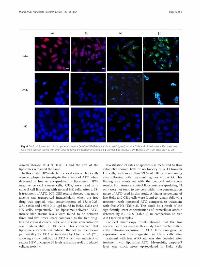
Analysis of Protein Expression of E6 and Caspase-3HeLa, C33a and HK cells were double immunostainedfor HPV16/18 E6 and caspase-3 after a 48-h exposure to5 μM free ATO or liposomal ATO. All cell lines wereexamined under confocal microscopy. No HPV16/18 E6expression was observed in C33a or HK cells as
Fig. 1 Percentage of ATO encapsulated in the liposome over a1-month period of storage at 4 °C. Data are shown asmeans ± standard deviations
Fig. 2 Diameter and zeta potential of the liposome formulations over a 1-month period. Data are means ± standard deviations of three replicatemeasurements in one representative experiment of at least two independent experiments. Using unpaired t test (p > 0.05), no significant changein diameter or zeta potential of liposomes was observed after the 1-month storage at 4 °C
Table 2 Arsenic concentrations in three cell lines determinedby ICP-OES after a 48-h treatment. Four samples are control(cell media), LIP (control liposome), ATO5 and ATO5 + LIP
Samples μg/L (mean ± SEM)
HeLa C33a HK
Control ND ND ND
LIP ND ND ND
ATO5 18.4 ± 0.23 3.81 ± 0.08 1.95 ± 0.11
ATO5 + LIP 4.25 ± 0.13 1.54 ± 0.16 ND
Data presented are means ± standard deviations of three replicate treatmentsin one representative experimentND lower than detection limit
Wang et al. Nanoscale Research Letters (2016) 11:94 Page 4 of 8
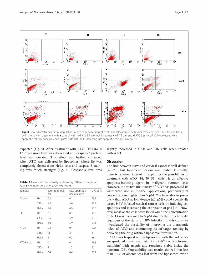
expected (Fig. 4). After treatment with ATO, HPV16/18E6 expression level was decreased and caspase-3 proteinlevel was elevated. This effect was further enhancedwhen ATO was delivered by liposomes, where E6 wascompletely absent from HeLa cells and caspase-3 stain-ing was much stronger (Fig. 4). Caspase-3 level was
slightly increased in C33a and HK cells when treatedwith ATO.
DiscussionThe link between HPV and cervical cancer is well defined[26–29], but treatment options are limited. Currently,there is renewed interest in exploring the possibilities oftreatment with ATO [14, 30, 31], which is an effectiveapoptosis-inducing agent in malignant tumour cells.However, the systematic toxicity of ATO has prevented itswidespread use in medical applications, particularly atconcentrations higher than 5 μM. We have shown previ-ously that ATO at low dosage (≤2 μM) could specificallytarget HPV-infected cervical cancer cells by inducing cellapoptosis and increasing the expression of p53 [14]. How-ever, most of the cells were killed when the concentrationof ATO was increased to 5 μM due to the drug toxicity,regardless of the status of HPV infection. In this study, weinvestigated the possibility of improving the therapeuticindex of ATO and eliminating its off-target toxicity bydelivering the drug within a liposomal formulation.ATO was trapped within liposomes with the aid of co-
encapsulated transition metal ions (Ni2+) which formed‘nanobins’ with arsenic and remained stable inside theliposome [25]. Our stability test results showed that lessthan 15 % of arsenic was lost from the liposomes over a
Fig. 3 Flow cytometry analysis of populations of live cells, early apoptotic cells and late/necrotic cells from three cell lines (HK, C33a and HeLacells) after a 48-h treatment with a control (cell media), b LIP (control liposome), c ATO 5 μm, and d ATO 5 μm+ LIP. FL1—detecting earlyapoptotic cells by Annexin-V conjugated with FITC; FL3—detecting late apoptotic cells by DNA dye PI
Table 3 Flow cytometry analysis showing different stages ofcells from three cell lines after treatment
Samples Early apoptoticcells
Late apoptotic/necrotic cells
Live cells
Control HK 0.2 0.1 99.7
C33a 1.3 5.6 93.4
HeLa 1.8 4.9 93.3
LIP HK 0.1 0.1 99.8
C33a 0.6 5.9 93.5
HeLa 3.0 5.9 91.1
ATO5 HK 0.2 0.4 99.4
C33a 93 2 5
HeLa 2 60 38
ATO5 + Lip HK 0.1 0.1 99.8
C33a 4 10 86
HeLa 3.4 8.4 88.2
Wang et al. Nanoscale Research Letters (2016) 11:94 Page 5 of 8
4-week storage at 4 °C (Fig. 1) and the size of theliposomes remained the same.In this study, HPV-infected cervical cancer HeLa cells
were employed to investigate the effects of ATO whendelivered in free or encapsulated in liposomes. HPV-negative cervical cancer cells, C33a, were used as acontrol cell line along with normal HK cells. After a 48-h treatment of ATO, ICP-OES results showed that morearsenic was transported intracellularly when the freedrug was applied, with concentrations of 18.4 ± 0.23,3.81 ± 0.08 and 1.95 ± 0.11 μg/l found in HeLa, C33a andHK cells, respectively. For liposomal-delivered ATO,intracellular arsenic levels were found to be betweenthree and five times lower compared to the free drug-treated cervical cancer cells, and arsenic concentrationwas undetectable in HK cells. This confirmed thatliposome encapsulation reduced the cellular membranepermeability to ATO as indicated by Chen et al. [25],allowing a slow build-up of ATO which was sufficient toreduce HPV oncogene E6 levels and also result in reducedcellular toxicity.
Investigation of rates of apoptosis as measured by flowcytometry showed little or no toxicity of ATO towardsHK cells, with more than 99 % of HK cells remainingalive following both treatment regimes with ATO. Thisfinding was consistent with the confocal microscopyresults. Furthermore, control liposomes encapsulating Nionly were not toxic to any cells within the concentrationrange of ATO used in this study. A higher percentage oflive HeLa and C33a cells were found to remain followingtreatment with liposomal ATO compared to treatmentwith free ATO (Table 3). This could be a result of thesignificantly lower concentrations of intracellular arsenicdetected by ICP-OES (Table 2) in comparison to freeATO-treated samples.Confocal microscopy results showed that the two
cervical cell lines used in this study have reacted differ-ently following exposure to ATO. HPV oncogene E6expression was down-regulated in HeLa cells aftertreatment with free ATO and was also depleted aftertreatment with liposomal ATO. Meanwhile, caspase-3level was much more up-regulated in HeLa cells
Fig. 4 Confocal fluorescent microscopic examinations (×400) of HPV-E6 (red) and caspase-3 (green) in HeLa, C33a and HK cells after a 48-h treatment.Cells were counter-stained with DAPI (blue) to reveal the nuclear/DNA location. a Control. b LIP. c ATO 5 μM. d ATO 5 μM+ LIP. Scale bar= 20 μm
Wang et al. Nanoscale Research Letters (2016) 11:94 Page 6 of 8
incubated with liposomal ATO compared to that withfree ATO. These findings were supported by the resultsfrom flow cytometry which showed a decreased percent-age of late apoptotic cells and an increased percentage oflive cells after a 48-h exposure to liposomal ATO com-pared with exposure to free ATO. This suggests thatATO can specifically target HPV-infected cells and,when delivered by liposomes, it may be able to alterthe features of the cells, converting them from cancercells to normal cells without causing any toxicity, al-though more research needs to be carried out to elu-cidate the mechanism involved.C33a cells appear to be more sensitive to the high
concentration of free ATO with the majority of cells foundto be in the stage of early apoptosis (Table 3). However,drug toxicity was greatly reduced when ATO was in anencapsulated form, with most of the cells revived fromearly apoptotic stage. It was also observed that C33a cellsformed small clusters following treatment with liposomalATO, which differed from the pattern seen with HeLacells. This suggests that different mechanisms of ATOaction may be involved in this process, which could beHPV relevant. As shown by flow cytometry, no cytotox-icity was observed from control liposomes which was alsoconfirmed by confocal microscopy. Normal HK cells werefound not to react with either the free or liposome-delivered drug, which was supported by both fluorescentconfocal microscopy and flow cytometer analysis.Recent published work by Cao and the others [19]
demonstrated that using targeted ligand can enhance theuptake of delivered agents by the targeted cells, whichwill accumulate in the cells and therefore will leadhigher cellular toxicity locally. Our study can be furtherextended to investigate the effects of ATO on differentsolid tumours by using targeting liposomes which candeliver the drug to a specific cellular compartment toenhance its efficacy and reduce its systemic toxicity.Furthermore, co-administered drugs may improve thepharmacokinetics profiling for the liposomal-delivereddrug in breast cancer treatment [18], which can befurther explored in our prospective study.
ConclusionsWe have shown a promising strategy for targeting HPV-infected cells using liposomal-encapsulated ATO. Thiscarrier system can potentially provide a therapeutic ap-proach that may act to revert the HPV-infected cancer cellsback to a healthier cell population with reduced levels ofHPV-E6 oncogene. ATO can be stably encapsulated intoliposomes with transition metal ions (Ni2+), and the result-ing liposomes revealed higher anti-HPV efficacy againstHeLa cells, which are less sensitive to free ATO. Furtherstudies are needed to elucidate the mechanisms involved.
Competing InterestsThe authors declare that they have no competing interests.
Authors’ ContributionsXSW conceived and co-designed the study, coordinated with other authorsand carried out flow cytometry study, and drafted the manuscript. XYWparticipated in the design of the study, was part of the ICP-OES work andwas involved in drafting the manuscript. DL carried out the cell culture andimmunostaining work. LG carried out part of the cell culture work andprovided valuable support for sorting out the problems during the process.RX was involved in the design of the study and carried out the statisticalanalysis. LP carried out the majority of the ICP-OES experiment. HG providedthe technical guidance, valuable support for ICP-MS and final drafting. CBparticipated in the initial design for the study, some statistical analysis workand final draft editing. All authors read and approved the final manuscript.
Author details1Centre for Investigative and Diagnostic Oncology, Middlesex University,London NW4 4BT, UK. 2Jiangsu Key Laboratory for Organic Electronics andInformation Displays (KLOEID), Institute of Advanced Materials (IAM), NanjingUniversity of Posts and Telecommunications (NJUPT), 9 Wenyuan Road,Nanjing 210046, People’s Republic of China. 3Department of NaturalSciences, School of Science and Technology, Middlesex University, TheBurroughs NW4 4BT, UK.
Received: 26 November 2015 Accepted: 8 February 2016
References1. Ho PC (2005) 33As metallotherapeutic arsenic compounds. In: Gielen M,
Tiekink ER (eds) Metallotherapeutic Drugs and Metal-Based DiagnosticAgents. Jonn Wiley & Sons Ltd, Chichester, pp 297–309
2. Zhang TD, Chen GQ, Wang ZG, Wang ZY, Chen SJ, Chen Z (2001) Arsenictrioxide, a therapeutic agent for APL. Oncogene 20:7146–7153
3. Wang Z, Sun G, Shen Z, Chen S, Chen Z (1999) Differentiation therapy foracute promyelocyticleukemia with all-trans retinoic acid: 10-year experienceof its clinical application. Chin Med J 112:963–967
4. Soignet SL, Maslak P, Wang ZG, Jhanwar S, Calleja E, Dardashti LJ, Corso D,DeBlasio A, Gabrilove J, Scheinberg DA, Pandolfi PP, Warrell RP (1998)Complete remission after treatment of acute promyelocyticleukemia witharsenic trioxide. N Engl J Med 339:1341–1348
5. Lam SK, Mak JC, Zheng CY, Li YY, Kwong YL, Ho JC (2014) Downregulationof thymidylate synthase with arsenic trioxide in lung adenocarcinoma. Int JOncol 44:2093–2102
6. Wang X, Jiang F, Mu J, Ye X, Si L, Ning S, Li Z, Li Y (2014) Arsenic trioxideattenuates the invasion potential of human liver cancer cells through thedemethylation-activated microRNA-491. Toxicol Lett 227:75–83
7. Zekri A, Ghaffari SH, Yousefi M, Ghanizadeh-Vesali S, Mojarrad M,Alimoghaddam K, Ghavamzadeh A (2013) Autocrine human growthhormone increases sensitivity of mammary carcinoma cell to arsenictrioxide-induced apoptosis. Mol Cell Endocrinol 377:84–92
8. Dilda PJ, Hogg PJ (2007) Arsenical-based cancer drugs. Cancer TreatRev 33:542–564
9. Kito M, Matsumoto K, Wada N, Sera K, Futatsugawa S, Naoe T, Nozawa Y,Akao Y (2003) Antitumor effect of arsenic trioxide in murine xenograftmodel. Cancer Sci 94:1010–1014
10. Maeda H, Hori S, Nishitoh H, Ichijo H, Ogawa O, Kakehi Y, KakizukaA (2001) Tumor growth inhibition by arsenic trioxide (As2O3) in theorthotopic metastasis model of androgen-independent prostate cancer.Cancer Res 61:5432–5440
11. Berenson JR, Yeh HS (2006) Arsenic compounds in the treatment ofmultiple myeloma: a new role for a historical remedy. Clin LymphomaMyeloma 7:192–198
12. Liu B, Pan S, Dong X, Qiao H, Jiang H, Krissansen GW, Sun X (2006)Opposing effects of arsenic trioxide on hepatocellular carcinomas in mice.Cancer Sci 97:675–681
13. Evens AM, Tallman MS, Gartenhaus RB (2004) The potential of arsenictrioxide in the treatment of malignant disease: past, present, and future.Leuk Res 28:891–900
Wang et al. Nanoscale Research Letters (2016) 11:94 Page 7 of 8
14. Wen X, Li D, Zhang Y, Liu S, Ghali L, Iles RK (2012) Arsenic trioxide inducescervical cancer apoptosis, but specifically targets human papillomavirus-infected cell populations. Anticancer Drugs 23:280–287
15. Fang JY, Hwang TL, Huang YL (2006) Liposomes as vehicles for enhancingdrug delivery via skin routes. Curr Nano 2:55–70
16. Du D, Chang N, Sun S, Li M, Yu H, Liu M, Liu X, Wang G, Li H, Liu X, Geng S,Wang Q, Peng H (2014) The role of glucose transporters in the distributionof p-aminophenyl-α-d-mannopyranoside modified liposomes within micebrain. J Control Release 182:99–110
17. Li M, Deng H, Peng H, Wang Q (2014) Functional nanoparticles in targetingglioma diagnosis and therapies. J Nanosci Nanotechnol 14:415–432
18. Li MH, Yu H, Wang TF, Chang ND, Zhang JQ, Du D, Liu MF, Sun SL, Wang R,Tao HQ, Geng SL, Shen ZY, Wang Q, Peng HS (2014) Tamoxifen embeddedin lipid bilayer improved the oncotarget of liposomal daunorubicin in vivo.J Mater Chem B 2:1619–1625
19. Cao J, Wang R, Gao N, Ming L, Tian X, Yang W, Ruan Y, Zhou C, Wang G, LiuX, Tang S, Yu Y, Liu, Sun G, Peng H, Wang Q (2015) A7RC peptide modifiedpaclitaxel liposomes dually target breast cancer. Biomater Sci 3:1545–1554
20. Ikoba U, Peng H, Li H, Miller C, Yu C, Wang Q (2015) Nanocarriers in therapyof infectious and inflammatory diseases. Nanoscale 7:4291–4305
21. Liu M, Li M, Sun S, Li B, Du D, Sun J, Cao F, Li H, Jia F, Wang T, Chang N, YuH, Wang Q, Peng H (2014) The use of antibody modified liposomes loadedwith AMO-1 to deliver oligonucleotides to ischemic myocardium forarrhythmia therapy. Biomaterials 35:3697–3707
22. Liu M, Li M, Wang G, Liu X, Liu D, Peng H, Wang Q (2014) Heart-targetednanoscale drug delivery systems. J Biomed Nanotechnol 10:2038–2062
23. Peng H, Liu X, Wang G, Li M, Bratlie KM, Cochrana E, Wang Q (2015)Polymeric multifunctional nanomaterials for theranostics. J Mater ChemB 3:6856–6870
24. Chen H, Ahn R, Van den Bossche J, Thompson DH, O'Halloran TV (2009)Folate-mediated intracellular drug delivery increases the anticancer efficacy ofnanoparticulate formulation of arsenic trioxide. Mol Cancer Ther 8:1955–1963
25. Chen H, MacDonald RC, Li S, Krett NL, Rosen ST, O'Halloran TV (2006) Lipidencapsulation of arsenic trioxide attenuates cytotoxicity and allows forcontrolled anticancer drug release. J Am Chem Soc 128:13348–13349
26. Dillman RO, Oldham RK (2009) Principles of cancer biotherapy, 5th edn.Springer, Dordrech
27. Walboomers JM, Jacobs MV, Manos MM, Bosch FX, Kummer JA, Shah KV,Snijders PJ, Peto J, Meijer CJ, Muñoz N (1999) Human papillomavirus is anecessary cause of invasive cervical cancer worldwide. J Pathol 189:12–19
28. Montero JA, Larkin JA, Houston SH, Toney J (1997) Examining the complexrelationship of human papillomavirus to cervical dysplasia and carcinoma.Medscape Womens Health 2:1
29. Munoz N, Bosch FX (1996) The causal link between HPV cervical cancerits implications for prevention of cervical cancer. Bull Pan Am HealthOrgan 30:362–377
30. Wang H, Gao P, Zheng J (2014) Arsenic trioxide inhibits cell proliferationand human papillomavirus oncogene expression in cervical cancer cells.Biochem Biophys Res Commun 451:556–561
31. Yu J, Qian H, Li Y, Wang Y, Zhang X, Liang X, Fu M, Lin C (2007) Therapeuticeffect of arsenic trioxide (As2O3) on cervical cancer in vitro and In vivothrough apoptosis induction. Cancer Biol Ther 6:580–586
Submit your manuscript to a journal and benefi t from:
7 Convenient online submission
7 Rigorous peer review
7 Immediate publication on acceptance
7 Open access: articles freely available online
7 High visibility within the fi eld
7 Retaining the copyright to your article
Submit your next manuscript at 7 springeropen.com
Wang et al. Nanoscale Research Letters (2016) 11:94 Page 8 of 8