Embed Size (px)
Citation preview
Hindawi Publishing CorporationMetal-Based DrugsVolume 2008, Article ID 260146, 13 pagesdoi:10.1155/2008/260146
Review ArticleArsenic-Based Antineoplastic Drugs andTheir Mechanisms of Action
Stephen John Ralph
School of Medical Sciences, Griffith University, Parklands Drive, Southport, Queensland 4215, Australia
Correspondence should be addressed to Stephen John Ralph, [email protected]
Received 1 April 2007; Revised 3 July 2007; Accepted 17 August 2007
Recommended by Rafael Moreno-Sanchez
Arsenic-based compounds have become accepted agents for cancer therapy providing high rates of remission of some cancers suchas acute promyelocytic leukemia (APL). The mechanisms by which arsenic-containing compounds kill cells and reasons for selec-tive killing of only certain types of cancer cells such as APLs have recently been delineated. This knowledge was gained in parallelwith increasing understanding and awareness of the importance of intracellular redox systems and regulation of the production ofreactive oxygen species (ROS) by controlling mitochondrial function. Many of the targets for the arsenic-containing compoundsare mitochondrial proteins involved in regulating the production of ROS. Inhibition of these proteins by disulfide linkage of vicinalthiol groups often leads to increased production of ROS and induction of apoptotic signalling pathways. Sensitivity or resistanceto the actions of arsenic-containing compounds on cancer cells and normal cells depends on the levels of transport systems fortheir uptake or efflux from the cells as well as their redox defence mechanisms. The exact mechanisms of arsenic toxicity as well asits anticancer properties are likely to be related and these aspects of arsenic metabolism are covered in this review. Greater under-standing of the mechanisms of action of arsenic will help determine the risks of human exposure to this chemical. Novel organicarsenic-containing compounds and the lessons learned from studying their selective sensitivity in targeting dividing endothelialcells to inhibit angiogenesis raise the future possibility for designing better targeted antineoplastic arsenic-containing compoundswith less toxicity to normal cells.
Copyright © 2008 Stephen John Ralph. This is an open access article distributed under the Creative Commons Attribution License,which permits unrestricted use, distribution, and reproduction in any medium, provided the original work is properly cited.
1. ARSENIC AND ARSENIC-CONTAININGCOMPOUNDS IN THE NATURAL ENVIRONMENTOR AS A RESULT OF HUMAN APPLICATION
Arsenic is a toxic metalloid [1, 2] that exists throughout na-ture in organic and inorganic forms. It is commonly presentin soils on average at several mg/kg, as well as in marinesediments, and is enriched in mineral deposits as oxidesand sulfides. The basic element, arsenic, exists in either ofthree allotropic forms: yellow, black, or grey with the stable,semimetallic form having a silver/steely-grey colour as a brit-tle, crystalline solid. The semimetallic form oxidises rapidlyin air, and at high temperatures produces a white cloud ofarsenic trioxide (As2O3). Arsenic, with its variety of chemi-cal forms and oxidation states, is listed in Table 1. The cur-rent IUPAC nomenclature is listed in [3] as well as the morecommon names of the arsenic-based compounds which aremainly used throughout this review.
When absorbed at toxic levels, arsenic causes severehealth problems, including cancer. Acceptable levels werelowered from 50 μg/L to 10 μg/L in drinking water by theWHO [4]. Arsenic-containing compounds are applied inplant pesticides and insecticides and arsenic environmentalcontamination represents a global health problem, particu-larly from leaching into ground water. When the soluble lev-els exceed 50 μg/L in drinking water as in many regions ofBangladesh, arsenic becomes a particular health concern asit has recently been associated with increased cancer ratesappearing after consumption with a lag time up to 10–20years [5].
Whilst the carcinogenic aspects of arsenic compoundsare not the focus of this review, nevertheless this undesirableaspect needs to be raised. Thus, arsenic and its methylatedspecies are known carcinogens but this description is proba-bly inaccurate as they act more as cocarcinogens by facilitat-ing/promoting the induction of tumors of the skin, urinary
2 Metal-Based Drugs
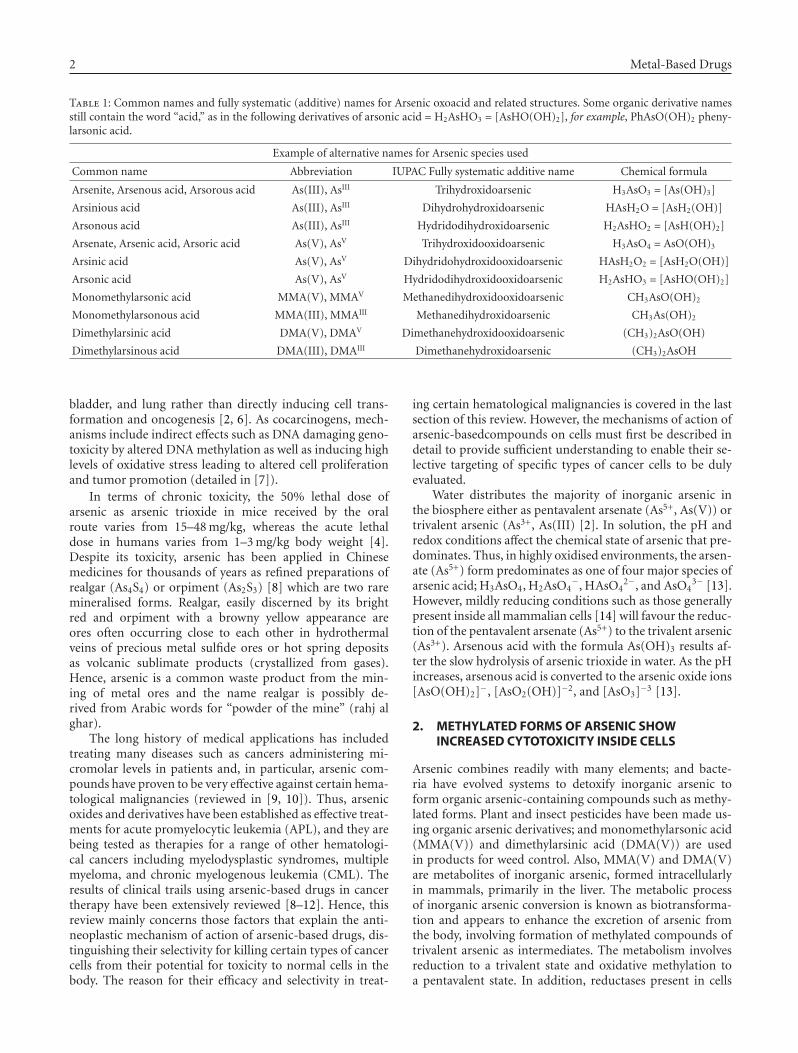
Table 1: Common names and fully systematic (additive) names for Arsenic oxoacid and related structures. Some organic derivative namesstill contain the word “acid,” as in the following derivatives of arsonic acid = H2AsHO3 = [AsHO(OH)2], for example, PhAsO(OH)2 pheny-larsonic acid.
Example of alternative names for Arsenic species used
Common name Abbreviation IUPAC Fully systematic additive name Chemical formula
Arsenite, Arsenous acid, Arsorous acid As(III), AsIII Trihydroxidoarsenic H3AsO3 = [As(OH)3]
Arsinious acid As(III), AsIII Dihydrohydroxidoarsenic HAsH2O = [AsH2(OH)]
Arsonous acid As(III), AsIII Hydridodihydroxidoarsenic H2AsHO2 = [AsH(OH)2]
Arsenate, Arsenic acid, Arsoric acid As(V), AsV Trihydroxidooxidoarsenic H3AsO4 = AsO(OH)3
Arsinic acid As(V), AsV Dihydridohydroxidooxidoarsenic HAsH2O2 = [AsH2O(OH)]
Arsonic acid As(V), AsV Hydridodihydroxidooxidoarsenic H2AsHO3 = [AsHO(OH)2]
Monomethylarsonic acid MMA(V), MMAV Methanedihydroxidooxidoarsenic CH3AsO(OH)2
Monomethylarsonous acid MMA(III), MMAIII Methanedihydroxidoarsenic CH3As(OH)2
Dimethylarsinic acid DMA(V), DMAV Dimethanehydroxidooxidoarsenic (CH3)2AsO(OH)
Dimethylarsinous acid DMA(III), DMAIII Dimethanehydroxidoarsenic (CH3)2AsOH
bladder, and lung rather than directly inducing cell trans-formation and oncogenesis [2, 6]. As cocarcinogens, mech-anisms include indirect effects such as DNA damaging geno-toxicity by altered DNA methylation as well as inducing highlevels of oxidative stress leading to altered cell proliferationand tumor promotion (detailed in [7]).
In terms of chronic toxicity, the 50% lethal dose ofarsenic as arsenic trioxide in mice received by the oralroute varies from 15–48 mg/kg, whereas the acute lethaldose in humans varies from 1–3 mg/kg body weight [4].Despite its toxicity, arsenic has been applied in Chinesemedicines for thousands of years as refined preparations ofrealgar (As4S4) or orpiment (As2S3) [8] which are two raremineralised forms. Realgar, easily discerned by its brightred and orpiment with a browny yellow appearance areores often occurring close to each other in hydrothermalveins of precious metal sulfide ores or hot spring depositsas volcanic sublimate products (crystallized from gases).Hence, arsenic is a common waste product from the min-ing of metal ores and the name realgar is possibly de-rived from Arabic words for “powder of the mine” (rahj alghar).
The long history of medical applications has includedtreating many diseases such as cancers administering mi-cromolar levels in patients and, in particular, arsenic com-pounds have proven to be very effective against certain hema-tological malignancies (reviewed in [9, 10]). Thus, arsenicoxides and derivatives have been established as effective treat-ments for acute promyelocytic leukemia (APL), and they arebeing tested as therapies for a range of other hematologi-cal cancers including myelodysplastic syndromes, multiplemyeloma, and chronic myelogenous leukemia (CML). Theresults of clinical trails using arsenic-based drugs in cancertherapy have been extensively reviewed [8–12]. Hence, thisreview mainly concerns those factors that explain the anti-neoplastic mechanism of action of arsenic-based drugs, dis-tinguishing their selectivity for killing certain types of cancercells from their potential for toxicity to normal cells in thebody. The reason for their efficacy and selectivity in treat-
ing certain hematological malignancies is covered in the lastsection of this review. However, the mechanisms of action ofarsenic-basedcompounds on cells must first be described indetail to provide sufficient understanding to enable their se-lective targeting of specific types of cancer cells to be dulyevaluated.
Water distributes the majority of inorganic arsenic inthe biosphere either as pentavalent arsenate (As5+, As(V)) ortrivalent arsenic (As3+, As(III) [2]. In solution, the pH andredox conditions affect the chemical state of arsenic that pre-dominates. Thus, in highly oxidised environments, the arsen-ate (As5+) form predominates as one of four major species ofarsenic acid; H3AsO4, H2AsO4
−, HAsO42−, and AsO4
3− [13].However, mildly reducing conditions such as those generallypresent inside all mammalian cells [14] will favour the reduc-tion of the pentavalent arsenate (As5+) to the trivalent arsenic(As3+). Arsenous acid with the formula As(OH)3 results af-ter the slow hydrolysis of arsenic trioxide in water. As the pHincreases, arsenous acid is converted to the arsenic oxide ions[AsO(OH)2]−, [AsO2(OH)]−2, and [AsO3]−3 [13].
2. METHYLATED FORMS OF ARSENIC SHOWINCREASED CYTOTOXICITY INSIDE CELLS
Arsenic combines readily with many elements; and bacte-ria have evolved systems to detoxify inorganic arsenic toform organic arsenic-containing compounds such as methy-lated forms. Plant and insect pesticides have been made us-ing organic arsenic derivatives; and monomethylarsonic acid(MMA(V)) and dimethylarsinic acid (DMA(V)) are usedin products for weed control. Also, MMA(V) and DMA(V)are metabolites of inorganic arsenic, formed intracellularlyin mammals, primarily in the liver. The metabolic processof inorganic arsenic conversion is known as biotransforma-tion and appears to enhance the excretion of arsenic fromthe body, involving formation of methylated compounds oftrivalent arsenic as intermediates. The metabolism involvesreduction to a trivalent state and oxidative methylation toa pentavalent state. In addition, reductases present in cells
Stephen John Ralph 3
and other reactions facilitate the reduction of arsenic acid[As(V)] to the arsenous [As(III)] form including methyla-tion of the arsenous form, mainly via the liver, to producemono-, di-, and trimethylated species [2]. The main en-zyme involved in methylation of As(III) is arsenic methyl-transferase (As3MT) that requires glutathione (GSH) to pro-mote the reaction and a cycling redox system such as thiore-doxin (reviewed in [15]) to detoxify arsenic-containing com-pounds. Many animal species convert inorganic arsenic-containing compounds into mono-, di- and tri-methylatedarsenic species which are mostly then secreted in the urine[16]. This is because the methylated forms are not as well ab-sorbed by cells compared to the inorganic forms, althoughthey have much greater cytotoxicity if they do enter cells[15, 17].
The reductive metabolism of arsenic has an importantrole in its toxicity. The trivalent arsenic-containing com-pounds, including the methylated organic forms have muchgreater potency than the pentavalent arsenic-containingcompounds as cytotoxics and carcinogens [18–23]. For ex-ample, in cytotoxicity assays, the IC50 values for culturesof primary hepatocytes, keratinocytes, and epithelial cellsranged from 3 μM to well over 20 μM for trihydroxidoarsenicsalts whereas for monomethylarsonous acid [MMA(III)],the values were consistently much lower at only severalμM for the equivalent normal cell types [24–26]. Organicarsenic-based ingredients are commonly used as feed ad-ditives in poultry farming to increase weight gain by pre-venting bacterial and parasitic infections, thereby increas-ing feed efficiency and improving pigmentation. The threemajor arsenic-containing compounds used in this man-ner are arsenilic acid (p-aminophenyl arsonic acid), roxar-sone (4-hydroxy-3-nitrophenylarsonic acid), and nitarsone(4-nitro-phenylarsonic acid) [22, 23]. The metabolism ofthese arsenic-containing compounds and waste productsproduced by birds and mammals consuming them is still un-certain at present and what the global environmental impactmight be.
3. CELLULAR ACTIONS OF ARSENIC-CONTAININGCOMPOUNDS
3.1. Inhibitors of energy metabolism: effects onglycolysis and oxidative phosphorylation
At the biochemical level, inorganic arsenic in the pentavalentstate (As(V), arsenate, AsO4) resembles a phosphate (PO4)group in structure and it can replace phosphate in many re-actions. The mitochondria is a major intracellular site wherearsenate is metabolised, taken up As(V), rapidly reducing it,and exporting the As(III) product back into the cytosol [27].The specific location of the site(s) for arsenic reduction inthe mitochondria has not yet been defined. However, arse-nate can affect oxidative phosphorylation by binding to theFo/F1 ATP synthase [28]. Arsenate can be used by the ATPsynthase more efficiently than phosphate depending on theCa2+ levels [29], producing ADP-arsenate which unlike ATPbecomes rapidly hydrolysed and unable to form stable high-energy compounds [30]. It is suggested that the structure and
charge similarities of PO43−, AsO4
3−, and SO42− result in in-
discriminate binding to at least two sites located in the mi-tochondrial matrix [31]. At one site, occupation by any ofthese three anions results in protection against uncouplingof the mitochondrial proton gradient by K+; at the secondsite, in the Fo/F1 ATP synthase, AsO4
3− and SO42− compete
for binding against PO43−, leading to the inhibition of ATP
production [31].Intriguingly, As2O3, whilst shown to have no effect on
oxidative phosphorylation levels in HeLa [10 μM] and AS-30D [100 μM] hepatoma cancer cells, significantly inhibitedglycolysis, particularly during the exponential growth phaseof these cells when they were actively respiring and pro-ducing 70% of their ATP from mitochondria [32]. Severalother studies have also shown that arsenic compounds donot affect oxidative phosphorylation. Thus, with submito-chondrial particles in the presence of an ATP regeneratingsystem, 20 mM arsenate had no effect on NADH formation,ATP hydrolysis, and Pi rightarrow H20 exchange [33]. Ina related study with isolated liver mitochondria, As2O3 wasagain shown to have no effect on oxygen consumption, or therespiratory control ratio at a concentration (10 μM) foundto be maximally effective in promoting apoptosis in wholecells [34]. However, at higher concentrations (> 50 μM),pyruvate/malate-supported respiration (via complex I) be-came blocked, but there was no effect on either complex IIor IV. The inhibitory effect on complex I was reversed bythe addition of the reducing agent, dithiothreitol, indicat-ing that direct oxidative damage was involved. In addition,it was shown that even with the block in complex I, the cellscontinued to maintain cellular ATP levels through glycolysis,and hence, depletion of cellular ATP was not the cause forthe cytotoxicity of As2O3 [34]. Probably the most definitiveevidence for the importance of mitochondria in mediatingAs2O3 killing of cancer cells comes from studying cells lack-ing mitochondrial function [35]. A subclone of mitochon-drial respiration deficient cells was derived from the HL-60human leukaemia cell line by growth in the presence of ethid-ium bromide to mutate the mitochondrial genome, and thesecells are known as HL-60 “ρ0” cells [35]. Due to the lack ofmitochondrial respiration, ρ0cells depend on glycolysis fortheir energy source and, as would be expected, produced sub-stantially less superoxide radicals (∼20% of the control cells).When these ρ0 cells were incubated with ∼10 μM As2O3,they were resistant to the drug, revealing that mitochondrialrespiratory function is required for the cytotoxic actions ofAs2O3 [35].
The inhibition of glycolysis by arsenic-based drugs ap-pears unlikely to be a significant factor involved in the drug-induced killing of cancer cells. Thus, incubating cells inglucose-deficient medium to block glycolysis had no signif-icant effect on the As2O−
3 [30 uM] mediated levels of celldeath in the Jurkat cell line [36]. However, when glycoly-sis was blocked and mitochondrial respiration inhibited us-ing oligomycin A, the cells became very sensitive to As2O3−
mediated cell death. In this regard, it is also worth not-ing that studies of individual glycolytic enzymes analysedin purified form in vitro have shown arsenite and arsen-ate to be relatively weak inhibitors. Hence, for hexokinase
4 Metal-Based Drugs
(IC50: 15 mM for arsenate [37, 38]), phosphofructokinase(IC50 > 5 mM for arsenite; [39]) and pyruvate dehydroge-nase (PDH IC50: 80–120 μM arsenite; [40]), relatively largeconcentrations were required to inhibit these enzymes. Infact, arsenate has been shown to stimulate the activities ofthe two important glycolytic enzymes, hexokinase [37] andGAPDH [41], by overcoming product inhibition in these re-actions. In the case of GAPDH, arsenate acts catalytically topromote the oxidation of phosphoglycerate [42] and the re-action involved the formation of an arsenate analogue of thephosphate ester as an intermediate which rapidly hydrolysed,helping to drive the reaction forward [33, 43]. This processhas been commonly described in relation to the effects ofarsenate on numerous enzymatic reactions involving phos-phate and has been termed “arsenolysis” [33].
Given the relative insensitivity to the direct effects of ar-senic compounds shown by the glycolytic pathway, it followsthat it is unlikely that glycolytic inhibition results from di-rect binding and modification of the enzymes in this path-way by arsenic-based drugs. More likely, the inhibition ofglycolysis results from an indirect effect, caused by the ac-tions of arsenic compounds in modifying mitochondrial res-piration leading to production of ROS which then acts toinhibit the glycolytic enzymes. Additional support for themitochondrial-mediated ROS involvement in the action ofarsenic to inhibit glycolysis comes from studies where As2O3
was found to be ∼38 times more potent in cells than inthe pure preparation at inhibiting pyruvate dehydrogenase[40]. Also, inhibiting mitochondrial respiration suppressedthe resulting inhibition of pyruvate dehydrogenase activityand H2O2 production by this drug. Furthermore, the inhibi-tion of pyruvate dehydrogenase by As2O3 was shown to re-quire the Fenton reaction occurring via hydroxyl radical in-termediates [40]. The mitochondrial effects of arsenic com-pounds are detailed later in this review and to reiterate at thispoint, the evidence indicates that the actions of arsenic com-pounds on glycolysis are not the main cause for the cytotoxiceffects of these drugs at clinically relevant concentrations (1–6 μM) required in plasma for the killing of cancer cells in APLpatients [8, 44].
3.2. Interconversion of As(III) rightarrow As(V) in cells
Under conditions of high mitochondrial respiration insidecells, it is possible that trivalent arsenicals inducing signif-icant production of ROS as superoxide, peroxide, and hy-droxyl radicals can also result in oxidation to produce arsen-ate (AsO4
3−) ionic species [45, 46]. Consequently, the impactthat arsenic compounds will have on any given cell will mostlikely depend on the state of cellular respiration and pro-duction of ROS affecting the arsenic speciation and whetherthe cell is dependant on glycolysis versus mitochondrial res-piration for its ATP synthesis. Glyceraldehyde-3-phosphatedehydrogenase (GAPDH), the glycolytic enzyme abundantlyfound in all cells and especially blood cells and liver, is amajor intracellular arsenate reductase [47] requiring GSH,NAD, and glycolytic substrate [48, 49]. Given that the levelsand specific activity of GAPDH is much higher in malignantcells than in normal cells [50], this could contribute to the
rapid reduction of As(V) species in their cytosol into moretoxic As(III) forms.
In the GAPDH reaction, As(V) reduction may take placeduring, or as a consequence of the arsenolytic cleavage ofthe thioester bond formed between the enzyme’s Cys149residue and the 3-phosphoglyceroyl moiety of the substrate.Hydrolysis of 1-arseno-3-phosphoglycerate is at least 2000times faster than hydrolysis of the normal substrate 1,3-diphosphoglycerate under the same conditions [51]. Hence,GAPDH is proposed as one of the key cellular converting en-zymes for reducing As(V) to As(III). Although purine nu-cleoside phosphorylase was proposed to be an arsenate re-ductase [52], this was later refuted [53]. The other majorclass of As(V) reductases in cells are the glutathione S trans-ferases and of these, the omega form or GSTO1 appears tobe most important. Thus, GSTO1 can reduce arsenate to ar-senite, MMA(V) to MMA(III), and DMA(V) to DMA(III)and deletion of the GSTO1 gene in mice reduced the ex-tent of biotransformation by 30–80% in most tissues exam-ined [54].
3.3. Structure and reactivity of arsenic-containingcompounds with reduced thiols
Arsenic has a high affinity for sulfur and hence, reactivesulfur-containing molecules such as reduced thiols with anavailable sulfur atom have a significant propensity for bind-ing to arsenic [55, 56]. As(III)-containing compounds existas trigonal pyramidal structures and this is also the structureformed upon binding of arsenic ions to cellular proteins invivo where the sulphur atoms of thiolate groups act as co-ordinating ligands. The resulting arsenic-thiol linkages aremainly responsible for the ability of arsenic to modulate thefunction of various key molecules, enzymes, and ion trans-porters inside cells and this intracellular action of arsenic isdiscussed in detail in this section. Arsenic-containing com-pounds react with mono- and dithiols, particularly the lat-ter when two thiols are located in close proximity, acting tocross-link the thiols together.
Some debate exists about the structures and speciation ofarsenic-containing compounds (both inorganic and organicforms) in solution. In the absence of sulfide, As(III) hydrox-ide complexes are the major arsenic-containing species andthese structures probably adopt a trigonal pyramidal struc-ture with the arsenic atom at the apex [57–59]. This trigonalpyramidal structure provides the potential for As(III) to co-ordinate linkages with several proximal thiol groups. This isbelieved to be the case in bacterial enzyme systems such asthe ArsR repressor protein where it is likely to bind three Cysatoms [60]. Thus, in the more toxic form as the trivalent state(As(III)) inorganic and organic (methylated) arsenic reactswith critical thiols in proteins, inhibiting their function, as isthe case with the bacterial ArsR protein. For example, As(III)was shown to target the reactive sulfhydryl group at the activesite of thiolase(s) involved in ketogenesis from acetyl Coen-zyme A [61].
The pentavalent species of inorganic arsenate (AsO43−)
favoured to exist in oxidised environments, as well as organicforms of As(V) have a different structure with a trigonal-bipyramidal shape where the As(V) atom is located at the
Stephen John Ralph 5
centre, co-ordinating to three equatorial and two polar atoms[62, 63]. In comparing the two different structural statesof As(III) and As(V), it is not yet clear why the trivalentmethylarsenic-containing compounds show a much highertoxicity than their pentavalent analogues [24, 64]. However,it could relate to cellular uptake as trivalent organoarseniccompounds are more membrane permeable than the pen-tavalent species [65]. Also, trivalent arsenic bonded at aphenyl ring is able to form much more stable covalentcross-links to cysteine residues compared to arsenic in smallmolecules such as arsenious acid or arsenite [66]. Further-more, the organic trivalent arsenic-containing compound,phenylarsine oxide [0.1–0.5 μM], is much more potent thanthe simple arsenite [1–10 μM] in its cytotoxic activity in APLcells [67].
One major drawback with phenylarsine oxide as a poten-tial cancer therapy is its high toxicity in vivo and its nonse-lectivity for cancer versus normal cells, resulting in cytotox-icity in normal endothelial cells in the same concentrationrange (0.2 μM) [68]. Thus, phenylarsine oxide, is precludedfrom application in clinical cancer therapy, without beingfurther modified as a drug. The greater reactivity of pheny-larsene oxide and associated cytotoxicity is in agreementwith the results outlined above [66] where mass spectromet-ric analysis of different complexes of peptides and proteinswith arsenic-containing species revealed that inorganic ar-senite or arsenates did not interact well with cysteine or glu-tathione, whereas the organic phenylarsine (3+) oxide did. Inaddition, three different phenyl arsenic acids and dimethy-larsinic acid that all contained As(V) also formed complexeswith glutathione [66]. Hence, the bulky hydrophobic groupswith electron withdrawing π orbitals (in the case of phenylgroups) may promote more stable bonds between the ar-senic atom and sulfur groups inside cells, modulating a largerrange of enzymes and proteins with important functions formaintaining cell viability.
In cells, the most common reactive species that are avail-able for interaction with arsenic are the abundant free thiolmoieties in the tripeptide glutathione (γGlu-Cys-Gly, GSH)and the free amino acid, cysteine. Thus, arsenic-based drugscan react by coordinating binding to free (reduced) thiolgroups such as those on cysteine, particularly those of thiore-doxin and glutathione as the major intracellular thiol speciesimportant in cellular redox regulation. It was observed earlyon that arsenite and phenylarsine oxide in particular, butnot arsenate, reacted with vicinal thiol groups on proteins[69, 70]. Since demonstrating that phenylarsine oxide wasparticularly effective at cross-linking vicinal thiols in the ac-tive site of tyrosine phosphatases [71], the range of proteins-containing vicinal cysteine residues with which phenylar-sine oxide reacts is increasing. Recent examples include thesmall GTP binding Rho protein family [72] and the mito-chondrial carnitine/acylcarnitine transporter [73]. Phenylar-sine oxide, as a strong inhibitor of tyrosine phosphataseswould increase tyrosine phosphorylation levels of enzymesin cellular growth signalling pathways. Whether the inhibi-tion of tyrosine phosphatases and other enzymes contributesto the toxic effects of arsenic-containing compounds in nor-mal cells is not clear, but is likely an important contributing
factor to its general cytotoxicity making phenylarsine oxideunsuitable for cancer therapy.
Analysis of the interactions of As(III) with glutathione orcysteine in vitro in aqueous solutions by equilibrium bindingand use of biophysical techniques including NMR, electronicspectroscopy, and potentiometry revealed that As(III) bindseither of glutathione or cysteine with similar equilibriumconstants [74]. However, several analytical studies by massspectroscopy have revealed that arsenate and arsenite do notcomplex readily with glutathione or cysteine, but prefer toreact with the thiols on reduced thioredoxin molecules [re-viewed in [66]]. This was confirmed by analysis of more bio-logically relevant samples from the intracellular environmentof HeLa cells where cytosolic thioredoxin 1 (TRX1) and, inparticular thioredoxin 2 (TRX2) in the mitochondria, wasshown to be highly reactive with arsenite (10 μM) whereaslittle reactivity was detected with cellular GSH/GSSG [75].
Studies with thioredoxin reductase (TxR) purified frommouse liver showed that arsenic-containing compounds withAs(III) and arsinothiols (complexes of As(III) with GSHor L-cysteine) were extremely potent inhibitors of this en-zyme [24]. Methylarsenic(III)oxide was most potent with aKi∼100 nM, as an irreversible competitive inhibitor. The ef-fects on purified glutathione reductase (GR) showed thatthe levels of inhibition were not as marked with inorganicAs(III) and As(V) oxides showing IC50s in the 10–50 mMrange, whereas for methylarsinic(III)oxide it was ∼9 μM.Studies on this enzyme in whole cells as hepatocytes showedthe IC50 to be reduced to ∼3 μM for methylarsinic(III) ox-ide and for As2O3 > 100 μM [24]. Hence, these observationsstrongly support a role for the components thioredoxins andthe thioredoxin reductase system as providing cellular targetsthat are very sensitive to inhibition by arsenic-based drugs inthe low micromolar range. It also explains the importance ofstudying the chemistry of arsenic-containing compounds inthe context of both purified enzymes as well as whole cellsas biological systems in order to obtain meaningful results.Thus, although the GSH/glutathione transferase system un-doubtedly plays an important role in arsenic sensitivity ofcells (see below in Section 4 for details), it would appear thatthe thioredoxin system might represent the most immediatepoint of sensitive reactivity in relation to cytotoxicity.
3.4. The major mechanism of action ofarsenic-containing compounds: modifyingmitochondrial function and redox regulationof the production of reactive oxygen species
Mitochondria are a main source of ROS in cells (reviewed in[76]). Thioredoxin (TRX), NADPH, and thioredoxin reduc-tase (TxR) comprise the thioredoxin system that has mul-tiple functions in cells including in redox signalling via in-teractions with other proteins, in transcriptional regulation,control of the reduced intracellular redox environment, cellgrowth, defense against oxidative stress and control of apop-tosis (reviewed in [77]). As outlined in the previous sec-tion, the thioredoxin system is very sensitive to arsenic-baseddrugs and may well be the basis for one of the importantmechanisms for their actions in inducing cancer cell death.
6 Metal-Based Drugs
Cytosol
VDAC VDAC
ANT ANT
SH SHSH SH
As
IIIPrx III
SHSH
Trx2
SH
SH
ASK1
AsIII
As
III
Trx1
SH
SH
Outer membrane
H+ H+H+
H+H+
H+H+
As
III
Inner membrane
Electron transport chain
C I e− C II e−UbQ
C III
C IV
NADHNAD+
SuccinateFumarate
ATP synthaseFactor B
SH
SHH2O
AsIII
ADP + Pi ATPH+
2H
++1/
2O 2
Mitochondrial matrix
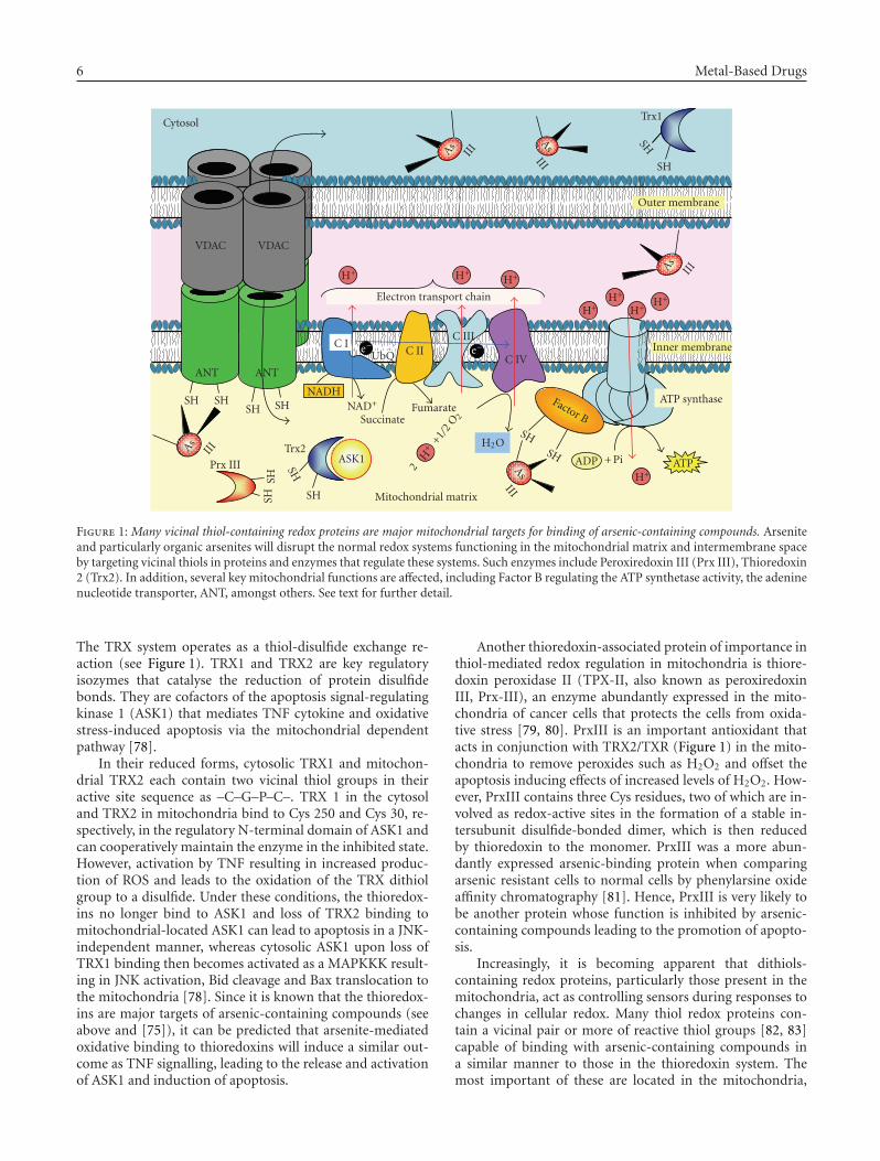
Figure 1: Many vicinal thiol-containing redox proteins are major mitochondrial targets for binding of arsenic-containing compounds. Arseniteand particularly organic arsenites will disrupt the normal redox systems functioning in the mitochondrial matrix and intermembrane spaceby targeting vicinal thiols in proteins and enzymes that regulate these systems. Such enzymes include Peroxiredoxin III (Prx III), Thioredoxin2 (Trx2). In addition, several key mitochondrial functions are affected, including Factor B regulating the ATP synthetase activity, the adeninenucleotide transporter, ANT, amongst others. See text for further detail.
The TRX system operates as a thiol-disulfide exchange re-action (see Figure 1). TRX1 and TRX2 are key regulatoryisozymes that catalyse the reduction of protein disulfidebonds. They are cofactors of the apoptosis signal-regulatingkinase 1 (ASK1) that mediates TNF cytokine and oxidativestress-induced apoptosis via the mitochondrial dependentpathway [78].
In their reduced forms, cytosolic TRX1 and mitochon-drial TRX2 each contain two vicinal thiol groups in theiractive site sequence as –C–G–P–C–. TRX 1 in the cytosoland TRX2 in mitochondria bind to Cys 250 and Cys 30, re-spectively, in the regulatory N-terminal domain of ASK1 andcan cooperatively maintain the enzyme in the inhibited state.However, activation by TNF resulting in increased produc-tion of ROS and leads to the oxidation of the TRX dithiolgroup to a disulfide. Under these conditions, the thioredox-ins no longer bind to ASK1 and loss of TRX2 binding tomitochondrial-located ASK1 can lead to apoptosis in a JNK-independent manner, whereas cytosolic ASK1 upon loss ofTRX1 binding then becomes activated as a MAPKKK result-ing in JNK activation, Bid cleavage and Bax translocation tothe mitochondria [78]. Since it is known that the thioredox-ins are major targets of arsenic-containing compounds (seeabove and [75]), it can be predicted that arsenite-mediatedoxidative binding to thioredoxins will induce a similar out-come as TNF signalling, leading to the release and activationof ASK1 and induction of apoptosis.
Another thioredoxin-associated protein of importance inthiol-mediated redox regulation in mitochondria is thiore-doxin peroxidase II (TPX-II, also known as peroxiredoxinIII, Prx-III), an enzyme abundantly expressed in the mito-chondria of cancer cells that protects the cells from oxida-tive stress [79, 80]. PrxIII is an important antioxidant thatacts in conjunction with TRX2/TXR (Figure 1) in the mito-chondria to remove peroxides such as H2O2 and offset theapoptosis inducing effects of increased levels of H2O2. How-ever, PrxIII contains three Cys residues, two of which are in-volved as redox-active sites in the formation of a stable in-tersubunit disulfide-bonded dimer, which is then reducedby thioredoxin to the monomer. PrxIII was a more abun-dantly expressed arsenic-binding protein when comparingarsenic resistant cells to normal cells by phenylarsine oxideaffinity chromatography [81]. Hence, PrxIII is very likely tobe another protein whose function is inhibited by arsenic-containing compounds leading to the promotion of apopto-sis.
Increasingly, it is becoming apparent that dithiols-containing redox proteins, particularly those present in themitochondria, act as controlling sensors during responses tochanges in cellular redox. Many thiol redox proteins con-tain a vicinal pair or more of reactive thiol groups [82, 83]capable of binding with arsenic-containing compounds ina similar manner to those in the thioredoxin system. Themost important of these are located in the mitochondria,
Stephen John Ralph 7
AsIII As
III
AsIII As
III
-SH -SH
AsIIINADP+
NADPH
Trx2 -(SH)2
Trx2 -S-S-2GSH
GS-SG
2H2O
2GSH
GS-SG
TrxR2
GRGrx2
Prx III
H2O2
-S-S protein ox
Protein red
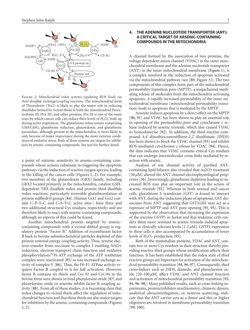
GPx
Figure 2: Mitochondrial redox systems regulating ROS levels viathiol-disulfide exchange/coupling reactions. The mitochondrial formof Thioredoxin (Trx2) is likely to play the major role in reducingdisulfides formed by vicinal thiols in both the mitochondrial Perox-iredoxin III (Prx III) and other proteins. Prx III is one of the mainways by which cancer cells can reduce their levels of H2O2 built upduring active respiration. The glutathione redox system comprisingGSH/GSSG, glutathione reductase, glutaredoxin, and glutathioneperoxidase, although present in the mitochondria, is more likely toonly become of major importance during the more extreme condi-tions of oxidative stress. Both of these systems are targets for inhibi-tion by arsenic-containing compounds. See text for further detail.
a point of extreme sensitivity to arsenic-containing com-pounds whose actions culminate in triggering the apoptoticpathways via the induction of reactive oxygen species, leadingto the killing of the cancer cells (Figures 1, 2). For example,two members of the glutaredoxin (GRX) family, includingGRX2 located primarily in the mitochondria, catalyse GSH-dependent TRX-disulfide redox and protein thiol-disulfideredox reactions, particularly reversible glutathionylation ofprotein sulfhydryl groups [84]. Human Grx1 and Grx2 con-tain C–P–Y–C and C–S–Y–C active sites ; have three andtwo additional structural Cys residues, respectively; and aretherefore likely to react with arsenic-containing compounds,although no reports of this could be found.
Another mitochondrial protein targeted by arsenic-containing compounds with a vicinal dithiol group is reg-ulatory protein “Factor B.” Addition of recombinant factorB back to bovine submitochondrial particles depleted of thisprotein restored energy coupling activity. Thus, reverse elec-tron transfer from succinate to complex I enabling NAD+reduction, electron transport chain function and oxidativephosphorylation/32Pi-ATP exchange of the ATP synthetasecomplex were reactivated [85] as was increased exchange ac-tivity of complex V [86]. Thus, the F0-F1 ATPase activity re-quires Factor B coupled to it for full activation. However,factor B contains six thiols and Cys 92 and Cys 94 in thebovine form were shown to bind phenylarsine oxide [87] andphenylarsine oxide or arsenite inhibit factor B coupling ac-tivity [88]. From all of these studies, it is becoming clear thatredox changes to vicinal thiols affect the regulation of mito-chondrial function and that these thiols are also major targetsfor inhibition by the arsenic-containing compounds (Figures1, 2).
4. THE ADENINE NUCLEOTIDE TRANSPORTER (ANT):A CRITICAL TARGET OF ARSENIC-CONTAININGCOMPOUNDS IN THE MITOCHONDRIA
A channel formed by the association of two proteins, thevoltage dependent anion channel (VDAC) in the outer mito-chondrial membrane and the adenine nucleotide transporter(ANT) in the inner mitochondrial membrane (Figure 1), isa complex involved in the induction of apoptosis activatedvia the mitochondrial pathway (see [89, Figure 3]). The twocomponents of this complex form part of the mitochondrialpermeability transition pore (MPTP), a megachannel medi-ating release of molecules from the mitochondria activatingapoptosis. A rapidly increased permeability of the inner mi-tochondrial membrane (mitochondrial permeability transi-tion) leads to apoptosis that is mediated by the MPTP.
Arsenite induces apoptosis by a direct effect on the MPTP[90, 91] and VDAC has been shown to play an essential rolein opening of the permeability pore and cytochrome c re-lease induced by arsenic trioxide, which also caused VDACto homodimerise [92]. In addition, the thiol-reactive com-pound 4,4’-diisothiocyanostilbene-2,2’-disulfonate (DIDS)has been shown to block the VDAC channel [93] and inhibitROS-mediated cytochrome c release by VDAC [94]. Hence,the data indicates that VDAC contains critical Cys residuesthat can undergo intermolecular cross-links mediated by re-action with arsenic.
Analysis of ion channel activity of purified ANT-containing lipid bilayers also revealed that As2O3 treatment[30 μM] altered the ANT channel electrophysiological prop-erties [36]. Interestingly, glutathione depletion leading to in-creased ROS may play an important role in the action ofarsenic trioxide [91]. Whereas in both normal and cancercells, glutathione S transferase (GST) is found to interactwith ANT, during the induction phase of apoptosis, GST dis-sociates from ANT suggesting that GST/GSH may act as arepressor of MPTP and ANT pore opening [91, 95]. This issupported by the observation that increasing the expressionof the enzyme GSTP1 in Jurkat and Raji leukemic cells ren-ders them more resistant to arsenic trioxide-induced apop-tosis at clinically relevant levels [1-2 μM]. GSTP1 expressionin these cells is also accompanied by accumulation of lowerlevels of H2O2 production [95].
Both of the mammalian proteins, VDAC and ANT, con-tain two or more Cys residues in their structure thereby pro-viding reactive thiol groups whose modification affects theirfunction. It has been established that the redox state of thiolreactive groups are important for activation of the mitochon-drial permeability transition [94, 96, 97]. Consequently, thiolcross-linkers such as DIDS, diamide, and phenylarsine ox-ide [20–100 μM] affect VDAC and ANT channel functionand activation of mitochondrial permeability transition [93,94, 96–98]. Many published results, such as cross-linking ex-periments, protein/inhibitor stoichiometry, chimeric dimers,analytical ultracentrifugation, or neutron scattering, indi-cate that the ANT carrier acts as a dimer and this or higheroligomers are involved in membrane permeability transition[99, 100].
8 Metal-Based Drugs
Facing out into the mitochondrial matrix, ANT has threeexposed loop regions containing a conserved repeat structurewith one Cys residue in each loop. These Cys residues areimportant to the process of ANT dimerisation, but it is notclear how this operates and whether the Cys residues formintermolecular disulfide bonds or not [101]. Nevertheless,copper-o-phenanthroline is able to dimerise ANT by inter-molecular cross-linking of Cys 56 (in the first matrix loop)[98]. In addition, phenylarsine oxide, eosin 5-maleimide,and diamide form intramolecular cross-links between Cys160 and Cys 257 on the other two matrix loops, restrictingANT in the open conformation, promoting mitochondrialpermeability transition [98]. Arsenic trioxide is much weakerthan phenylarsine oxide at binding to the ANT Cys residues[96, 97] and this may explain the greater sensitivity exhib-ited by APL cells to phenylarsine oxide [IC50: 0.1 μM] thanto As2O3 [IC50: 4 μM] [67].
Single thiol interacting compounds such as N-ethylmaleimide (NEM) can inhibit the mitochondrialpermeability transition and this could be either the result ofdirect interaction with the key Cys residues on the matrixloops of ANT or indirectly via reaction with GSH andthereby preventing GSH from being oxidized and catalysingdisulfide bridging between the adjacent thiol groups inthe ANT loops [98]. NEM or monobromobimane, in the25–50 μM range, preferentially react with GSH, leadingto its modification in mitochondria and thereby preventsGSH from being oxidised. As a result, NEM inhibits mi-tochondrial permeability transition activation by the thiolreactive compounds, diamide or t-butylhydroperoxide,implying a role for GSSG in the action of these agents on thepermeability transition [96, 97]. Arsenites, albeit that muchhigher concentrations would be required given their loweraffinity for glutathione interaction, could have a similaraction. Thus, high levels of arsenites could modify and in-hibit glutathione redox control such that glutathione-basedenzymes are unable to function, as well as directly mediatingdisulfide cross-linking of ANT, leading to increases in cellu-lar ROS production, MPTP, and apoptosis. However, giventheir low reactivity with glutathione systems, this appearsto be unlikely as opposed to the indirect action via themitochondrial effects leading to increased ROS productionwhich then reduces cellular GSH levels.
The multidrug resistance (MDR) protein MRP1/ABCC1has been shown to transport AsIII out of cells as a tri-GSHconjugate (As-(GSH)3), and glutathione S-transferase (GST)probably facilitates the process [102]. This is likely to be partof the normal cell and cancer cell resistance mechanismsagainst the cytotoxic effects of arsenic-based compounds.GSH-depleted cells are more sensitive to killing by arsenic-containing compounds [103] and transfection of cells to ex-press glutathione S transferase protects them from arseniteinducing death by promoting arsenite transport from thecells and decreasing ROS levels [104, 105]. In support ofthis proposal, the long-term exposure of cells to arsenic-containing compounds induced increased expression of glu-tathione S transferase and MRPs [106]. Arsenic levels do notattain very high levels in blood plasma of patients, rapidlybecoming eliminated [8, 44, 107]. This removal probably re-
sults from efficient uptake of As-(GSH)3 via MRP2 in theproximal tubules of the kidneys as part of the detoxificationprocess during the excretion of arsenic-based drugs predom-inantly into the urine [108, 109]. The remainder is mostly re-moved via uptake in the liver and secretion as bile [110–112].As-(GSH)3 may be an important part of the metabolic pro-cess for converting inorganic As(III) to methylated speciesduring detoxification in the liver by ASMT1/Cyt19 [113].
5. MODIFIED ORGANIC ARSENICSTRUCTURES WITH INCREASED POTENCYAS ANTINEOPLASTIC AGENTS
Arsenic-containing compounds substituted with organicgroups such as modified phenylarsine oxides have been syn-thesized and examined for their cytotoxic effect on humanleukemic cells and breast cancer cells in culture. Some ofthese compounds were found to exhibit potent cytotoxic an-ticancer activity, particularly against human breast cancerand leukemic cell lines, including primary leukemia cells,at micromolar concentrations. One of these compounds,the novel glutathionyl peptide trivalent arsenic-containingcompound para 4-[N-(S-glutathionylacetyl)amino]pheny-larsenoxide (p-GSAO) shows promise as a novel antineoplas-tic drug and is now in clinical trials. P-GSAO, like pheny-larsine oxide, inactivates ANT-mediated ATP/ADP trans-port and triggers Ca2+-dependent MPTP opening by cross-linking the critical Cys residues of ANT. This leads to in-creased production of cellular ROS, ATP depletion, mito-chondrial depolarization, and apoptosis of angiogenic en-dothelial cells and inhibition of tumor growth in mice withno apparent toxicity [114]. However, the action of p-GSAOwas indirect, and did not appear to be as a result of selectivetumor cell toxicity. Rather, p-GSAO inhibited the proliferat-ing, but not growth-quiescent endothelial cells in vitro andangiogenesis in vivo and thus acted to eliminate tumors byblocking their blood supply [114, 115]. The trivalent arsenic-containing moiety of p-GSAO was shown to cross-link thematrix facing Cys160 and Cys257 thiols of ANT [114] and ef-fectively locks ANT into the open configuration. Inactivationof ANT by p-GSAO causes an increase in superoxide levels,proliferation arrest, ATP depletion, mitochondrial depolar-ization, and apoptosis in the dividing endothelial cells.
It is likely that the arsenic-containing moiety of p-GSAOreacts similarly as does arsenite (see above) with one or twomolecules of glutathione before it is removed from the cellby MRPs [116]. Tumor cells export p-GSAO much more effi-ciently than endothelial cells because they have higher MRP1or MRP2 activity and cellular glutathione levels [116] andthis may explain why p-GSAO is not highly effective at in-hibiting tumor cell growth in vivo. In addition, the greaterwater soluble properties of p-GSAO than other arsenic-containing compounds, particularly organic species shouldhelp to retain p-GSAO in the intravascular system where it ismore likely to affect endothelial cells and inhibit tumor an-giogenesis.
Interestingly, although the para form of GSAO revealedno apparent toxicity in treated animals and inhibited tumorgrowth leading to phase I clinical trials as an anticancer agent

Stephen John Ralph 9
[114], the ortho form (o-GSAO) was toxic and this was pro-posed to result from increased accumulation of the drug incells, including normal cells due to loss of multidrug resis-tance efflux [116]. Consequently, o-GSAO is unlikely to be ofmuch further interest as an antineoplastic agent, whereas theclinical efficacy of p-GSAO is eagerly awaited.
6. TARGETING OF CANCER CELLS: SELECTIVEUPTAKE AND DELIVERY INTO SPECIFIC TYPESOF CANCER CELLS
As(III) as the anhydrous form of As(OH)3 (Trisenox, CellTherapeutics, Seattle, Wash, USA) received FDA approvalin 2000 as a chemotherapeutic agent for the treatment ofAPL [117]. Acute promyelocytic leukemia (APL) is associ-ated with reciprocal and balanced chromosomal transloca-tions always involving the retinoic acid receptor alpha (RAR-alpha) gene on chromosome 17 and variable partner geneson distinct chromosomes. RARalpha fuses to the promyeloc-tyic leukemia (PML) gene in the majority of APL cases (re-viewed in [118]). Arsenic trioxide is particularly effective atkilling APL cells and this was proposed to be the direct resultof its ability to induce the relocalization and degradation ofthe nuclear body protein PML, as well as the degradation ofPML-RARalpha in APL cells [119–122]. However, this seemsunlikely to be the main mechanism of action for arsenic tri-oxide given that no differences in sensitivity to growth inhi-bition and killing by apoptosis have been observed betweenwild-type and PML−/− cells [123].
Arsenic trioxide as a single agent has provided 86% com-plete hematologic remission with minimal toxicity in APLpatients [124], equal to any of the current standards ofcare for treating APL, including the combination of all-transretinoic acid (ATRA) plus chemotherapy [125]. This raisesthe question why APL cells are very sensitive to arsenic-containing compounds like arsenic trioxide. It would appearthat the reason is because APL cells express the transmem-brane transporter protein, aquaglyceroporin 9 (AQP9) in-volved in arsenic uptake [126] at much higher levels in APLcells than in other leukemic cell types and that correlates witharsenite sensitivity [127]. In this regard, it is worth notingthat aquaglyceroporins AQP7 and AQP9 are present in nor-mal cell types. Interestingly, AQP9 is primarily expressed inhuman lung, liver, and leukocytes [128] and this may helpexplain arsenic toxicity, given that liver is one of the main or-gans affected. The fact that AQP9 provides APL cancer cellspecificity with high response rates suggests that if arsenic-containing compounds could be targeted for specific deliv-ery into cancer cells, then they would represent outstandingagents for killing these cells. However, further modificationswill be required to provide suitable drug targeting for im-proved delivery of arsenic-containing compounds to cancercells.
7. CONCLUSIONS
Vicinal thiols located in key enzymes and proteins providetargets for reaction with arsenic-containing compounds, par-ticularly organic derivatives such as phenylarsenic-contain-
ing compounds that favour intramolecular cross-linking be-tween adjacent thiols. Intriguingly, most of the key intra-cellular targets for this reaction have been identified to in-clude the main REDOX regulatory systems in the mitochon-dria, including thioredoxin and peroxiredoxin systems andthe adenine nucleotide transporter, all of whose function isadversely affected. The net result is the activation of sev-eral independent pathways including ROS production to fa-cilitate the induction of apoptosis. One main pathway op-erates via the opening of the MPTP, the other via activa-tion of ASK1 kinase, and the JNK/Bid/Bax pathway of chan-nel formation in MOM. In the case of APL, cell selectiv-ity for sensitive responses to these drugs is facilitated byselective transport systems such as provided by the AQP9transporter. Low MDR levels present in dividing endothelialcells also provides selective targeting by specially substitutedphenylarsenic-containing compounds like p-GSAO, leadingto decreased blood supply into tumors, with some toxicity tocancer cells, but little toxicity on normal cells. Hence, a com-bination of selective delivery and retention provides the nec-essary targeting of arsenic-containing compounds to tumorsand provides scope for additional modifications to be madeto enhance the antineoplastic activity of arsenic-containingcompounds, given their range of actions and efficiency inkilling cancer cells.
ACKNOWLEDGMENT
The author would like to thank Professor R. K. Ralph forhelpful comments and editing of the manuscript.
REFERENCES
[1] J. C. Saha, A. K. Dikshit, M. Bandyopadhyay, and K. C. Saha,“A review of arsenic poisoning and its effects on humanhealth,” Critical Reviews in Environmental Science and Tech-nology, vol. 29, pp. 281–313, 1999.
[2] P. Roy and A. Saha, “Metabolism and toxicity of arsenic: ahuman carcinogen,” Current Science, vol. 82, no. 1, pp. 38–45, 2002.
[3] N. G. Connelly, T. Damhus, R. M. Hartshorn, and A. T. Hut-ton, Eds., Nomenclature of Inorganic Chemistry—IUPAC Rec-ommendations, The Royal Society of Chemistry, Cambridge,UK, 2005.
[4] International Agency for Research on Cancer, “IARC Mono-graphs on the Evaluation of Carcinogenic Risks to Humans.Some Drinking-water Disinfectants and Contaminants, In-cluding Arsenic,” vol. 84, 2004.
[5] J. H. Lubin, L. E. Beane Freeman, and K. P. Cantor, “Inor-ganic arsenic in drinking water: an evolving public healthconcern,” Journal of the National Cancer Institute, vol. 99,no. 12, pp. 906–907, 2007.
[6] T. G. Bredfeldt, B. Jagadish, K. E. Eblin, E. A. Mash, and A.J. Gandolfi, “Monomethylarsonous acid induces transforma-tion of human bladder cells,” Toxicology and Applied Pharma-cology, vol. 216, no. 1, pp. 69–79, 2006.
[7] M. Valko, C. J. Rhodes, J. Moncol, M. Izakovic, and M.Mazur, “Free radicals, metals and antioxidants in oxida-tive stress-induced cancer,” Chemico-Biological Interactions,vol. 160, no. 1, pp. 1–40, 2006.
10 Metal-Based Drugs
[8] D.-P. Lu, J.-Y. Qiu, B. Jiang, et al., “Tetra-arsenic tetra-sulfidefor the treatment of acute promyelocytic leukemia: a pilot re-port,” Blood, vol. 99, no. 9, pp. 3136–3143, 2002.
[9] W.-C. Chou and C. V. Dang, “Acute promyelocytic leukemia:recent advances in therapy and molecular basis of responseto arsenic therapies,” Current Opinion in Hematology, vol. 12,no. 1, pp. 1–6, 2005.
[10] J. Hu, J. Fang, Y. Dong, S. J. Chen, and Z. Chen, “Arsenic incancer therapy,” Anti-Cancer Drugs, vol. 16, no. 2, pp. 119–127, 2005.
[11] S. Amadori, P. Fenaux, H. Ludwig, M. O’Dwyer, and M. Sanz,“Use of arsenic trioxide in haematological malignancies: in-sight into the clinical development of a novel agent,” CurrentMedical Research and Opinion, vol. 21, no. 3, pp. 403–411,2005.
[12] D. Douer and M. S. Tallman, “Arsenic trioxide: new clinicalexperience with an old medication in hematologic malignan-cies,” Journal of Clinical Oncology, vol. 23, no. 10, pp. 2396–2410, 2005.
[13] J. F. Ferguson and J. Gavis, “Review of the arsenic cycle innatural waters,” Water Research, vol. 6, no. 11, pp. 1259–1274,1972.
[14] C. T. Dooley, T. M. Dore, G. T. Hanson, W. C. Jackson,S. J. Remington, and R. Y. Tsien, “Imaging dynamic redoxchanges in mammalian cells with green fluorescent proteinindicators,” The Journal of Biological Chemistry, vol. 279,no. 21, pp. 22284–22293, 2004.
[15] D. J. Thomas, J. Li, S. B. Waters, et al., “Arsenic (+3 oxidationstate) methyltransferase and the methylation of arsenicals,”Experimental Biology and Medicine, vol. 232, no. 1, pp. 3–13,2007.
[16] S. M. Cohen, L. L. Arnold, M. Eldan, A. S. Lewis, and B. D.Beck, “Methylated arsenicals: the implications of metabolismand carcinogenicity studies in rodents to human risk assess-ment,” Critical Reviews in Toxicology, vol. 36, no. 2, pp. 99–133, 2006.
[17] M. Vahter and E. Marafante, “Intracellular interaction andmetabolic fate of arsenite and arsenate in mice and rab-bits,” Chemico-Biological Interactions, vol. 47, no. 1, pp. 29–44, 1983.
[18] T.-C. Wang, K.-Y. Jan, A. S. S. Wang, and J.-R. Gurr, “Triva-lent arsenicals induce lipid peroxidation, protein carbonyla-tion, and oxidative DNA damage in human urothelial cells,”Mutation Research, vol. 615, no. 1-2, pp. 75–86, 2007.
[19] T. Sakurai, C. Kojima, Y. Kobayashi, et al., “Toxicity of atrivalent organic arsenic compound, dimethylarsinous glu-tathione in a rat liver cell line (TRL 1215),” British Journal ofPharmacology, vol. 149, no. 7, pp. 888–897, 2006.
[20] P.-F. Su, Y.-J. Hu, I.-C. Ho, Y.-M. Cheng, and T.-C. Lee,“Distinct gene expression profiles in immortalized humanurothelial cells exposed to inorganic arsenite and its methy-lated trivalent metabolites,” Environmental Health Perspec-tives, vol. 114, no. 3, pp. 394–403, 2006.
[21] K. E. Eblin, T. G. Bredfeldt, S. Buffington, and A. J. Gandolfi,“Mitogenic signal transduction caused by monomethylar-sonous acid in human bladder cells: role in arsenic-inducedcarcinogenesis,” Toxicological Sciences, vol. 95, no. 2, pp. 321–330, 2007.
[22] B. P. Jackson, P. M. Bertsch, M. L. Cabrera, J. J. Camberato,J. C. Seaman, and C. W. Wood, “Trace element speciationin poultry litter,” Journal of Environmental Quality, vol. 32,no. 2, pp. 535–540, 2003.
[23] F. T. Jones, “A broad view of arsenic,” Poultry Science, vol. 86,no. 1, pp. 2–14, 2007.
[24] D. J. Thomas, M. Styblo, and S. Lin, “The cellular metabolismand systemic toxicity of arsenic,” Toxicology and AppliedPharmacology, vol. 176, no. 2, pp. 127–144, 2001.
[25] J. S. Petrick, F. Ayala-Fierro, W. R. Cullen, D. E. Carter, and H.Vasken Aposhian, “Monomethylarsonous acid (MMAIII) ismore toxic than arsenite in Chang human hepatocytes,” Tox-icology and Applied Pharmacology, vol. 163, no. 2, pp. 203–207, 2000.
[26] J. S. Petrick, B. Jagadish, E. A. Mash, and H. Vasken Aposhian,“Monomethylarsonous acid (MMAIII) and arsenite: LD50 inhamsters and in vitro inhibition of pyruvate dehydrogenase,”Chemical Research in Toxicology, vol. 14, no. 6, pp. 651–656,2001.
[27] B. Nemeti and Z. Gregus, “Mitochondria work as reactors inreducing arsenate to arsenite,” Toxicology and Applied Phar-macology, vol. 182, no. 3, pp. 208–218, 2002.
[28] T. V. Zharova and A. D. Vinogradov, “Energy-linked bind-ing of Pi is required for continuous steady-state proton-translocating ATP hydrolysis catalyzed by F0.F1 ATP syn-thase,” Biochemistry, vol. 45, no. 48, pp. 14552–14558, 2006.
[29] R. Moreno-Sanchez, “Contribution of the translocator ofadenine nucleotides and the ATP synthase to the controlof oxidative phosphorylation and arsenylation in liver mi-tochondria,” The Journal of Biological Chemistry, vol. 260,no. 23, pp. 12554–12560, 1985.
[30] S. A. Moore, D. M. Moennich, and M. J. Gresser, “Synthesisand hydrolysis of ADP-arsenate by beef heart submitochon-drial particles,” The Journal of Biological Chemistry, vol. 258,no. 10, pp. 6266–6271, 1983.
[31] P. Cortes, V. Castrejon, J. G. Sampedro, and S. Uribe, “Inter-actions of arsenate, sulfate and phosphate with yeast mito-chondria,” Biochimica et Biophysica Acta, vol. 1456, no. 2-3,pp. 67–76, 2000.
[32] S. Rodrıguez-Enrıquez, P. A. Vital-Gonzalez, F. L. Flores-Rodrıguez, A. Marin-Hernandez, L. Ruiz-Azuara, and R.Moreno-Sanchez, “Control of cellular proliferation by mod-ulation of oxidative phosphorylation in human and rodentfast-growing tumor cells,” Toxicology and Applied Pharmacol-ogy, vol. 215, no. 2, pp. 208–217, 2006.
[33] E. G. DeMaster and R. A. Mitchell, “A comparison of ar-senate and vanadate as inhibitors or uncouplers of mito-chondrial and glycolytic energy metabolism,” Biochemistry,vol. 12, no. 19, pp. 3616–3621, 1973.
[34] L. K. Nutt, V. Gogvadze, W. Uthaisang, B. Mirnikjoo, D. J.McConkey, and S. Orrenius, “Indirect effects of Bax and Bakinitiate the mitochondrial alterations that lead to cytochromec release during arsenic trioxide-induced apoptosis,” CancerBiology and Therapy, vol. 4, no. 4, pp. 459–467, 2005.
[35] H. Pelicano, L. Feng, Y. Zhou, et al., “Inhibition of mi-tochondrial respiration: a novel strategy to enhance drug-induced apoptosis in human leukemia cells by a reactive oxy-gen species-mediated mechanism,” The Journal of BiologicalChemistry, vol. 278, no. 39, pp. 37832–37839, 2003.
[36] A.-S. Belzacq, C. El Hamel, H. L. A. Vieira, et al., “Adeninenucleotide translocator mediates the mitochondrial mem-brane permeabilization induced by lonidamine, arsenite andCD437,” Oncogene, vol. 20, no. 52, pp. 7579–7587, 2001.
[37] T. K. White and J. E. Wilson, “Isolation and characteriza-tion of the discrete N- and C-terminal halves of rat brainhexokinase: retention of full catalytic activity in the isolatedC-terminal half,” Archives of Biochemistry and Biophysics,vol. 274, no. 2, pp. 375–393, 1989.
[38] T. K. White and J. E. Wilson, “Binding of nucleoside triphos-phates, inorganic phosphate, and other polyanionic ligands
Stephen John Ralph 11
to the N-terminal region of rat brain hexokinase: relation-ship to regulation of hexokinase activity by antagonistic in-teractions between glucose 6-phosphate and inorganic phos-phate,” Archives of Biochemistry and Biophysics, vol. 277,no. 1, pp. 26–34, 1990.
[39] R. Poon and I. Chu, “Effects of potassium antimony tartrateon rat erythrocyte phosphofructokinase activity,” Journal ofBiochemical and Molecular Toxicology, vol. 12, no. 4, pp. 227–233, 1998.
[40] T. Samikkannu, C.-H. Chen, L.-H. Yih, et al., “Reactive oxy-gen species are involved in arsenic trioxide inhibition ofpyruvate dehydrogenase activity,” Chemical Research in Toxi-cology, vol. 16, no. 3, pp. 409–414, 2003.
[41] S. M. Dagher and W. C. Deal Jr., “Glyceraldehyde-3-phosphate dehydrogenase from pig liver,” Methods in Enzy-mology, vol. 89, part D, pp. 310–316, 1982.
[42] O. Warburg and W. Christian, “Isolierung und Kristallisationdes Proteins des oxydierenden Garungsferments,” Biochemis-che Zeitschrift, vol. 303, pp. 40–68, 1939.
[43] D. H. Slocum and J. E. Varner, “Transfer of O18 in arsenolysisreactions,” The Journal of Biological Chemistry, vol. 235, no. 2,pp. 492–495, 1960.
[44] Z.-X. Shen, G.-Q. Chen, J.-H. Ni, et al., “Use of arsenic triox-ide (As2O3) in the treatment of acute promyelocytic leukemia(AFL)—II: clinical efficacy and pharmacokinetics in relapsedpatients,” Blood, vol. 89, no. 9, pp. 3354–3360, 1997.
[45] K. E. Eblin, M. E. Bowen, D. W. Cromey, et al., “Arsenite andmonomethylarsonous acid generate oxidative stress responsein human bladder cell culture,” Toxicology and Applied Phar-macology, vol. 217, no. 1, pp. 7–14, 2006.
[46] M. Pettine, L. Campanella, and F. J. Millero, “Arsenite ox-idation by H2O2 in aqueous solutions,” Geochimica et Cos-mochimica Acta, vol. 63, no. 18, pp. 2727–2735, 1999.
[47] B. Nemeti, I. Csanaky, and Z. Gregus, “Effect of an inacti-vator of glyceraldehyde-3-phosphate dehydrogenase, a fortu-itous arsenate reductase, on disposition of arsenate in rats,”Toxicological Sciences, vol. 90, no. 1, pp. 49–60, 2006.
[48] B. Nemeti and Z. Gregus, “Reduction of arsenate to ar-senite by human erythrocyte lysate and rat liver cytosol—characterization of a glutathione- and NAD-dependent ar-senate reduction linked to glycolysis,” Toxicological Sciences,vol. 85, no. 2, pp. 847–858, 2005.
[49] Z. Gregus and B. Nemeti, “The glycolytic enzymeglyceraldehyde-3-phosphate dehydrogenase works as anarsenate reductase in human red blood cells and rat livercytosol,” Toxicological Sciences, vol. 85, no. 2, pp. 859–869,2005.
[50] S. Bagui, M. Ray, and S. Ray, “Glyceraldehyde-3-phosphatedehydrogenase from Ehrlich ascites carcinoma cells: its pos-sible role in the high glycolysis of malignant cells,” EuropeanJournal of Biochemistry, vol. 262, no. 2, pp. 386–395, 1999.
[51] L. D. Byers, H. S. She, and A. Alayoff, “Interaction of phos-phate analogues with glyceraldehyde-3-phosphate dehydro-genase,” Biochemistry, vol. 18, no. 12, pp. 2471–2480, 1979.
[52] Z. Gregus and B. Nemeti, “Purine nucleoside phosphory-lase as a cytosolic arsenate reductase,” Toxicological Sciences,vol. 70, no. 1, pp. 13–19, 2002.
[53] B. Nemeti and Z. Gregus, “Glutathione-dependent reductionof arsenate in human erythrocytes—a process independentof purine nucleoside phosphorylase,” Toxicological Sciences,vol. 82, no. 2, pp. 419–428, 2004.
[54] U. K. Chowdhury, R. A. Zakharyan, A. Hernandez, M.D. Avram, M. J. Kopplin, and H. Vasken Aposhian,“Glutathione-S-transferase-omega [MMA(V) reductase]
knockout mice: enzyme and arsenic species concentrationsin tissues after arsenate administration,” Toxicology andApplied Pharmacology, vol. 216, no. 3, pp. 446–457, 2006.
[55] W. H. Miller Jr., H. M. Schipper, J. S. Lee, J. Singer, and S.Waxman, “Mechanisms of action of arsenic trioxide,” CancerResearch, vol. 62, no. 14, pp. 3893–3903, 2002.
[56] S. Stauder, B. Raue, and F. Sacher, “Thioarsenates in sul-fidic waters,” Environmental Science and Technology, vol. 39,no. 16, pp. 5933–5939, 2005.
[57] G. Pokrovski, R. Gout, J. Schott, A. Zotov, and J.-C. Harri-choury, “Thermodynamic properties and stoichiometry ofAs (III) hydroxide complexes at hydrothermal conditions,”Geochimica et Cosmochimica Acta, vol. 60, no. 5, pp. 737–749,1996.
[58] J. A. Tossell, “Theoretical studies on arsenic oxide andhydroxide species in minerals and in aqueous solution,”Geochimica et Cosmochimica Acta, vol. 61, no. 8, pp. 1613–1623, 1997.
[59] R. M. Minyaev and V. I. Minkin, “Unusual low-barrier in-version of the trigonal-pyramidal bond configuration of thearsenic atom,” Doklady Chemistry, vol. 375, no. 4–6, pp. 277–280, 2000.
[60] W. Shi, J. Dong, R. A. Scott, M. Y. Ksenzenko, and B. P. Rosen,“The role of arsenic-thiol interactions in metalloregulation ofthe ars operon,” The Journal of Biological Chemistry, vol. 271,no. 16, pp. 9291–9297, 1996.
[61] K. A. Rein, B. Borrebaek, and J. Bremer, “Arsenite inhibitsβ-oxidation in isolated rat liver mitochondria,” Biochimica etBiophysica Acta, vol. 574, no. 3, pp. 487–494, 1979.
[62] T. B. Brill and G. G. Long, “Studies of pentavalentorganoarsenic, -antimony, and -bismuth halide compoundsby nuclear quadrupole resonance spectroscopy,” InorganicChemistry, vol. 9, no. 9, pp. 1980–1985, 1970.
[63] S. C. B. Myneni, S. J. Traina, G. A. Waychunas, and T. J. Lo-gan, “Experimental and theoretical vibrational spectroscopicevaluation of arsenate coordination in aqueous solutions,solids, and at mineral-water interfaces,” Geochimica et Cos-mochimica Acta, vol. 62, no. 19-20, pp. 3285–3300, 1998.
[64] M. F. Hughes, “Arsenic toxicity and potential mechanisms ofaction,” Toxicology Letters, vol. 133, no. 1, pp. 1–16, 2002.
[65] E. Dopp, L. M. Hartmann, A.-M. Florea, et al., “Uptake ofinorganic and organic derivatives of arsenic associated withinduced cytotoxic and genotoxic effects in Chinese hamsterovary (CHO) cells,” Toxicology and Applied Pharmacology,vol. 201, no. 2, pp. 156–165, 2004.
[66] A.-C. Schmidt, J. Koppelt, M. Neustadt, and M. Otto, “Massspectrometric evidence for different complexes of peptidesand proteins with arsenic(III), arsenic(V), copper(II), andzinc(II) species,” Rapid Communications in Mass Spectrom-etry, vol. 21, no. 2, pp. 153–163, 2006.
[67] N. Sahara, A. Takeshita, M. Kobayashi, et al., “Pheny-larsine oxide (PAO) more intensely induces apoptosis inacute promyelocytic leukemia and As2O3-resistant APL celllines than As2O3 by activating the mitochondrial pathway,”Leukemia and Lymphoma, vol. 45, no. 5, pp. 987–995, 2004.
[68] S. Hirano, Y. Kobayashi, T. Hayakawa, et al., “Accumula-tion and toxicity of monophenyl arsenicals in rat endothelialcells,” Archives of Toxicology, vol. 79, no. 1, pp. 54–61, 2005.
[69] S. Robert Adamson, J. A. Robinson, and K. J. Stevenson, “In-hibition of pyruvate dehydrogenase multienzyme complexfrom Escherichia coli with a radiolabeled bifunctional ar-senoxide: evidence for an essential histidine residue at the ac-tive site of lipoamide dehydrogenase,” Biochemistry, vol. 23,no. 6, pp. 1269–1274, 1984.
12 Metal-Based Drugs
[70] F. C. Knowles, “Reactions of lipoamide dehydrogenase andglutathione reductase with arsonic acids and arsonous acids,”Archives of Biochemistry and Biophysics, vol. 242, no. 1, pp. 1–10, 1985.
[71] Z.-Y. Zhang, J. P. Davis, and R. L. Van Etten, “Covalent mod-ification and active site-directed inactivation of a low molec-ular weight phosphotyrosyl protein phosphatase,” Biochem-istry, vol. 31, no. 6, pp. 1701–1711, 1992.
[72] R. Gerhard, H. John, K. Aktories, and I. Just, “Thiol-modifying phenylarsine oxide inhibits guanine nucleotidebinding of Rho but not of Rac GTPases,” Molecular Pharma-cology, vol. 63, no. 6, pp. 1349–1355, 2003.
[73] A. Tonazzi, N. Giangregorio, C. Indiveri, and F. Palmieri,“Identification by site-directed mutagenesis and chemicalmodification of three vicinal cysteine residues in rat mito-chondrial carnitine/acylcarnitine transporter,” The Journal ofBiological Chemistry, vol. 280, no. 20, pp. 19607–19612, 2005.
[74] N. A. Rey, O. W. Howarth, and E. C. Pereira-Maia, “Equilib-rium characterization of the As(III)-cysteine and the As(III)-glutathione systems in aqueous solution,” Journal of InorganicBiochemistry, vol. 98, no. 6, pp. 1151–1159, 2004.
[75] J. M. Hansen, H. Zhang, and D. P. Jones, “Differential ox-idation of thioredoxin-1, thioredoxin-2, and glutathione bymetal ions,” Free Radical Biology and Medicine, vol. 40, no. 1,pp. 138–145, 2006.
[76] A. Yu. Andreyev, Yu. E. Kushnareva, and A. A. Starkov,“Mitochondrial metabolism of reactive oxygen species,” Bio-chemistry, vol. 70, no. 2, pp. 200–214, 2005.
[77] E. S. Arner and A. Holmgren, “The thioredoxin system incancer,” Seminars in Cancer Biology, vol. 16, no. 6, pp. 420–426, 2006.
[78] R. Zhang, R. Al-Lamki, L. Bai, et al., “Thioredoxin-2 inhibitsmitochondria-located ASK1-mediated apoptosis in a JNK-independent manner,” Circulation Research, vol. 94, no. 11,pp. 1483–1491, 2004.
[79] L. Nonn, M. Berggren, and G. Powis, “Increased expressionof mitochondrial peroxiredoxin-3 (thioredoxin peroxidase-2) protects cancer cells against hypoxia and drug-inducedhydrogen peroxide-dependent apoptosis,” Molecular CancerResearch, vol. 1, no. 9, pp. 682–689, 2003.
[80] T.-S. Chang, C.-S. Cho, S. Park, S. Yu, S. W. Kang, and S.G. Rhee, “Peroxiredoxin III, a mitochondrion-specific per-oxidase, regulates apoptotic signaling by mitochondria,” TheJournal of Biological Chemistry, vol. 279, no. 40, pp. 41975–41984, 2004.
[81] K. N. Chang, T. C. Lee, M. F. Tam, et al., “Identificationof galectin I and thioredoxin peroxidase II as two arsenic-binding proteins in Chinese hamster ovary cells,” BiochemicalJournal, vol. 371, no. 2, pp. 495–503, 2003.
[82] D. E. Fomenko and V. N. Gladyshev, “Identity and functionsof CxxC-derived motifs,” Biochemistry, vol. 42, no. 38, pp.11214–11225, 2003.
[83] D. E. Fomenko, W. Xing, B. M. Adair, D. J. Thomas, and V.N. Gladyshev, “High-throughput identification of catalyticredox-active cystein residues,” Science, vol. 315, no. 5810, pp.387–389, 2007.
[84] C. Berndt, C. H. Lillig, and A. Holmgren, “Thiol-basedmechanisms of the thioredoxin and glutaredoxin systems:implications for diseases in the cardiovascular system,” Amer-ican Journal of Physiology—Heart and Circulatory Physiology,vol. 292, no. 3, pp. H1227–H1236, 2007.
[85] G. I. Belogrudov and Y. Hatefi, “Factor B and the mitochon-drial ATP synthase complex,” The Journal of Biological Chem-istry, vol. 277, no. 8, pp. 6097–6103, 2002.
[86] G. I. Belogrudov, “Factor B is essential for ATP synthesisby mitochondria,” Archives of Biochemistry and Biophysics,vol. 406, no. 2, pp. 271–274, 2002.
[87] G. I. Belogrudov, “Bovine factor B: cloning, expression, andcharacterization,” Archives of Biochemistry and Biophysics,vol. 451, no. 1, pp. 68–78, 2006.
[88] S. Joshi and J. B. Hughes, “Inhibition of coupling factor Bactivity by cadmium ion, arsenite-2,3-dimercaptopropanol,and phenylarsine oxide, and preferential reactivation bydithiols,” The Journal of Biological Chemistry, vol. 256, no. 21,pp. 11112–11116, 1981.
[89] S. J. Ralph, P. Low, L. Dong, A. Lawen, and J. Neuzil, “Mito-cans: mitochondrial targeted anti-cancer drugs as improvedtherapies and related patent documents,” Recent Patents onAnti-Cancer Drug Discovery, vol. 1, no. 3, pp. 327–346, 2006.
[90] N. Larochette, D. Decaudin, E. Jacotot, et al., “Arsenite in-duces apoptosis via a direct effect on the mitochondrialpermeability transition pore,” Experimental Cell Research,vol. 249, no. 2, pp. 413–421, 1999.
[91] F. Verrier, A. Deniaud, M. LeBras, et al., “Dynamic evolutionof the adenine nucleotide translocase interactome duringchemotherapy-induced apoptosis,” Oncogene, vol. 23, no. 49,pp. 8049–8064, 2004.
[92] Y. Zheng, Y. Shi, C. Tian, et al., “Essential role of the voltage-dependent anion channel (VDAC) in mitochondrial perme-ability transition pore opening and cytochrome c release in-duced by arsenic trioxide,” Oncogene, vol. 23, no. 6, pp. 1239–1247, 2004.
[93] I. Shafir, W. Feng, and V. Shoshan-Barmataz, “Voltage-dependent anion channel proteins in synaptosomes of theTorpedo electric organ: immunolocalization, purification,and characterization,” Journal of Bioenergetics and Biomem-branes, vol. 30, no. 5, pp. 499–510, 1998.
[94] M. Madesh and G. Hajnoczky, “VDAC-dependent permeabi-lization of the outer mitochondrial membrane by superoxideinduces rapid and massive cytochrome c release,” The Journalof Cell Biology, vol. 155, no. 6, pp. 1003–1016, 2001.
[95] L. Zhou, Y. Jing, M. Styblo, Z. Chen, and S. Waxman,“Glutathione-S-transferase π inhibits As2O3-induced apop-tosis in lymphoma cells: involvement of hydrogen peroxidecatabolism,” Blood, vol. 105, no. 3, pp. 1198–1203, 2005.
[96] P. Costantini, B. V. Chernyak, V. Petronilli, and P. Bernardi,“Modulation of the mitochondrial permeability transitionpore by pyridine nucleotides and dithiol oxidation at twoseparate sites,” The Journal of Biological Chemistry, vol. 271,no. 12, pp. 6746–6751, 1996.
[97] P. Costantini, A.-S. Belzacq, H. L. A. Vieira, et al., “Oxidationof a critical thiol residue of the adenine nucleotide transloca-tor enforces Bcl-2-independent permeability transition poreopening and apoptosis,” Oncogene, vol. 19, no. 2, pp. 307–314, 2000.
[98] G. P. McStay, S. J. Clarke, and A. P. Halestrap, “Role of criticalthiol groups on the matrix surface of the adenine nucleotidetranslocase in the mechanism of the mitochondrial perme-ability transition pore,” Biochemical Journal, vol. 367, no. 2,pp. 541–548, 2002.
[99] H. Nury, C. Dahout-Gonzalez, V. Trezeguet, G. J.-M.Lauquin, G. Brandolin, and E. Pebay-Peyroula, “Rela-tions between structure and function of the mitochondrialADP/ATP carrier,” Annual Review of Biochemistry, vol. 75, pp.713–741, 2006.
[100] C. Dahout-Gonzalez, H. Nury, V. Trezeguet, G. J.-M.Lauquin, E. Pebay-Peyroula, and G. Brandolin, “Molecular,functional, and pathological aspects of the mitochondrial
Stephen John Ralph 13
ADP/ATP carrier,” Physiology, vol. 21, no. 4, pp. 242–249,2006.
[101] S. D. Dyall, S. C. Agius, C. De Marcos Lousa, V. Trezeguet,and K. Tokatlidis, “The dynamic dimerization of the yeastADP/ATP carrier in the inner mitochondrial membrane isaffected by conserved cysteine residues,” The Journal of Bio-logical Chemistry, vol. 278, no. 29, pp. 26757–26764, 2003.
[102] E. M. Leslie, A. Haimeur, and M. P. Waalkes, “Arsenictransport by the human multidrug resistance protein 1(MRP1/ABCC1): evidence that a tri-glutathione conjugateis required,” The Journal of Biological Chemistry, vol. 279,no. 31, pp. 32700–32708, 2004.
[103] T. Sakurai, M. Ochiai, C. Kojima, et al., “Preventive mech-anism of cellular glutathione in monomethylarsonic acid-induced cytolethality,” Toxicology and Applied Pharmacology,vol. 206, no. 1, pp. 54–65, 2005.
[104] H.-F. Wang and T.-C. Lee, “Glutathione-S-transferase π fa-cilitates the excretion of arsenic from arsenic-resistant Chi-nese hamster ovary cells,” Biochemical and Biophysical Re-search Communications, vol. 192, no. 3, pp. 1093–1099, 1993.
[105] L. Zhou, Y. Jing, M. Styblo, Z. Chen, and S. Waxman,“Glutathione-S-transferase π inhibits As2O3-induced apop-tosis in lymphoma cells: involvement of hydrogen peroxidecatabolism,” Blood, vol. 105, no. 3, pp. 1198–1203, 2005.
[106] C. Kojima, W. Qu, M. P. Waalkes, S. Himeno, and T. Saku-rai, “Chronic exposure to methylated arsenicals stimulatesarsenic excretion pathways and induces arsenic tolerance inrat liver cells,” Toxicological Sciences, vol. 91, no. 1, pp. 70–81,2006.
[107] M. F. Hughes, “Biomarkers of exposure: a case study with in-organic arsenic,” Environmental Health Perspectives, vol. 114,no. 11, pp. 1790–1796, 2006.
[108] S. V. Kala, G. Kala, C. I. Prater, A. C. Sartorelli, and M.W. Lieberman, “Formation and urinary excretion of arsenictriglutathione and methylarsenic diglutathione,” ChemicalResearch in Toxicology, vol. 17, no. 2, pp. 243–249, 2004.
[109] D. S. Miller, J. R. Shaw, C. R. Stanton, et al., “MRP2 and ac-quired tolerance to inorganic arsenic in the kidney of killifish(Fundulus heteroclitus),” Toxicological Sciences, vol. 97, no. 1,pp. 103–110, 2007.
[110] C. G. Dietrich, R. Ottenhoff, D. R. de Waart, and R. P. J. OudeElferink, “Role of MRP2 and GSH in intrahepatic cycling oftoxins,” Toxicology, vol. 167, no. 1, pp. 73–81, 2001.
[111] G.-X. Li, Q.-L. Pei, Y. Gao, et al., “Protective effects of hep-atocellular canalicular conjugate export pump (MRP2) onsodium arsenite-induced hepatic dysfunction in rats,” Exper-imental and Toxicologic Pathology, vol. 58, no. 6, pp. 447–453,2007.
[112] Y. Kobayashi, X. Cui, and S. Hirano, “Stability of arsenicmetabolites, arsenic triglutathione [As(GS)3] and methy-larsenic diglutathione [CH3As(GS)2], in rat bile,” Toxicology,vol. 211, no. 1-2, pp. 115–123, 2005.
[113] T. Hayakawa, Y. Kobayashi, X. Cui, and S. Hirano, “A newmetabolic pathway of arsenite: arsenic-glutathione com-plexes are substrates for human arsenic methyltransferaseCyt19,” Archives of Toxicology, vol. 79, no. 4, pp. 183–191,2005.
[114] A. S. Don, O. Kisker, P. Dilda, et al., “A peptide trivalent ar-senical inhibits tumor angiogenesis by perturbing mitochon-drial function in angiogenic endothelial cells,” Cancer Cell,vol. 3, no. 5, pp. 497–509, 2003.
[115] J. Folkman, “Fundamental concepts of the angiogenic pro-cess,” Current Molecular Medicine, vol. 3, no. 7, pp. 643–651,2003.
[116] P. J. Dilda, A. S. Don, K. M. Tanabe, et al., “Mecha-nism of selectivity of an angiogenesis inhibitor from screen-ing a genome-wide set of Saccharomyces cerevisiae deletionstrains,” Journal of the National Cancer Institute, vol. 97,no. 20, pp. 1539–1547, 2005.
[117] K. H. Antman, “Introduction: the history of arsenic trioxidein cancer therapy,” The Oncologist, vol. 6, supplement 2, pp.1–2, 2001.
[118] P. P. Scaglioni and P. P. Pandolfi, “The theory of APL re-visited,” Current Topics in Microbiology and Immunology,vol. 313, pp. 85–100, 2007.
[119] G.-Q. Chen, J. Zhu, X.-G. Shi, et al., “In vitro studies on cel-lular and molecular mechanisms of arsenic trioxide (As2O3)in the treatment of acute promyelocytic leukemia: As2O3
induces NB4 cell apoptosis with downregulation of Bcl-2expression and modulation of PML-RARα/PML proteins,”Blood, vol. 88, no. 3, pp. 1052–1061, 1996.
[120] J. Zhu, M. H. M. Koken, F. Quignon, et al., “Arsenic-inducedPML targeting onto nuclear bodies: implications for thetreatment of acute promyelocytic leukemia,” Proceedings ofthe National Academy of Sciences of the United States of Amer-ica, vol. 94, no. 8, pp. 3978–3983, 1997.
[121] W. Shao, M. Fanelli, F. F. Ferrara, et al., “Arsenic trioxide as aninducer of apoptosis and loss of PML/RARα protein in acutepromyelocytic leukemia cells,” Journal of the National CancerInstitute, vol. 90, no. 2, pp. 124–133, 1998.
[122] S. Muller, W. H. Miller Jr., and A. Dejean, “Trivalent antimo-nials induce degradation of the PML-RARα oncoprotein andreorganization of the promyelocytic leukemia nuclear bodiesin acute promyelocytic leukemia NB4 cells,” Blood, vol. 92,no. 11, pp. 4308–4316, 1998.
[123] Z.-G. Wang, R. Rivi, L. Delva, et al., “Arsenic trioxideand melarsoprol induce programmed cell death in myeloidleukemia cell lines and function in a PML and PML-RARαindependent manner,” Blood, vol. 92, no. 5, pp. 1497–1504,1998.
[124] V. Mathews, B. George, K. M. Lakshmi, et al., “Single-agentarsenic trioxide in the treatment of newly diagnosed acutepromyelocytic leukemia: durable remissions with minimaltoxicity,” Blood, vol. 107, no. 7, pp. 2627–2632, 2006.
[125] D. Douer, “ATO: the forefront of APL treatment?” Blood,vol. 107, no. 7, pp. 2588–2589, 2006.
[126] Z. Liu, J. M. Carbrey, P. Agre, and B. P. Rosen, “Arsenic triox-ide uptake by human and rat aquaglyceroporins,” Biochemi-cal and Biophysical Research Communications, vol. 316, no. 4,pp. 1178–1185, 2004.
[127] J. Leung, A. Pang, W.-H. Yuen, Y.-L. Kwong, and E. W. C. Tse,“Relationship of expression of aquaglyceroporin 9 with ar-senic uptake and sensitivity in leukemia cells,” Blood, vol. 109,no. 2, pp. 740–746, 2007.
[128] H. Tsukaguchi, S. Weremowicz, C. C. Morton, and M. A.Hediger, “Functional and molecular characterization of thehuman neutral solute channel aquaporin-9,” American Jour-nal of Physiology, vol. 277, no. 5, pp. F685–F696, 1999.