Embed Size (px)
Citation preview

Arrangement and Mobility of the Voltage Sensor Domain inProkaryotic Voltage-gated Sodium Channels*□S
Received for publication, September 18, 2010, and in revised form, December 16, 2010 Published, JBC Papers in Press, December 22, 2010, DOI 10.1074/jbc.M110.186510
Takushi Shimomura‡, Katsumasa Irie‡§, Hitoshi Nagura‡, Tomoya Imai¶, and Yoshinori Fujiyoshi‡§1
From the ‡Department of Biophysics, Graduate School of Science, Kyoto University, Oiwake, Kitashirakawa, Sakyo-ku, Kyoto606-8502, Japan, the §Japan Biological Informatics Consortium, Oiwake, Kitashirakawa, Sakyo-ku, Kyoto 606-8502, Japan,and the ¶Research Institute for Sustainable Humanosphere, Kyoto University, Gokasho, Uji, Kyoto 611-0011, Japan
Prokaryotic voltage-gated sodium channels (NaVs) form homo-tetramers with each subunit contributing six transmembrane�-helices (S1–S6). Helices S5 and S6 form the ion-conductingpore, and helices S1-S4 function as the voltage sensor with helixS4 thought to be the essential element for voltage-dependent acti-vation. Although the crystal structures have provided insight intovoltage-gated K channels (KVs), revealing a characteristic domainarrangement in which the voltage sensor domain of one subunitis close to the pore domain of an adjacent subunit in the tetramer,the structural and functional information onNaVs remains lim-ited. Here, we show that the domain arrangement in NaChBac, afirstly cloned prokaryotic NaV, is similar to that in KVs. Cysteinesubstitutions of three residues in helix S4, Q107C, T110C, andR113C, effectively induced intersubunit disulfide bond formationwith a cysteine introduced in helix S5,M164C, of the adjacentsubunit. In addition, substituting two acidic residues with lysine,E43K andD60K, shifted the activation of the channel tomorepositivemembrane potentials and consistently shifted the prefer-entially formed disulfide bond fromT110C/M164C toQ107C/M164C. Because Gln-107 is located closer to the extracellularside of helix S4 than Thr-110, this finding suggests that the func-tional shift in the voltage dependence of activation is related to arestriction of the position of helix S4 in the lipid bilayer. The do-main arrangement and vertical mobility of helix S4 in NaChBacindicate that the structure and themechanism of voltage-depen-dent activation in prokaryotic NaVs are similar to those in canoni-cal KVs.
Voltage-gated ion channels play essential roles in electricsignaling, muscle contraction, and other important physio-logic processes (1). Mammalian voltage-gated sodium chan-nels (NaVs)2 are formed by a single, long polypeptide (�2000amino acids) that contains four homologous domains (2).
Prokaryotic NaVs are simpler than mammalian NaVs, com-prising shorter polypeptides of �300 amino acids that formhomotetramers (3–6). Each subunit, corresponding to onehomologous domain in mammalian NaVs, contains six trans-membrane �-helices (S1–S6). Helices S5 and S6 form the ion-conducting pore in the center of the tetrameric channel, andhelices S1-S4 form voltage sensors that surround the poredomain and detect the membrane potential. Helix S4 featuresa series of positively charged residues that are essential forvoltage-dependent gating (7, 8). It is thought that changes inthe membrane potential cause some of these charges to movevertically in the lipid bilayer (9).NaChBac is a prokaryotic NaV cloned from Bacillus halo-
durans. Its function has been studied by expression in mam-malian cells and confirmed to be a Na�-selective channel (3),providing insight into gating charge movements related tovoltage-dependent gating (10), and C-type inactivation (6, 11).Different prokaryotic NaVs differ in their voltage dependenceand ion conduction kinetics (5, 6). The structural simplicityand functional diversity of prokaryotic NaVs make them anideal model for studying the structure and function of otherNaVs.The best studied voltage-gated ion channels are voltage-gated
K channels (KVs). The arrangement of subunits in KV tetramerswas initially investigated by introducing double cysteine muta-tions at the extracellular side of helices S4 and S5 in the Shakerchannel fromDrosophila melanogaster (12–14). Some doublecysteine mutation pairs result in the formation of intersubunitdisulfide bonds, showing that helix S4 of one subunit is in closeproximity to helix S5 of an adjacent subunit. The proximity ofthe residues identified in these studies was subsequently verifiedwith the crystal structures of the rat KV1.2 and KV1.2/2.1 chi-mera channels (15, 16), which showed that helix S4 of the voltagesensor domain indeed faces helix S5 of the pore domain of anadjacent subunit in the KV tetramer.
The domains in tetramers formed by prokaryotic NaVs arethought to have an arrangement similar to that of tetramericKVs. Confirming that KVs and prokaryotic NaVs have similardomain arrangements would allow findings obtained fromanalyses of prokaryotic NaVs to be generalized to all tetra-meric voltage-gated ion channels. Although recent distancemeasurements by luminescence resonance energy transfersuggested that NaChBac and KvAP, a prokaryotic KV, share asimilar subunit organization (17), direct evidence for a similardomain arrangement in KVs and prokaryotic NaVs is stillmissing.
* This work was supported by Grants-in-Aid for Scientific Research, the Ja-pan New Energy and Industrial Technology Development Organization,and the Kyoto University Global COE Program, Formation of a StrategicBase for Biodiversity and Evolutionary Research: From Genome toEcosystem.
□S The on-line version of this article (available at http://www.jbc.org) con-tains supplemental Figs. S1–S4.
1 To whom correspondence should be addressed: Dept. of Biophysics, Grad-uate School of Science, Kyoto University, Oiwake, Kitashirakawa, Sakyo-ku, Kyoto 606-8502, Japan. Tel.: 81-75-753-4216; Fax: 81-75-753-4218;E-mail: [email protected].
2 The abbreviations used are: NaV, voltage-gated sodium channel; KV, volt-age-gated potassium channel; QC, Q107C/R113A/M164C mutant; TC,T110C/R113A/M164C mutant; RC, R113C/M164C mutant.
THE JOURNAL OF BIOLOGICAL CHEMISTRY VOL. 286, NO. 9, pp. 7409 –7417, March 4, 2011© 2011 by The American Society for Biochemistry and Molecular Biology, Inc. Printed in the U.S.A.
MARCH 4, 2011 • VOLUME 286 • NUMBER 9 JOURNAL OF BIOLOGICAL CHEMISTRY 7409
by guest on April 10, 2018
http://ww
w.jbc.org/
Dow
nloaded from

Using the same double cysteine mutagenesis approach pre-viously used for the Shaker KV, we confirmed the proximitybetween helix S4 and helix S5 of an adjacent subunit inNaChBac. The double mutants that formed disulfide-bondedtetramers were consistent with previous results obtained forKVs. These results suggest that NaChBac has the same do-main arrangement as KVs. We also show that substitutingGln-107, Thr-110, and Arg-113 in helix S4 with cysteine is themost efficient method of forming a disulfide bond withM164C in helix S5. Helix S4 is thought to move vertically dur-ing voltage-dependent activation (18–20). The finding thatmultiple residues in helix S4 can form disulfide bonds withthe same residue in helix S5 indicates that helix S4 is very mo-bile in the vertical direction. To examine the relationship be-tween the mobility of helix S4 and voltage-dependent activa-tion, we assessed the effect of mutations in helices S1 and S2,which shifted the activation of NaChBac to a more positivemembrane potential. The additional mutations resulted inM164C forming disulfide bonds preferentially with residuesin helix S4 closer to the extracellular surface. These resultsdemonstrate that the vertical position of helix S4 depends onthe charges surrounding the voltage sensor domain and thatchanges in the electrostatic environment shift the voltage de-pendence of activation of NaVs.
EXPERIMENTAL PROCEDURES
Molecular Biology, Protein Expression, and Western BlotAnalysis—Construction of vectors carrying each NaChBacmutant and expression of the wild-type and mutant NaChBacvectors were performed as previously reported with slightmodification (6). For expression in Escherichia coli, cDNAencoding wild-type and mutant NaChBac was inserted intothe pQE-80L vector (Qiagen) using the BamHI and HindIIIsites or into the pET-21b vector (Novagen) using the NdeIand SalI sites.NaChBac proteins were expressed using the pQE-80L vec-
tor in E. coli BL21 (Invitrogen). Cell cultures (15 ml) weregrown for 72 h at 37 °C after induction with 0.5 mM isopropyl1-thio-�-D-galactopyranoside (Wako) to allow for disulfidebond formation. The cells were pelleted and resuspended in 1ml of TBS buffer (20 mM Tris-HCl, pH 7.4, 300 mM NaCl).The cell samples were mixed with electrophoresis samplebuffer containing 20 mM iodoacetamide for nonreducing con-ditions or 2% �-mercaptoethanol for reducing conditions.After boiling for 5 min at 70 °C, the samples were analyzed bySDS-PAGE using 10 to 20% gradient gels (Wako). For West-ern blotting, the proteins were transferred onto nitrocellulosemembranes that were treated with blocking buffer (PBS buffercontaining 0.25% gelatin, 2.5% BSA, and 0.01% NaN3) for 30min at room temperature and then incubated with Penta-Hisantibody (Qiagen) for �12 h at 4 °C. The antibody was de-tected by alkaline phosphatase-conjugated anti-mouse anti-body (Promega) and developed with 5-bromo-4-chloro-3-indolyl phosphate/nitro blue tetrazolium solution (Promega).Quantification of the Efficiency of Disulfide Bond Formation—
Mutant NaChBac proteins were expressed using the pET-21bvector in E. coli BL21. The collected cells were incubated for1 h with 10 �g/ml egg lysozyme (Wako) in 1 ml of TBS buffer
and then with 15 �g/ml DNase I (Wako) and 15 mM MgSO4for 30 min at 4 °C. The cells were sonicated, and insolublematerial was removed by centrifugation at 12,000 � g for 20min at 4 °C. The supernatant was subjected to SDS-PAGE,and the separated proteins were transferred to polyvinylidenedifluoride membranes (Bio-Rad). The membranes weretreated with Blocking One solution (Nacalai Tesque) at roomtemperature for 30 min and incubated with a monoclonalantibody against NaChBac at 4 °C for �12 h. The antibodywas detected by horseradish peroxidase-conjugated secondaryantibody (Promega) and visualized using ECL Plus (GEHealthcare). The bands were scanned and quantified using anLAS-3000 image analyzer (Fuji Film). Disulfide bond forma-tion efficiency was calculated as the intensity of the band rep-resenting the disulfide-bonded tetramer divided by sum of theintensity of all bands. The efficiency of disulfide bond forma-tion of each mutant was compared using Tukey’s test.Electrophysiology—CHO-K1 or HEK-293 cells were grown
in DMEM (Invitrogen) supplemented with 10% FBS (Bio-whittaker), 100 units/ml penicillin (Invitrogen), and 100�g/ml streptomycin (Invitrogen) at 37 °C under a 5% CO2atmosphere. The cells were transfected with NaChBac andpEGFP DNA using a calcium phosphate transfection kit(Invitrogen) and plated on coverslips. HEK-293 cells wereused to confirm the sodium currents of mutant channelsunder reducing and nonreducing conditions, and CHO-K1cells were used to analyze the voltage dependence of acti-vation. The cells were voltage-clamped with an EPC10 am-plifier (HEKA Elektronik, Lambrecht, Germany). Wholecell currents were recorded 24–48 h after transfection.The pipette solution was 10 mM HEPES-NaOH (pH 7.4),105 mM CsF, 35 mM NaCl, and 10 mM EGTA, and the bathsolution was 10 mM HEPES-NaOH (pH 7.4), 150 mM NaCl,1.5 mM CaCl2, 1 mM MgCl2, 2 mM KCl, and 10 mM glucose.Patch pipette resistances were 2–3.5 M�. For measure-ments under reducing conditions, the cells were treatedwith bath solution containing 5 mM DTT for 30 min. Thedata were collected using Pulse (HEKA). All of the experi-ments were conducted at 25 � 2 °C.
Current traces were recorded for a 500-ms depolarizationfrom a holding potential of �120 mV. Voltage was stepped in10-mV increments at 15-s intervals. The voltage dependenceof NaChBac and mutant channel activation was determinedfrom deactivation tail currents. To measure tail currents, aseries of depolarizing test pulse potentials from a holding po-tential of �120 mV were given in 10-mV steps for 40 ms fol-lowed by repolarization to �90 mV. The amplitudes of thetail currents were normalized to the maximal amplitude andfitted with a simple Boltzmann distribution, 1/(1 � exp (V �V1⁄2)/�), where � is the slope factor, and V1⁄2 is the voltage atwhich half-activation was reached.
RESULTS
Identification of Paired Cysteine Mutations in Helices S4and S5 That Lead to the Formation of Intersubunit DisulfideBonds—To investigate whether the domain arrangements inprokaryotic NaVs and KVs are similar, we systematically intro-duced double cysteine mutations in NaChBac to identify pairs
Domain Arrangement of Prokaryotic NaVs
7410 JOURNAL OF BIOLOGICAL CHEMISTRY VOLUME 286 • NUMBER 9 • MARCH 4, 2011
by guest on April 10, 2018
http://ww
w.jbc.org/
Dow
nloaded from

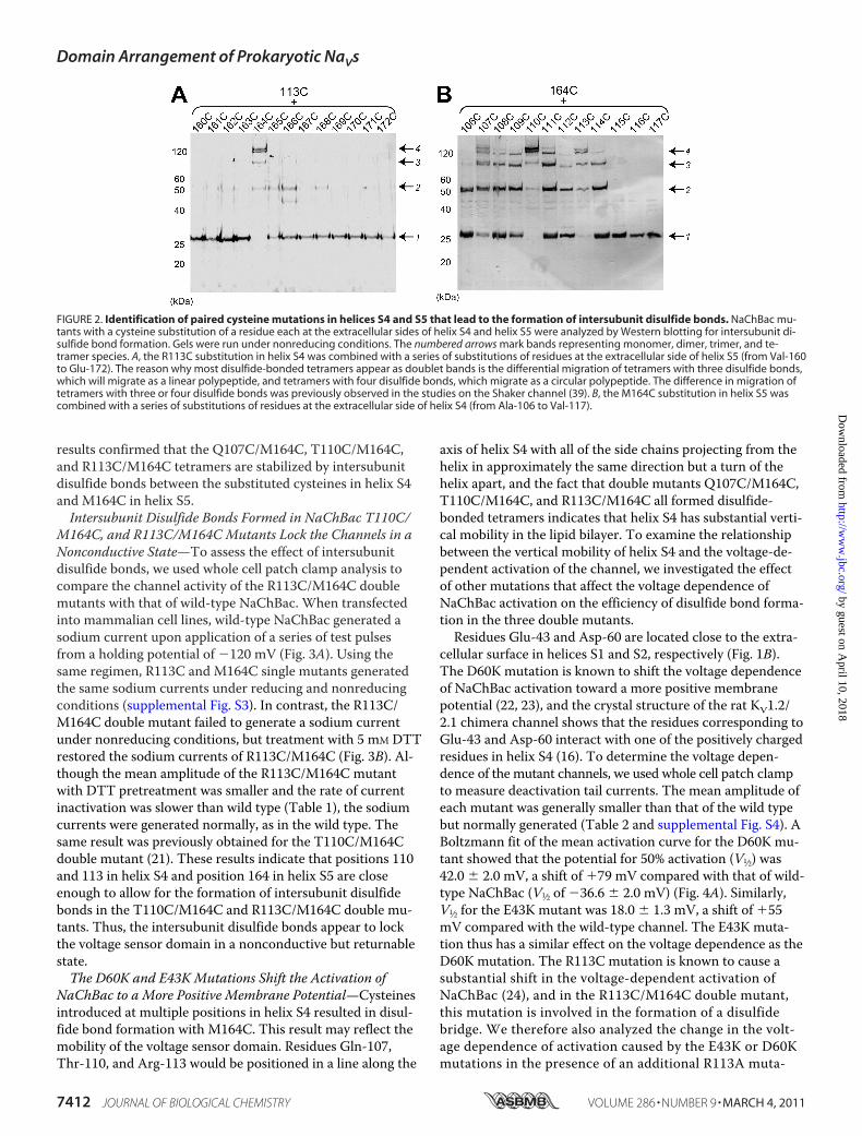
that would allow the formation of intersubunit disulfidebonds. A previous study of Shaker KV showed that the cys-teine substitutions of both Arg-362 in helix S4 and Ala-419 inhelix S5 resulted in disulfide-bonded channel tetramers (12),indicating that residues Arg-362 and Ala-419 of the adjacentsubunit are sufficiently close to each other to allow for theformation of a disulfide bond. If NaChBac has a similar do-main arrangement as the Shaker channel, double cysteinesubstitution of the corresponding residues in helices S4 andS5 should allow for the formation of a disulfide-bondedNaChBac tetramer (Fig. 1).To determine what double cysteine mutants would form
disulfide-bonded tetramers, we expressed them in E. coli, be-cause it is an easy and convenient expression system thatmakes it feasible to screen a large number of mutants. Ofnote, NaChBac contains no cysteine residues. We first intro-duced a cysteine substitution for residue Arg-113 in helix S4of NaChBac, the residue that corresponds to residue Arg-362in the Shaker channel. We then generated 13 mutants with anadditional cysteine substitution at positions 160–172 inhelix S5 (Fig. 1), and the E. coli cells expressing the variousNaChBac mutants were analyzed by Western blotting. Al-though most mutants showed a band consistent with the mo-lecular mass of a monomer (29 kDa; arrow 1 in Fig. 2A), theR113C/M164C double mutant migrated as a band of highermolecular mass (arrow 4 in Fig. 2A). With a molecular massof �120 kDa, approximately four times the molecular mass ofa NaChBac monomer, the band should represent a disulfide-
bonded tetramer. Thus, among the 13 residues tested, Met-164 was the residue in helix S5 closest to Arg-113.After identifying Met-164 in helix S5, we combined the
M164C mutation with 12 cysteine mutations at positions106–117 in helix S4 (Fig. 1). Western blot analysis of the dou-ble mutants showed that the T110C/M164C mutant was themost efficient in forming disulfide-bonded tetramers migrat-ing at a molecular mass of �120 kDa, whereas the R113C/M164C mutant was the second most efficient in forming di-sulfide-bonded tetramers (Fig. 2B). Of the 12 mutants tested,eight showed a band corresponding to the disulfide-bondedtetramer (cysteine residues introduced from positions 107 to114). The Q107C/M164C mutant was slightly more efficientin forming disulfide-bonded tetramers than the other mutantsbut less efficient than the T110C/M164C and R113C/M164Cmutants. By combining the T110C substitution in helix S4with the 13 cysteine substitutions in helix S5, we confirmedthat M164C most efficiently formed a disulfide bond withT110C (supplemental Fig. S1). M164C also most efficientlyformed disulfide bonds with V111C and L112C (supplementalFig. S1). Thr-110 and Met-164 thus appear to be the closestpair of residues between helices S4 and S5. SDS-PAGE analy-sis of wild-type NaChBac; the Q107C, T110C, R113C, andM164C single mutants; and the Q107C/M164C, T110C/M164C, and R113C/M164C double mutants showed that allvariants migrated mainly as monomers under reducing condi-tions, and only three double mutants migrated as tetramersunder nonreducing conditions (supplemental Fig. S2). These
FIGURE 1. Amino acid sequence and predicted topology of NaChBac. A, alignment of the sequence of NaChBac with some KVs obtained with clustalW1.81. NaChBac, NaV from B. halodurans C-125 (GI: 10174118); KvAP, KV from Aeropyrum pernix K1 (GI: 5104624); KV1.2/2.1, �-subunit of the chimeric KV1.2/KV2.1 channel (GI: 160877792); Shaker, �-subunit of the Shaker KV from D. melanogaster (GI: 288442). The residue numbers at the top and bottom are basedon the sequences of NaChBac and Shaker, respectively. The arrowheads indicate residues reported to be close between helices S4 and S5 in the Shakerchannel, Arg-362, Phe-416, and Ala-419 (12), and the brown arrows mark the residues mutated to cysteine in the present study, Ala-106 to Val-117 in S4 andVal-160 to Glu-172 in S5. B, putative transmembrane topology of NaChBac. Helices S1–S4 form the voltage sensor domain, and helices S5-S6 form the poredomain. The ovals indicate residues Gln-107, Thr-110, and Arg-113 in S4 and Met-164 in S5, which were particularly efficient in forming intersubunit disul-fide bonds. The boxes indicate residues Glu-43 and Asp-60, which were mutated to lysine. C, putative domain arrangement in a NaV tetramer with eachmonomer shown in a different color. The four pore domains form an ion-conducting pore at the center of the channel, and each voltage sensor domain islocated near the pore domain of the adjacent subunit.
Domain Arrangement of Prokaryotic NaVs
MARCH 4, 2011 • VOLUME 286 • NUMBER 9 JOURNAL OF BIOLOGICAL CHEMISTRY 7411
by guest on April 10, 2018
http://ww
w.jbc.org/
Dow
nloaded from
results confirmed that the Q107C/M164C, T110C/M164C,and R113C/M164C tetramers are stabilized by intersubunitdisulfide bonds between the substituted cysteines in helix S4and M164C in helix S5.Intersubunit Disulfide Bonds Formed in NaChBac T110C/
M164C, and R113C/M164C Mutants Lock the Channels in aNonconductive State—To assess the effect of intersubunitdisulfide bonds, we used whole cell patch clamp analysis tocompare the channel activity of the R113C/M164C doublemutants with that of wild-type NaChBac. When transfectedinto mammalian cell lines, wild-type NaChBac generated asodium current upon application of a series of test pulsesfrom a holding potential of �120 mV (Fig. 3A). Using thesame regimen, R113C and M164C single mutants generatedthe same sodium currents under reducing and nonreducingconditions (supplemental Fig. S3). In contrast, the R113C/M164C double mutant failed to generate a sodium currentunder nonreducing conditions, but treatment with 5 mM DTTrestored the sodium currents of R113C/M164C (Fig. 3B). Al-though the mean amplitude of the R113C/M164C mutantwith DTT pretreatment was smaller and the rate of currentinactivation was slower than wild type (Table 1), the sodiumcurrents were generated normally, as in the wild type. Thesame result was previously obtained for the T110C/M164Cdouble mutant (21). These results indicate that positions 110and 113 in helix S4 and position 164 in helix S5 are closeenough to allow for the formation of intersubunit disulfidebonds in the T110C/M164C and R113C/M164C double mu-tants. Thus, the intersubunit disulfide bonds appear to lockthe voltage sensor domain in a nonconductive but returnablestate.The D60K and E43K Mutations Shift the Activation of
NaChBac to a More Positive Membrane Potential—Cysteinesintroduced at multiple positions in helix S4 resulted in disul-fide bond formation with M164C. This result may reflect themobility of the voltage sensor domain. Residues Gln-107,Thr-110, and Arg-113 would be positioned in a line along the
axis of helix S4 with all of the side chains projecting from thehelix in approximately the same direction but a turn of thehelix apart, and the fact that double mutants Q107C/M164C,T110C/M164C, and R113C/M164C all formed disulfide-bonded tetramers indicates that helix S4 has substantial verti-cal mobility in the lipid bilayer. To examine the relationshipbetween the vertical mobility of helix S4 and the voltage-de-pendent activation of the channel, we investigated the effectof other mutations that affect the voltage dependence ofNaChBac activation on the efficiency of disulfide bond forma-tion in the three double mutants.Residues Glu-43 and Asp-60 are located close to the extra-
cellular surface in helices S1 and S2, respectively (Fig. 1B).The D60K mutation is known to shift the voltage dependenceof NaChBac activation toward a more positive membranepotential (22, 23), and the crystal structure of the rat KV1.2/2.1 chimera channel shows that the residues corresponding toGlu-43 and Asp-60 interact with one of the positively chargedresidues in helix S4 (16). To determine the voltage depen-dence of the mutant channels, we used whole cell patch clampto measure deactivation tail currents. The mean amplitude ofeach mutant was generally smaller than that of the wild typebut normally generated (Table 2 and supplemental Fig. S4). ABoltzmann fit of the mean activation curve for the D60K mu-tant showed that the potential for 50% activation (V1⁄2) was42.0 � 2.0 mV, a shift of �79 mV compared with that of wild-type NaChBac (V1⁄2 of �36.6 � 2.0 mV) (Fig. 4A). Similarly,V1⁄2 for the E43K mutant was 18.0 � 1.3 mV, a shift of �55mV compared with the wild-type channel. The E43K muta-tion thus has a similar effect on the voltage dependence as theD60K mutation. The R113C mutation is known to cause asubstantial shift in the voltage-dependent activation ofNaChBac (24), and in the R113C/M164C double mutant,this mutation is involved in the formation of a disulfidebridge. We therefore also analyzed the change in the volt-age dependence of activation caused by the E43K or D60Kmutations in the presence of an additional R113A muta-
FIGURE 2. Identification of paired cysteine mutations in helices S4 and S5 that lead to the formation of intersubunit disulfide bonds. NaChBac mu-tants with a cysteine substitution of a residue each at the extracellular sides of helix S4 and helix S5 were analyzed by Western blotting for intersubunit di-sulfide bond formation. Gels were run under nonreducing conditions. The numbered arrows mark bands representing monomer, dimer, trimer, and te-tramer species. A, the R113C substitution in helix S4 was combined with a series of substitutions of residues at the extracellular side of helix S5 (from Val-160to Glu-172). The reason why most disulfide-bonded tetramers appear as doublet bands is the differential migration of tetramers with three disulfide bonds,which will migrate as a linear polypeptide, and tetramers with four disulfide bonds, which migrate as a circular polypeptide. The difference in migration oftetramers with three or four disulfide bonds was previously observed in the studies on the Shaker channel (39). B, the M164C substitution in helix S5 wascombined with a series of substitutions of residues at the extracellular side of helix S4 (from Ala-106 to Val-117).
Domain Arrangement of Prokaryotic NaVs
7412 JOURNAL OF BIOLOGICAL CHEMISTRY VOLUME 286 • NUMBER 9 • MARCH 4, 2011
by guest on April 10, 2018
http://ww
w.jbc.org/
Dow
nloaded from

tion. V1⁄2 values for the R113A and D60K/R113A mutantswere 2.59 � 3.6 and 80.3 � 0.9 mV, respectively, a differ-ence between them of �78 mV (Fig. 4B). The V1⁄2 for theE43K/R113A mutant was 24.5 � 2.5 mV, a shift of �22 mVcompared with the R113A mutant (Fig. 4). Hence, theE43K or D60K mutations shift the voltage dependence ofactivation toward a more positive membrane potential,with or without the additional R113A mutation.The D60K and E43K Mutations Change Which Residues in
the Double Cysteine Mutants Preferentially Form DisulfideBonds—We tested whether the D60K and E43K mutationswould affect the disulfide bond formation of the three mu-
tants, Q107C/R113A/M164C (QC), T110C/R113A/M164C(TC), and R113C/M164C (RC), in which residue Arg-113 wasneutralized by substitution to alanine or cysteine. The mu-tants were expressed in E. coli, and disulfide bond formationin the proteins was assessed by quantitative Western blotanalysis (Fig. 5A). The efficiency of disulfide bond formationwas determined as the intensity of the band representing thedisulfide-bonded tetramer divided by the sum of the intensityof all bands (Fig. 5B). The QC mutant formed significantlyfewer disulfide-bonded tetramers than the TC (p � 0.01) andRC (p � 0.05) mutants. Thus, neutralization of Arg-113 bysubstitution to alanine or cysteine did not change the orderconcerning the efficiency with which the three double cys-teine mutants formed disulfide bonds. We next quantified theefficiency with which the three double cysteine mutantsformed disulfide bonds when we introduced an additionalD60K or E43K mutation. The D60K/QC mutant formed di-sulfide bonds significantly more efficiently than the D60K/TC(p � 0.01) and D60K/RC (p � 0.01) mutants, and theE43K/QC mutant more efficiently than the E43K/RC mutant(p � 0.05). With both the E43K and D60K mutations, the QCmutant was most efficient in forming disulfide bonds, fol-lowed by the TC mutant and finally the RC mutant. Theseresults suggest that in the presence of the E43K and D60Kmutations, the residue in helix S4 closest to Met-164 in helixS5 is no longer Thr-110 but rather Gln-107, and that thecharges surrounding the voltage sensor domain affect the pre-ferred vertical position of helix S4.
FIGURE 3. Intersubunit disulfide bonds formed in NaChBac T110C/M164C and R113C/M164C mutants lock the channels in a nonconduc-tive state. The representative current traces for wild type (A) and R113C/M164C mutant (B) were generated by a series of test pulses in HEK-293 cells.The currents were recorded without (top panels) or with (bottom panels) a30-min preincubation with 5 mM DTT. The panels below each current traceare averaged I-V curves derived from each experimental condition. The errorbars correspond to the S.E. of the mean.
TABLE 1The time constants of NaChBac cysteine mutantsEach mutant was expressed in HEK-293 cells. The time constant of activation(�act.) is the time from 10 to 90% of the peak current. The time constant ofinactivation (�inact.) is the time from the peak current to 1/e of the peak current.The �act. and �inact. were calculated at a membrane potential of �10 mV. All of theresults are expressed as the means � S.E. NR, nonreducing condition; R, reducingcondition; ND, not detectable.
�act. �inact. n
WT NR 6.11 � 0.9 91.3 � 11 9R 4.70 � 1.2 74.4 � 5.3 6
R113C NR 8.46 � 1.1 141 � 11 8R 5.95 � 0.7 122 � 12 10
M164C NR 12.7 � 1.4 138 � 8 6R 10.6 � 1.3 165 � 28 6
R113C/M164C (RC) NR ND NDR 11.2 � 0.4 84.1 � 11 7
TABLE 2The kinetic constants of NaChBac mutantsEach mutant was expressed in CHO-K1 cells. The time constant of activation(�act.) is the time from 10 to 90% of peak current. The time constant ofinactivation (�inact.) is the time from the peak current to 1/e of the peak current.The �act. and �inact. of WT were calculated at a membrane potential of �10 mV,and those of E43K, D60K, and R113A were calculated at a membrane potential of�60 mV. V1⁄2 of activation is the potential of 50% activation. � is the slope factor.All of the results are expressed as the means � S.E. ND, not determined.
�act. �inact. V1⁄2 � n
mV mV/e-foldWT 4.56 � 0.7 86.1 � 0.7 �36.6 � 2.0 10.4 � 1.6 7E43K 3.30 � 0.5 114 � 20 18.0 � 1.3 11.0 � 1.0 6D60K ND ND 42.0 � 2.0 17.8 � 1.5 6R113A 9.02 � 2.5 172 � 13 2.59 � 3.6 18.6 � 2.3 10E43K/R113A ND ND 24.5 � 2.5 22.1 � 1.9 6D60K/R113A ND ND 80.3 � 0.9 17.4 � 0.7 6
Domain Arrangement of Prokaryotic NaVs
MARCH 4, 2011 • VOLUME 286 • NUMBER 9 JOURNAL OF BIOLOGICAL CHEMISTRY 7413
by guest on April 10, 2018
http://ww
w.jbc.org/
Dow
nloaded from

DISCUSSION
The Domain Arrangement in NaChBac Is Similar to that inKVs—By generating double cysteine mutants, we confirmedthat in NaChBac, helix S4 is close to helix S5 of an adjacentsubunit. The observation of disulfide-bonded tetramers forthe T110C/M164C and R113C/M164C mutants indicates thatresidues Thr-110 and Arg-113 in helix S4 are close to residueMet-164 in helix S5 of the adjacent subunit (Fig. 2), suggest-ing that the putative domain arrangement shown in Fig. 1C islikely correct for prokaryotic NaVs. Our electrophysiologicanalysis shows that the T110C/M164C and R113C/M164Cmutants do not generate currents under nonreducing condi-tions, but activity is recovered under reducing conditions (Fig.3) (21). The intersubunit disulfide bonds presumably preventconformational changes of the voltage sensor domains andkeep the mutant channels in a nonconductive state. Becausealignment of the NaChBac and Shaker channel sequencesshows that the T110C/M164C mutations in NaChBac corre-spond to the R362C/F416C mutations in the Shaker channel,previously shown to result in the formation of an intersubunitdisulfide bond (Fig. 1A) (12), these results are strong evidencethat the subunit arrangement in NaChBac is similar to that inthe Shaker channel (Fig. 1C).The efficiency of disulfide bond formation of the mu-
tants with the M164C substitution depended on which po-sition in helix S4 was mutated to cysteine (Fig. 2B). Thedifference in efficiency reflects both the vertical position aswell as the rotational orientation of the side chains of theresidues. Disulfide bond formation was most efficient whenresidues Gln-107, Thr-110, and Arg-113 were mutated tocysteine. Because these residues are three positions apart,the approximate distance of a helical turn, they presumablyform a line along helix S4 that faces residue Met-164 inhelix S5 of the adjacent subunit. The Q107C/M164C mu-tant was less efficient in forming disulfide bonds than theT110C/M164C and Arg-113C/M164C mutants, suggestingthat Thr-110 and Arg-113 are closer to Met-164 than Gln-
FIGURE 5. The D60K and E43K mutations change which residues in thedouble cysteine mutants preferentially form disulfide bonds. ResiduesAsp-60 or Glu-43 were mutated to lysine in the Q107C/R113A/M164C (QC),T110C/R113A/M164C (TC), and R113C/M164C (RC) mutants. The mem-branes isolated from E. coli expressing these mutants were analyzed byquantitative Western blotting. A, representative Western blot of NaChBacdouble cysteine mutants with no additional mutation (left panel), with theD60K mutation (middle panel), and the E43K mutation (right panel). Thenumbered arrows indicate the bands representing the monomer, dimer,trimer, and tetramer species. B, quantification of the amount of disulfide-bonded tetramer that formed with the various mutants. The presented val-ues are the means of the intensity of the tetramer band divided by the sumof the intensity of all protein bands (no addition, n � 9; D60K, n � 9; E43K,n � 6). The error bars correspond to the S.E. of the mean. The asterisks indi-cate the differences from control (*, p � 0.05; **, p � 0.01). Comparisonswithout asterisks were not statistically significant.
FIGURE 4. The D60K and E43K mutations shift the activation of NaChBac to a more positive membrane potential. Normalized curves of the voltage-dependent activation of NaChBac mutants derived from CHO-K1 cells. The voltage dependence was determined by measuring deactivation tail currents.The error bars correspond to the S.E. of the mean. A, activation curves for wild-type NaChBac (closed circles) and the E43K (open circles) and D60K (closed tri-angles) mutants. B, activation curves for the R113A single mutant (closed circles) and the E43K/R113A (open circles) and D60K/R113A (closed triangles) doublemutants.
Domain Arrangement of Prokaryotic NaVs
7414 JOURNAL OF BIOLOGICAL CHEMISTRY VOLUME 286 • NUMBER 9 • MARCH 4, 2011
by guest on April 10, 2018
http://ww
w.jbc.org/
Dow
nloaded from

107. In the structure of the KV1.2/2.1 chimera channel, theresidues corresponding to Gln-107, Thr-110, and Arg-113(Arg-287, Gln-290, and Arg-293, respectively) also form aline along helix S4 (16), and residues Gln-290 and Arg-293 inhelix S4 are closest to Phe-344 in helix S5, the residue corre-sponding toMet-164 in NaChBac. In the structure of the KV1.2/2.1 chimera channel, the distances from the C� carbon of Phe-344 to those of Arg-287, Gln-290, and Arg-293 are 11.5, 8.4, and8.1 Å, respectively. Thus, the locations of residues Gln-107, Thr-110, and Arg-113 in NaChBac predicted from our disulfidecross-linking analysis are consistent with the positions of thecorresponding residues in the crystal structure of Kv1.2/2.1.These results further support a similar domain arrangement inNaChBac and KVs.
The proximity of helices S4 and S5 of an adjacent sub-unit has also been observed in other six-transmembranetetrameric ion channels. In KvAP, cysteine substitution ofArg-117 and Tyr-169, the residues corresponding to Arg-113 and Thr-163 in NaChBac, also results in the formationof intersubunit disulfide bonds (25). Furthermore, the crys-tal structure of MlotiK1, a prokaryotic cyclic nucleotide-gated ion channel, showed that helix S4 is close to helix S5of the adjacent subunit (26). The proximity of helix S4 tohelix S5 of an adjacent subunit thus appears to be a charac-teristic feature of six-transmembrane tetrameric ion chan-nels. The role of the interaction between helices S4 and S5has not yet been firmly established, but some studies sug-gest that the interaction is required for channel function,particularly for voltage-dependent activation and inactiva-tion (27–30).Helix S4 Is Highly Mobile—The efficient formation of disul-
fide bonds between M164C and cysteines introduced at posi-tions 107, 110, and 113 indicates that all three of these resi-dues on helix S4 can come close to Met-164 in helix S5 (Fig.2B). This result implies that helix S4 has substantial mobility,as much as 7.2 Å, the approximate distances of two helicalturns, in the vertical direction. Of the 12 cysteine substitu-tions in helix S4 of NaChBac, eight formed some disulfide-bonded tetramers with M164C (from Q107C to I114C) (Fig.2B). Disulfide bond formation between substituted cysteineresidues is a well established method for analyzing proteinstructure (31). Because of its helical structure, a rotationalmovement of helix S4 would allow residues in helix S4 otherthan Gln-107, Thr-110, and Arg-113 to form disulfide bondswith Met-164, but this appears to occur rarely. In contrast, ofthe 13 cysteine substitutions in helix S5 (from Val-160 to Glu-172), only M164C fully formed disulfide bonds with residuesin helix S4 (Fig. 2A and supplemental Fig. S1). This result in-dicates that helix S4 is highly mobile, appearing to move bothvertically and to some degree rotationally, in the lipid bilayer,whereas helix S5 was more static.The Vertical Position of Helix S4 Is Related to the Voltage
Dependence of Activation—Introducing the additional D60Kmutation changed the preferentially formed disulfide bond inNaChBac from TC to QC (Fig. 5). Because the D60K muta-tion shifts the voltage dependence of activation toward amore positive membrane potential (Fig. 4), the change in theclosest residues between helices S4 and S5 may be caused by
the shift in activation potential. In the Shaker channel, thenegative charge of the residue corresponding to Asp-60 inNaChBac assists the charge transfer during voltage-depen-dent activation by forming a salt bridge with a positivelycharged residue in helix S4 (32, 33). Substitution of Asp-60with lysine prevents the formation of this salt bridge, thussuppressing the vertical movement of helix S4. As a result,Gln-107, which is closer to the extracellular surface than Thr-110, would become more accessible for disulfide bond forma-tion with Met-164 and could be the cause of the shift in thevoltage dependence of activation to a more positive mem-brane potential.The effect of the E43K mutation on the voltage depen-
dence of activation and the preferentially formed disulfidebridge was similar to those of the D60K mutation (Figs. 4and 5). In the structure of the rat KV1.2/2.1 chimera, theresidue corresponding to Glu-43 forms a salt bridge withthe third positively charged residue in helix S4, whereasthe residue corresponding to Asp-60 interacts with thefourth positively charged residue (16). The role of residueGlu-43 in voltage-dependent activation may thus be similarto that of Asp-60, and the E43K mutation may thus alsointerfere with the vertical movement of helix S4. The roleof negative charges in the vertical movement of helix S4was recently demonstrated in NaChBac, because residuesAsp-60 and Glu-70 were shown to interact sequentiallywith positive charges in S4 during voltage dependent acti-vation (34). E43 also interacts with Arg-113 and Arg-116during channel activation in NaChBac (35). The involvednegative charges, including Glu-43, might work coopera-tively to assist the vertical movement of helix S4. Theseresults imply that the range of the vertical mobility of helixS4 should be more suppressed in ion channels that are acti-vated at higher membrane potentials (Fig. 6). Although thecharged residues in the voltage sensor domain are generallywell conserved, the residues at the positions correspondingto those of Glu-43 and Asp-60 in NaChBac are less con-served (36). The diversity in amino acids at these positionsmight thus contribute to differences in the voltage depen-dence of activation among voltage-gated ion channels.Indications for the Voltage-dependentMovement of Helix S4—
Helix S4 has been proposed to underlie voltage-dependentactivation by moving vertically in the lipid bilayer uponchanges in membrane potential (18–20, 37). Movement ofhelix S4 is thought to result in a conformational changeof the voltage sensor domain and eventually the opening ofthe pore gate (9). Double cysteine mutagenesis and struc-tural studies of KVs showed that in the activated state theresidues corresponding to Thr-110 or Arg-113 are close tohelix S5 (12, 16), whereas in the resting state residues inthe more extracellular part of helix S4 are close to residuesin helix S5 (13). The D60K and E43K mutations increasethe energy needed to activate the channels so that thesetwo mutations keep the channel in the resting state as thedisulfide bonds are formed. Therefore, the change causedby the E43K or D60K mutations in the efficiency withwhich disulfide bonds are formed in QC, TC, and RCmight indicate a shift in the residues that are close to each
Domain Arrangement of Prokaryotic NaVs
MARCH 4, 2011 • VOLUME 286 • NUMBER 9 JOURNAL OF BIOLOGICAL CHEMISTRY 7415
by guest on April 10, 2018
http://ww
w.jbc.org/
Dow
nloaded from

other during voltage-dependent gating (Fig. 5). ResidueGln-107, which is closer to the extracellular surface thanresidues Thr-110 or Arg-113, might be close to Met-164 inthe resting state or in the early phase of voltage-dependentactivation. These results imply that helix S4 moves verti-cally with Gln-107, Thr-110, and Arg-113 facing Met-164and that the positive charges move from the intracellulartoward the extracellular side during voltage-dependentgating.The voltage-dependent movement of helix S4 in NaChBac
suggested by our double cysteine mutagenesis and additionallysine substitution of residues Glu-43 and Asp-60 is consis-tent with previously proposed models for voltage-dependentgating. For example, the helical screw and paddle models bothrequire a large movement of helix S4 along its axis (20, 37). Itwas recently shown that the voltage sensor of NaChBac hasan overall structure similar to KVs and changes its conforma-tion during activation in the same manner as well (38). Therotational and vertical movement of helix S4 in NaChBac in-dicated by our cysteine mutagenesis agrees well with the heli-cal screw model, which requires that helix S4 moves both ver-tically and rotationally. Our results thus indicate that themovement of helix S4 in NaChBac is similar to that of canoni-cal voltage-gated ion channels. Our study only shows, how-ever, that helix S4 is mobile. Structural studies are needed toreveal its detailed movement during voltage-dependent acti-vation of NaChBac.
Acknowledgments—We thank Dr. Chikara Sato (Advanced Indus-trial Science and Technology, Neuroscience Research Institute) forkindly providing the NaChBac DNA. We are grateful to Dr. ThomasWalz for critical reading of the manuscript.
REFERENCES1. Hille, B. (2001) Ion Channels of Excitable Membranes, 3rd Ed., Sinauer
Associates Sunderland, MA2. Catterall, W. A. (2000) Neuron 26, 13–253. Ren, D., Navarro, B., Xu, H., Yue, L., Shi, Q., and Clapham, D. E. (2001)
Science 294, 2372–23754. Ito, M., Xu, H., Guffanti, A. A., Wei, Y., Zvi, L., Clapham, D. E., and
Krulwich, T. A. (2004) Proc. Natl. Acad. Sci. U.S.A. 101, 10566–105715. Koishi, R., Xu, H., Ren, D., Navarro, B., Spiller, B. W., Shi, Q., and
Clapham, D. E. (2004) J. Biol. Chem. 279, 9532–95386. Irie, K., Kitagawa, K., Nagura, H., Imai, T., Shimomura, T., and Fujiyoshi,
Y. (2010) J. Biol. Chem. 285, 3685–36947. Aggarwal, S. K., and MacKinnon, R. (1996) Neuron 16, 1169–11778. Seoh, S. A., Sigg, D., Papazian, D. M., and Bezanilla, F. (1996) Neuron 16,
1159–11679. Armstrong, C. M., and Bezanilla, F. (1974) J. Gen. Physiol. 63,
533–55210. Kuzmenkin, A., Bezanilla, F., and Correa, A. M. (2004) J. Gen. Physiol.
124, 349–35611. Pavlov, E., Bladen, C., Winkfein, R., Diao, C., Dhaliwal, P., and French,
R. J. (2005) Biophys. J. 89, 232–24212. Laine, M., Lin, M. C., Bannister, J. P., Silverman, W. R., Mock, A. F.,
Roux, B., and Papazian, D. M. (2003) Neuron 39, 467–48113. Neale, E. J., Elliott, D. J., Hunter, M., and Sivaprasadarao, A. (2003)
J. Biol. Chem. 278, 29079–2908514. Broomand, A., Mannikko, R., Larsson, H. P., and Elinder, F. (2003)
J. Gen. Physiol. 122, 741–74815. Long, S. B., Campbell, E. B., and Mackinnon, R. (2005) Science 309,
897–90316. Long, S. B., Tao, X., Campbell, E. B., and MacKinnon, R. (2007) Nature
450, 376–38217. Richardson, J., Blunck, R., Ge, P., Selvin, P. R., Bezanilla, F., Papazian,
D. M., and Correa, A. M. (2006) Proc. Natl. Acad. Sci. U.S.A. 103,15865–15870
18. Chanda, B., Asamoah, O. K., Blunck, R., Roux, B., and Bezanilla, F.(2005) Nature 436, 852–856
19. Posson, D. J., Ge, P., Miller, C., Bezanilla, F., and Selvin, P. R. (2005) Na-ture 436, 848–851
20. Ruta, V., Chen, J., and MacKinnon, R. (2005) Cell 123, 463–47521. Nagura, H., Irie, K., Imai, T., Shimomura, T., Hige, T., and Fujiyoshi, Y.
(2010) Biochem. Biophys. Res. Commun. 399, 341–34622. Blanchet, J., Pilote, S., and Chahine, M. (2007) Biophys. J. 92,
3513–352323. Zhao, Y., Scheuer, T., and Catterall, W. A. (2004) Proc. Natl. Acad. Sci.
U.S.A. 101, 17873–1787824. Chahine, M., Pilote, S., Pouliot, V., Takami, H., and Sato, C. (2004) J.
Membr. Biol. 201, 9–2425. Lee, S. Y., Lee, A., Chen, J., and MacKinnon, R. (2005) Proc. Natl. Acad.
Sci. U.S.A. 102, 15441–1544626. Clayton, G. M., Altieri, S., Heginbotham, L., Unger, V. M., and Morais-
Cabral, J. H. (2008) Proc. Natl. Acad. Sci. U.S.A. 105, 1511–151527. Li-Smerin, Y., Hackos, D. H., and Swartz, K. J. (2000) Neuron 25,
411–42328. Elinder, F., Mannikko, R., and Larsson, H. P. (2001) J. Gen. Physiol. 118,
1–1029. Larsson, H. P., and Elinder, F. (2000) Neuron 27, 573–58330. Lai, H. C., Grabe, M., Jan, Y. N., and Jan, L. Y. (2005) Neuron 47,
395–40631. Wahlberg, E., and Hard, T. (2006) J. Am. Chem. Soc. 128, 7651–766032. Papazian, D. M., Shao, X. M., Seoh, S. A., Mock, A. F., Huang, Y., and
Wainstock, D. H. (1995) Neuron 14, 1293–1301
FIGURE 6. Schematic drawing of the position of helix S4 relative tohelix S5 in NaChBac. Interface between helix S4 (blue) and helix S5 ofthe adjacent subunit (yellow). In the wild-type channel, helix S4 has sig-nificant vertical mobility (indicated by a long black arrow in the leftpanel), which is facilitated by ionic interactions between the negativelycharged residues Glu-43 or Asp-60 with positive charges in helix S4. TheE43K or D60K mutations introduce a positive charge, which repulses thepositive charges in helix S4 and may thus restrict the mobility of helix S4(indicated by the shorter red arrow in the right panel). As a result, highermembrane potentials are required to activate the E43K and D60K mu-tants (bottom panel).
Domain Arrangement of Prokaryotic NaVs
7416 JOURNAL OF BIOLOGICAL CHEMISTRY VOLUME 286 • NUMBER 9 • MARCH 4, 2011
by guest on April 10, 2018
http://ww
w.jbc.org/
Dow
nloaded from

33. Tiwari-Woodruff, S. K., Lin, M. A., Schulteis, C. T., and Papazian, D. M.(2000) J. Gen. Physiol. 115, 123–138
34. DeCaen, P. G., Yarov-Yarovoy, V., Sharp, E. M., Scheuer, T., and Cat-terall, W. A. (2009) Proc. Natl. Acad. Sci. U.S.A. 106, 22498–22503
35. Paldi, T., and Gurevitz, M. (2010) Biophys. J. 99, 456–46336. Yu, F. H., and Catterall, W. A. (2004) Sci. STKE 2004, re15
37. Yarov-Yarovoy, V., Baker, D., and Catterall, W. A. (2006) Proc. Natl.Acad. Sci. U.S.A. 103, 7292–7297
38. Chakrapani, S., Sompornpisut, P., Intharathep, P., Roux, B., and Perozo,E. (2010) Proc. Natl. Acad. Sci. U.S.A. 107, 5435–5440
39. Schulteis, C. T., Nagaya, N., and Papazian, D. M. (1996) Biochemistry 35,12133–12140
Domain Arrangement of Prokaryotic NaVs
MARCH 4, 2011 • VOLUME 286 • NUMBER 9 JOURNAL OF BIOLOGICAL CHEMISTRY 7417
by guest on April 10, 2018
http://ww
w.jbc.org/
Dow
nloaded from

FujiyoshiTakushi Shimomura, Katsumasa Irie, Hitoshi Nagura, Tomoya Imai and Yoshinori
Voltage-gated Sodium ChannelsArrangement and Mobility of the Voltage Sensor Domain in Prokaryotic
doi: 10.1074/jbc.M110.186510 originally published online December 22, 20102011, 286:7409-7417.J. Biol. Chem.
10.1074/jbc.M110.186510Access the most updated version of this article at doi:
Alerts:
When a correction for this article is posted•
When this article is cited•
to choose from all of JBC's e-mail alertsClick here
Supplemental material:
http://www.jbc.org/content/suppl/2011/01/03/M110.186510.DC1
http://www.jbc.org/content/286/9/7409.full.html#ref-list-1
This article cites 38 references, 19 of which can be accessed free at
by guest on April 10, 2018
http://ww
w.jbc.org/
Dow
nloaded from