Embed Size (px)
Citation preview

RESEARCH ARTICLE Open Access
Architecture of the Short External RotatorMuscles of the HipKevin C. Parvaresh1, Charles Chang1, Ankur Patel1, Richard L. Lieber1,2, Scott T. Ball1 and Samuel R. Ward1,2,3*
Abstract
Background: Muscle architecture, or the arrangement of sarcomeres and fibers within muscles, defines functionalcapacity. There are limited data that provide an understanding of hip short external rotator muscle architecture. Thepurpose of this study was thus to characterize the architecture of these small hip muscles.
Methods: Eight muscles from 10 independent human cadaver hips were used in this study (n= 80 muscles).Architectural measurements were made on pectineus, piriformis, gemelli, obturators, quadratus femoris, and gluteusminimus. Muscle mass, fiber length, sarcomere length, and pennation angle were used to calculate the normalizedmuscle fiber length, which defines excursion, and physiological cross-sectional area (PCSA), which defines force-producingcapacity.
Results: Gluteus minimus had the largest PCSA (8.29 cm2) followed by obturator externus (4.54 cm2), whereassuperior gemellus had the smallest PCSA (0.68 cm2). Fiber lengths clustered into long (pectineus - 10.38 cmand gluteus minimus - 10.30 cm), moderate (obturator internus - 8.77 cm and externus - 8.04 cm), or short(inferior gemellus - 5.64 and superior gemellus - 4.85). There were no significant differences among musclesin pennation angle which were all nearly zero. When the gemelli and obturators were considered as asingle functional unit, their collective PCSA (10.00 cm2) exceeded that of gluteus minimus as a substantialforce-producing group.
Conclusions: The key findings are that these muscles have relatively small individual PCSAs, short fiberlengths, and low pennation angles. The large collective PCSA and short fiber lengths of the gemelli andobturators suggest that they primarily play a stabilizing role rather than a joint rotating role.
Keywords: Hip, Muscle, Rotators, Architecture, Stability, Joint, Biomechanics, Fiber length
BackgroundThere is growing interest in hip joint function and path-ology that has been accompanied by recent technologicalprogress in biomechanical research. Much of the litera-ture has focused on bony [1–3], labral [4–6], and capsu-lar [7–9] morphology. Prior studies have shown thatspecific movement patterns are related to each of thesepathologies [10–13]. Detailed architectural properties ofthe muscles surrounding the hip and the neighboringbony structures (Fig. 1) are essential to understandingthe functional biomechanics of hip movement andstability.
Skeletal muscle architecture is defined as the arrange-ment of muscle fibers relative to the axis of force gener-ation [14, 15]. Understanding muscle architecture isparticularly important as it provides the best anatomicalinsight to predict muscle function [15]. To our know-ledge, there is only one prior study that has evaluatedthe muscle architecture for the small rotational musclesof the hip [16]. In this study, Friedrich and Brand re-ported measurements for fiber length and physiologicalcross-sectional area (PCSA) for selected small rotationalmuscles of the hip. However, the study was extremelylimited as it involved only two cadaveric specimens anddid not normalize measurements based on sarcomerelength. Sarcomere length measurement is critical be-cause it allows normalized fiber length (Lf) and PCSA tobe calculated. Without sarcomere length measurements,
© The Author(s). 2019 Open Access This article is distributed under the terms of the Creative Commons Attribution 4.0International License (http://creativecommons.org/licenses/by/4.0/), which permits unrestricted use, distribution, andreproduction in any medium, provided you give appropriate credit to the original author(s) and the source, provide a link tothe Creative Commons license, and indicate if changes were made. The Creative Commons Public Domain Dedication waiver(http://creativecommons.org/publicdomain/zero/1.0/) applies to the data made available in this article, unless otherwise stated.
* Correspondence: [email protected] of Orthopaedic Surgery, University of California, 9500 GilmanDrive, La Jolla, San Diego, CA 92093-0863, USA2Departments of Bioengineering, University of California, San Diego, USAFull list of author information is available at the end of the article
Parvaresh et al. BMC Musculoskeletal Disorders (2019) 20:611 https://doi.org/10.1186/s12891-019-2995-0

muscle measurements are distorted based on the fixationposition of the cadaver. We previously showed thatsarcomere length measurements eliminate this problem.These metrics are the only ones that are proportional tomuscle excursion [17] and force generating capacity[18], respectively. Normalized architectural measure-ments are therefore required to understanding the rolethese muscles play in coordinating hip motion.The purpose of this study was to measure the architec-
tural properties of selected small muscles of the hip includ-ing the gluteus minimus, pectineus, piriformis, gemelli,obturators, and quadratus femoris. Our goal was to definethe architectural properties of these muscles in order tobetter understand their functional role in hip joint bio-mechanics. We hypothesized that the architecture of thesemuscles would support their putative role in controllingjoint position and providing stability.
MethodsWhole cadaveric lower extremity specimens were ob-tained from the University of California, San Diego’sbody donations program and were bisected along the
midline. The regions of the hip and thigh were dissectedthrough the deep fascia, and each muscle was visualizedand obtained by removal from its most proximal originto distal tendon attachment. Eight muscles (Table 1)from each of 10 formaldehyde-fixed human lower ex-tremities (mean age ± standard deviation; 83 ± 9 years;male:female ratio, 5:5; height, 168.4 ± 9.3 cm; mass,82.7 ± 15.3 kg, femoral head diameter 49.43 ± 1.1 mm)were carefully excised and stored in 1X phosphate-buffered saline (PBS).Muscle architectural measurements were made based
on the same methods described by Sacks and Roy [19],modified by Lieber et al. [20], and adapted for the lowerextremity by Ward et al. [21]. Briefly, muscle length(Lm) was measured as the distance from most proximalfibers to the most distal fibers. Raw fiber length (Lf′)was measured for each muscle in 3 regions; proximal,middle, and distal; using a digital caliper (accuracy, 0.01mm). Surface pennation angle was measured with agoniometer as the angle between the fibers and the distaltendon. Values for normalized fiber length (Lf) were cal-culated based on the following equation [22]:
Lf ¼ Lf02:7 μm=Lsð Þ
where Ls is the measured sarcomere length and 2.7 μmis the optimum sarcomere length for human muscle[22]. Normalizing fiber length is key as it allows forcomparisons among muscles fixed in various degrees oftension and sarcomere lengths [23]. Normalized Lm wascalculated using a similar equation. The Lf/Lm ratio wasalso determined to assess excursion design comparisonsacross muscles [15]. PCSA was calculated according tothe following equation: [18].
PCSA cm2� � ¼ M gð Þ � cosθð Þ= ρ g=cm3
� �� Lf cmð Þ� �
where M is mass, θ is pennation angle, and ρ is muscledensity (1.056 g/cm3) [24], accounting for dehydrationthat occurs during fixation.Multiple measurements were made on each muscle
(n= > 3), then averaged for each sample, yielding grandmeans which are presented. All data are reported asmean ± standard error unless otherwise noted. Between-muscle and between-muscle group comparisons of mass,mean fiber length, and total PCSA were made with one-way ANOVAs after confirming the assumptions of nor-mality and homogeneity of variances were met. Compar-isons to gluteus medius and maximus were made usingpreviously reported data from similarly aged specimens[21]. Post hoc Tukey’s tests were used to identify specificmuscle differences. All analyses were performed usingSPSS® software (Version 20.0; SPSS Inc., Chicago, IL).Significance was set to p < 0.05 for the ANOVA and posthoc tests.
Fig. 1 Lateral view of the hip with the selected muscles of the hipand gluteus medius illustrated
Parvaresh et al. BMC Musculoskeletal Disorders (2019) 20:611 Page 2 of 6
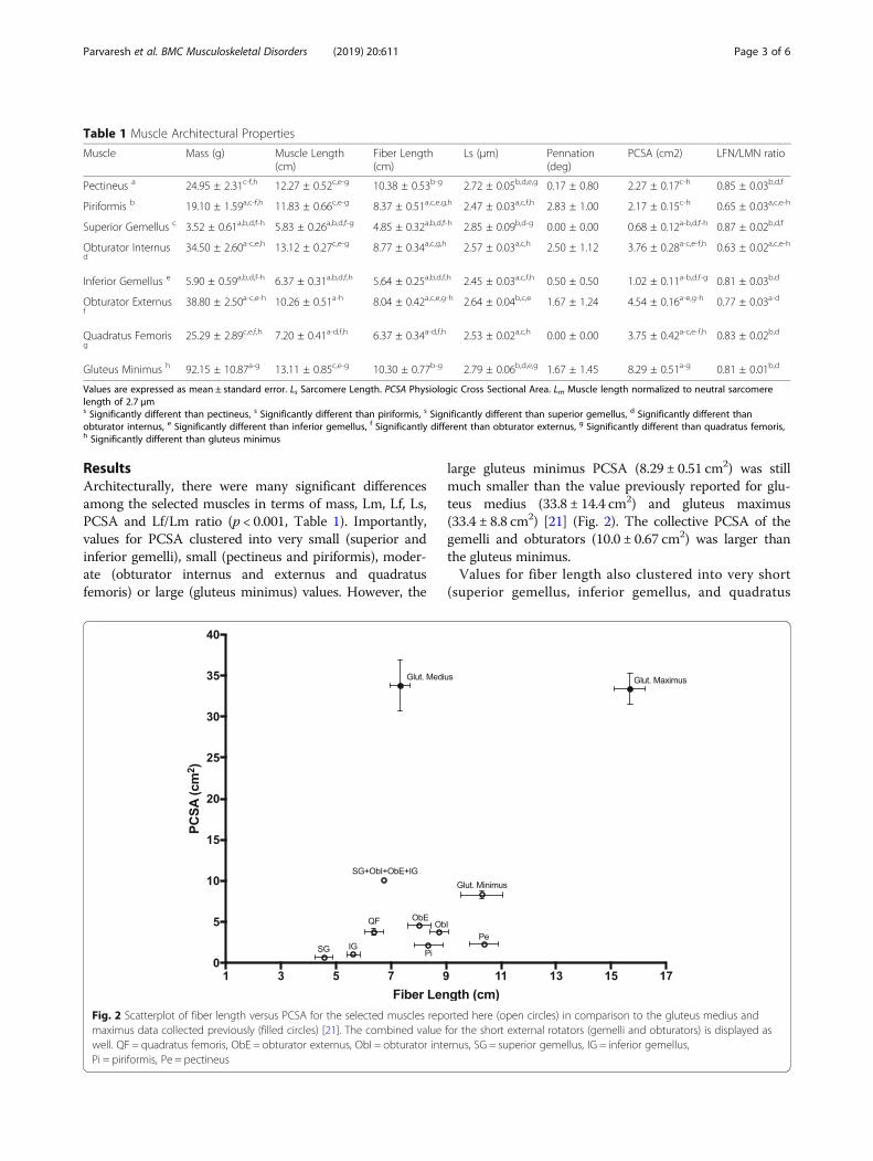
ResultsArchitecturally, there were many significant differencesamong the selected muscles in terms of mass, Lm, Lf, Ls,PCSA and Lf/Lm ratio (p < 0.001, Table 1). Importantly,values for PCSA clustered into very small (superior andinferior gemelli), small (pectineus and piriformis), moder-ate (obturator internus and externus and quadratusfemoris) or large (gluteus minimus) values. However, the
large gluteus minimus PCSA (8.29 ± 0.51 cm2) was stillmuch smaller than the value previously reported for glu-teus medius (33.8 ± 14.4 cm2) and gluteus maximus(33.4 ± 8.8 cm2) [21] (Fig. 2). The collective PCSA of thegemelli and obturators (10.0 ± 0.67 cm2) was larger thanthe gluteus minimus.Values for fiber length also clustered into very short
(superior gemellus, inferior gemellus, and quadratus
Table 1 Muscle Architectural Properties
Muscle Mass (g) Muscle Length(cm)
Fiber Length(cm)
Ls (μm) Pennation(deg)
PCSA (cm2) LFN/LMN ratio
Pectineus a 24.95 ± 2.31c-f,h 12.27 ± 0.52c,e-g 10.38 ± 0.53b-g 2.72 ± 0.05b,d,e,g 0.17 ± 0.80 2.27 ± 0.17c-h 0.85 ± 0.03b,d,f
Piriformis b 19.10 ± 1.59a,c-f,h 11.83 ± 0.66c,e-g 8.37 ± 0.51a,c,e,g,h 2.47 ± 0.03a,c,f,h 2.83 ± 1.00 2.17 ± 0.15c-h 0.65 ± 0.03a,c,e-h
Superior Gemellus c 3.52 ± 0.61a,b,d,f-h 5.83 ± 0.26a,b,d,f-g 4.85 ± 0.32a,b,d,f-h 2.85 ± 0.09b,d-g 0.00 ± 0.00 0.68 ± 0.12a-b,d,f-h 0.87 ± 0.02b,d,f
Obturator Internusd
34.50 ± 2.60a-c,e,h 13.12 ± 0.27c,e-g 8.77 ± 0.34a,c,g,h 2.57 ± 0.03a,c,h 2.50 ± 1.12 3.76 ± 0.28a-c,e-f,h 0.63 ± 0.02a,c,e-h
Inferior Gemellus e 5.90 ± 0.59a,b,d,f-h 6.37 ± 0.31a,b,d,f,h 5.64 ± 0.25a,b,d,f,h 2.45 ± 0.03a,c,f,h 0.50 ± 0.50 1.02 ± 0.11a-b,d,f-g 0.81 ± 0.03b,d
Obturator Externusf
38.80 ± 2.50a-c,e-h 10.26 ± 0.51a-h 8.04 ± 0.42a,c,e,g-h 2.64 ± 0.04b,c,e 1.67 ± 1.24 4.54 ± 0.16a-e,g-h 0.77 ± 0.03a-d
Quadratus Femorisg
25.29 ± 2.89c,e,f,h 7.20 ± 0.41a-d,f,h 6.37 ± 0.34a-d,f,h 2.53 ± 0.02a,c,h 0.00 ± 0.00 3.75 ± 0.42a-c,e-f,h 0.83 ± 0.02b,d
Gluteus Minimus h 92.15 ± 10.87a-g 13.11 ± 0.85c,e-g 10.30 ± 0.77b-g 2.79 ± 0.06b,d,e,g 1.67 ± 1.45 8.29 ± 0.51a-g 0.81 ± 0.01b,d
Values are expressed as mean ± standard error. Ls Sarcomere Length. PCSA Physiologic Cross Sectional Area. Lm Muscle length normalized to neutral sarcomerelength of 2.7 μms Significantly different than pectineus, s Significantly different than piriformis, s Significantly different than superior gemellus, d Significantly different thanobturator internus, e Significantly different than inferior gemellus, f Significantly different than obturator externus, g Significantly different than quadratus femoris,h Significantly different than gluteus minimus
Fig. 2 Scatterplot of fiber length versus PCSA for the selected muscles reported here (open circles) in comparison to the gluteus medius andmaximus data collected previously (filled circles) [21]. The combined value for the short external rotators (gemelli and obturators) is displayed aswell. QF = quadratus femoris, ObE = obturator externus, ObI = obturator internus, SG = superior gemellus, IG = inferior gemellus,Pi = piriformis, Pe = pectineus
Parvaresh et al. BMC Musculoskeletal Disorders (2019) 20:611 Page 3 of 6

femoris), short (piriformis, obturator internus andexternus) or moderate (pectineus and gluteus mini-mus) lengths. On average, these values were similar tothe short fiber length observed for gluteus medius(7.33 ± 1.57 cm) and shorter than the long fiber lengthobserved for gluteus maximus (15.69 ± 2.57 cm) [21](Fig. 2). Average Ls (2.63 μm) tended to be shorterthan optimal length (2.7 μm), which was not surprisinggiven the externally rotated position of the hips at thetime of fixation. Fiber length to muscle lengths ratiostended to be long with the exception of piriformis(0.65 ± 0.03) and obturator internus (0.63 ± 0.02).There were no significant differences in pennation an-gles which were nearly zero for all muscles.
DiscussionIn this study we report the detailed the architecturalproperties of the rotational muscles of the hip, includingpectineus, piriformis, gemelli, obturator internus andexternus, quadratus femoris, and gluteus minimus, cor-rected for muscle sarcomere length. The key findingswere the relatively small individual PCSAs, short fiberlengths, short sarcomere lengths, and uniformly lowpennation angles. To our knowledge there are no priorpublications on the properties of these selected hip mus-cles utilizing sarcomere length to provide normalizedvalues for fiber length and PCSA.In a comprehensive literature review of the short exter-
nal rotators of the hip, Yoo et al. [25] found only one pub-lished article containing quantitative values of hip musclearchitecture [16]. In that study, Friederich and Brand re-ported architectural measurements from two cadavericspecimens without sarcomere normalization. Regardingthe short external rotators, they found the PCSAs of thepiriformis (20.54 cm2) and quadratus femoris (21.00 cm2)were the largest, by approximately a 4 fold greater magni-tude. Our data showed a similar range of distribution ofPCSAs between the short external rotator muscles, butour PCSA values were significantly smaller (Fig. 2). Thelack of sarcomere length measurements from their studymakes it difficult to reconcile our data, highlighting theimportance of this normalizing measurement for compar-ing muscle architecture because most sarcomere lengthsthey measured were below optimum.This concept may be supported by comparing our data
to the normalized data of Wickiewicz et al. [26]. In theirpilot study, they report normalizing muscle architecturedata to sarcomere length. Although their sarcomerelength was based on an average from a separate studyand not a direct measurement, it still provides a baselinefor comparison. Their average measurements for pecti-neus muscle length (12.30 cm), fiber length (10.43 cm),pennation angle (0 degrees), and PCSA (2.9) are nearlyidentical to our values (Table 1). Although pectineus
was the only muscle available for direct comparison,these findings underscore the importance of normalizingdata in comparing muscle architecture.Our data provide a number of insights into the design
of the hip short external rotators. All muscles exhibitedan almost parallel pennation angle of 0 degrees, suggest-ing force generation is maximized to act in a single axisof rotation. This may be helpful in maintaining lowmuscle mass and PCSA in constrained regions of the hipwhile still allowing sufficient force generation. Fromgross dissection, we know that the superior gemellus,obturator internus, inferior gemellus, and obturatorexternus are essentially fused. If these muscles are con-sidered as a single functional unit, their collective PCSAbecomes functionally relevant (Fig. 2). In fact, their com-bined PCSA exceeds that of gluteus minimus. With theaddition of quadratus femoris and piriformis, the collect-ive “short external rotators” become a substantial force-producing unit. Considering them as a unit with a largePCSA and short fiber lengths, their design features cor-respond to a stabilizing role [27].Although we did not directly measure joint geom-
etry, these architectural data may be combined withknown values previously reported to evaluate theirrole in muscle-joint kinematics. Due to the short ex-ternal rotators’ close proximity to the axis of rota-tion, muscle length does not change substantiallyrelative to joint position and moment arms remainoriented toward external rotation [28]. Unlike thegluteal muscles, the short external rotators maytherefore rotate the hip relatively independent of sa-gittal and coronal motion. Such independent move-ment provides valuable rotational control withoutotherwise affecting joint position. When combinedwith their rotational antagonists (gluteus minimus,pectineus, and adductors), these muscles appear toprovide a stabilizing role to the hip joint [29]. Withsimultaneous internal and external rotational con-traction, a medial compressive force is created tobalance the lateralizing force of the abductors. Suchbalance may facilitate dynamic stabilization of thehip joint, though further studies are necessary to val-idate these hypotheses.Additionally, our findings have implications for
current surgical approaches to the hip. Decisions torelease the short external rotators during hip surgeryshould represent a balance between achieving ad-equate surgical exposure and preserving soft tissueanatomy, which may lead to less post-operative pain,faster rehabilitation, and a more stable joint [30].During traditional posterior approaches to the hip,the short external rotators are often sacrificed. Earlyin the practice of total hip arthroplasties, leaving theshort external rotators unrepaired was believed to
Parvaresh et al. BMC Musculoskeletal Disorders (2019) 20:611 Page 4 of 6

have no adverse effect on hip stability [8, 31]. Theimportance of these structures has become apparentin the recent years, however, as other reports haveshown that adequate repair of the posterior struc-tures greatly decreases the future risk of hip instabil-ity caused by soft-tissue attenuation [32, 33]. Whilerecent meta-analyses have shown that surgical ap-proach does not affect dislocation rate [34], fewstudies directly measured muscle function followingsurgery. Evolving techniques including the direct su-perior approach, which spares the external rotators,may offer a functional advantage [35], but long-termcomparison studies are lacking. Further researchshould be dedicated to assessment of hip musclefunction following hip surgery.This study has several limitations. Fixation position
was in an externally rotated joint configuration and maynot reflect the clinically accepted definition of a neutralhip joint angle which is 0° abduction, flexion, and rota-tion. However, normalization of results with sarcomerelength removes variation associated with position andtherefore positioning should not significantly affect theresults. Second, the advanced age of the cadaveric speci-mens may have led to lower PCSAs than would other-wise be observed in younger patients, but still likelyprovide a baseline for functional predictions and arecomparable among muscles. Future studies may expandon these data for functional evaluation such as electro-myographic studies of activation patterns during variousmovements and activities.
ConclusionsIn summary, these findings characterize the architectureof selected muscles of the hip. These data support the hy-pothesis that these muscles act as dynamic stabilizers.Moreover, they highlight the functional importance ofthese muscles relative to hip pathology, surgery, and re-habilitation. We suggest that these data be expanded inthe future to characterize the dynamic interactions amongthese muscles and other extra- and intra-articular struc-tures as well as muscle adaptations to immobilization andpathology to further our knowledge of hip biomechanics.
AbbreviationsANOVA: Analysis of variance; Lf: Normalized fiber length; Lf’: Raw fiber length;Lm: Muscle length; Ls: Sarcomere length; M: Mass; PBS: Phosphate-bufferedsaline; PCSA: Physiological cross-sectional area; θ: Pennation angle; ρ: Muscledensity
AcknowledgementsWe would like to acknowledge our cadaveric specimen donors for providingthe means for scientific exploration and advancement.
Authors’ contributionsAll authors made considerable contributions to constitute authorship for thisstudy as defined in the journal’s criteria for authorship. KP drafted themanuscript and interpreted the data. CC designed the project, acquired, andanalyzed the data. AP acquired and analyzed the data. RL assisted with
project design, data interpretation, and manuscript review. SB assisted withproject design and data interpretation. SW assisted with project design, dataanalysis and interpretation, and manuscript revision. All authors read andapproved the final manuscript and have agreed to be personallyaccountable for their own contributions.
FundingThis manuscript was funded in part by a grant from the National Institutes ofHealth (PI: Lieber) (R01) HD048501. This work was supported (or supported inpart) by Research Career Scientist Award Award Number IK6 RX003351 fromthe United States (U.S.) Department of Veterans Affairs Rehabilitation R&D(Rehab RD) Service.
Availability of data and materialsThe datasets analyzed during the current study are available as a supportingfile or from the corresponding author on reasonable request.
Ethics approval and consent to participateEach author certifies that his or her institution has approved the protocol forthis investigation and that all investigations were conducted in conformitywith ethical principles of research. As this was a cadaveric study, no formalethics approval was required.
Consent for publicationAll authors give their consent to publish this manuscript.
Competing interestsAll authors verify there are no competing interests.
Author details1Departments of Orthopaedic Surgery, University of California, 9500 GilmanDrive, La Jolla, San Diego, CA 92093-0863, USA. 2Departments ofBioengineering, University of California, San Diego, USA. 3Departments ofRadiology, University of California, 9500 Gilman Drive, La Jolla, San Diego, CA92093-0863, USA.
Received: 15 August 2019 Accepted: 9 December 2019
References1. Ito K, Minka M, Leunig M, Werlen S, Ganz R. Femoroacetabular
impingement and the cam-effect: a MRI-based quantitative anatomicalstudy of the femoral head-neck offset. J Bone Jt Surg Br. 2001;83(2):171–6.
2. Beck M, Kalhor M, Leunig M, Ganz R. Hip morphology influences the patternof damage to the acetabular cartilage. J Bone Jt Surg Br. 2005;87(7):1012–8.
3. Ganz R, Parvizi J, Beck M, Leunig M, Nötzli H, Siebenrock K.Femoroacetabular impingement: a cause for osteoarthritis of the hip. ClinOrthop. 2003;417:112–20.
4. Ferguson SJ, Bryant JT, Ito K. The material properties of the bovineacetabular labrum. J Orthop Res. 2001;19(5):887–96.
5. Ferguson SJ, Bryant JT, Ganz R, Ito K. An in vitro investigation of theacetabular labral seal in hip joint mechanics. J Biomech. 2003;36(2):171–8.
6. Dy CJ, Thompson MT, Crawford MJ, Alexander JW, McCarthy JC, Noble PC.Tensile strain in the anterior part of the acetabular labrum duringprovocative maneuvering of the normal hip. J Bone Jt Surg Am. 2008;90(7):1464–72.
7. Lloyd-Roberts GC. The role of capsular changes in osteoarthritis of the hipjoint. J Bone Jt Surg Br. 1953;35-B(4):627–42.
8. Woo RY, Morrey BF. Dislocations after total hip arthroplasty. J Bone Jt SurgAm. 1982;64(9):1295–306.
9. Hewitt J, Guilak F, Glisson R, Parker VT. Regional material properties of thehuman hip joint capsule ligaments. J Orthop Res. 2001;19(3):359–64.
10. Kubiak-Langer M, Tannast M, Murphy SB, Siebenrock KA, Langlotz F. Rangeof motion in anterior femoroacetabular impingement. Clin Orthop. 2007;458:117–24.
11. Austin AB, Souza RB, Meyer JL, Powers CM. Identification of abnormal hipmotion associated with acetabular labral pathology. J Orthop Sports PhysTher. 2008;38(9):558–65.
12. Kennedy MJ, Lamontagne M, Beaulé PE. Femoroacetabular impingementalters hip and pelvic biomechanics during gait: walking biomechanics ofFAI. Gait Posture. 2009;30(1):41–4.
Parvaresh et al. BMC Musculoskeletal Disorders (2019) 20:611 Page 5 of 6

13. Casartelli NC, Maffiuletti NA, Item-Glatthorn JF, Staehli S, Bizzini M,Impellizzeri FM, et al. Hip muscle weakness in patients with symptomaticfemoroacetabular impingement. Osteoarthr Cartil. 2011;19(7):816–21.
14. Gans C, Gaunt AS. Muscle architecture in relation to function. J Biomech.1991;24(S1):53–65.
15. Lieber RL, Friden J. Functional and clinical significance of skeletal musclearchitecture. Muscle Nerve. 2000;23(11):1647–66.
16. Friederich JA, Brand RA. Muscle fiber architecture in the human lower limb.J Biomech. 1990;23(1):91–5.
17. Winters TM, Takahashi M, Lieber RL, Ward SR. Whole muscle length-tensionrelationships are accurately modeled as scaled sarcomeres in rabbithindlimb muscles. J Biomech. 2011;44(1):109–15.
18. Powell PL, Roy RR, Kanim P, Bello M, Edgerton VR. Predictability of skeletalmuscle tension from architectural determinations in Guinea pig hindlimbs. JAppl Physiol. 1984;57(6):1715–21.
19. Sacks RD, Roy RR. Architecture of the hind limb muscles of cats: functionalsignificance. J Morphol. 1982;173(2):185–95.
20. Lieber RL, Fazeli BM, Botte MJ. Architecture of selected wrist flexor andextensor muscles. J Hand Surg. 1990;15(2):244–50.
21. Ward SR, Eng CM, Smallwood LH, Lieber RL. Are current measurements oflower extremity muscle architecture accurate? Clin Orthop. 2009;467:1074–82.
22. Lieber RL, Loren GJ, Friden J. In vivo measurement of human wrist extensormuscle sarcomere length changes. J Neurophysiol. 1994;71(3):874–81.
23. Felder A, Ward SR, Lieber RL. Sarcomere length measurement permits highresolution normalization of muscle fiber length in architectural studies. JExp Biol. 2005;208(17):3275–9.
24. Ward SR, Lieber RL. Density and hydration of fresh and fixed human skeletalmuscle. J Biomech. 2005;38(11):2317–20.
25. Yoo S, Dedova I, Pather N. An appraisal of the short lateral rotators of thehip joint. Clin Anat. 2015;28:800–12.
26. Wickiewicz TL, Roy RR, Powell PL, Edgerton VR. Muscle architecture of thehuman lower limb. Clin Orthop. 1983;179:275–83.
27. Ward SR, Winters TM, Blemker SS. The architectural design of the glutealmuscle group: implications for movement and rehabilitation. J OrthopSports Phys Ther. 2010;40(2):95–102.
28. Delp SL, Hess WE, Hungerford DS, Jones LC. Variation of rotation momentarms with hip flexion. J Biomech. 1999;32(5):493–501.
29. Giphart JE, Stull JD, Laprade RF, Wahoff MS, Philippon MJ. Recruitment andactivity of the pectineus and piriformis muscles during hip rehabilitationexercises: an electromyography study. Am J Sports Med. 2012;40(7):1654–63.
30. Solomon LB, Lee YC, Callary SA, Beck M, Howie DW. Anatomy of piriformis,obturator internus and obturator externus: implications for the posteriorsurgical approach to the hip. J Bone Jt Surg Br. 2010;92(9):1317–24.
31. Stähelin T, Vienne P, Hersche O. Failure of reinserted short external rotatormuscles after total hip arthroplasty. J Arthroplast. 2002;17(5):604–7.
32. Robinson RP, Robinson HJJ, Salvati E. Comparison of the transtrochanteric andposterior approaches for total hip replacement. Clin Orthop. 1980;147:143–7.
33. Pine J, Binns M, Wright P, Soames R. Piriformis and obturator internusmorphology: a cadaveric study. Clin Anat. 2011;24(1):70–6.
34. Higgins BT, Barlow DR, Heagerty NE, Lin TJ. Anterior vs. posterior approachfor Total hip Arthroplasty, a systematic review and meta-analysis. JArthroplast. 2015 Mar 1;30(3):419–34.
35. Barrett AA, Ezzibdeh RM, Horst PK, Roger DJ, Amanatullah DF. Directsuperior approach to the hip for Total hip Arthroplasty. JBJS Essent SurgTech. 2019;9(2):e17.
Publisher’s NoteSpringer Nature remains neutral with regard to jurisdictional claims inpublished maps and institutional affiliations.
Parvaresh et al. BMC Musculoskeletal Disorders (2019) 20:611 Page 6 of 6