Embed Size (px)
Citation preview

SoutheaSt aSian J trop Med public health
142 Vol 45 No. 1 January 2014
Correspondence: Noppadol Larbcharoensub, Department of Pathology, Faculty of Medicine Ramathibodi Hospital, Mahidol University, 270 Rama VI Road, Ratchathewi, Bangkok 10400, Thailand.Tel: +66 (0) 2354 7277; Fax: +66 (0) 2354 7266E-mail: [email protected]
CASE REPORT
ARCANOBACTERIUM PYOGENES ENDOCARDITIS: A CASE REPORT AND LITERATURE REVIEW
Supavit Chesdachai1, Noppadol Larbcharoensub2, Tharintorn Chansoon2,Panas Chalermsanyakorn2, Pitak Santanirand3, Darunee Chotiprasitsakul4,
Disya Ratanakorn5 and Sarana Boonbaichaiyapruck6
1Medical Student, 2Division of Anatomical Pathology, 3Division of Microbiology, Department of Pathology, 4Division of Infectious Disease, 5Division of Neurology,
6Division of Cardiology, Department of Medicine, Faculty of Medicine Ramathibodi Hospital, Mahidol University, Bangkok, Thailand
Abstract. We report the case of a 64-year-old man with Arcanobacterium pyogenes endocarditis. The patient presented with dyspnea and asymmetrical progressive quadriparesis. A transthoracic echocardiogram revealed mobile vegetations on both leaflets of his mitral valve measuring 0.5 x 3 cm, thickening of the mitral valve with severe mitral regurgitation due to dehiscence of the papillary muscle to the posterior mitral leaflet. He also had aortic sclerosis with a vegetation measuring 0.5 x 1 cm causing aortic valve dehiscence and free flow aortic regurgitation. An initial hemoculture grew out pleomorphic, gram-positive, non-motile, anaerobic to microaerophilic bacilli. A diagnosis of infective endocarditis was made using modified Duke criteria. He was treated with intravenous ampicillin and gentamicin. Four days after admission, he developed acute respiratory failure and succumbed to the disease. A pre-mortem hemoculture and post-mortem heart valve culture grew Arcanobacterium pyogenes. Septic thromboemboli involving the brain, kidneys, lungs and spleen were documented. The patient also had ischemic vasculopathy with focal spinal arteriolitis and bilateral demyelination of the cervical corticospi-nal tracts. There are three published reports of human A. pyogenes endocarditis in the literature. Neurological involvement with ischemic spinal vasculopathy and demyelination has not been reported. We report the first autopsy proven case of A. pyogenes infective endocarditis with ischemic spinal vasculopathy. We review the clinicopathologic features of systemic A. pyogenes infection.
Keyword: Arcanobacterium pyogenes, endocarditis, ischemic spinal vasculopathy, clinicopathologic features
INTRODUCTION
Arcanobacterium pyogenes is a pleo-morphic, gram-positive, non-motile, anaerobic to microaerophilic bacillus, that is commensal on mucosal surfaces of cattle, sheep, swine and occasionally other

ArcAnobActerium pyogenes endocarditiS
Vol 45 No. 1 January 2014 143
domestic animals (Lipsky et al, 1982; Yeru-ham et al, 2002). It was formerly known as Corynebacterium pyogenes and later as Actinomyces pyogenes (Plamondon et al, 2007). Since 1997, it has been known as A. pyogenes (Ramos et al, 1997). A. pyogenes is primarily an animal pathogen causing pyo-genic infections in cattle, including wound infections, pneumonia, endocarditis, endo-metritis and mastitis (Meyer and Reboli, 2009). A. pyogenes infections have rarely been reported in humans and occur almost exclusively in a rural setting (Gahrn-Han-sen and Frederiksen, 1992; Plamondon et al, 2007; Meyer and Reboli, 2009). There are two main well-defined clinical manifesta-tions of A. pyogenes infection in humans. The most common is localized infection, characterized by localized pain, swelling and redness of the infected site, typically the extremities (Kotrajaras and Tagami, 1987). The most serious manifestation of A. pyogenes infection is systemic infection, including septicemia, where endocarditits has been reported (Jootar et al, 1978; Reddy et al, 1997; Plamondon et al, 2007). Although systemic infection is relatively uncommon, this type of A. pyogenes infection appears to be increasing in incidence. Endocarditis caused by A. pyogenes has high morbid-ity and mortality rates (Jootar et al, 1978; Reddy et al, 1997; Plamondon et al, 2007). There are three published reports of human A. pyogenes infective endocarditis (IE) in the literature (Table 1). However, neurological involvement with ischemic spinal vascu-lopathy and demyelination has not been reported. We report here the clinicopatho-logical features and autopsy findings of a patient with A. pyogenes endocarditis and ischemic spinal vasculopathy.
CASE REPORTA 64-year-old man with underlying
type 2 diabetes mellitus was admitted to
Ramathibodi Hospital, Bangkok, Thailand, in December 2011 with a one day history of dyspnea and asymmetrical progressive quadriparesis. In October 2011, he had a two-week history of high grade fever, paraparesis and weight loss. He went to the hospital and on physical examination in October was noted to have proximal muscle weakness of all extremities with left sided muscular atrophy, hyperreflexia and increased muscle tone of both lower extremities. His left foot was swollen and tender and he was given the diagnosis of cellulitis and treated with oral dicloxacil-lin 500 mg every 6 hours for ten days. Magnetic resonance imaging (MRI) of the cervical spine was planned. He was lost to follow-up because of the Thai floods that hindered transportation.
In December 2011, he developed dys-pnea on exertion along with fever. He had a temperature of 38ºC, a respiratory rate of 28/min, a pulse rate of 160/min, and a blood pressure of 85/50 mmHg.
Physical examination revealed a grade 4/6 pansystolic murmur at the right upper parasternal border radiating to the apex with a left ventricular heave. Both lungs were clear to auscultation. The cellulitis of his left foot had resolved. No lymphade-nopathy was detected. The neurological finding was the same as on the first visit. He had no conjunctival hemorrhages, Jane-way’s lesions, Osler’s nodes, or splinter hemorrhages. Ophthalmoscopic examina-tion revealed no Roth’s spot. A provisional diagnosis was infective endocarditis.
He was intubated and initially treated with intravenous ceftriaxone. Relevant laboratory investigations included: a he-moglobin of 6.8 g/dl, a hematocrit of 23%, a mean corpuscular volume of 55 fl, a white blood cell count of 22,710 per mm3 with 94% neutrophils, 4% lymphocytes and 2% monocytes. Hemoglobin electrophoresis

SoutheaSt aSian J trop Med public health
144 Vol 45 No. 1 January 2014
Tabl
e 1
Lite
ratu
re re
view
of p
atie
nts
with
A. p
yoge
nes s
yste
mic
infe
ctio
n.
F : M
, fem
ale
: mal
e; C
VA, c
ereb
rova
scul
ar a
ccid
ent;
OA
, ost
eoar
thrit
is.
Refe
renc
e Ye
ar N
umbe
r A
ge
Sex
Type
of i
nfec
tion
Und
erly
ing
dise
ase
Trea
tmen
t O
utco
mes
of c
ases
Joot
ar et
al,
1978
1
20
F A
cute
end
ocar
ditis
N
ot k
now
n.
Peni
cilli
n, g
enta
mic
in.
Die
dG
ahrn
-Han
sen
and
1992
2
42
F A
bdom
inal
abs
cess
C
ervi
cal c
arci
nom
a.
Surg
ery,
pen
icill
in,
Cur
edFr
eder
ikse
n
su
lfona
mid
e.
68
M
Si
gmoi
ditis
, cys
titis
D
iver
ticul
itis.
Peni
cilli
n.
Cur
edD
ranc
ourt
et al
, 19
93
2 82
M
Su
bcut
aneo
us lu
mba
r N
one.
Su
rger
y, a
mpi
cilli
n.
Cur
ed
absc
ess
56
M
In
fect
ed fo
ot u
lcer
, D
iabe
tes m
ellit
us.
Surg
ery,
cef
otax
ime,
C
ured
ba
cter
emia
oflox
acin
, met
roni
dazo
le,
am
oxic
illin
/cla
vula
nic
acid
.Re
ddy
et al
, 19
97
1 64
M
Su
bacu
te e
ndoc
ardi
tis
Aor
tic st
enos
is.
Cef
otax
ime,
gen
tam
icin
, D
ied
am
anta
dine
, cef
tria
xone
,
vanc
omyc
in, a
mpi
cilli
n,
pe
nici
llin.
N
icho
lson
et al
, 19
98
1 46
M
Se
ptic
art
hriti
s D
iabe
tes m
ellit
us.
Clo
xaci
llin,
pen
icill
in,
Cur
ed
ampi
cilli
n, ri
fam
pici
n.
Her
mid
a A
mej
eira
s et a
l, 20
04
1 81
M
Pn
eum
onia
N
one.
C
efot
axim
e, c
larit
hrom
yxin
. C
ured
Ide
et al
, 20
06
1 56
M
Sp
ondy
lodi
sciti
s C
VA, O
A k
nees
, Pe
nici
llin,
clin
dam
ycin
. C
ured
hype
rcho
lest
erol
emia
.Pl
amon
don
et al
, 20
07
1 57
M
A
cute
end
ocar
ditis
D
iabe
tes m
ellit
us,
Pipe
raci
llin/
Tazo
bact
am,
Die
d
ci
rrho
sis.
Vanc
omyc
in, p
enic
illin
. Le
vy et
al,
2009
1
27
M
Otit
is m
edia
, sep
sis
Non
e.
Cef
epim
e, a
mpi
cilli
n,
Cur
ed
gent
amic
in.
Pres
ent c
ase
2014
1
64
M
Acu
te e
ndoc
ardi
tis
Dia
bete
s mel
litus
. A
mpi
cilli
n, g
enta
mic
in.
Die
d
ArcAnobActerium pyogenes endocarditiS
Vol 45 No. 1 January 2014 145
revealed he had homozygous hemoglobin E. An arterial blood gas showed a wide gap metabolic acidosis with a pH of 7.176, a pCO2 of 15.9 mmHg and a HCO3
- 5.9 mmol/l. His blood urea nitrogen level was 58 mg/dl and serum creatinine level was 2.6 mg/dl. Microscopic hematuria was detected. He was seronegative for human immunodeficiency virus (HIV). Gram stain of the organism obtained with the hemoculture showed non-spore forming, gram-positive, pleomorphic bacilli with an irregular shape. Blood agar showed white round colonies with a large b-hemolysis zone. The organism was negative on the catalase test. Standard biochemical bac-teriological testing confirmed Arcanobac-terium pyogenes with the negative reverse CAMP test, the ability to produce acid from xylose, to hydrolyze gelatin and a posi-tive b-glucuronidase test. An API Coryne Kit (bioMerieux, Marcy l’Etoile, France) revealed A. pyogenes.
A transthoracic echocardiogram re-vealed mobile vegetations on both mitral leaflets, measuring 0.5 x 3 cm and thicken-ing of the mitral valve with severe mitral regurgitation due to dehiscence of the papillary muscle to the posterior mitral leaflet. He also had aortic sclerosis with a vegetation measuring 0.5 x 1 cm caus-ing aortic valve dehiscence and free flow aortic regurgitation. Pulmonary hyperten-sion was present. A diagnosis of infective endocarditis was made using modified Duke criteria. He was treated with intra-venous ampicillin and gentamicin. Four days after admission, he developed acute respiratory failure and then succumbed to the disease. An autopsy was performed. His social history revealed he lived in Bangkok and had run a restaurant. The patient had no previous history of intra-venous drug use, animal contacts or recent travel history.
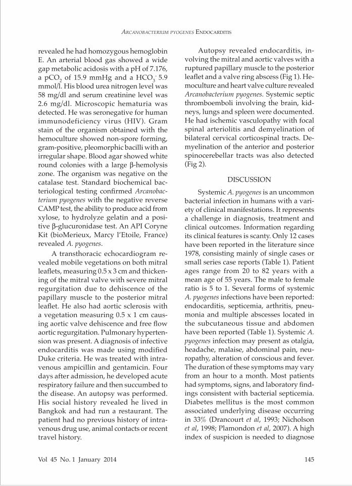
Autopsy revealed endocarditis, in-volving the mitral and aortic valves with a ruptured papillary muscle to the posterior leaflet and a valve ring abscess (Fig 1). He-moculture and heart valve culture revealed Arcanobacterium pyogenes. Systemic septic thromboemboli involving the brain, kid-neys, lungs and spleen were documented. He had ischemic vasculopathy with focal spinal arteriolitis and demyelination of bilateral cervical corticospinal tracts. De-myelination of the anterior and posterior spinocerebellar tracts was also detected (Fig 2).
DISCUSSION
Systemic A. pyogenes is an uncommon bacterial infection in humans with a vari-ety of clinical manifestations. It represents a challenge in diagnosis, treatment and clinical outcomes. Information regarding its clinical features is scanty. Only 12 cases have been reported in the literature since 1978, consisting mainly of single cases or small series case reports (Table 1). Patient ages range from 20 to 82 years with a mean age of 55 years. The male to female ratio is 5 to 1. Several forms of systemic A. pyogenes infections have been reported: endocarditis, septicemia, arthritis, pneu-monia and multiple abscesses located in the subcutaneous tissue and abdomen have been reported (Table 1). Systemic A. pyogenes infection may present as otalgia, headache, malaise, abdominal pain, neu-ropathy, alteration of conscious and fever. The duration of these symptoms may vary from an hour to a month. Most patients had symptoms, signs, and laboratory find-ings consistent with bacterial septicemia. Diabetes mellitus is the most common associated underlying disease occurring in 33% (Drancourt et al, 1993; Nicholson et al, 1998; Plamondon et al, 2007). A high index of suspicion is needed to diagnose

SoutheaSt aSian J trop Med public health
146 Vol 45 No. 1 January 2014
Fig 1–Multiple vegetations on aortic valves (asterisk), mitral valve leaflet (arrowhead) and ruptured chordae tendineae (arrow) (A). The tricuspid valve was intact (B). Acute inflammatory cells and multiple bacterial organisms on the endocardial
surface of the aortic valve (C, x40). Gram stain showing pleomorphic gram-positive bacilli (D, x1,000).
Fig 2–A section of the brain showing a brain abscess with men-ingitis (A, x40). A section of the spinal cord showing focal loss of myelinated fibers (decreased staining) in bilateral corticospinal tracts without degeneration of anterior horn cells (B, x100). Focal spinal arteriolitis (C, x200) and vacuolar myelopathy (D, x200) are seen.
this disease, but it should be considered in a patient with a history of animal contacts. A case of fatal endocarditis in a patient with no animal contacts has been reported (Plamondon et al, 2007). In our case, the patient had no history of animal contacts.
Endocarditis is a seri-ous infection characterized by colonization or invasion of the heart valves or the mural endocardium by a microbe. This leads to the formation of vegetations composed of thrombotic debris and organisms, often associated with destruction of the underlying cardiac tissues (Halder and O’Gara, 2011). The diagnosis of bacterial endocarditis is made using modified Duke criteria, and is confirmed by demonstration of the organism with tissue reac-tion on the valvular leaf-lets. Endocarditis is most frequently caused by bac-teria. Streptococcus viridans is the most common caus-ative organism (Halder and O’Gara, 2011). A. pyogenes infection is an uncommon cause of endocarditis. There are three reported cases of endocarditis caused by A. pyogenes in the literature: one with subacute and two with acute infection (Jootar et al, 1978; Reddy et al, 1997; Plamondon et al, 2007). The patient we report here had acute endocarditis.
ArcAnobActerium pyogenes endocarditiS
Vol 45 No. 1 January 2014 147
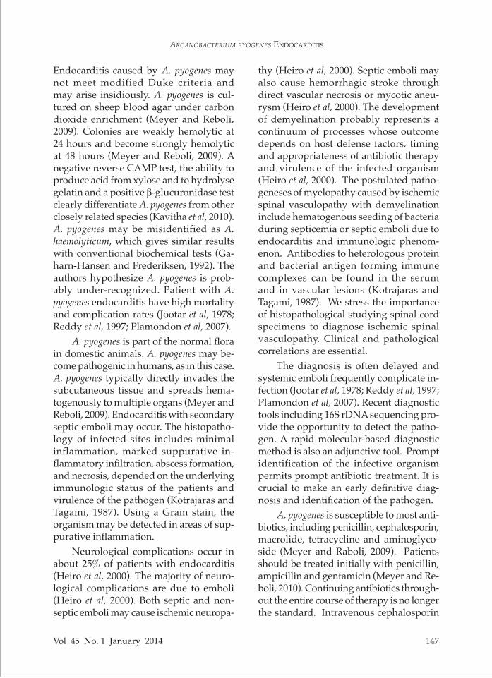
Endocarditis caused by A. pyogenes may not meet modified Duke criteria and may arise insidiously. A. pyogenes is cul-tured on sheep blood agar under carbon dioxide enrichment (Meyer and Reboli, 2009). Colonies are weakly hemolytic at 24 hours and become strongly hemolytic at 48 hours (Meyer and Reboli, 2009). A negative reverse CAMP test, the ability to produce acid from xylose and to hydrolyse gelatin and a positive b-glucuronidase test clearly differentiate A. pyogenes from other closely related species (Kavitha et al, 2010). A. pyogenes may be misidentified as A. haemolyticum, which gives similar results with conventional biochemical tests (Ga-harn-Hansen and Frederiksen, 1992). The authors hypothesize A. pyogenes is prob-ably under-recognized. Patient with A. pyogenes endocarditis have high mortality and complication rates (Jootar et al, 1978; Reddy et al, 1997; Plamondon et al, 2007).
A. pyogenes is part of the normal flora in domestic animals. A. pyogenes may be-come pathogenic in humans, as in this case. A. pyogenes typically directly invades the subcutaneous tissue and spreads hema-togenously to multiple organs (Meyer and Reboli, 2009). Endocarditis with secondary septic emboli may occur. The histopatho- logy of infected sites includes minimal inflammation, marked suppurative in-flammatory infiltration, abscess formation, and necrosis, depended on the underlying immunologic status of the patients and virulence of the pathogen (Kotrajaras and Tagami, 1987). Using a Gram stain, the organism may be detected in areas of sup-purative inflammation.
Neurological complications occur in about 25% of patients with endocarditis (Heiro et al, 2000). The majority of neuro-logical complications are due to emboli (Heiro et al, 2000). Both septic and non-septic emboli may cause ischemic neuropa-
thy (Heiro et al, 2000). Septic emboli may also cause hemorrhagic stroke through direct vascular necrosis or mycotic aneu-rysm (Heiro et al, 2000). The development of demyelination probably represents a continuum of processes whose outcome depends on host defense factors, timing and appropriateness of antibiotic therapy and virulence of the infected organism (Heiro et al, 2000). The postulated patho-geneses of myelopathy caused by ischemic spinal vasculopathy with demyelination include hematogenous seeding of bacteria during septicemia or septic emboli due to endocarditis and immunologic phenom-enon. Antibodies to heterologous protein and bacterial antigen forming immune complexes can be found in the serum and in vascular lesions (Kotrajaras and Tagami, 1987). We stress the importance of histopathological studying spinal cord specimens to diagnose ischemic spinal vasculopathy. Clinical and pathological correlations are essential.
The diagnosis is often delayed and systemic emboli frequently complicate in-fection (Jootar et al, 1978; Reddy et al, 1997; Plamondon et al, 2007). Recent diagnostic tools including 16S rDNA sequencing pro-vide the opportunity to detect the patho-gen. A rapid molecular-based diagnostic method is also an adjunctive tool. Prompt identification of the infective organism permits prompt antibiotic treatment. It is crucial to make an early definitive diag-nosis and identification of the pathogen.
A. pyogenes is susceptible to most anti-biotics, including penicillin, cephalosporin, macrolide, tetracycline and aminoglyco-side (Meyer and Raboli, 2009). Patients should be treated initially with penicillin, ampicillin and gentamicin (Meyer and Re-boli, 2010). Continuing antibiotics through-out the entire course of therapy is no longer the standard. Intravenous cephalosporin

SoutheaSt aSian J trop Med public health
148 Vol 45 No. 1 January 2014
is recommended as alternative treatment of systemic A. pyogenes infection (Meyer and Raboli, 2009). However, no patient has been successfully treated for A. pyogenes endocarditis (Jootar et al, 1978; Reddy et al, 1997; Plamondon et al, 2007).
ACKNOWLEDGEMENTS
The authors thank the attending physi-cians for managing the reported case.
REFERENCESDrancourt M, Oules O, Bouche V, Peloux Y.
Two cases of Actinomyces pyogenes infection in humans. Eur J Clin Microbiol Infect Dis 1993; 12: 55-7.
Gahrn-Hansen B, Frederiksen W. Human infec-tions with Actinomyces pyogenes (Corynebac-terium pyogenes). Diagn Microbiol Infect Dis 1992; 15: 349-54.
Halder SM, O’Gara P. Infective endocarditis. In: Fuster V, Walsh RA, Harrington RA, eds. Hurst’s the heart. 13th ed. New York: McGrrawHill, 2011: 1940-69.
Heiro M, Nikoskelainen J, Engblom E, Kotilain-en E, Marttila R, Kotilainen P. Neurologic manifestations of infective endocarditis: a 17-year experience in a teaching hospital in Finland. Arch Intern Med 2000; 160: 2781-7.
Hermida Amejeiras A, Romero Jung P, Cabar-cos Ortiz De Barrón A, Treviño Castallo M. One case of pneumonia with Arcano-bacterium pyogenes. An Med Interna 2004; 21: 334-6.
Ide L, Decostere A, Stuer A, et al. Arcanobacte-rium pyogenes spondylodiscitis in a vet-erinary surgeon: a plea for cooperation between medical and veterinary microbi-ologists in identification of causal agents of zoonotic infections. Clin Microbiol Newsl 2006; 28: 163-7.
Jootar P, Gherunpong V, Saitanu K. Corynebacte-rium pyogenes endocarditis report of a case with necropsy and review of the literature. J Med Assoc Thai 1978; 61: 596-601.
Kavitha K, Latha R, Udayashankar C, Jayanthi K, Oudeacoumar P. Three cases of Arcano-bacterium pyogenes –associated soft tissue infection. J Med Microbiol 2010; 59: 736-9.
Kotrajaras R, Tagami H. Corynebacterium pyo-genes. Its pathogenic mechanism in epi-demic leg ulcers in Thailand. Int J Dermatol 1987; 26: 45-50.
Levy CE, Pedro RJ, Von Nowakonski A, Hol-anda LM, Brocchi M, Ramo MC. Arcano-bacterium pyogenes sepsis in farmer, Brazil. Emerg Infect Dis 2009; 15: 1131-2.
Lipsky BA, Goldberger AC, Tompkins LS, Plorde JJ. Infections caused by nondiph-theria corynebacteria. Rev Infect Dis 1982; 4: 1220-35.
Meyer DK, Reboli AC. Other Coryneform bacteria and Rhodococci. In: Mandell GL, Bennett JE, Dolin R, eds. Mandell, Douglas and Bennett’s principles and practice of infectious diseases. 7th ed. Philadelphia: Saunders Elsevier, 2009: 2700-1.
Nicholson P, Kiely P, Street J, Mahalingum K. Septic arthritis due to Actinomyces pyo-genes. Injury 1998; 29: 640-2.
Plamondon M, Martinez G, Raynal L, Touchette M, Valiquette L. A fatal case of Arcanobac-terium pyogenes endocarditis in a man with no identified animal contact: case report and review of the literature. Eur J Clin Microbiol Infect Dis 2007; 26: 663-6.
Ramos CP, Foster G, Collins MD. Phylogenetic analysis of the genus Actinomyces based on 16S rRNA gene sequences: descrip-tion of Arcanobacterium phocae sp. nov., Arcanobacterium bernardiae comb. nov., and Arcanobacterium pyogenes comb. nov. Int J Syst Bacteriol 1997; 47: 46-53.
Reddy I, Ferguson DA Jr, Sarubbi FA. Endo-carditis due to Actinomyces pyogenes. Clin Infect Dis 1997; 25: 1476-7.
Yeruham I, Orgad U, Avidar Y, Elad D. Pituitary abscess and high urea concentration as causes of neurological signs in a cow: A ret-rospective analysis and literature review. Revue Méd Vét 2002; 12: 829-31.