Embed Size (px)
Citation preview
Arabidopsis CALCIUM-DEPENDENT PROTEIN KINASE8 andCATALASE3 Function in Abscisic Acid-MediatedSignaling and H2O2 Homeostasis in Stomatal GuardCells under Drought StressOPEN
Jun-Jie Zou,1,2 Xi-Dong Li,1 Disna Ratnasekera,1,3 Cun Wang,1 Wen-Xin Liu, Lian-Fen Song, Wen-Zheng Zhang,4
and Wei-Hua Wu5
State Key Laboratory of Plant Physiology and Biochemistry, College of Biological Sciences, National Plant Gene Research Centre(Beijing), China Agricultural University, Beijing 100193, China
ORCID IDs: 0000-0002-4824-400X (J.-J.Z.); 0000-0002-0077-4466 (X.-D.L.); 0000-0003-3176-6888 (D.R.); 0000-0003-0583-9510(W.-X.L.); 0000-0001-5497-5768 (L.-F.S.); 0000-0002-0642-5523 (W.-H.W.)
Drought is a major threat to plant growth and crop productivity. Calcium-dependent protein kinases (CDPKs, CPKs) arebelieved to play important roles in plant responses to drought stress. Here, we report that Arabidopsis thaliana CPK8functions in abscisic acid (ABA)- and Ca2+-mediated plant responses to drought stress. The cpk8 mutant was more sensitiveto drought stress than wild-type plants, while the transgenic plants overexpressing CPK8 showed enhanced tolerance todrought stress compared with wild-type plants. ABA-, H2O2-, and Ca2+-induced stomatal closing were impaired in cpk8mutants. Arabidopsis CATALASE3 (CAT3) was identified as a CPK8-interacting protein, confirmed by yeast two-hybrid,coimmunoprecipitation, and bimolecular fluorescence complementation assays. CPK8 can phosphorylate CAT3 at Ser-261and regulate its activity. Both cpk8 and cat3 plants showed lower catalase activity and higher accumulation of H2O2 comparedwith wild-type plants. The cat3mutant displayed a similar drought stress-sensitive phenotype as cpk8mutant. Moreover, ABAand Ca2+ inhibition of inward K+ currents were diminished in guard cells of cpk8 and cat3 mutants. Together, these resultsdemonstrated that CPK8 functions in ABA-mediated stomatal regulation in responses to drought stress through regulation ofCAT3 activity.
INTRODUCTION
In terrestrial plants, calcium plays a vital role as a secondmessenger in plant responses to various environmental stimuli(White and Broadley, 2003; Kudla et al., 2010). Calcium signalstriggered by different stimuli are recognized by specific calciumsensors in living plant cells. There are three major familiesof calcium sensors in terrestrial plants, including calmodulins(CaMs) and CaM-like proteins (Zielinski, 1998; McCormacket al., 2005), calcineurin B-like (CBL) proteins (Kolukisaogluet al., 2004; Luan, 2009; Weinl and Kudla, 2009), and calcium-dependent protein kinases (CDPKs or CPKs) (Harmon et al.,2000; Cheng et al., 2002; Harper and Harmon, 2005). CaMs andCBLs do not have enzymatic activities and so do not directly
transmit the Ca2+ signals (Luan et al., 2002). By contrast,CDPKs, which harbor a CaM-like domain as well as a catalyticSer/Thr kinase domain, can sense calcium signals and directlymediate a variety of cellular responses (Harmon et al., 2000;Cheng et al., 2002).CDPKs are encoded by multigene families and found only in
plants and some protists (Harmon et al., 2000; Cheng et al.,2002). The CDPKs exhibit different subcellular localizations, in-cluding cytosol, nucleus, the plasma membrane, endoplasmicreticulum, peroxisome, mitochondrial outer membrane, and oilbodies (Harper et al., 2004), likely reflecting diverse functions.CDPKs are believed to be important regulators in multiple plantsignal transduction pathways downstream of cytosolic Ca2+
([Ca2+]cyt) elevations (Harmon et al., 2000; Cheng et al., 2002;Ludwig et al., 2004; Boudsocq and Sheen, 2013; Romeis andHerde, 2014).The Arabidopsis thaliana genome encodes 34 CDPKs, and
a number of studies have shown that CDPKs are involvedin plant development and in responses to biotic and abioticstresses. Arabidopsis CDPKs can act as positive or negativeregulators in response to abiotic stress and abscisic acid (ABA)signaling. Arabidopsis CPK32 has been characterized as anABA signaling component that regulates ABA-responsive geneexpression via ABF4 (Choi et al., 2005). Arabidopsis CPK3 andCPK6 function as positive transducers in plant ion channelregulation and stomatal ABA signaling (Mori et al., 2006), as wellas salt and/or drought stress(es) (Mehlmer et al., 2010; Xu et al.,
1 These authors contributed equally to this work.2 Current address: Key Laboratory of Plant Molecular Physiology, Instituteof Botany, Chinese Academy of Sciences, Beijing 100093, China.3 Current address: Department of Agricultural Biology, Faculty of Agri-culture, University of Ruhuna, Matara 81100, Sri Lanka.4 Current address: The Samuel Roberts Noble Foundation, 2510 SamNoble Parkway, Ardmore, OK 73401.5 Address correspondence to [email protected] author responsible for distribution of materials integral to the findingspresented in this article in accordance with the policy described in theInstructions for Authors (www.plantcell.org) is: Wei-Hua Wu ([email protected]).OPENArticles can be viewed online without a subscription.www.plantcell.org/cgi/doi/10.1105/tpc.15.00144
The Plant Cell, Vol. 27: 1445–1460, May 2015, www.plantcell.org ã 2015 American Society of Plant Biologists. All rights reserved.
2010). Arabidopsis CPK4 and CPK11 have been identified asimportant positive regulators in CDPK/calcium-mediated ABAsignaling (Zhu et al., 2007). By contrast, Arabidopsis CPK21functions as a negative regulator in abiotic stress signal trans-duction (Franz et al., 2011). Furthermore, CPK12 serves asa negative ABA-signaling regulator in seed germination andpostgermination growth, which is different from the function ofits homologs, CPK4 and CPK11 (Zhao et al., 2011). In additionto abiotic stress, some Arabidopsis CDPKs have been reportedto be involved in the plant innate immune response (CPKs 4,5, 6, and 11 [Boudsocq et al., 2010], CPK1 [Coca and SanSegundo, 2010], and CPK5 [Dubiella et al., 2013]), herbivory-induced signaling network (CPK3 and CPK13; Kanchiswamyet al., 2010), regulation of pollen tube growth (CPK17 andCPK34 [Myers et al., 2009], CPK11 and CPK24 [Zhao et al.,2013], CPK2 and CPK20 [Gutermuth et al., 2013], and CPK32[Zhou et al., 2014]), and stem elongation and vascular de-velopment (CPK28; Matschi et al., 2013).
We previously demonstrated that Arabidopsis CPK10 func-tions in ABA- and Ca2+-mediated stomatal regulation in re-sponse to drought stress (Zou et al., 2010). Here, we report thatCPK8 (At5g19450) as well as its interacting protein CATALASE3(CAT3; At1g20620) play important roles in ABA- and H2O2-mediated signal transduction and in maintaining H2O2 homeo-stasis in response to drought stress.
RESULTS
CPK8 Acts as a Positive Regulator in Response to DroughtStress and Involving in ABA- and H2O2-MediatedStomatal Movement
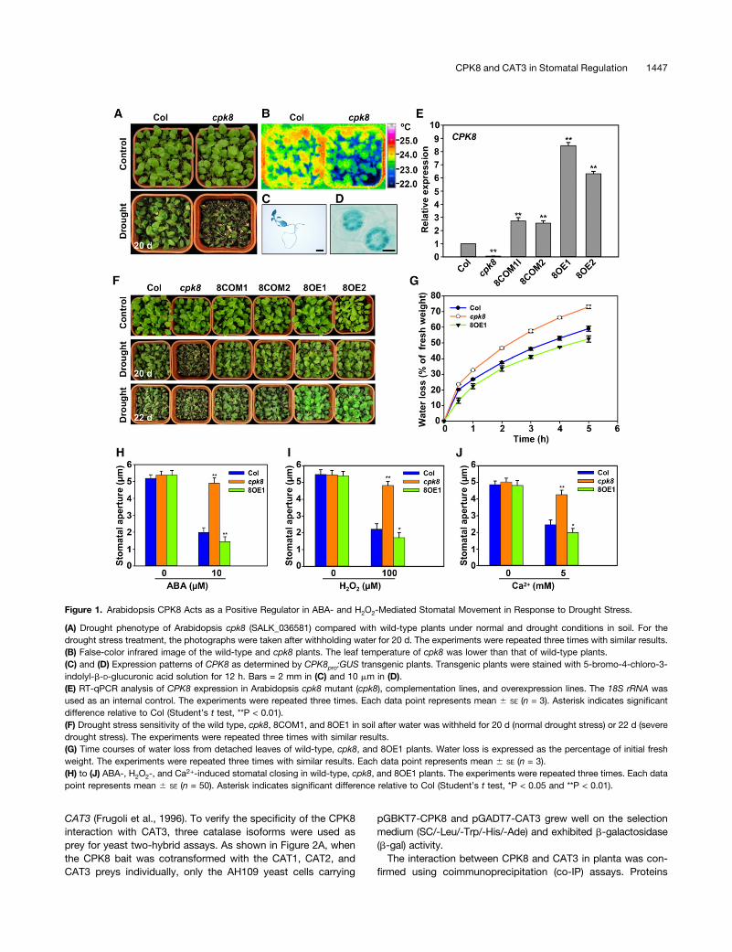
After a 20-d period of drought stress, the CPK8 T-DNA insertionmutant plants (cpk8, SALK_036581; Supplemental Figure 1A)showed more sensitive phenotype compared with wild-typeplants (Figure 1A), while there was no obvious morphologicaldifference observed between cpk8 mutant and wild-type plantsunder normal growth condition (Figure 1A). When 3-week-oldplants grown under normal conditions were subjected todrought stress for about 1 week, the leaf temperature of cpk8mutant plants was lower than that in the wild-type plants (Figure1B), suggesting that water loss of the cpk8 plants may be fasterthan that of the wild-type plants. b-Glucuronidase (GUS) stain-ing of CPK8pro:GUS transgenic plants showed that the CPK8promoter drove expression in leaves and roots (Figure 1C) andparticularly in stomatal guard cells (Figure 1D), suggestinga potential role of CPK8 in regulation of stomatal movement. Theresults of reverse transcriptase quantitative PCR (RT-qPCR)showed that CPK8 transcript accumulation was induced bydrought (Supplemental Figure 1B), ABA (Supplemental Figure1C), and H2O2 (Supplemental Figure 1D) treatments.
To further test whether the drought sensitivity of cpk8 plantsresulted from CPK8 disruption, CPK8 overexpression lines(ecotype Columbia [Col] + CPK8; 8OE) and complementationlines (cpk8 + CPK8; 8COM) were generated. The RT-qPCRresults showed that transcription of CPK8 was disrupted inthe cpk8 homozygous plants (Figure 1E). When subjected to
drought stress for 20 d, the cpk8 mutant plants were moresensitive to drought stress, whereas the CPK8 overexpressionlines (8OE1 and 8OE2) showed enhanced tolerance to droughtstress compared with wild-type plants (Figure 1F). The over-expression lines (8OE1 and 8OE2) grew well and the rosetteleaves remained green even after 22 d without watering (Figure1F). The cpk8 complementation lines (8COM1 and 8COM2)showed a similar phenotype as wild-type plants after thedrought stress treatment (Figure 1F). These results indicate thatdisruption of CPK8 expression increases plant sensitivity todrought stress and that increased CPK8 expression enhancesplant tolerance to drought stress.The stomatal pores control most of the water loss via tran-
spiration to the atmosphere (Schroeder et al., 2001). To test thehypothesis that CPK8 may be involved in regulation of stomatalmovements under drought stress, water loss assays of de-tached leaves were conducted. As shown in Figure 1G, waterloss of the detached rosette leaves of cpk8 plants was muchfaster than that of wild-type leaves, while water loss of the CPK8overexpression (8OE1) leaves was slower than that of wild-typeleaves under dehydration conditions.H2O2 is one of the major reactive oxygen species (ROS) and
serves as an important messenger in ABA-regulated stomatalmovement (Pei et al., 2000; Murata et al., 2001; Zhang et al.,2001a; Kwak et al., 2003; Miao et al., 2006). Accumulation ofH2O2 in stomatal guard cell can cause [Ca2+]cyt elevationsand result in stomatal closure (Pei et al., 2000; Murata et al.,2001; Kwak et al., 2003). We further hypothesized that CPK8may serve as a calcium sensor and function in ABA- andH2O2-mediated regulation of stomatal movement. To testthis hypothesis, stomatal aperture assays were conducted toexamine stomatal responses of different lines to ABA, H2O2,and Ca2+. As shown in Figures 1H to 1J, stomatal closureinduced by ABA (Figure 1H), H2O2 (Figure 1I), or Ca2+ (Figure1J) was impaired in the cpk8 mutant plants. These resultssuggest that CPK8 may serve as an important signalingcomponent to mediate ABA as well as H2O2 and Ca2+ signalsin regulation of stomatal movement.
Identification of CAT3 as a CPK8-Interacting Protein
We next undertook identification of CPK8-interacting protein(s)to gain insight into CPK8 might function in ABA and H2O2
signaling. In previous studies, a number of experimental ap-proaches had been employed to identify CDPK-interactingproteins (Patharkar and Cushman, 2000; Lee et al., 2003; Shaoand Harmon, 2003; Rodriguez Milla et al., 2006; Böhmer andRomeis, 2007; Vlad et al., 2008; Uno et al., 2009). Among thesedifferent approaches, yeast two-hybrid screening with baitscontaining only the kinase domain of the protein had beendemonstrated to be successful for identification of CDPK sub-strates, such as for ice plant (Mesembryanthemum crystallinum)CPK1 (Patharkar and Cushman, 2000) and tobacco (Nicotianatabacum) CDPK1 (Lee et al., 2003). Here, using the kinase do-main of CPK8 as bait, a catalase protein encoded by At1g20620(CAT3) was identified as a potential CPK8-interacting protein(Figure 2A). There are three catalase genes in the Arabidopsisgenome, including CAT1 (At1g20630), CAT2 (At4g35090), and
1446 The Plant Cell
CAT3 (Frugoli et al., 1996). To verify the specificity of the CPK8interaction with CAT3, three catalase isoforms were used asprey for yeast two-hybrid assays. As shown in Figure 2A, whenthe CPK8 bait was cotransformed with the CAT1, CAT2, andCAT3 preys individually, only the AH109 yeast cells carrying
pGBKT7-CPK8 and pGADT7-CAT3 grew well on the selectionmedium (SC/-Leu/-Trp/-His/-Ade) and exhibited b-galactosidase(b-gal) activity.The interaction between CPK8 and CAT3 in planta was con-
firmed using coimmunoprecipitation (co-IP) assays. Proteins
Figure 1. Arabidopsis CPK8 Acts as a Positive Regulator in ABA- and H2O2-Mediated Stomatal Movement in Response to Drought Stress.
(A) Drought phenotype of Arabidopsis cpk8 (SALK_036581) compared with wild-type plants under normal and drought conditions in soil. For thedrought stress treatment, the photographs were taken after withholding water for 20 d. The experiments were repeated three times with similar results.(B) False-color infrared image of the wild-type and cpk8 plants. The leaf temperature of cpk8 was lower than that of wild-type plants.(C) and (D) Expression patterns of CPK8 as determined by CPK8pro:GUS transgenic plants. Transgenic plants were stained with 5-bromo-4-chloro-3-indolyl-b-D-glucuronic acid solution for 12 h. Bars = 2 mm in (C) and 10 mm in (D).(E) RT-qPCR analysis of CPK8 expression in Arabidopsis cpk8 mutant (cpk8), complementation lines, and overexpression lines. The 18S rRNA wasused as an internal control. The experiments were repeated three times. Each data point represents mean 6 SE (n = 3). Asterisk indicates significantdifference relative to Col (Student’s t test, **P < 0.01).(F) Drought stress sensitivity of the wild type, cpk8, 8COM1, and 8OE1 in soil after water was withheld for 20 d (normal drought stress) or 22 d (severedrought stress). The experiments were repeated three times with similar results.(G) Time courses of water loss from detached leaves of wild-type, cpk8, and 8OE1 plants. Water loss is expressed as the percentage of initial freshweight. The experiments were repeated three times with similar results. Each data point represents mean 6 SE (n = 3).(H) to (J) ABA-, H2O2-, and Ca2+-induced stomatal closing in wild-type, cpk8, and 8OE1 plants. The experiments were repeated three times. Each datapoint represents mean 6 SE (n = 50). Asterisk indicates significant difference relative to Col (Student’s t test, *P < 0.05 and **P < 0.01).
CPK8 and CAT3 in Stomatal Regulation 1447
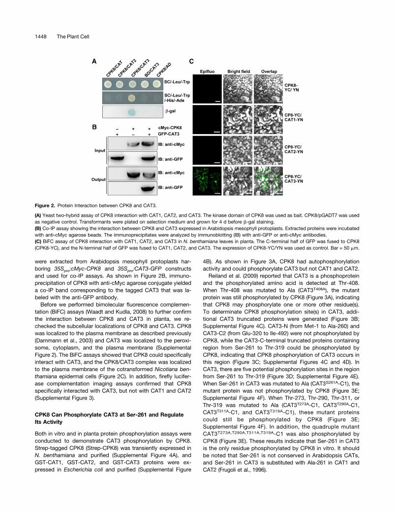
were extracted from Arabidopsis mesophyll protoplasts har-boring 35Spro:cMyc-CPK8 and 35Spro:CAT3-GFP constructsand used for co-IP assays. As shown in Figure 2B, immuno-precipitation of CPK8 with anti-cMyc agarose conjugate yieldeda co-IP band corresponding to the tagged CAT3 that was la-beled with the anti-GFP antibody.
Before we performed bimolecular fluorescence complemen-tation (BiFC) assays (Waadt and Kudla, 2008) to further confirmthe interaction between CPK8 and CAT3 in planta, we re-checked the subcellular localizations of CPK8 and CAT3. CPK8was localized to the plasma membrane as described previously(Dammann et al., 2003) and CAT3 was localized to the peroxi-some, cytoplasm, and the plasma membrane (SupplementalFigure 2). The BiFC assays showed that CPK8 could specificallyinteract with CAT3, and the CPK8/CAT3 complex was localizedto the plasma membrane of the cotransformed Nicotiana ben-thamiana epidermal cells (Figure 2C). In addition, firefly lucifer-ase complementation imaging assays confirmed that CPK8specifically interacted with CAT3, but not with CAT1 and CAT2(Supplemental Figure 3).
CPK8 Can Phosphorylate CAT3 at Ser-261 and RegulateIts Activity
Both in vitro and in planta protein phosphorylation assays wereconducted to demonstrate CAT3 phosphorylation by CPK8.Strep-tagged CPK8 (Strep-CPK8) was transiently expressed inN. benthamiana and purified (Supplemental Figure 4A), andGST-CAT1, GST-CAT2, and GST-CAT3 proteins were ex-pressed in Escherichia coli and purified (Supplemental Figure
4B). As shown in Figure 3A, CPK8 had autophosphorylationactivity and could phosphorylate CAT3 but not CAT1 and CAT2.Reiland et al. (2009) reported that CAT3 is a phosphoprotein
and the phosphorylated amino acid is detected at Thr-408.When Thr-408 was mutated to Ala (CAT3T408A), the mutantprotein was still phosphorylated by CPK8 (Figure 3A), indicatingthat CPK8 may phosphorylate one or more other residue(s).To determinate CPK8 phosphorylation site(s) in CAT3, addi-tional CAT3 truncated proteins were generated (Figure 3B;Supplemental Figure 4C). CAT3-N (from Met-1 to Ala-260) andCAT3-C2 (from Glu-320 to Ile-492) were not phosphorylated byCPK8, while the CAT3-C-terminal truncated proteins containingregion from Ser-261 to Thr-319 could be phosphorylated byCPK8, indicating that CPK8 phosphorylation of CAT3 occurs inthis region (Figure 3C; Supplemental Figures 4C and 4D). InCAT3, there are five potential phosphorylation sites in the regionfrom Ser-261 to Thr-319 (Figure 3D; Supplemental Figure 4E).When Ser-261 in CAT3 was mutated to Ala (CAT3S261A-C1), themutant protein was not phosphorylated by CPK8 (Figure 3E;Supplemental Figure 4F). When Thr-273, Thr-290, Thr-311, orThr-319 was mutated to Ala (CAT3T273A-C1, CAT3T290A-C1,CAT3T311A-C1, and CAT3T319A-C1), these mutant proteinscould still be phosphorylated by CPK8 (Figure 3E;Supplemental Figure 4F). In addition, the quadruple mutantCAT3T273A,T290A,T311A,T319A-C1 was also phosphorylated byCPK8 (Figure 3E). These results indicate that Ser-261 in CAT3is the only residue phosphorylated by CPK8 in vitro. It shouldbe noted that Ser-261 is not conserved in Arabidopsis CATs,and Ser-261 in CAT3 is substituted with Ala-261 in CAT1 andCAT2 (Frugoli et al., 1996).
Figure 2. Protein Interaction between CPK8 and CAT3.
(A) Yeast two-hybrid assay of CPK8 interaction with CAT1, CAT2, and CAT3. The kinase domain of CPK8 was used as bait. CPK8/pGADT7 was usedas negative control. Transformants were plated on selection medium and grown for 4 d before b-gal staining.(B) Co-IP assay showing the interaction between CPK8 and CAT3 expressed in Arabidopsis mesophyll protoplasts. Extracted proteins were incubatedwith anti-cMyc agarose beads. The immunoprecipitates were analyzed by immunoblotting (IB) with anti-GFP or anti-cMyc antibodies.(C) BiFC assay of CPK8 interaction with CAT1, CAT2, and CAT3 in N. benthamiana leaves in planta. The C-terminal half of GFP was fused to CPK8(CPK8-YC), and the N-terminal half of GFP was fused to CAT1, CAT2, and CAT3. The expression of CPK8-YC/YN was used as control. Bar = 50 mm.
1448 The Plant Cell
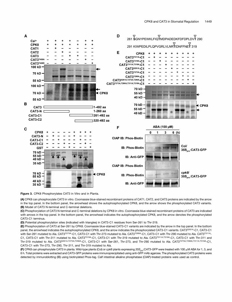
Figure 3. CPK8 Phosphorylates CAT3 in Vitro and in Planta.
(A) CPK8 can phosphorylate CAT3 in vitro. Coomassie blue-stained recombinant proteins of CAT1, CAT2, and CAT3 proteins are indicated by the arrowin the top panel. In the bottom panel, the arrowhead shows the autophosphorylated CPK8, and the arrow shows the phosphorylated CAT3 variants.(B) Model of CAT3 N-terminal and C-terminal deletions.(C) Phosphorylation of CAT3 N-terminal and C-terminal deletions by CPK8 in vitro. Coomassie blue-stained recombinant proteins of CAT3 are indicatedwith arrows in the top panel. In the bottom panel, the arrowhead indicates the autophosphorylated CPK8, and the arrow denotes the phosphorylatedCAT3-C1 terminus.(D) Potential phosphorylation sites (indicated with triangles) in CAT3-C1 residues from Ser-261 to Thr-319.(E) Phosphorylation of CAT3 at Ser-261 by CPK8. Coomassie blue-stained CAT3-C1 variants are indicated by the arrow in the top panel. In the bottompanel, the arrowhead indicates the autophosphorylated CPK8, and the arrow indicates the phosphorylated CAT3-C1 variants. CAT3S261A-C1, CAT3-C1with Ser-261 mutated to Ala. CAT3T273A-C1, CAT3-C1 with Thr-273 mutated to Ala. CAT3T290A-C1, CAT3-C1 with Thr-290 mutated to Ala. CAT3T311A-C1, CAT3-C1 with Thr-311 mutated to Ala. CAT3T319A-C1, CAT3-C1 with Thr-319 mutated to Ala. CAT3T311A,T319A-C1, CAT3-C1 with Thr-311 andThr-319 mutated to Ala. CAT3S261A,T273A,T290A-C1, CAT3-C1 with Ser-261, Thr-273, and Thr-290 mutated to Ala. CAT3T273A,T290A,T311A,T319A-C1,CAT3-C1 with Thr-273, Thr-290, Thr-311, and Thr-319 mutated to Ala.(F) CPK8 can phosphorylate CAT3 in planta. Wild-type plants (Col) or cpk8 plants expressing 35Spro:CAT3-GFPwere treated with 100 mM ABA for 1, 3, and6 h. Total proteins were extracted and CAT3-GFP proteins were immunoprecipitated using anti-GFP mAb agarose. The phosphorylated CAT3 proteins weredetected by immunoblotting (IB) using biotinylated Phos-tag. Calf intestinal alkaline phosphatase (CIAP)-treated proteins were used as control.
CPK8 and CAT3 in Stomatal Regulation 1449
To determine whether CAT3 is phosphorylated by CPK8 inplanta, transgenic lines (Col/35Spro:CAT3-GFP and cpk8/35Spro:CAT3-GFP) were generated. The CAT3 phosphorylation levelwas detected using Phos-Biotin technology as described pre-viously (Kinoshita-Kikuta et al., 2007). Under normal conditions,CAT3 phosphorylation was very low in Col/35Spro:CAT3-GFPand cpk8/35Spro:CAT3-GFP plants (Figure 3F). After ABA treat-ment, CAT3 phosphorylation was obviously enhanced in Col/35Spro:CAT3-GFP (Figure 3F). However, this ABA-induced CAT3phosphorylation was weakened in cpk8/35Spro:CAT3-GFP, in-dicating that ABA-induced CAT3 phosphorylation is CPK8 de-pendent (Figure 3F).
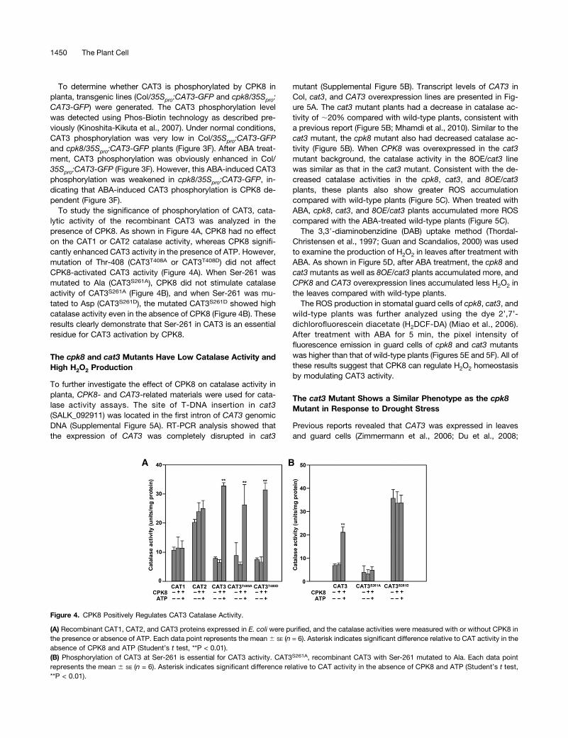
To study the significance of phosphorylation of CAT3, cata-lytic activity of the recombinant CAT3 was analyzed in thepresence of CPK8. As shown in Figure 4A, CPK8 had no effecton the CAT1 or CAT2 catalase activity, whereas CPK8 signifi-cantly enhanced CAT3 activity in the presence of ATP. However,mutation of Thr-408 (CAT3T408A or CAT3T408D) did not affectCPK8-activated CAT3 activity (Figure 4A). When Ser-261 wasmutated to Ala (CAT3S261A), CPK8 did not stimulate catalaseactivity of CAT3S261A (Figure 4B), and when Ser-261 was mu-tated to Asp (CAT3S261D), the mutated CAT3S261D showed highcatalase activity even in the absence of CPK8 (Figure 4B). Theseresults clearly demonstrate that Ser-261 in CAT3 is an essentialresidue for CAT3 activation by CPK8.
The cpk8 and cat3 Mutants Have Low Catalase Activity andHigh H2O2 Production
To further investigate the effect of CPK8 on catalase activity inplanta, CPK8- and CAT3-related materials were used for cata-lase activity assays. The site of T-DNA insertion in cat3(SALK_092911) was located in the first intron of CAT3 genomicDNA (Supplemental Figure 5A). RT-PCR analysis showed thatthe expression of CAT3 was completely disrupted in cat3
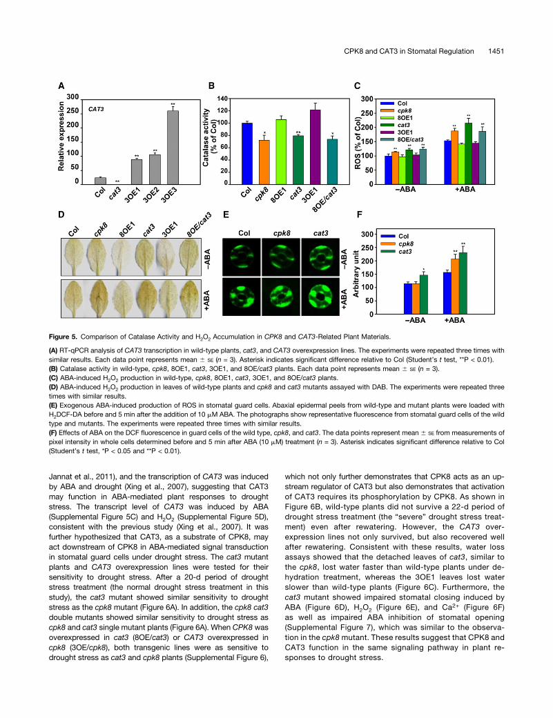
mutant (Supplemental Figure 5B). Transcript levels of CAT3 inCol, cat3, and CAT3 overexpression lines are presented in Fig-ure 5A. The cat3 mutant plants had a decrease in catalase ac-tivity of ;20% compared with wild-type plants, consistent witha previous report (Figure 5B; Mhamdi et al., 2010). Similar to thecat3 mutant, the cpk8 mutant also had decreased catalase ac-tivity (Figure 5B). When CPK8 was overexpressed in the cat3mutant background, the catalase activity in the 8OE/cat3 linewas similar as that in the cat3 mutant. Consistent with the de-creased catalase activities in the cpk8, cat3, and 8OE/cat3plants, these plants also show greater ROS accumulationcompared with wild-type plants (Figure 5C). When treated withABA, cpk8, cat3, and 8OE/cat3 plants accumulated more ROScompared with the ABA-treated wild-type plants (Figure 5C).The 3,39-diaminobenzidine (DAB) uptake method (Thordal-
Christensen et al., 1997; Guan and Scandalios, 2000) was usedto examine the production of H2O2 in leaves after treatment withABA. As shown in Figure 5D, after ABA treatment, the cpk8 andcat3mutants as well as 8OE/cat3 plants accumulated more, andCPK8 and CAT3 overexpression lines accumulated less H2O2 inthe leaves compared with wild-type plants.The ROS production in stomatal guard cells of cpk8, cat3, and
wild-type plants was further analyzed using the dye 2’,7’-dichlorofluorescein diacetate (H2DCF-DA) (Miao et al., 2006).After treatment with ABA for 5 min, the pixel intensity offluorescence emission in guard cells of cpk8 and cat3 mutantswas higher than that of wild-type plants (Figures 5E and 5F). All ofthese results suggest that CPK8 can regulate H2O2 homeostasisby modulating CAT3 activity.
The cat3 Mutant Shows a Similar Phenotype as the cpk8Mutant in Response to Drought Stress
Previous reports revealed that CAT3 was expressed in leavesand guard cells (Zimmermann et al., 2006; Du et al., 2008;
Figure 4. CPK8 Positively Regulates CAT3 Catalase Activity.
(A) Recombinant CAT1, CAT2, and CAT3 proteins expressed in E. coli were purified, and the catalase activities were measured with or without CPK8 inthe presence or absence of ATP. Each data point represents the mean 6 SE (n = 6). Asterisk indicates significant difference relative to CAT activity in theabsence of CPK8 and ATP (Student’s t test, **P < 0.01).(B) Phosphorylation of CAT3 at Ser-261 is essential for CAT3 activity. CAT3S261A, recombinant CAT3 with Ser-261 mutated to Ala. Each data pointrepresents the mean 6 SE (n = 6). Asterisk indicates significant difference relative to CAT activity in the absence of CPK8 and ATP (Student’s t test,**P < 0.01).
1450 The Plant Cell
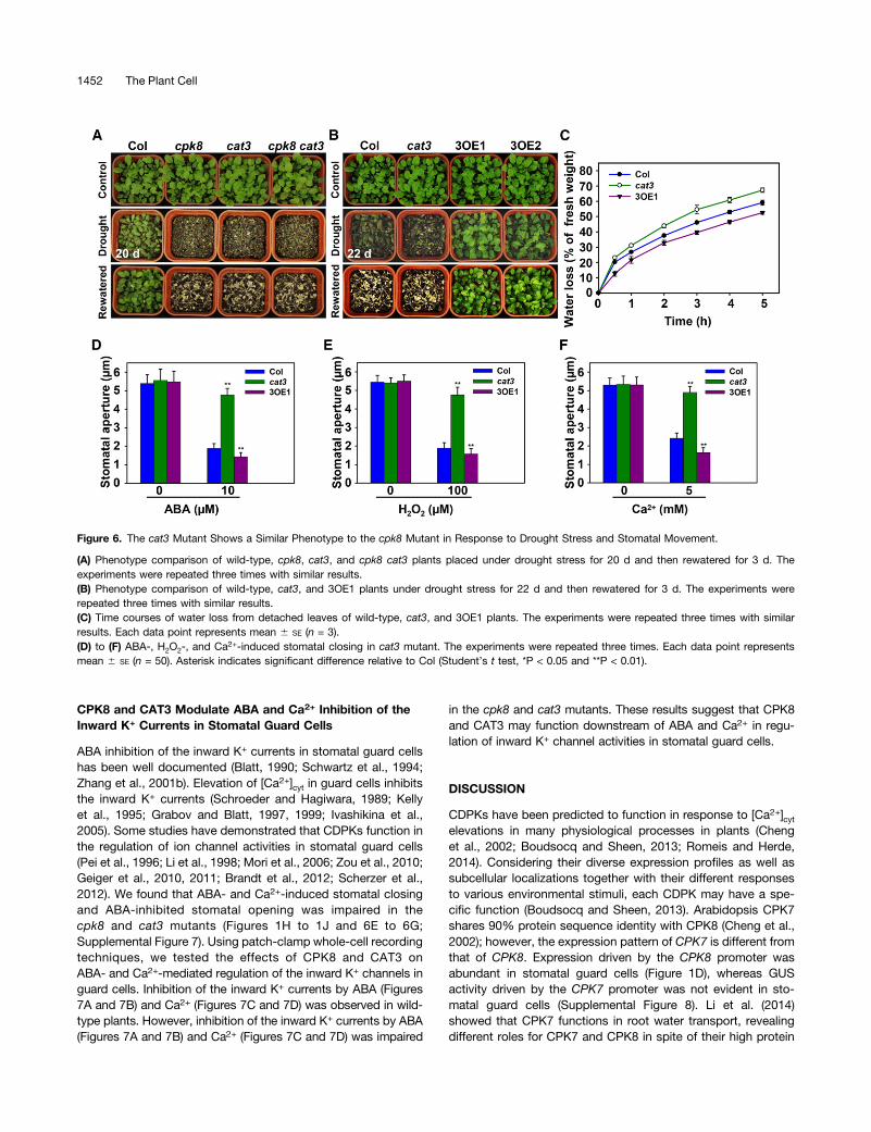
Jannat et al., 2011), and the transcription of CAT3 was inducedby ABA and drought (Xing et al., 2007), suggesting that CAT3may function in ABA-mediated plant responses to droughtstress. The transcript level of CAT3 was induced by ABA(Supplemental Figure 5C) and H2O2 (Supplemental Figure 5D),consistent with the previous study (Xing et al., 2007). It wasfurther hypothesized that CAT3, as a substrate of CPK8, mayact downstream of CPK8 in ABA-mediated signal transductionin stomatal guard cells under drought stress. The cat3 mutantplants and CAT3 overexpression lines were tested for theirsensitivity to drought stress. After a 20-d period of droughtstress treatment (the normal drought stress treatment in thisstudy), the cat3 mutant showed similar sensitivity to droughtstress as the cpk8 mutant (Figure 6A). In addition, the cpk8 cat3double mutants showed similar sensitivity to drought stress ascpk8 and cat3 single mutant plants (Figure 6A). When CPK8 wasoverexpressed in cat3 (8OE/cat3) or CAT3 overexpressed incpk8 (3OE/cpk8), both transgenic lines were as sensitive todrought stress as cat3 and cpk8 plants (Supplemental Figure 6),
which not only further demonstrates that CPK8 acts as an up-stream regulator of CAT3 but also demonstrates that activationof CAT3 requires its phosphorylation by CPK8. As shown inFigure 6B, wild-type plants did not survive a 22-d period ofdrought stress treatment (the “severe” drought stress treat-ment) even after rewatering. However, the CAT3 over-expression lines not only survived, but also recovered wellafter rewatering. Consistent with these results, water lossassays showed that the detached leaves of cat3, similar tothe cpk8, lost water faster than wild-type plants under de-hydration treatment, whereas the 3OE1 leaves lost waterslower than wild-type plants (Figure 6C). Furthermore, thecat3 mutant showed impaired stomatal closing induced byABA (Figure 6D), H2O2 (Figure 6E), and Ca2+ (Figure 6F)as well as impaired ABA inhibition of stomatal opening(Supplemental Figure 7), which was similar to the observa-tion in the cpk8 mutant. These results suggest that CPK8 andCAT3 function in the same signaling pathway in plant re-sponses to drought stress.
Figure 5. Comparison of Catalase Activity and H2O2 Accumulation in CPK8 and CAT3-Related Plant Materials.
(A) RT-qPCR analysis of CAT3 transcription in wild-type plants, cat3, and CAT3 overexpression lines. The experiments were repeated three times withsimilar results. Each data point represents mean 6 SE (n = 3). Asterisk indicates significant difference relative to Col (Student’s t test, **P < 0.01).(B) Catalase activity in wild-type, cpk8, 8OE1, cat3, 3OE1, and 8OE/cat3 plants. Each data point represents mean 6 SE (n = 3).(C) ABA-induced H2O2 production in wild-type, cpk8, 8OE1, cat3, 3OE1, and 8OE/cat3 plants.(D) ABA-induced H2O2 production in leaves of wild-type plants and cpk8 and cat3 mutants assayed with DAB. The experiments were repeated threetimes with similar results.(E) Exogenous ABA-induced production of ROS in stomatal guard cells. Abaxial epidermal peels from wild-type and mutant plants were loaded withH2DCF-DA before and 5 min after the addition of 10 mM ABA. The photographs show representative fluorescence from stomatal guard cells of the wildtype and mutants. The experiments were repeated three times with similar results.(F) Effects of ABA on the DCF fluorescence in guard cells of the wild type, cpk8, and cat3. The data points represent mean 6 SE from measurements ofpixel intensity in whole cells determined before and 5 min after ABA (10 mM) treatment (n = 3). Asterisk indicates significant difference relative to Col(Student’s t test, *P < 0.05 and **P < 0.01).
CPK8 and CAT3 in Stomatal Regulation 1451
CPK8 and CAT3 Modulate ABA and Ca2+ Inhibition of theInward K+ Currents in Stomatal Guard Cells
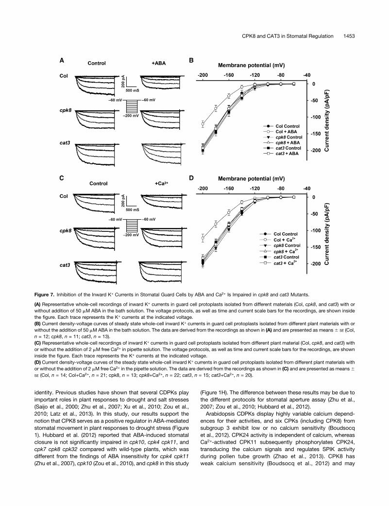
ABA inhibition of the inward K+ currents in stomatal guard cellshas been well documented (Blatt, 1990; Schwartz et al., 1994;Zhang et al., 2001b). Elevation of [Ca2+]cyt in guard cells inhibitsthe inward K+ currents (Schroeder and Hagiwara, 1989; Kellyet al., 1995; Grabov and Blatt, 1997, 1999; Ivashikina et al.,2005). Some studies have demonstrated that CDPKs function inthe regulation of ion channel activities in stomatal guard cells(Pei et al., 1996; Li et al., 1998; Mori et al., 2006; Zou et al., 2010;Geiger et al., 2010, 2011; Brandt et al., 2012; Scherzer et al.,2012). We found that ABA- and Ca2+-induced stomatal closingand ABA-inhibited stomatal opening was impaired in thecpk8 and cat3 mutants (Figures 1H to 1J and 6E to 6G;Supplemental Figure 7). Using patch-clamp whole-cell recordingtechniques, we tested the effects of CPK8 and CAT3 onABA- and Ca2+-mediated regulation of the inward K+ channels inguard cells. Inhibition of the inward K+ currents by ABA (Figures7A and 7B) and Ca2+ (Figures 7C and 7D) was observed in wild-type plants. However, inhibition of the inward K+ currents by ABA(Figures 7A and 7B) and Ca2+ (Figures 7C and 7D) was impaired
in the cpk8 and cat3 mutants. These results suggest that CPK8and CAT3 may function downstream of ABA and Ca2+ in regu-lation of inward K+ channel activities in stomatal guard cells.
DISCUSSION
CDPKs have been predicted to function in response to [Ca2+]cytelevations in many physiological processes in plants (Chenget al., 2002; Boudsocq and Sheen, 2013; Romeis and Herde,2014). Considering their diverse expression profiles as well assubcellular localizations together with their different responsesto various environmental stimuli, each CDPK may have a spe-cific function (Boudsocq and Sheen, 2013). Arabidopsis CPK7shares 90% protein sequence identity with CPK8 (Cheng et al.,2002); however, the expression pattern of CPK7 is different fromthat of CPK8. Expression driven by the CPK8 promoter wasabundant in stomatal guard cells (Figure 1D), whereas GUSactivity driven by the CPK7 promoter was not evident in sto-matal guard cells (Supplemental Figure 8). Li et al. (2014)showed that CPK7 functions in root water transport, revealingdifferent roles for CPK7 and CPK8 in spite of their high protein
Figure 6. The cat3 Mutant Shows a Similar Phenotype to the cpk8 Mutant in Response to Drought Stress and Stomatal Movement.
(A) Phenotype comparison of wild-type, cpk8, cat3, and cpk8 cat3 plants placed under drought stress for 20 d and then rewatered for 3 d. Theexperiments were repeated three times with similar results.(B) Phenotype comparison of wild-type, cat3, and 3OE1 plants under drought stress for 22 d and then rewatered for 3 d. The experiments wererepeated three times with similar results.(C) Time courses of water loss from detached leaves of wild-type, cat3, and 3OE1 plants. The experiments were repeated three times with similarresults. Each data point represents mean 6 SE (n = 3).(D) to (F) ABA-, H2O2-, and Ca2+-induced stomatal closing in cat3 mutant. The experiments were repeated three times. Each data point representsmean 6 SE (n = 50). Asterisk indicates significant difference relative to Col (Student’s t test, *P < 0.05 and **P < 0.01).
1452 The Plant Cell
identity. Previous studies have shown that several CDPKs playimportant roles in plant responses to drought and salt stresses(Saijo et al., 2000; Zhu et al., 2007; Xu et al., 2010; Zou et al.,2010; Latz et al., 2013). In this study, our results support thenotion that CPK8 serves as a positive regulator in ABA-mediatedstomatal movement in plant responses to drought stress (Figure1). Hubbard et al. (2012) reported that ABA-induced stomatalclosure is not significantly impaired in cpk10, cpk4 cpk11, andcpk7 cpk8 cpk32 compared with wild-type plants, which wasdifferent from the findings of ABA insensitivity for cpk4 cpk11(Zhu et al., 2007), cpk10 (Zou et al., 2010), and cpk8 in this study
(Figure 1H). The difference between these results may be due tothe different protocols for stomatal aperture assay (Zhu et al.,2007; Zou et al., 2010; Hubbard et al., 2012).Arabidopsis CDPKs display highly variable calcium depend-
ences for their activities, and six CPKs (including CPK8) fromsubgroup 3 exhibit low or no calcium sensitivity (Boudsocqet al., 2012). CPK24 activity is independent of calcium, whereasCa2+-activated CPK11 subsequently phosphorylates CPK24,transducing the calcium signals and regulates SPIK activityduring pollen tube growth (Zhao et al., 2013). CPK8 hasweak calcium sensitivity (Boudsocq et al., 2012) and may
Figure 7. Inhibition of the Inward K+ Currents in Stomatal Guard Cells by ABA and Ca2+ Is Impaired in cpk8 and cat3 Mutants.
(A) Representative whole-cell recordings of inward K+ currents in guard cell protoplasts isolated from different materials (Col, cpk8, and cat3) with orwithout addition of 50 mM ABA in the bath solution. The voltage protocols, as well as time and current scale bars for the recordings, are shown insidethe figure. Each trace represents the K+ currents at the indicated voltage.(B) Current density-voltage curves of steady state whole-cell inward K+ currents in guard cell protoplasts isolated from different plant materials with orwithout the addition of 50 mM ABA in the bath solution. The data are derived from the recordings as shown in (A) and are presented as means6 SE (Col,n = 12; cpk8, n = 11; cat3, n = 13).(C) Representative whole-cell recordings of inward K+ currents in guard cell protoplasts isolated from different plant material (Col, cpk8, and cat3) withor without the addition of 2 mM free Ca2+ in pipette solution. The voltage protocols, as well as time and current scale bars for the recordings, are showninside the figure. Each trace represents the K+ currents at the indicated voltage.(D) Current density-voltage curves of the steady state whole-cell inward K+ currents in guard cell protoplasts isolated from different plant materials withor without the addition of 2 mM free Ca2+ in the pipette solution. The data are derived from the recordings as shown in (C) and are presented as means6SE (Col, n = 14; Col+Ca2+, n = 21; cpk8, n = 13; cpk8+Ca2+, n = 22; cat3, n = 15; cat3+Ca2+, n = 20).
CPK8 and CAT3 in Stomatal Regulation 1453
phosphorylate its substrate CAT3 even without addition of cal-cium in the reaction buffer (Figure 3A). However inhibition of theinward K+ currents by Ca2+ (Figures 7C and 7D) was impaired inthe cpk8, suggesting that CPK8 may function downstream ofCa2+. These results indicate that CPK8 may be indirectly regu-lated by Ca2+ signals and that other Ca2+-dependent proteinsmay transduce Ca2+ signals to CPK8. One example of a similarmechanism is Ca2+-independent CPK24, which is activated(phosphorylated) by Ca2+-dependent CPK11 (Zhao et al., 2013).
Ca2+-Mediated Protein Phosphorylation: A UniversalRegulatory Mechanism for Plant Ion channelsand Transporters
A number of CDPKs have been reported to regulate ion channelactivities in stomatal movement (Pei et al., 1996; Li et al., 1998;Mori et al., 2006; Geiger et al., 2010, 2011; Zou et al., 2010;Brandt et al., 2012; Scherzer et al., 2012), plant responses to saltstress (Latz et al., 2013), and pollen tube growth (Gutermuthet al., 2013; Zhao et al., 2013; Zhou et al., 2014). In this report,the plasma membrane localized-CPK8 showed abundant ex-pression in stomatal guard cells (Figure 1D; Supplemental Figure2), indicating its potential role in regulation of stomatal move-ment and ion channel activity. As shown in Figure 7, ABA andCa2+ inhibition of inward K+ currents were impaired in guardcells of cpk8 mutant plants. So far, at least 11 ArabidopsisCDPKs have been reported to function in regulation of ionchannel activity in plant cells (Hwang et al., 2000; Mori et al., 2006;Geiger et al., 2010, 2011; Zou et al., 2010; Brandt et al., 2012;Scherzer et al., 2012; Latz et al., 2013; Gutermuth et al., 2013;Zhao et al., 2013; Zhou et al., 2014). The previous reports haverevealed that plant ion channels and transporters are regulated bymembers of the CIPK (CBL-interacting protein kinase) family,another Ca2+-regulated protein kinase family, such as AKT1regulation by CIPK23 (Xu et al., 2006; Wang and Wu, 2013) andNa+/H+ antiporter (SOS1) regulation by CIPK24 (Xiong et al.,2002). Together with the results in this study, these findingssupport the notion that regulation of various plant ion channelsand transporters via their Ca2+-mediated phosphorylation repre-sents a universal mechanism.
Identification of a Downstream Target of CPK8
Identification and functional characterization of the specifictarget (or substrate) of a CDPK is a key step to understandCDPK functions in plant signaling (Boudsocq and Sheen, 2013).Some substrates of CDPKs have been identified in the last de-cade, revealing their different roles in plant development andimmune and stress signaling (Boudsocq and Sheen, 2013;Schulz et al., 2013). For example, Arabidopsis CPK4 and CPK11phosphorylate ABF1 and ABF4 during ABA signal transduction(Zhu et al., 2007). Arabidopsis CPK5 induces ROS production bydirectly phosphorylating the NADPH oxidase RBOHB duringinnate immune responses (Dubiella et al., 2013). ArabidopsisCPK11 and CPK24 modulate the activity of shaker pollen inwardK+ channel (SPIK, also named AKT6) during pollen tube growth(Zhao et al., 2013). In this study, yeast two-hybrid, co-IP, BiFC,and protein phosphorylation assays demonstrated specific
interaction between CPK8 and CAT3 (Figures 2 and 3A). Inaddition, CPK8 specifically enhanced CAT3 activity throughphosphorylation at Ser-261 (Figures 2 to 4). The cpk8 cat3double mutants showed similar drought stress sensitivity as thecpk8 and cat3 single mutants (Figure 6A). Similar to CPK8, CAT3functions in the regulation of stomatal movement, most likelythrough regulation of the inward K+ channel activities in stomatalguard cells (Figures 6D to 6F and 7). Our data reveal importantroles of CPK8 and CAT3-mediated ABA stomatal signal trans-duction in response to drought stress.
Roles of CPK8-CAT3 Interaction in H2O2 Homeostasis
Excess ROS accumulation in living plant cells is toxic to cellularactivities, so the cytosolic concentration of ROS must be strin-gently regulated (Mittler, 2002; Apel and Hirt, 2004). ROS playa dual role in plant responses to abiotic stresses, acting as toxicby-products of aerobic metabolism and as key regulators ofgrowth, development, and defense pathways (Mittler et al.,2004; Miller et al., 2010). ROS-scavenging pathways are re-sponsible for maintaining a low steady state level of ROS onwhich the different signals can be registered (Mittler et al., 2004;Miller et al., 2010). Rice (Oryza sativa) CPK12-overexpressingplants exhibit enhanced tolerance to salt stress, possibly asa result of decreased ROS accumulation (Asano et al., 2012).Arabidopsis glutathione peroxidase GPX3 plays dual roles, thefirst in the general control of H2O2 homeostasis and the secondin relaying ABA and H2O2 signaling in stomatal movement (Miaoet al., 2006).As an important H2O2-scavenging enzyme, the expression or
activity of Arabidopsis catalase is also regulated by othercomponents. The expression profiles and cellular localizationsof Arabidopsis catalases are varied, suggesting their potentialroles in response to environmental stimuli in addition to H2O2
decomposition (Zimmermann et al., 2006; Bueso et al., 2007;Xing et al., 2007; Du et al., 2008). ABA-induced CAT1 expressiondepends on the production of H2O2, which is triggered byMKK1-MPK6 signaling pathway, revealing the roles of CAT1 inROS scavenging and its feedback regulation of the H2O2 sig-naling (Xing et al., 2007, 2008). CAT2 has been reported to beinvolved in crosstalk between oxidative stress, cation homeo-stasis, and ethylene (Bueso et al., 2007). Arabidopsis CAT3 caninteract with several proteins, such as CaM PCM6 in potato(Solanum tuberosum) (Yang and Poovaiah, 2002) and NDK1(Fukamatsu et al., 2003), SOS2 (CIPK24; Verslues et al., 2007),and LSD1 (Li et al., 2013) in Arabidopsis, consistent with rolesfor CAT3 in regulating H2O2 homeostasis and signaling in re-sponse to ROS stress. This work showed that H2O2 can inducethe expression of CPK8 and CAT3 (Supplemental Figures 1Dand 5D). The cpk8 and cat3 mutants had lower catalase activi-ties and accumulated more ROS in leaves and stomatal guardcells compared with wild-type plants when treated with ABA(Figures 5C to 5F), whereas CPK8 and CAT3 overexpressionlines had lower ROS accumulation compared with wild-typeplants when treated with ABA (Figures 5C and 5D).In summary, the results presented in this study demonstrate
that CPK8 can specifically phosphorylate CAT3 and regulate itsactivity. The CPK8-CAT3 interaction not only acts as a positive
1454 The Plant Cell
regulatory component in ABA-mediated regulation of stomatalmovement in plant responses to drought stress, but also playsimportant role in maintaining H2O2 homeostasis in living plant cells.
METHODS
Plant Materials and Growth Conditions
Arabidopsis thaliana ecotype Columbia and Nicotiana benthamiana wereused in this study. The T-DNA insertion lines cpk8 (SALK_036581) and cat3(SALK_092911)were obtained from theABRC (http://www.arabidopsis.org/abrc/).
Seeds were surface sterilized and placed in the Petri dishes containingMurashige and Skoog agar (0.8%, w/v) medium and incubated for 2 d at4°C before transfer to 22°C for germination. After 7 d under constantillumination at 60 mmol m22 s21 at 22°C, the seedlings were transplantedto pots containing soil mixture (rich soil:vermiculite, 2:1, v/v) and kept ingrowth chambers at 22°C with illumination at 120 mmol m22 s21 for a 16-hdaily light period. The relative humidity was approximate 70% (65%).
N. benthamiana seeds were planted in the potting soil mixture (rich soil:vermiculite, 2:1, v/v) and kept in growth chambers at 23°C with illumi-nation at 120 mmol m22 s21 for a 16-h daily light period. After 3 weeksgrowth, the plants were used for transformation.
Vector Constructions and Generation of Transgenic Plants
The CPK8pro:GUS construct was generated by introducing the CPK8promoter fragment (1.96 kb) in front of the GUS coding sequence in theEcoRI and SalI sites of pCAMBIA1381 vector. The GUS staining assayswere performed as described previously (Xu et al., 2006).
For Superpro:CPK8 and Superpro:CAT3 constructs, the coding se-quence of CPK8 was introduced into the KpnI and XhoI sites ofpCAMBIA1300 vector under the Super promoter, and the coding se-quence of CAT3 was introduced into the XbaI and XhoI sites ofpCAMBIA1300 vector under the Super promoter (Chen et al., 2009),respectively. Primers for Superpro:CPK8 and Superpro:CAT3 constructsare listed in Supplemental Table 1. The Superpro:CPK8 and Superpro:CAT3constructs were introduced into wild-type plants for generation of CPK8and CAT3 overexpression lines, respectively. To generate the constructfor complementation of cpk8, a genomic DNA fragment of CPK8 wasamplified and cloned into SacI and KpnI sites of pCAMBIA1300 vector.Arabidopsis transformation with Agrobacterium tumefaciens (strainGV3101) was performed by the floral dip method (Clough and Bent, 1998).Homozygous T3 transgenic lines were used for further analyses.
RT-qPCR
Total RNA was extracted from plants with Trizol reagent (Invitrogen)following the manufacturer’s protocols. For RT-qPCR analyses, total RNAtreatedwith DNase I (Takara) was used for cDNA synthesis by SuperScriptII reverse transcriptase (Invitrogen). The quantitative PCR analysis wasperformed using an Applied Biosystems 7500 real-time PCR system. TheSYBR Premix Ex Taq kit (Applied Biosystems) was used for reactionaccording to the manufacturer’s instruction. Arabidopsis 18S rRNA wasquantified as an internal control. The primer sequences of CPK8, CAT3and 18S rRNA for RT-qPCR are listed in Supplemental Table 1.
Drought Treatment and Water Loss Measurements
For drought treatment, plants grown onMurashige and Skoogmedium for7 d were transplanted into soil for about 1 week with sufficient wateringfollowed by a 20-d drought stress (withholding irrigation) as describedbefore (Zou et al., 2010), with slight modifications. The seedlings were
grown in a growth room with 12-h daily light. Normally watered plantswere used as the control. For drought stress treatment, water waswithheld for 20 d (normal drought stress) or 22 d (severe drought stress).Two independent transgenic lines were used for drought stress.
For water loss measurement, rosette leaves were detached from4-week-old plants, weighed, and placed on the laboratory bench (therelative humidity was between 40 and 50%) at 22°C (61°C). Weight lossof the detached leaves was monitored at the indicated time intervals.Water loss was expressed as the percentage of initial fresh weight.
Thermal Imaging
Thermal imaging of plants was performed as described previously (Hua et al.,2012). Three-week-old plants grown under normal conditions were subjectedto drought stress for about 1 week. Thermal images were obtained usingVarioCAM HD and leaf temperature was calculated using IRBIS 3 software.
Stomatal Aperture Measurements
Stomatal closure assays were conducted as described before (Zou et al.,2010). After treatment, the abaxial epidermis was peeled and photo-graphed using a Leica microscope (Leica DFC320). Stomatal apertureswere measured using ImageJ software (National Institutes of Health).
Yeast Two-Hybrid Assay
The GAL4-based two-hybrid system was used for yeast two-hybridscreening (Clontech). The CPK8-pGBKT7 construct was generated byfusing the sequence encoding the CPK8 kinase domain (from 169 to 945bp of the coding sequence) with that encoding the GAL4 binding domainin the EcoRI and SalI sites of pGBKT7, and the resulting construct wastransformed into the AH109 yeast strain. For screening, the reporter yeastwas transformed with library plasmid DNA from CD4-22 library (ABRC).Yeast transformation, growth conditions, and assays for b-gal activitywere performed according to the manufacturer’s instructions (Clontech).Positive clones were selected for sequencing. Three coding sequences ofcatalases were inserted into pGADT7 prey plasmid containing the GAL4activating domain and cotransformed into AH109 with CPK8 bait for b-galactivity assays, respectively. The primer sequences and restriction sitesare listed in Supplemental Table 1.
Co-IP Assays
The coding sequence ofCPK8with a cMyc tag (cMYC-CPK8) was clonedinto the SpeI and KpnI sites of pCAMBIA1307 vector under control of the35S promoter. The coding sequence of CAT3with a GFP tag (GFP-CAT3)was cloned into the XbaI and SalI sites of pSAT6-GFP-N1 vector undercontrol of the 35S promoter. The primer sequences and restriction sitesare listed in Supplemental Table 1. The combinations of cMyc-CPK8 andGFP-CAT3 were cotransformed into Arabidopsis mesophyll protoplastsas described before (Ren et al., 2013). After incubation overnight at 4°C,the protoplasts were lysed, sonicated, and centrifuged at 12,000g for10 min at 4°C. The supernatant was incubated with 10 mL anti-cMyc agaroseconjugate (Sigma-Aldrich) for 6 h at 4°C. The co-IP products were washedbriefly with extraction buffer for five times at 4°C and then detected viaimmunoblot analysis. Both anti-GFP (Sigma-Aldrich) and anti-cMyc(Sigma-Aldrich) antibodies were used at 1:5000 dilutions, and chemi-luminescence signals were detected using Fusion Solo.
Subcellular Localizations of CPK8 and CAT3
For generation of GFP-fusion protein, the coding sequence of CPK8 wasfused with GFP in the XbaI and KpnI sites of pCAMBIA1300 vector. The
CPK8 and CAT3 in Stomatal Regulation 1455
coding sequence ofCAT3was fusedwithGFP in theAscI andPacI sites ofpCM1205-C-GFP vector (Zhou et al., 2012). The coding sequence forpotassium transporter AKT1 (Xu et al., 2006) was fused with mCherry inthe SUPERR:sXVE:mCherryC:Bar vector (Schlücking et al., 2013) andused as the plasma membrane marker. PX-RK (CD3-83; ABRC) was usedas peroxisome marker. All constructs were transformed into the Agro-bacterium strain GV3101. The Agrobacterium lines were infiltrated intoleaves of N. benthamiana. Plants were grown at 23°C and allowed torecover for 3 d. The fluorescence of GFP or mCherry in the leaves wasimaged using a confocal laser scanning microscope (Leica sp5).
BiFC Assays
BiFC assays were performed as described previously (Waadt and Kudla,2008). For generation of the BiFC vectors, the coding region of CPK8 wascloned via BamHI-SalI into pSPYCEM, resulting in CPK8-YC, and thecoding region of CAT1, CAT2, or CAT3 was cloned via BamHI-SalI intopSPYNE173, resulting in CAT1-YN, CAT2-YN and CAT3-YN, re-spectively. The gene-specific primer pairs forCPK8 andCATs are listed inSupplemental Table 1. All constructs were transformed into the Agro-bacterium strain GV3101. An equal volume of Agrobacterium harboringCPK8-YC, CAT1-YN (or CAT2-YN, CAT3-YN) and P19 was mixed toa final concentration of OD600 = 0.8. Agrobacterium lines were infiltratedleaves of N. benthamiana. Plants were grown at 23°C and allowed torecover for 3 d; then, the fluorescence of YFP in the leaves was imagedusing a confocal laser scanning microscope (Leica sp5).
Firefly Luciferase Complementation Image Assay
Firefly luciferase complementation imaging assays were performed asdescribed before (Chen et al., 2008). For generation of the assayvectors, the coding region of CPK8 was cloned via BamHI-SalI intopCAMBIA1300-cLuc, resulting in CPK8-CLuc, and the coding region ofCAT1,CAT2, orCAT3was cloned via BamHI-SalI into pCAMBIA1300-nLuc,resulting in CAT1-nLuc, CAT2-nLuc, or CAT3-nLuc, respectively.The gene-specific primer pairs for CPK8 and CATs are shown inSupplemental Table 1.
All constructs were transformed into the Agrobacterium strain GV3101.An equal volume of Agrobacterium harboring pCAMBIA-nLuc, pCAMBIA-cLuc, and P19 was mixed to a final concentration of OD600 = 0.8. Threedifferent combinations of Agrobacterium were infiltrated into three dif-ferent positions at the same leaves of N. benthamiana. Plants were grownat 23°C and allowed to recover for 3 d. A low-light cooled CCD imagingapparatus (fluorescence/chemiluminescence imaging system) was usedto capture the LUC image.
In Vitro Kinase Assay
To prepare recombinant CPK8, the full-length coding sequence of CPK8was amplified and cloned into the XcmI sites of pCXSN vector, resulting inpCXSN-Strep-CPK8. The gene-specific primer pairs of CPK8 are listed inSupplemental Table 1. The Strep-CPK8 was purified as described pre-viously (Witte et al., 2004).
To prepare recombinant CAT1, CAT2, and CAT3 truncated proteins,the corresponding coding sequences of CATs were amplified and clonedinto BamHI and SalI sites of pGEX-4T-1 vector, resulting in pGEX-4T-1-CATs constructs, respectively. CAT3 mutant proteins were made by site-directed mutagenesis using a QuickChange kit (Agilent Technologies; theprimers are listed in Supplemental Table 1). The constructs were thenintroduced into BL21 cells by transformation and used to purify GST-CATproteins.
In vitro kinase assays were performed as described previously(Rodriguez Milla et al., 2006), with slight modification. In brief, phos-phorylation was initiated by adding 0.5 mCi [g-32P] to reaction buffer
containing 25 mM Tris-HCl (pH 7.5), 10 mMMgCl2, 1 mMDTT, and 10 mMATP, followed by incubation for 20 min at 25°C. The proteins wereseparated by 10% SDS-PAGE, and the phosphorylated proteins werevisualized by autoradiography.
In Planta Kinase Assay
In planta kinase assays were used to test the phosphorylation of CAT3.Total proteins in Col/35Spro:CAT3-GFP or cpk8/35Spro:CAT3-GFP beforeand after treatment with ABA were extracted. CAT3-GFP proteins wereimmunoprecipitated using Anti-GFP mAb agarose (MBL). CAT3-GFPproteins were then separated by 10% SDS-PAGE, and phosphorylatedproteins were detected by immunoblotting using Biotinylated Phos-tag asdescribed previously (Kinoshita-Kikuta et al., 2007). Calf intestinal alkalinephosphatase-treated (Takara) proteins were used as control.
Measurements of H2O2 Production
H2O2 was detected by DAB staining as described previously (Thordal-Christensen et al., 1997; Guan and Scandalios, 2000). Fully expandedleaves were excised and incubated in water containing 100 mM ABA for30 min in dark at 28°C. The leaves were then incubated in DAB solution(1 mg mL21, pH 3.8; Sigma-Aldrich) for 8 h in dark at 28°C. The leaveswere immersed in boiling 80% (v/v) ethanol for 10 min to terminate thestaining and to decolor the leaves (except for the deep brown poly-merization product produced by the reaction of DAB with H2O2). Aftercooling, the leaves were extracted with 80% (v/v) ethanol and preservedat 4°C in 80% (v/v) ethanol before photographed.
ROS production in guard cells was examined by loading abaxialepidermal strips with H2DCF-DA (Miao et al., 2006). The detached leaveswere floated in incubation buffer for 2.5 h to induce stomatal opening, andthe epidermal strips were peeled and placed into loading buffer (10 mMTris and 50 mM KCl, pH 7.2) containing 50 mM H2DCF-DA in the dark for10 min. Excess dye was removed by washing the samples three timeswith distilled water. Epidermal tissues were then incubated in loadingbuffer for 5 min with 50 mMABA or with an equal volume of ethanol addedas a control. Examinations of peel fluorescence were performed witha confocal laser scanningmicroscope (Nikon TE2000-E). To quantitativelyanalyze the data, pixel values were averaged over rectangular regions(4 mm2) manually located on each image. The pixel intensity from at least150 guard cells was recorded.
Measurement of Catalase Activity
The activity of CAT was determined with a CAT assay kit according tothe manufacturer’s instructions (Beyotime). Briefly, samples weretreated with excess H2O2 for decomposition by catalase for 5 min,and the remaining H2O2 coupled with a substrate was treated withperoxidase to generate a red product, N-4-antipyryl-3-chloro-5-sulfonate-p-benzoquinonemonoimine, which absorbs maximally at520 nm. Catalase activity was thus determined by measuring thedecomposed H2O2. All values were normalized by the total proteinconcentration of the same sample.
Patch-Clamp Experiments
Arabidopsis guard cell protoplasts were isolated as described previously(Pei et al., 1997; Wang et al., 2001). Standard whole-cell recordingtechniques were applied in this study (Hamill et al., 1981). All experimentswere conducted at room temperature (;22°C) under dim light. The whole-cell currents were recorded as described before (Zou et al., 2010). ThepCLAMP software (version 6.0.4; Axon Instruments) was used to acquireand analyze the whole-cell currents. SigmaPlot software was used todraw current-voltage plots and for data analysis.
1456 The Plant Cell
Accession Numbers
Sequence data from this article can be found in the Arabidopsis GenomeInitiative or GenBank/EMBL databases under the following accessionnumbers: At5g19450 for CPK8, At1g20630 for CAT1, At4g35090 forCAT2, and At1g20620 for CAT3.
Supplemental Data
Supplemental Figure 1. Position of T-DNA Insertion in cpk8 andExpression Levels of CPK8 in Response to Abiotic Stresses.
Supplemental Figure 2. Subcellular Localizations of CPK8 and CAT3.
Supplemental Figure 3. Firefly Luciferase Complementation ImageAssay of CPK8 Interaction with CAT3 in N. benthamiana Leaves.
Supplemental Figure 4. Identification of CPK8 Phosphorylation Sitesin CAT3.
Supplemental Figure 5. Position of T-DNA Insertion in CAT3 andExpression Levels of CAT3 in Response to ABA and H2O2.
Supplemental Figure 6. Phenotype Observation of Various CPK8-and CAT3-Related Plant Materials under Drought Stress.
Supplemental Figure 7. ABA Inhibition of Stomatal Opening IsImpaired in the cpk8 and cat3 Mutants.
Supplemental Figure 8. Expression Patterns of CPK7 as Determinedfrom CPK7pro:GUS Transgenic Plants.
Supplemental Table 1. Primer Sequences Used in This Study.
ACKNOWLEDGMENTS
This work was financially supported by grants from the “973” Project(2011CB915401 and 2012CB114203 to W.-H.W.), the “111” Project (No.B06003 to W.-H.W.), and the National Natural Science Foundation ofChina (31000119 to J.-J.Z.). We thank Jörg Kudla (Institut für Botanik,Universität Münster, Germany) for providing the vectors SUPERR:sXVE:mCherryC:Bar for AKT1-mCherry construct and pSPYCEM andpSPYNE173 for BiFC assays. We thank Yan Guo (China AgriculturalUniversity) for providing pCM1205-C-GFP and pSAT6-GFP-N1 vectors.
AUTHOR CONTRIBUTIONS
J.-J.Z. and W.-H.W. designed the research. J.-J.Z., D.R., X.-D.L., C.W.,W.-X.L., L.-F.S., and W.-Z.Z. performed research and analyzed data.J.-Z.Z. and W.-H.W. wrote the article. J.-J.Z., X.-D.L., and W.-H.W.revised the article.
Received February 15, 2015; revised April 11, 2015; accepted April 23,2015; published May 12, 2015.
REFERENCES
Apel, K., and Hirt, H. (2004). Reactive oxygen species: metabolism, oxida-tive stress, and signal transduction. Annu. Rev. Plant Biol. 55: 373–399.
Asano, T., Hayashi, N., Kobayashi, M., Aoki, N., Miyao, A.,Mitsuhara, I., Ichikawa, H., Komatsu, S., Hirochika, H.,Kikuchi, S., and Ohsugi, R. (2012). A rice calcium-dependentprotein kinase OsCPK12 oppositely modulates salt-stress toleranceand blast disease resistance. Plant J. 69: 26–36.
Böhmer, M., and Romeis, T. (2007). A chemical-genetic approach toelucidate protein kinase function in planta. Plant Mol. Biol. 65: 817–827.
Blatt, M.R. (1990). Potassium channel currents in intact stomatal guardcells: rapid enhancement by abscisic acid. Planta 180: 445–455.
Boudsocq, M., Droillard, M.J., Regad, L., and Laurière, C. (2012).Characterization of Arabidopsis calcium-dependent protein kina-ses: activated or not by calcium? Biochem. J. 447: 291–299.
Boudsocq, M., and Sheen, J. (2013). CDPKs in immune and stresssignaling. Trends Plant Sci. 18: 30–40.
Boudsocq, M., Willmann, M.R., McCormack, M., Lee, H., Shan, L.,He, P., Bush, J., Cheng, S.H., and Sheen, J. (2010). Differentialinnate immune signalling via Ca(2+) sensor protein kinases. Nature464: 418–422.
Brandt, B., Brodsky, D.E., Xue, S., Negi, J., Iba, K., Kangasjärvi, J.,Ghassemian, M., Stephan, A.B., Hu, H., and Schroeder, J.I.(2012). Reconstitution of abscisic acid activation of SLAC1 anionchannel by CPK6 and OST1 kinases and branched ABI1 PP2Cphosphatase action. Proc. Natl. Acad. Sci. USA 109: 10593–10598.
Bueso, E., Alejandro, S., Carbonell, P., Perez-Amador, M.A.,Fayos, J., Bellés, J.M., Rodriguez, P.L., and Serrano, R. (2007).The lithium tolerance of the Arabidopsis cat2 mutant revealsa cross-talk between oxidative stress and ethylene. Plant J. 52:1052–1065.
Chen, H., Zou, Y., Shang, Y., Lin, H., Wang, Y., Cai, R., Tang, X.,and Zhou, J.M. (2008). Firefly luciferase complementation imagingassay for protein-protein interactions in plants. Plant Physiol. 146:368–376.
Cheng, S.-H., Willmann, M.R., Chen, H.-C., and Sheen, J. (2002).Calcium signaling through protein kinases. The Arabidopsiscalcium-dependent protein kinase gene family. Plant Physiol. 129:469–485.
Chen, Y.-F., Li, L.-Q., Xu, Q., Kong, Y.-H., Wang, H., and Wu, W.-H.(2009). The WRKY6 transcription factor modulates PHOSPHATE1expression in response to low Pi stress in Arabidopsis. Plant Cell21: 3554–3566.
Choi, H.I., Park, H.-J., Park, J.H., Kim, S., Im, M.-Y., Seo, H.-H.,Kim, Y.-W., Hwang, I., and Kim, S.Y. (2005). Arabidopsis calcium-dependent protein kinase AtCPK32 interacts with ABF4, a tran-scriptional regulator of abscisic acid-responsive gene expression,and modulates its activity. Plant Physiol. 139: 1750–1761.
Clough, S.J., and Bent, A.F. (1998). Floral dip: a simplified method forAgrobacterium-mediated transformation of Arabidopsis thaliana.Plant J. 16: 735–743.
Coca, M., and San Segundo, B. (2010). AtCPK1 calcium-dependentprotein kinase mediates pathogen resistance in Arabidopsis. PlantJ. 63: 526–540.
Dammann, C., Ichida, A., Hong, B., Romanowsky, S.M., Hrabak,E.M., Harmon, A.C., Pickard, B.G., and Harper, J.F. (2003).Subcellular targeting of nine calcium-dependent protein kinaseisoforms from Arabidopsis. Plant Physiol. 132: 1840–1848.
Du, Y.-Y., Wang, P.-C., Chen, J., and Song, C.-P. (2008). Compre-hensive functional analysis of the catalase gene family in Arabi-dopsis thaliana. J. Integr. Plant Biol. 50: 1318–1326.
Dubiella, U., Seybold, H., Durian, G., Komander, E., Lassig, R.,Witte, C.-P., Schulze, W.X., and Romeis, T. (2013). Calcium-dependent protein kinase/NADPH oxidase activation circuit is re-quired for rapid defense signal propagation. Proc. Natl. Acad. Sci.USA 110: 8744–8749.
Franz, S., Ehlert, B., Liese, A., Kurth, J., Cazalé, A.-C., and Romeis,T. (2011). Calcium-dependent protein kinase CPK21 functions inabiotic stress response in Arabidopsis thaliana. Mol. Plant 4: 83–96.
Frugoli, J.A., Zhong, H.H., Nuccio, M.L., McCourt, P., McPeek,M.A., Thomas, T.L., and McClung, C.R. (1996). Catalase is
CPK8 and CAT3 in Stomatal Regulation 1457
encoded by a multigene family in Arabidopsis thaliana (L.) Heynh.Plant Physiol. 112: 327–336.
Fukamatsu, Y., Yabe, N., and Hasunuma, K. (2003). ArabidopsisNDK1 is a component of ROS signaling by interacting with threecatalases. Plant Cell Physiol. 44: 982–989.
Geiger, D., Scherzer, S., Mumm, P., Marten, I., Ache, P., Matschi,S., Liese, A., Wellmann, C., Al-Rasheid, K.A.S., Grill, E., Romeis,T., and Hedrich, R. (2010). Guard cell anion channel SLAC1 isregulated by CDPK protein kinases with distinct Ca2+ affinities.Proc. Natl. Acad. Sci. USA 107: 8023–8028.
Geiger, D., Maierhofer, T., Al-Rasheid, K.A., Scherzer, S., Mumm,P., Liese, A., Ache, P., Wellmann, C., Marten, I., Grill, E., Romeis,T., and Hedrich, R. (2011). Stomatal closure by fast abscisic acidsignaling is mediated by the guard cell anion channel SLAH3 andthe receptor RCAR1. Sci. Signal. 4: ra32.
Grabov, A., and Blatt, M.R. (1997). Parallel control of the inward-rectifier K+ channel by cytosolic free Ca2+ and pH in Vicia guardcells. Planta 201: 84–95.
Grabov, A., and Blatt, M.R. (1999). A steep dependence of inward-rectifying potassium channels on cytosolic free calcium concen-tration increase evoked by hyperpolarization in guard cells. PlantPhysiol. 119: 277–288.
Guan, L.M., and Scandalios, J.G. (2000). Hydrogen-peroxide-mediated catalase gene expression in response to wounding.Free Radic. Biol. Med. 28: 1182–1190.
Gutermuth, T., Lassig, R., Portes, M.T., Maierhofer, T., Romeis, T.,Borst, J.W., Hedrich, R., Feijó, J.A., and Konrad, K.R. (2013).Pollen tube growth regulation by free anions depends on the interactionbetween the anion channel SLAH3 and calcium-dependent proteinkinases CPK2 and CPK20. Plant Cell 25: 4525–4543.
Hamill, O.P., Marty, A., Neher, E., Sakmann, B., and Sigworth, F.J.(1981). Improved patch-clamp techniques for high-resolution cur-rent recording from cells and cell-free membrane patches. PflugersArch. 391: 85–100.
Harmon, A.C., Gribskov, M., and Harper, J.F. (2000). CDPKs - a ki-nase for every Ca2+ signal? Trends Plant Sci. 5: 154–159.
Harper, J.F., and Harmon, A. (2005). Plants, symbiosis and parasites:a calcium signalling connection. Nat. Rev. Mol. Cell Biol. 6: 555–566.
Harper, J.F., Breton, G., and Harmon, A. (2004). Decoding Ca(2+)signals through plant protein kinases. Annu. Rev. Plant Biol. 55:263–288.
Hua, D., Wang, C., He, J., Liao, H., Duan, Y., Zhu, Z., Guo, Y., Chen,Z., and Gong, Z. (2012). A plasma membrane receptor kinase,GHR1, mediates abscisic acid- and hydrogen peroxide-regulatedstomatal movement in Arabidopsis. Plant Cell 24: 2546–2561.
Hubbard, K.E., Siegel, R.S., Valerio, G., Brandt, B., and Schroeder,J.I. (2012). Abscisic acid and CO2 signalling via calcium sensitivitypriming in guard cells, new CDPK mutant phenotypes and a methodfor improved resolution of stomatal stimulus-response analyses.Ann. Bot. (Lond.) 109: 5–17.
Hwang, I., Sze, H., and Harper, J.F. (2000). A calcium-dependentprotein kinase can inhibit a calmodulin-stimulated Ca2+ pump(ACA2) located in the endoplasmic reticulum of Arabidopsis. Proc.Natl. Acad. Sci. USA 97: 6224–6229.
Ivashikina, N., Deeken, R., Fischer, S., Ache, P., and Hedrich, R.(2005). AKT2/3 subunits render guard cell K+ channels Ca2+ sen-sitive. J. Gen. Physiol. 125: 483–492.
Jannat, R., Uraji, M., Morofuji, M., Islam, M.M., Bloom, R.E.,Nakamura, Y., McClung, C.R., Schroeder, J.I., Mori, I.C., andMurata, Y. (2011). Roles of intracellular hydrogen peroxide accu-mulation in abscisic acid signaling in Arabidopsis guard cells. J.Plant Physiol. 168: 1919–1926.
Kanchiswamy, C.N., et al. (2010). Regulation of Arabidopsis defenseresponses against Spodoptera littoralis by CPK-mediated calciumsignaling. BMC Plant Biol. 10: 97.
Kelly, W.B., Esser, J.E., and Schroeder, J.I. (1995). Effects of cy-tosolic calcium and limited, possible dual, effects of G proteinmodulators on guard cell inward potassium channels. Plant J. 8:479–489.
Kinoshita-Kikuta, E., Aoki, Y., Kinoshita, E., and Koike, T. (2007).Label-free kinase profiling using phosphate affinity polyacrylamidegel electrophoresis. Mol. Cell. Proteomics 6: 356–366.
Kolukisaoglu, U., Weinl, S., Blazevic, D., Batistic, O., and Kudla, J.(2004). Calcium sensors and their interacting protein kinases: ge-nomics of the Arabidopsis and rice CBL-CIPK signaling networks.Plant Physiol. 134: 43–58.
Kudla, J., Batisti�c, O., and Hashimoto, K. (2010). Calcium signals: thelead currency of plant information processing. Plant Cell 22: 541–563.
Kwak, J.M., Mori, I.C., Pei, Z.M., Leonhardt, N., Torres, M.A.,Dangl, J.L., Bloom, R.E., Bodde, S., Jones, J.D., and Schroeder,J.I. (2003). NADPH oxidase AtrbohD and AtrbohF genes function inROS-dependent ABA signaling in Arabidopsis. EMBO J. 22: 2623–2633.
Latz, A., Mehlmer, N., Zapf, S., Mueller, T.D., Wurzinger, B., Pfister,B., Csaszar, E., Hedrich, R., Teige, M., and Becker, D. (2013). Saltstress triggers phosphorylation of the Arabidopsis vacuolar K+
channel TPK1 by calcium-dependent protein kinases (CDPKs). Mol.Plant 6: 1274–1289.
Lee, S.S., Cho, H.S., Yoon, G.M., Ahn, J.W., Kim, H.H., and Pai, H.S.(2003). Interaction of NtCDPK1 calcium-dependent protein kinasewith NtRpn3 regulatory subunit of the 26S proteasome in Nicotianatabacum. Plant J. 33: 825–840.
Li, G., Boudsocq, M., Hem, S., Vialaret, J., Rossignol, M., Maurel,C., and Santoni, V. (2014). The calcium-dependent protein kinaseCPK7 acts on root hydraulic conductivity. Plant Cell Environ.,10.1111/pce.12478.
Li, J., Lee, Y.-R.J., and Assmann, S.M. (1998). Guard cells possessa calcium-dependent protein kinase that phosphorylates the KAT1potassium channel. Plant Physiol. 116: 785–795.
Li, Y., Chen, L., Mu, J., and Zuo, J. (2013). LESION SIMULATINGDISEASE1 interacts with catalases to regulate hypersensitive celldeath in Arabidopsis. Plant Physiol. 163: 1059–1070.
Luan, S. (2009). The CBL-CIPK network in plant calcium signaling.Trends Plant Sci. 14: 37–42.
Luan, S., Kudla, J., Rodriguez-Concepcion, M., Yalovsky, S., andGruissem, W. (2002). Calmodulins and calcineurin B-like proteins:calcium sensors for specific signal response coupling in plants.Plant Cell 14 (Suppl): S389–S400.
Ludwig, A.A., Romeis, T., and Jones, J.D.G. (2004). CDPK-mediatedsignalling pathways: specificity and cross-talk. J. Exp. Bot. 55: 181–188.
Matschi, S., Werner, S., Schulze, W.X., Legen, J., Hilger, H.H., andRomeis, T. (2013). Function of calcium-dependent protein kinaseCPK28 of Arabidopsis thaliana in plant stem elongation and vas-cular development. Plant J. 73: 883–896.
McCormack, E., Tsai, Y.-C., and Braam, J. (2005). Handling calciumsignaling: Arabidopsis CaMs and CMLs. Trends Plant Sci. 10: 383–389.
Mehlmer, N., Wurzinger, B., Stael, S., Hofmann-Rodrigues, D., Csaszar,E., Pfister, B., Bayer, R., and Teige, M. (2010). The Ca2+-dependentprotein kinase CPK3 is required for MAPK-independent salt-stress accli-mation in Arabidopsis. Plant J. 63: 484–498.
Mhamdi, A., Queval, G., Chaouch, S., Vanderauwera, S., VanBreusegem, F., and Noctor, G. (2010). Catalase function inplants: a focus on Arabidopsis mutants as stress-mimic models. J.Exp. Bot. 61: 4197–4220.
Miao, Y., Lv, D., Wang, P., Wang, X.-C., Chen, J., Miao, C., andSong, C.-P. (2006). An Arabidopsis glutathione peroxidase
1458 The Plant Cell
functions as both a redox transducer and a scavenger in abscisicacid and drought stress responses. Plant Cell 18: 2749–2766.
Miller, G., Suzuki, N., Ciftci-Yilmaz, S., and Mittler, R. (2010). Re-active oxygen species homeostasis and signalling during droughtand salinity stresses. Plant Cell Environ. 33: 453–467.
Mittler, R. (2002). Oxidative stress, antioxidants and stress tolerance.Trends Plant Sci. 7: 405–410.
Mittler, R., Vanderauwera, S., Gollery, M., and Van Breusegem, F.(2004). Reactive oxygen gene network of plants. Trends Plant Sci. 9:490–498.
Mori, I.C., Murata, Y., Yang, Y., Munemasa, S., Wang, Y.-F.,Andreoli, S., Tiriac, H., Alonso, J.M., Harper, J.F., Ecker, J.R.,Kwak, J.M., and Schroeder, J.I. (2006). CDPKs CPK6 andCPK3 function in ABA regulation of guard cell S-type anion- andCa(2+)-permeable channels and stomatal closure. PLoS Biol. 4: e327.
Murata, Y., Pei, Z.-M., Mori, I.C., and Schroeder, J. (2001). Abscisicacid activation of plasma membrane Ca2+ channels in guard cellsrequires cytosolic NAD(P)H and is differentially disrupted upstream anddownstream of reactive oxygen species production in abi1-1 and abi2-1protein phosphatase 2C mutants. Plant Cell 13: 2513–2523.
Myers, C., Romanowsky, S.M., Barron, Y.D., Garg, S., Azuse, C.L.,Curran, A., Davis, R.M., Hatton, J., Harmon, A.C., and Harper,J.F. (2009). Calcium-dependent protein kinases regulate polarizedtip growth in pollen tubes. Plant J. 59: 528–539.
Patharkar, O.R., and Cushman, J.C. (2000). A stress-inducedcalcium-dependent protein kinase from Mesembryanthemum crys-tallinum phosphorylates a two-component pseudo-response regu-lator. Plant J. 24: 679–691.
Pei, Z.M., Kuchitsu, K., Ward, J.M., Schwarz, M., and Schroeder,J.I. (1997). Differential abscisic acid regulation of guard cell slowanion channels in Arabidopsis wild-type and abi1 and abi2 mutants.Plant Cell 9: 409–423.
Pei, Z.-M., Murata, Y., Benning, G., Thomine, S., Klüsener, B.,Allen, G.J., Grill, E., and Schroeder, J.I. (2000). Calcium channelsactivated by hydrogen peroxide mediate abscisic acid signalling inguard cells. Nature 406: 731–734.
Pei, Z.M., Ward, J.M., Harper, J.F., and Schroeder, J.I. (1996). Anovel chloride channel in Vicia faba guard cell vacuoles activated bythe serine/threonine kinase, CDPK. EMBO J. 15: 6564–6574.
Reiland, S., Messerli, G., Baerenfaller, K., Gerrits, B., Endler, A.,Grossmann, J., Gruissem, W., and Baginsky, S. (2009). Large-scale Arabidopsis phosphoproteome profiling reveals novel chlo-roplast kinase substrates and phosphorylation networks. PlantPhysiol. 150: 889–903.
Ren, X.L., Qi, G.N., Feng, H.Q., Zhao, S., Zhao, S.S., Wang, Y., andWu, W.H. (2013). Calcineurin B-like protein CBL10 directly interactswith AKT1 and modulates K+ homeostasis in Arabidopsis. Plant J.74: 258–266.
Rodriguez Milla, M.A., Uno, Y., Chang, I.-F., Townsend, J., Maher,E.A., Quilici, D., and Cushman, J.C. (2006). A novel yeast two-hybrid approach to identify CDPK substrates: characterization ofthe interaction between AtCPK11 and AtDi19, a nuclear zinc fingerprotein. FEBS Lett. 580: 904–911.
Romeis, T., and Herde, M. (2014). From local to global: CDPKs insystemic defense signaling upon microbial and herbivore attack.Curr. Opin. Plant Biol. 20: 1–10.
Saijo, Y., Hata, S., Kyozuka, J., Shimamoto, K., and Izui, K. (2000).Over-expression of a single Ca2+-dependent protein kinase confersboth cold and salt/drought tolerance on rice plants. Plant J. 23:319–327.
Schroeder, J.I., and Hagiwara, S. (1989). Cytosolic calcium regu-lates ion channels in the plasma membrane of Vicia faba guard cells.Nature 338: 427–430.
Schroeder, J.I., Allen, G.J., Hugouvieux, V., Kwak, J.M., andWaner, D. (2001). Guard cell signal transduction. Annu. Rev. PlantPhysiol. Plant Mol. Biol. 52: 627–658.
Scherzer, S., Maierhofer, T., Al-Rasheid, K.A., Geiger, D., andHedrich, R. (2012). Multiple calcium-dependent kinases modulateABA-activated guard cell anion channels. Mol. Plant 5: 1409–1412.
Schlücking, K., Edel, K.H., Köster, P., Drerup, M.M., Eckert, C.,Steinhorst, L., Waadt, R., Batisti�c, O., and Kudla, J. (2013). A newbeta-estradiol-inducible vector set that facilitates easy construction andefficient expression of transgenes reveals CBL3-dependent cytoplasmto tonoplast translocation of CIPK5. Mol. Plant 6: 1814–1829.
Schulz, P., Herde, M., and Romeis, T. (2013). Calcium-dependentprotein kinases: hubs in plant stress signaling and development.Plant Physiol. 163: 523–530.
Schwartz, A., Wu, W.H., Tucker, E.B., and Assmann, S.M. (1994).Inhibition of inward K+ channels and stomatal response by abscisicacid: an intracellular locus of phytohormone action. Proc. Natl.Acad. Sci. USA 91: 4019–4023.
Shao, J., and Harmon, A.C. (2003). In vivo phosphorylation ofa recombinant peptide substrate of CDPK suggests involvementof CDPK in plant stress responses. Plant Mol. Biol. 53: 691–700.
Thordal-Christensen, H., Zhang, Z., Wei, Y., and Collinge, D.B.(1997). Subcellular localization of H2O2 in plants. H2O2 accumulationin papillae and hypersensitive response during the barley-powderymildew interaction. Plant J. 11: 1187–1194.
Uno, Y., Rodriguez Milla, M.A., Maher, E., and Cushman, J.C.(2009). Identification of proteins that interact with catalytically activecalcium-dependent protein kinases from Arabidopsis. Mol. Genet.Genomics 281: 375–390.
Verslues, P.E., Batelli, G., Grillo, S., Agius, F., Kim, Y.-S., Zhu, J.,Agarwal, M., Katiyar-Agarwal, S., and Zhu, J.-K. (2007). In-teraction of SOS2 with nucleoside diphosphate kinase 2 and cat-alases reveals a point of connection between salt stress and H2O2
signaling in Arabidopsis thaliana. Mol. Cell. Biol. 27: 7771–7780.Vlad, F., Turk, B.E., Peynot, P., Leung, J., and Merlot, S. (2008). A
versatile strategy to define the phosphorylation preferences of plantprotein kinases and screen for putative substrates. Plant J. 55: 104–117.
Waadt, R., and Kudla, J. (2008). In planta visualization of protein in-teractions using bimolecular fluorescence complementation (BiFC).CSH Protoc. 2008: prot4995.
Wang, X.-Q., Ullah, H., Jones, A.M., and Assmann, S.M. (2001). Gprotein regulation of ion channels and abscisic acid signaling inArabidopsis guard cells. Science 292: 2070–2072.
Wang, Y., and Wu, W.H. (2013). Potassium transport and signaling inhigher plants. Annu. Rev. Plant Biol. 64: 451–476.
Weinl, S., and Kudla, J. (2009). The CBL-CIPK Ca2+-decoding sig-naling network: function and perspectives. New Phytol. 184: 517–528.
White, P.J., and Broadley, M.R. (2003). Calcium in plants. Ann. Bot.(Lond.) 92: 487–511.
Witte, C.P., Noël, L.D., Gielbert, J., Parker, J.E., and Romeis, T.(2004). Rapid one-step protein purification from plant material usingthe eight-amino acid StrepII epitope. Plant Mol. Biol. 55: 135–147.
Xing, Y., Jia, W., and Zhang, J. (2007). AtMEK1 mediates stress-induced gene expression of CAT1 catalase by triggering H2O2
production in Arabidopsis. J. Exp. Bot. 58: 2969–2981.Xing, Y., Jia, W., and Zhang, J. (2008). AtMKK1 mediates ABA-
induced CAT1 expression and H2O2 production via AtMPK6-coupled signaling in Arabidopsis. Plant J. 54: 440–451.
Xiong, L., Schumaker, K.S., and Zhu, J.-K. (2002). Cell signalingduring cold, drought, and salt stress. Plant Cell 14 (suppl.): S165–S183.
CPK8 and CAT3 in Stomatal Regulation 1459
Xu, J., Li, H.-D., Chen, L.-Q., Wang, Y., Liu, L.-L., He, L., and Wu,W.-H. (2006). A protein kinase, interacting with two calcineurinB-like proteins, regulates K+ transporter AKT1 in Arabidopsis.Cell 125: 1347–1360.
Xu, J., Tian, Y.-S., Peng, R.-H., Xiong, A.-S., Zhu, B., Jin, X.-F., Gao,F., Fu, X.-Y., Hou, X.-L., and Yao, Q.-H. (2010). AtCPK6, a func-tionally redundant and positive regulator involved in salt/droughtstress tolerance in Arabidopsis. Planta 231: 1251–1260.
Yang, T., and Poovaiah, B.W. (2002). Hydrogen peroxide homeo-stasis: activation of plant catalase by calcium/calmodulin. Proc.Natl. Acad. Sci. USA 99: 4097–4102.
Zhang, X., Zhang, L., Dong, F., Gao, J., Galbraith, D.W., and Song,C.-P. (2001a). Hydrogen peroxide is involved in abscisic acid-induced stomatal closure in Vicia faba. Plant Physiol. 126: 1438–1448.
Zhang, X., Miao, Y.C., An, G.Y., Zhou, Y., Shangguan, Z.P., Gao,J.F., and Song, C.P. (2001b). K+ channels inhibited by hydrogenperoxide mediate abscisic acid signaling in Vicia guard cells. CellRes. 11: 195–202.
Zhao, L.-N., Shen, L.-K., Zhang, W.-Z., Zhang, W., Wang, Y., andWu, W.-H. (2013). Ca2+-dependent protein kinase11 and 24 mod-ulate the activity of the inward rectifying K+ channels in Arabidopsispollen tubes. Plant Cell 25: 649–661.
Zhao, R., Sun, H.-L., Mei, C., Wang, X.-J., Yan, L., Liu, R., Zhang,X.-F., Wang, X.-F., and Zhang, D.-P. (2011). The Arabidopsis
Ca2+-dependent protein kinase CPK12 negatively regulates ab-scisic acid signaling in seed germination and post-germinationgrowth. New Phytol. 192: 61–73.
Zhou, H., Zhao, J., Yang, Y., Chen, C., Liu, Y., Jin, X., Chen, L., Li,X., Deng, X.W., Schumaker, K.S., and Guo, Y. (2012). Ubiquitin-specific protease16 modulates salt tolerance in Arabidopsis byregulating Na+/H+ antiport activity and serine hydroxymethyl-transferase stability. Plant Cell 24: 5106–5122.
Zhou, L., Lan, W., Jiang, Y., Fang, W., and Luan, S. (2014). Acalcium-dependent protein kinase interacts with and activatesa calcium channel to regulate pollen tube growth. Mol. Plant 7: 369–376.
Zhu, S.-Y., et al. (2007). Two calcium-dependent protein kinases,CPK4 and CPK11, regulate abscisic acid signal transduction inArabidopsis. Plant Cell 19: 3019–3036.
Zielinski, R.E. (1998). Calmodulin and calmodulin-binding proteins inplants. Annu. Rev. Plant Physiol. Plant Mol. Biol. 49: 697–725.
Zimmermann, P., Heinlein, C., Orendi, G., and Zentgraf, U. (2006).Senescence-specific regulation of catalases in Arabidopsis thaliana(L.) Heynh. Plant Cell Environ. 29: 1049–1060.
Zou, J.-J., Wei, F.-J., Wang, C., Wu, J.-J., Ratnasekera, D., Liu,W.-X., and Wu, W.-H. (2010). Arabidopsis calcium-dependentprotein kinase CPK10 functions in abscisic acid- and Ca2+-mediatedstomatal regulation in response to drought stress. Plant Physiol. 154:1232–1243.
1460 The Plant Cell
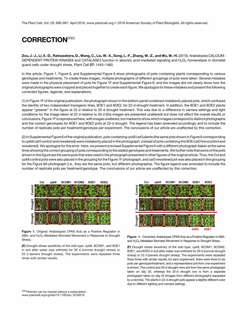
CORRECTIONOPEN
Zou, J.-J., Li, X.-D.,Ratnasekera,D.,Wang,C., Liu,W.-X., Song, L.-F., Zhang,W.-Z., andWu,W.-H. (2015). ArabidopsisCALCIUM-
DEPENDENT PROTEIN KINASE8 and CATALASE3 function in abscisic acid-mediated signaling and H2O2 homeostasis in stomatal
guard cells under drought stress. Plant Cell 27: 1445–1460.
In this article, Figure 1, Figure 6, and Supplemental Figure 6 show photographs of pots containing plants corresponding to various
genotypes and treatments. To create these images, multiple photographs of different groupings of pots were taken. Several mistakes
were made in the physical placement of pots for Figure 1F and Supplemental Figure 6, and the images did not clearly show how the
original photographswerecroppedandpieced together tocreateeachfigure.Weapologize for thesemistakesandpresent the following
corrected figures, legends, and explanations.
(1) In Figure 1F of the original publication, the photograph shown in the bottompanel containedmistakenly placed pots, which confused
the identity of two independent transgenic lines, 8OE1 and 8OE2, for 22-d drought treatment. In addition, the 8OE1 and 8OE2 plants
appear “greener” in the figure at 22 d relative to 20 d drought treatment. This was due to a difference in camera settings and light
conditions for the image taken at 22 d relative to 20 d (the images are presented unaltered) but does not affect the overall results or
conclusions. Figure1F is reproducedhere,with imagesunaltered,butmarked toshowwhich imagescorrespond todistinctphotographs
and the correct genotypes for 8OE1 and 8OE2 pots at 22-d drought. The legend has been amended accordingly and to include the
number of replicate pots per treatment/genotype per experiment. The conclusions of our article are unaffected by this correction.
(2) InSupplemental Figure 6of the original publication, pots containing cpk8/cat3plants (the samepots shown in Figure 6corresponding
to cpk8/cat3 control and rewatered)weremistakenly placed in the photograph, instead of pots containing the 8OE/cat3 line (control and
rewatered).Weapologize for this error. Here,wepresent a revisedSupplemental Figure 6with a different photograph (taken at the same
time) showing the correct grouping of pots corresponding to the stated genotypes and treatments.We further note that someof thepots
shown in this figure are the samepots thatwereused in thephotographpresented in other figures of theoriginal article. Thus, theCol and
cpk8 control potswere alsoplaced in the grouping for the Figure 1Fphotograph, and cat3 rewatered potwas alsoplaced in the grouping
for the Figure 6A photograph (i.e., they are the same pots, but different photographs). The figure legend was amended to include the
number of replicate pots per treatment/genotype. The conclusions of our article are unaffected by this correction.
Figure 1. Original: Arabidopsis CPK8 Acts as a Positive Regulator in
ABA- and H2O2-Mediated Stomatal Movement in Response to Drought
Stress.
(F) Drought stress sensitivity of the wild type, cpk8, 8COM1, and 8OE1
in soil after water was withheld for 20 d (normal drought stress) or
22 d (severe drought stress). The experiments were repeated three
times with similar results.
Figure 1. Corrected: Arabidopsis CPK8Acts as a Positive Regulator in ABA-
and H2O2-Mediated Stomatal Movement in Response to Drought Stress.
(F) Drought stress sensitivity of the wild type, cpk8, 8COM1, 8COM2,
8OE1, and 8OE2 in soil after water was withheld for 20 d (normal drought
stress) or 22 d (severe drought stress). The experiments were repeated
three times with similar results; for each experiment, there were three to six
pots per genotype/treatment, and a representative pot from one experiment
is shown. The control and 20-d drought rows are from the same photograph
taken on day 20, whereas the 22-d drought row is from a separate
photograph taken on day 22 (images from different photographs separated
by a red line). The plants in 22-d drought pots appear a slightly different color
due to different lighting and camera settings.
OPENArticles can be viewed without a subscription.www.plantcell.org/cgi/doi/10.1105/tpc.16.00210
The Plant Cell, Vol. 28: 990–991, April 2016, www.plantcell.org ã 2016 American Society of Plant Biologists. All rights reserved.
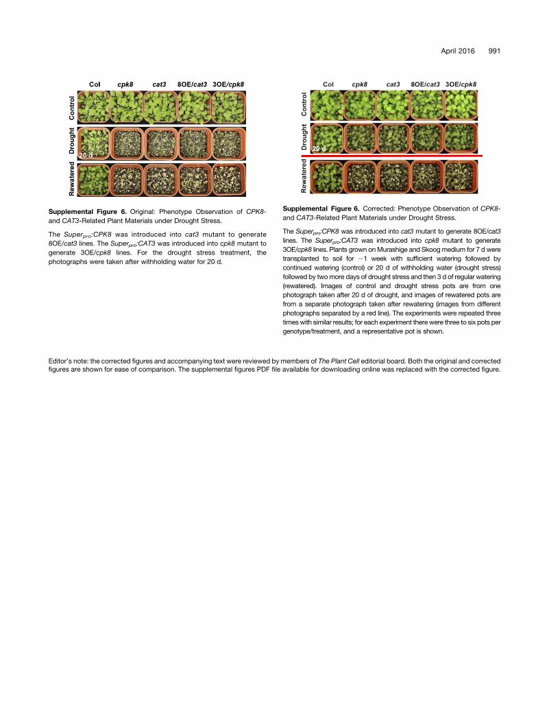
Editor’s note: the corrected figures and accompanying text were reviewed by members of The Plant Cell editorial board. Both the original and correctedfigures are shown for ease of comparison. The supplemental figures PDF file available for downloading online was replaced with the corrected figure.
Supplemental Figure 6. Original: Phenotype Observation of CPK8-
and CAT3-Related Plant Materials under Drought Stress.
The Superpro:CPK8 was introduced into cat3 mutant to generate
8OE/cat3 lines. The Superpro:CAT3 was introduced into cpk8 mutant to
generate 3OE/cpk8 lines. For the drought stress treatment, the
photographs were taken after withholding water for 20 d.
Supplemental Figure 6. Corrected: Phenotype Observation of CPK8-
and CAT3-Related Plant Materials under Drought Stress.
The Superpro:CPK8 was introduced into cat3 mutant to generate 8OE/cat3
lines. The Superpro:CAT3 was introduced into cpk8 mutant to generate
3OE/cpk8 lines. Plants grown onMurashige and Skoogmedium for 7 d were
transplanted to soil for ;1 week with sufficient watering followed by
continued watering (control) or 20 d of withholding water (drought stress)
followed by twomore days of drought stress and then 3 d of regular watering
(rewatered). Images of control and drought stress pots are from one
photograph taken after 20 d of drought, and images of rewatered pots are
from a separate photograph taken after rewatering (images from different
photographs separated by a red line). The experiments were repeated three
timeswith similar results; for each experiment therewere three to six pots per
genotype/treatment, and a representative pot is shown.
April 2016 991
DOI 10.1105/tpc.15.00144; originally published online May 12, 2015; 2015;27;1445-1460Plant Cell
Zhang and Wei-Hua WuJun-Jie Zou, Xi-Dong Li, Disna Ratnasekera, Cun Wang, Wen-Xin Liu, Lian-Fen Song, Wen-Zheng
Drought Stress Homeostasis in Stomatal Guard Cells under2O2Abscisic Acid-Mediated Signaling and H
Arabidopsis CALCIUM-DEPENDENT PROTEIN KINASE8 and CATALASE3 Function in
This information is current as of July 6, 2018
Supplemental Data /content/suppl/2015/04/29/tpc.15.00144.DC1.html
References /content/27/5/1445.full.html#ref-list-1
This article cites 101 articles, 38 of which can be accessed free at:
Permissions https://www.copyright.com/ccc/openurl.do?sid=pd_hw1532298X&issn=1532298X&WT.mc_id=pd_hw1532298X
eTOCs http://www.plantcell.org/cgi/alerts/ctmain
Sign up for eTOCs at:
CiteTrack Alerts http://www.plantcell.org/cgi/alerts/ctmain
Sign up for CiteTrack Alerts at:
Subscription Information http://www.aspb.org/publications/subscriptions.cfm
is available at:Plant Physiology and The Plant CellSubscription Information for
Errata
/content/28/4/990.full.pdfbe accessed at:An erratum has been published regarding this article. It is appended to this PDF and can also
ADVANCING THE SCIENCE OF PLANT BIOLOGY © American Society of Plant Biologists


![[3H]Nitrendipine-labeled calcium ... · [3H]Nitrendipine-labeled calciumchannelsdiscriminateinorganic ... Drugs that block these voltage-dependent calcium channels ... The subcellular](https://img.pdfslide.us/doc/110x75/5b92cae009d3f2d1448c069e/3hnitrendipine-labeled-calcium-3hnitrendipine-labeled-calciumchannelsdiscriminateinorganic.jpg)