Embed Size (px)
Citation preview

Calcium-dependent inactivation of thedihydropyridine-sensitive calcium channels inGH3 cellsDaniel Kalman, Emory UniversityP.H. Olague, University of California, Los AngelesC. Erxleben, University of California, Los AngelesD.L. Armstrong, University of California, Los Angeles
Journal Title: Journal of General PhysiologyVolume: Volume 92, Number 4Publisher: Rockefeller University Press | 1988-10-01, Pages 531-548Type of Work: Article | Final Publisher PDFPublisher DOI: 10.1085/jgp.92.4.531Permanent URL: https://pid.emory.edu/ark:/25593/rr536
Final published version: http://dx.doi.org/10.1085/jgp.92.4.531
Copyright information:© 1988 Rockefeller University PressThis is an Open Access work distributed under the terms of the CreativeCommons Attribution-NonCommercial-ShareAlike 3.0 Unported License(http://creativecommons.org/licenses/by-nc-sa/3.0/).
Accessed November 30, 2021 12:23 AM EST
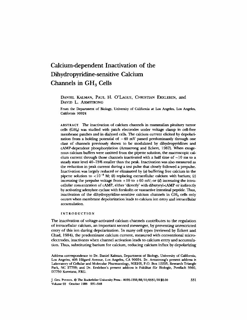
Calcium-dependent Inactivation of the Dihydropyridine-sensitive Calcium Channels in GHa Cells
DANIEL KALMAN, PAUL H. O'LAGUE, CHRISTIAN ERXLEBEN, a n d DAVID L. ARMSTRONG
From the Department of Biology, University of California at Los Angeles, Los Angeles, California 90024
ABSTRACT The inactivation of calcium channels in mammalian pituitary tumor cells (GHa) was studied with patch electrodes under voltage clamp in cell-free membrane patches and in dialyzed cells. The calcium current elicited by depolari- zation from a holding potential of - 4 0 mV passed predominantly through one class of channels previously shown to be modulated by dihydropyridines and cAMP-dependent phosphorylation (Armstrong and Eckert, 1987). When exoge- nous calcium buffers were omitted from the pipette solution, the macroscopic cal- cium current through those channels inactivated with a half time of ~ 10 ms to a steady state level 40-75% smaller than the peak. Inactivation was also measured as the reduction in peak current during a test pulse that closely followed a prepulse. Inactivation was largely reduced or eliminated by (a) buffering free calcium in the pipette solution to <10 -s M; (b) replacing extracellular calcium with barium; (c) increasing the prepulse voltage from + 10 to +60 mV; or (d) increasing the intra- cellular concentration of cAMP, either 'directly' with dibutyryl-cAMP or indirectly by activating adenylate cyclase with forskolin or vasoactive intestinal peptide. Thus, inactivation of the dihydropyridine-sensitive calcium channels in GH a cells only occurs when membrane depolarization leads to calcium ion entry and intracellular accumulation.
I N T R O D U C T I O N
The inactivation of voltage-activated calcium channels contributes to the regulation of intracellular calcium, an important second messenger, by preventing unrestricted entry of this ion during depolarization. In many cell types (reviewed by Eckert and Chad, 1984), the predominant calcium current, measured with conventional micro- electrodes, inactivates when channel activation leads to calcium entry and accumula- tion. Thus, substituting bar ium for calcium, reducing calcium influx by depolarizing
Address correspondence to Dr. Daniel Kalman, Department of Biology, University of California, Los Angeles, 405 Hilgard Avenue, Los Angeles, CA 90024. Dr. Armstrong's present address is Laboratory of Cellular and Molecular Pharmacology, NIEHS, P.O. Box 12233, Research Triangle Park, NC 27709; and Dr. Erxleben's present address is Fakiiltat fiir Biologle, Postfach 5560, D7750 Konstanz, FRG.
j. Gas. PHVSIOL �9 The Rockefeller University Press �9 0022-1295/88/10/0531/18 $2.00 531 Volume 92 October 1988 551-548
5 3 2 THE JOURNAL OF GENERAL PHYSIOLOGY �9 VOLUME 9 2 �9 1 9 8 8
close to the reversal potential for calcium, or buffering intracellular calcium concen- trations below 10 -s M all reduce or eliminate inactivation. In other cell types, cal- cium currents exhibit inactivation that appears to be voltage dependent as well as calcium dependent (Brown et al., 1981; Kass and Sanguinetti, 1984; Lee et al., 1985). This raises the possibility that for a single channel, these two mechanisms may not be mutually exclusive. Alternatively, the macroscopic current may reflect the activity of more than one class of channel, each of which inactivates by one of the two mechanisms (Deitmer, 1984, 1986).
In our laboratory, we are using patch-clamp techniques to study these mecha- nisms in cells o f the pituitary tumor line GHs. Recent patch-clamp studies on these cells (Armstrong and Matteson, 1985; Matteson and Armstrong, 1986; Cohen and McCarthy, 1987) as well as on other vertebrate cells (Carbone and Lux, 1984; Hess et al. 1984; Bean, 1985; Fedulova et al., 1985; Nilius et al., 1985; Nowycky et al., 1985), have revealed two classes of voltage-activated calcium channels. Channels of one class have a low threshold of activation (~ - 4 0 mV) and inactivate in a voltage- dependent manner. Channels of the other class activate at more positive voltages ( - - 20 mV) and are characterized by their sensitivity to dihydropyridines. Whether dihydropyridine-sensitive channels also inactivate, and if so by what mechanisms, has not been resolved. It is clear that these channels do not inactivate significantly when barium is the charge carrier, nor in whole cells dialyzed internally with exogenous calcium buffers (e.g., Matteson and Armstrong, 1986; Cohen and McCarthy, 1987). However, these experimental conditions are also ones that eliminate calcium-depen- dent inactivation. In fact, the presence of such a mechanism is suggested by the similarity between these channels and the calcium-inactivating channels in mollus- can neurons. Both lose activity or 'wash out ' when the cytoplasmic side of the mem- brane is exposed to minimal saline solutions (Byerly and Hagiwara, 1982; Fenwick et al., 1982; Cavalie et al., 1983; Matteson and Armstrong, 1986), an effect reversed by cAMP-dependent phosphorylation (Doroshenko et al., 1984; Chad and Eckert, 1986; Armstrong and Eckert, 1987).
Considering this similarity and that previous studies on these channels did not optimize conditions to detect calcium-dependent inactivation, we investigated whether the dihydropyridine-sensitive channels inactivate and whether they do so in a calcium-dependent manner. We report here that with calcium as the charge car- tier, and without including exogenous calcium buffers in the pipette solution, inac- tivation occurs when channel activation leads to calcium ion entry and accumula- tion. Furthermore, we find no evidence for direct effects of voltage on inactivation aside f rom those on calcium entry. Thus we conclude that the surface membrane of GH3 cells has calcium channels o f two classes, each of which inactivates by a separate mechanism, one dependent on calcium, the other on voltage. Some of the results have been published previously (Kalman et al., 1987a, b).
M E T H O D S
GHs cells, a mammalian cell line isolated from a rat pituitary tumor (rashjian, 1979), were obtained from American Type Culture Collection (Rockville, MD). Cells were grown in plastic tissue culture flasks (Coming Glass Works, Coming, NY) containing Ham's F10 supple- mented with 15% horse serum, 2.5% fetal calf serum, penicillin (50 ~g/ml), and streptomycin

~'r ~ . Calcium-dependent Inactivation 533
(50 U/ml) (GIBCO, Grand Island, NY) and incubated in a 5% CO~/95% air mixture at 37~ Cells were fed every 3 -4 d. For electrophysiological experiments, cells were mechanically dis- sociated and plated onto collagen-coated glass coverslips (No. 1, 12-ram diana; Fisher Scien- tific Co., Pittsburgh, PA). Each experiment was begun on a new coverslip of cells, and only cells grown on coverslips for longer than 1 d, but not more than 3 d were used in the exper- iments described here.
Cells (<20 ~tm in diameter) were voltage clamped in the whole-cell configuration and cell- free membrane patches were formed in the outside-out configuration as described by Hamill et al. (1981). Low resistance (<3 Mr0 patch pipettes were manufactured from Coming glass 7052, 7040, or 8161 (Garner Glass Co., Pomona, CA). Some of the results reported here were obtained with pipettes made from 8161 glass because they frequently made high resis- tance seals. Pipettes made of this glass have been reported to alter the kinetics of potassium currents in whole-cell recordings (Cota and Armstrong, 1988). We have observed that unitary barium currents are partially blocked in cell-attached recordings with 8161 glass (unpub- lished observations). However, in the whole-cell and outside-out patch recordings reported here, the kinetics of calcium currents appeared to be unaffected by this glass because identi- cal results were obtained with the other glasses.
Macroscopic currents through calcium channels were isolated from currents through volt- age-activated sodium and potassium channels as described by others (Hagiwara and Ohmori, 1982; Dubinsky and Oxford, 1984; Matteson and Armstrong, 1984, 1986). In most experi- ments, including those comparing barium and calcium currents, the bath contained either 25 mM CaCI~ or 25 mM BaCIi, 106 mM tetraethylammonium chloride (TEA-CI), 10 mM HEPES (pH 7.2), 1 mM MgClt, 5 mM KCI, 19 mM glucose, and 2 #M tetrodotoxin (TrX) (Sigma Chemical Co., St. Louis, MO), and the pipette was filled with a minimal saline solution con- sisting of 140 mM CsC1, 10 mM HEPES (pH 7.2), and 2 mM MgClz. In earlier experiments, the bath solution contained 130 mM NaCI, 10 mM CaCl~, 10 mM HEPES (pH 7.2), 2 mM MgCI~, 5 mM KC1, and 2 #M TTX. Where indicated, the free calcium concentration in the pipette solution was buffered to 10 -s M with 5 mM ethyleneglycol-biso(B-aminoethylether) N,N,N',N'otetraacetic acid (EGTA; Sigma Chemical Co.) or 5 mM bis-(o-aminophenoxy)- ethane-N,N,N'N' -tetraacetic acid (BAPTA, tetrasodium salt; Molecular Probes, Junction City, OR). For single-channel recordings from outside-out patches, the bath solution contained 90 mM BaC12, 20 mM TEA-CI, 10 mM HEPES (pH 7.2), and 2 ~M TIX, and the pipette was filled with 125 mM CsCI, 2 mM MgCI~, 5 mM EGTA (pCa = 8), and 40 mM HEPES (pH 7.2). In some experiments, the pipette solution was supplemented with 2 mM ATP-Mg and 2 ~g,/ ml purified catalytic subunit of cAMP-dependent protein kinase (cf., Armstrong and Eckert, 1987). Experiments were carrried out at room temperature (20-23~
Currents elicited every 6 s by depolarizing voltage steps were low-pass filtered at 2 kHz with an 8-pole Bessel filter, and digitized at 10 kHz for storage and analysis on an Indec 11-23 computer system (Indec Systems, Sunnyvale, CA). Linear leakage and uncancelled capacitive currents were subtracted digitally from the traces by a 1 / - 4 procedure. In this procedure the scaled average of currents elicited by four hyperpolarizing pulses of amplitude P/4, where P is the difference between the test and holding potential, was added to the test- pulse current. The subtraction procedure did not affect the kinetics of the calcium current during the step because leakage currents during the hyperpolarizing step were flat. More- over, leakage currents were always <5% of the current elicited by an equivalent depolarizing step.
Nimodipine (Miles Laboratories, New Haven, CT), a dihydropyridine antagonist of calcium channels in pituitary tumor cells (Armstrong and Eckert, 1987; Cohen and McCarthy, 1987), and forskolin (Calbiocbem, San Diego, CA), an activator of adenylate cyclase (Seamon et al., 1981), were dissolved in dimethylsulfoxide (DMSO) before their addition to the bath solu-
534 THE JOURNAL OF GENERAL PHYSIOLOGY �9 VOLUME 9 2 �9 1 9 8 8
tion. The final concentration of DMSO in the bath was always <0.1%, a concentration which produced no detectable effects on the calcium current. Vasoactive intestinal peptide ([k)eh- ringer Mannheim Biochemicals, Indianapolis, IN) and dibutyryl-c.AMP (Sigma Chemical Co.) were dissolved in the bath solution.
R E S U L T S
Separation of Two Classes of Calcium Channels
To study inactivation of dihydropyridine-sensitive channels in whole cell experi- ments, the current through those channels was isolated from that which went through dihydropyridine-insensitive channels. To do this, we took advantage of the finding that insensitive channels undergo voltage-dependent inactivation at holding potentials more positive than - 8 0 mV (Matteson and Armstrong, 1986; Cohen and McCarthy, 1987). We chose - 4 0 mV because it was the most positive holding potential below the activation threshold of either class of calcium channel, and we measured the extent to which dihydropyridine-insensitive channels inactivated at that holding potential.
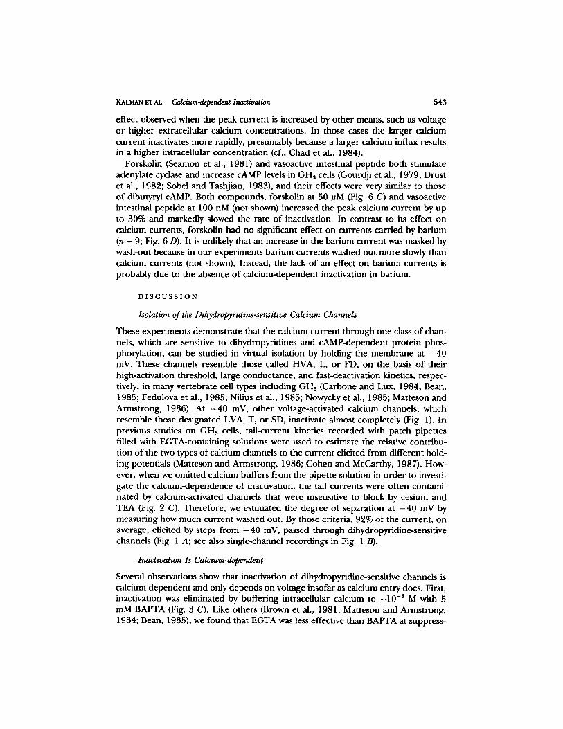
With barium as the charge carrier, we determined the relative contribution of the two types of calcium channels to the currents elicited from different holding poten- tials. This is illustrated in Fig. 1. In the whole-cell records in Fig. 1 A, the current elicited from a holding potential of - 4 0 mV (trace al) decreased by ~90% (i.e., washed out; trace bl) within 10 min of establishing the whole-cell recording with the minimal saline solution in the pipette. Under these conditions, wash-out was irre- versible (but see Chad and Eckert, 1986; and Armstrong and Eckert, 1987), that is, no further current could be elicited from - 4 0 mV despite holding at - 8 0 mV for several minutes. In addition, the current elicited from - 4 0 mV was almost com- pletely blocked by 1 izM nimodipine, a dihydropyridine antagonist (e.g., Fig. 2 B). In contrast, depolarization from a holding potential of - 8 0 mV elicited an additional transient current (Fig. 1, trace a2) that did not wash out (trace a2 ~ trace a~ + trace b2). The wash-out resistant component (trace b2) inactivated almost completely at a holding potential of - 4 0 mV (trace bl). Similar separation was obtained whether barium or calcium was the charge carrier. On average (n = 18), only 8% of the cur- rent elicited from ~40 mV was resistant to wash-out or nimodipine block.
At the single-channel level, we observed two classes of unitary barium currents during depolarizations of outside-out patches exposed to a similar minimal pipette solution. This is illustrated in Fig. 1 B. Channels of one class (three traces, a2 and b2) have a small conductance in 90 mM barium (~ 10 pS; measured previously by Arm- strong and Eckert, 1987) and a threshold of activation near - 4 0 mV. Channels of the other class (traces at) have a larger conductance in barium (~25 pS; measured previously) and a higher threshold of activation, near ~20 mV. Channels of both classes are blocked by 2 mM cobalt (not shown), but not by tetrodotoxin, and have extrapolated reversal potentials more positive than +40 mV. 6 rain after forming the outside-out patch with a minimal saline solution lacking reagents to support cAMP-dependent phosphorylation, the larger conductance channel stopped open- ing in response to depolarization (traces bt). In contrast, the smaller conductance channel continued to respond to depolarization (traces b2) as long as the seal
KALMAN ET AL. Ca/c/um-depeflde~ Inactivation 535
remained intact (~20 min). In the absence o f the larger conduc tance channel open- ings, the smaller conduc tance channels (traces b2) unde rgo nearly complete steady state inactivation at the holding potential o f - 4 0 mV (traces bt). Together , these results demons t ra te that the macroscopic cur ren t elicited f rom - 4 0 mV is primarily
A
B
OmV OmV Vl~=-40 mV', I . . . . . . . ~.80- mV- l l . . . . .
- - - b 2 after wash-out
al i5OPA .
20 ms
+lOmV OmV v = - 4 o ~v --1- . . . . . . . . . . . . . . . . . . L . . . . . . ' g 6 ~ . . . . . . . . . . . . . . . . . . - -
h - 8 0 mV _._1 L
a. Con t ro l : 2 m i n u t e s
a f t e r w a s h o u t : 6 m inu tes
..__J IpA 30ms
FIGURE 1. Whole-cell and single-channel barium currents before and after wash-out. Volt- age steps are shown above current records. (A) Whole-cell barium currents elicited by a depo- larizing step to 0 mV from a holding potential of - 40 mV (trace a j) washed out almost com- pletely (~90%) after 10 min (trace bt). In contrast, the current elicited from - 8 0 mV (trace a2) only partially washed out (~67%; trace b2). Note that the wash-out-resistant current elic- ited from - 8 0 mV (trace b2) was almost completely inactivated at a holding potential of - 4 0 mV (trace bt) because little current could be elicited by the step to 0 mV. (B) Unitary barium currents in an outside-out patch. (B, a2 and az) Current records taken at the indicated voltages (top traces) 2 min after excising the patch. (B, b~ and b2) The response to the same voltage steps as a, 4 rain later. Records were filtered at 2 kHz except those of b2, which were filtered at 1 kHz.
due to the high-threshold, la rge-conductance calcium channels that wash ou t entirely in minimal saline solutions.
Dihydropyridine-sensitive Channels Inactivate when Calcium Carries the Current
With calcium as charge carrier and with exogenous calcium buffers omi t ted f rom the pipette solution, currents elicited by steps f rom - 4 0 mV relaxed dur ing sus-
536 T H E J O U R N A L O F G E N E R A L P H Y S I O L O G Y �9 V O L U M E 9 ~ �9 1 9 8 8
0 mV
V"=-4OmV I I
A
15oo 25 ms
B b, + I # M nimodipine
25 ms
C 3, +20 mV
1, -20 mV V h : - 4 0 mV
I ] . . . . . . I
1 25 ms
FIGURE 2. Wash-out-sensitive calcium currents inactivate when no calcium buffers are included in the pipette solution. (A) Calcium current elicited by a step from - 4 0 to 0 mY. (B) Calcium current elicited by a step from - 4 0 to 0 mV (trace a) is blocked by 1 #M nimodipine (trace b). (C) Calcium currents elicited by voltage steps (top superimposed traces) to - 2 0 mV (trace 1), 0 mV (trace 2), and +20 mV (trace 3) from a holding potential of - 4 0 inV. Note the long-lasting inward tail currents seen after the steps to 0 mV and + 20 mV, which may result from nonselective cation channels or chloride channels activated by calcium entry dur- ing the step. Also note the marked inactivation in B despite the absence of the long-lasting inward tail current. The pipette solution contained cesium chloride but no calcium buffers.
KAt.MAN E'r AL. C a l c i u m ~ Inactivation 537
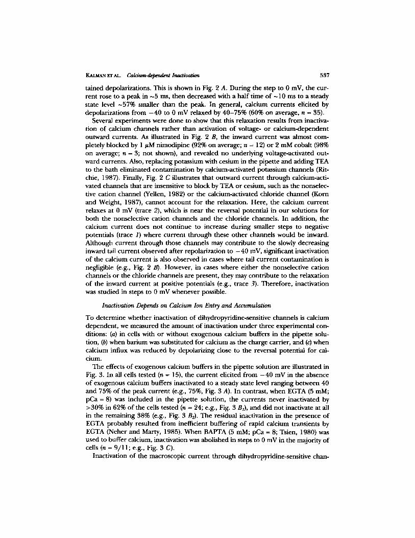
tained depolarizations. This is shown in Fig. 2 A. During the step to 0 mV, the cur- rent rose to a peak in ~5 ms, then decreased with a half time of ~10 ms to a steady state level ~57% smaller than the peak. In general, calcium currents elicited by depolarizations f rom - 4 0 to 0 mV relaxed by 40-75% (60% on average, n - 35).
Several experiments were done to show that this relaxation results f rom inactiva- tion of calcium channels ra ther than activation of voltage- or calcium-dependent outward currents. As illustrated in Fig. 2 B, the inward current was almost com- pletely blocked by 1 ttM nimodipine (92% on average; n = 12) or 2 mM cobalt (98% on average; n = 3; not shown), and revealed no underlying voltage-activated out- ward currents. Also, replacing potassium with cesium in the pipette and adding TEA to the bath eliminated contamination by calcium-activated potassium channels (Rit- chic, 1987). Finally, Fig. 2 C illustrates that outward current through calcium-acti- vated channels that are insensitive to block by TEA or cesium, such as the nonselec- tive cation channel (Yellen, 1982) or the calcium-activated chloride channel (Korn and Weight, 1987), cannot account for the relaxation. Here, the calcium current relaxes at 0 mV (trace 2), which is near the reversal potential in our solutions for both the nonselective cation channels and the chloride channels. In addition, the calcium current does not continue to increase during smaller steps to negative potentials (trace I) where current through these other channels would be inward. Although current through those channels may contribute to the slowly decreasing inward tail current observed after repolarization to - 4 0 mV, significant inactivation of the calcium current is also observed in cases where tail current contamination is negligible (e.g., Fig. 2 B). However, in cases where either the nonselective cation channels or the chloride channels are present, they may contribute to the relaxation of the inward current at positive potentials (e.g., trace 3). Therefore, inactivation was studied in steps to 0 mV whenever possible.
Inactivation Depends on Calcium Ion Entry and Accumulation
To determine whether inactivation of dihydropyridine-sensitive channels is calcium dependent , we measured the amount of inactivation under three experimental con- ditions: (a) in cells with or without exogenous calcium buffers in the pipette solu- tion, (b) when barium was substituted for calcium as the charge carrier, and (c) when calcium influx was reduced by depolarizing close to the reversal potential for cal- cium.
The effects o f exogenous calcium buffers in the pipette solution are illustrated in Fig. 3. In all cells tested (n = 15), the current elicited f rom - 4 0 mV in the absence of exogenous calcium buffers inactivated to a steady state level ranging between 40 and 75% of the peak current (e.g., 75%, Fig. 3 A). In contrast, when EGTA (5 raM; pCa = 8) was included in the pipette solution, the currents never inactivated by >30% in 62% of the cells tested (n = 24; e.g., Fig. 3 Bt), and did not inactivate at all in the remaining 38% (e.g., Fig. 3 B2). The residual inactivation in the presence of EGTA probably resulted f rom inefficient buffering of rapid calcium transients by EGTA (Neher and Marty, 1985). When BAPTA (5 mM; pCa = 8; Tsien, 1980) was used to buffer calcium, inactivation was abolished in steps to 0 mV in the majority of cells (n = 9/11; e.g., Fig. 3 C).
Inactivation of the macroscopic current through dihydropyridine-sensitive chan-

538 THE JOURNAL OF GENERAL PHYSIOLOGY �9 VOLUME 92 �9 1 9 8 8
nels did not occur when barium carried the current, even in the absence of exoge- nous buffers. Replacing extracellular calcium with equimolar barium caused the peak current to increase by up to threefold but did not produce any inactivation in voltage pulses lasting 160 ms (Fig. 4 A). A small relaxation was observed during longer steps (1,600 ms) to very positive potentials (e.g., +40 mV; Fig. 4 B, trace 2), even when BAPTA (5 mM) was included in the pipette solution to reduce intracellu- lar accumulation of barium. However, there was no inactivation of bar ium currents
A. No odded C0 buffers
~ 2 0 oA
20 ms
B]. EGTA B z. EGTA
50 pAI 40 pA i
40 ms
C. BAPTA
. . . . . . . . . F - - !
. ~ 1 0 0 oA
20 ms
FIGURE 3. Exogenous calcium buffers significantly slow or prevent inacfivadon. (A) Calcium current elicited by a step from a holding potential of - 40 to 0 mV in the absence of exoge- nous calcium buffers. Current inactivated by ~75%. (B) Effects of 5 mM EGTA in the pipette solution. Currents recorded in other cells from the same culture dish as in A showed substan- tially less inactivation (~30%; BI) or no inactivation at all (B2). (C) Effects of 5 mM BAPTA, a more efficient buffer of rapid calcium transients than EGTA.
during long pulses to less positive voltages (e.g., 0 mV; Fig. 4 B, trace 1), during which outward current contamination was less likely.
Further support for this lack of inactivation in bar ium can be seen in the single- channel records f rom cell-free patches in the outside-out configuration (Fig. 4, C and D). In these experiments, the dihydropyridine-sensitive channels were pre- vented f rom washing out by including Mg-ATP and the purified catalytic subunit o f the cAMP-dependent protein kinase in the pipette solution (Armstrong and Eckert, 1987). Fig. 4 C shows five representative traces of the unitary bar ium currents recorded during 160 ms depolarizations f rom - 4 0 to 0 mV, and an ensemble aver- age of all the records f rom that patch (bottom trace); with barium as the charge
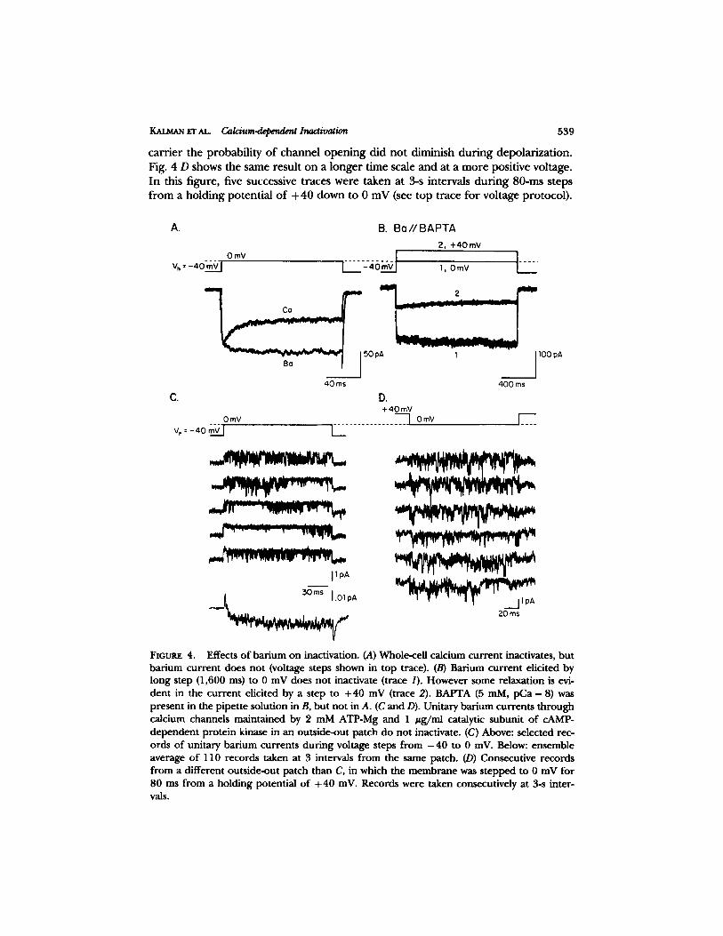
gr AL C~ldum.depend~ Inactivation 539
ca r r i e r the p robab i l i ty o f channe l o p e n i n g d id no t d iminish d u r i n g depo la r iza t ion . Fig. 4 D shows the same resul t on a l o n g e r t ime scale and at a m o r e posi t ive voltage. In this f igure, five successive t races were taken at 3-s intervals d u r i n g 80-ms s teps f rom a ho ld ing po ten t ia l o f + 40 down to 0 mV (see t op t race fo r vol tage pro tocol ) .
A. B. B o / / B A P T A
2, 4-40 mV
. . . . O m V . . . . I . . . . .
V.=-4OmVl I ,, OmV
4 0 ms 400 ms
D. + 40 mV
OmV . . . . . . . . . . . . . . . ~ OmV ] . . . .
v~ 4o~vl /
C.
I l i l l l I
I I oA
20 ms
FtGLrRE 4. Effects of barium on inactivation. (A) Whole-cell calcium current inactivates, but barium current does not (voltage steps shown in top trace). (B) Barium current elicited by long step (1,600 ms) to 0 mV does not inactivate (trace 1). However some relaxation is evi- dent in the current elicited by a step to +40 mV (trace 2). BAPTA (5 mM, pCa - 8) was present in the pipette solution in B, but not in A. (C and D). Unitary barium currents through calcium channels maintained by 2 mM ATP-Mg and 1 #g/ml catalytic subunit of cAMP- dependent protein kinase in an outride-out patch do not inactivate. (C) Above: selected rec- ords of unitary barium currents during voltage steps from - 4 0 to 0 inV. Below: ensemble average of 110 records taken at 3 intervals from the same patch. (D) Consecutive records from a different outside-out patch than C, in which the membrane was stepped to 0 mV for 80 ms from a holding potential of + 40 mV. Records were taken consecutively at 3-s inter- vals.
A + 6 0 mV
Vh = - 4 0 m V
Vpre Vtes t 4 0 ms I
B O~ �9
~D
. - - 0 . 2 5 -o N
"-- - 0.50 o
E o Z - 0.75
- I .0
-~,0
C 1.0 m
o ~. 0 . 7 5
" tD (D N
= 0 . 5 0 O
E o
Z O.25
Q
-b
0
D
0 0
I
0 2b Vpr e ( m Y )
Ca �9 " Bo
4b 6b
0 o 0 0
B0
Co
es
t i I
o o 2'0 40 6b V p , , ( m V )
FIGURE 5. Inactivation of dihydropyridine-sensitive current (whole-cell recording) depends on voltage only to the extent that calcium entry depends on voltage. (A) Paired-pulse proto- col: calcium or barium currents were elicited from a holding potential of - 4 0 mV (two dif- ferent cells). A 60-ms prepulse of various amplitudes between - 4 0 and +60 mV was fol- lowed after 20 ms by a 60-ms test pulse to 0 mV. (B) Peak current recorded during the pre- pulse is plotted as a function of the prepulse voltage. The currents are normalized to the maximum current for each charge carrier. Both calcium (m) and barium (Fq) currents peaked near +10 mV. As the prepulse voltage is increased from +10 to +60 mV, the prepulse current declines because of the decrease in driving force. (C) The peak current during the test pulse is plotted as a function of the prepulse voltage. The test-pulse current is normalized to the current recorded in the absence of a prepulse. The calcium current ( i ) during the test pulse is maximally suppressed at potentials where calcium entry during the prepulse is maxi- mal. Barium current (F-I) is not suppressed by prepulses to any voltage.
KALMAN El" AL. C t l / , c / u m ~ Inact/vat/on 541
Although the membrane was held continuously at + 40 mV between steps, activity at 0 mV was robust. Together, these results provide no evidence for a voltage-depen- dent component of inactivation.
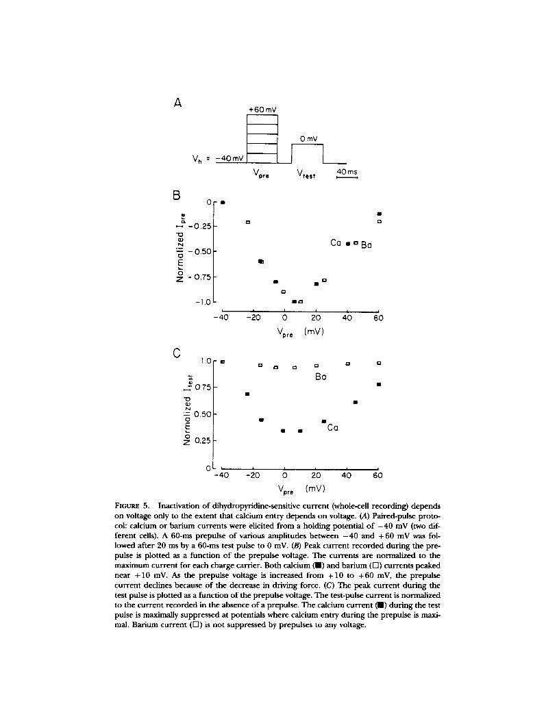
In addition, no voltage-dependence is indicated by the double-pulse experiments shown in Fig. 5. Here, inactivation was measured as the reduction in peak current during a test pulse that followed a prepulse. We used the paired-pulse protocol (Eckert and Tillotson, 1981) illustrated in Fig. 5 A to investigate whether inactiva- tion occurred as a result of calcium entry during the prepulse or as a result of the prepulse depolarization itself. The peak amplitudes of calcium (m, Fig. 5 B) and barium currents (D, Fig. 5 B) elicited by the prepulse in two different cells are plot- ted in Fig. 5 B as a function of the prepulse voltage. They show the typical current- voltage relation for the dihydropFridine-sensitive calcium channels: a threshold near - 2 0 mV, a maximum near + 10 mV, and an extrapolated reversal potential near +70 mV. As the prepulse voltage was increased from - 4 0 to +10 mV, calcium entry during the prepulse increased (Fig. 5 B), and the calcium current during the unchanging test pulse to 0 mV was correspondingly suppressed (m, Fig. 5 C). When the prepulse and the test pulse were equal (0 mV in this case), the peak current observed during the test pulse was approximately equal to the inactivated current amplitude at the end of prepulse. Thus, the inactivation produced by the prepulse showed little or no recovery during the 20-ms delay between the two pulses, and both the single- and paired-pulse protocols yielded equivalent measures of inactiva- tion.
As the prepulse voltage was increased further from + 10 to +60 mV (Fig. 5 B), less suppression occurred and the amplitude of the calcium current during the test pulse increased. This reduction in the amount of inactivation produced by the pre- pulse paralleled the decrease in calcium entry during the prepulse in the same volt- age range (compare Fig. 5 B with C). In contrast, when barium replaced calcium, currents during the prepulse did not inactivate, and little or no suppression during the test pulse was observed at any prepulse voltage (O, Fig. 5 C). I f inactivation were voltage dependent, higher prepulse voltages would have enhanced, not dimin- ished, inactivation of the test-pulse current carded by either calcium or barium. Such behavior is observed for isolated dihydropyridine-insensitive currents in steps from - 80 mV (not shown). Thus, inactivation of dihydropyridine-sensitive channels appears voltage dependent only to the extent that calcium entry is voltage depen- dent.
Raising Internal cAMP Slows Calcium-dependent Inactivation
The mechanism by which calcium produces inactivation is unknown. However, sev- eral previous observations have indicated a role for phosphorylation/dephosphory- lation reactions in the control of calcium channel activity in GH3 cells (Armstrong and Eckert, 1987; Chad et al., 1987; Armstrong and Kalman, 1988). Therefore, we tested the effect of increasing intracellular cAMP on the dihydropyridine-sensitive calcium current (Fig. 6). As seen in Fig. 6 A, even though the current was washing out, both the peak (m) and the steady state (F-l) current increased when a mem- brane-permeable analogue of cAMP (dibutyryl-cAMP, 2 mM) was added to the bath. These effects cannot have resulted simply from a shift in the current-voltage rela-

542 THE JOURNAL OF GENERAL PHYSIOLOGY �9 VOLUME 92 �9 1988
tion because similar effects were observed at all voltages (not shown). In all cells tested, the percentage increase in steady state cur ren t (e.g., 100% in Fig. 6 A) was substantially larger than the increase in peak cur ren t (e.g., 35% in Fig. 6 A). Thus, the net effect o f dibutyryl-cAMP was to decrease the effectiveness by which calcium p roduced inactivation. The cont inued wash-out o f peak cur ren t in the presence o f
B
80
60
40
20
+ 2 mM dibu~yryl cyclic AMP
I n I � 9
�9 8 1 l i d I
| ~ H | | ~ a | l | | i l
q Cg~ g ~am
0 i * |
0 60 120 180
T ime (s)
+ dibutyry l cyclic AMP
C J �9
~ I ~ ~, ~0~ C, 75 pA
+ forskol in _ _ 0,]50pA 20 ms
FIGURE 6. I n c r e a s i n g i n t r a -
cellular cAMP increases peak and steady state currents and slows calcium-dependent inac- tivation. (A) The peak ( I ) and steady state (I-I) amplitudes of calcium currents elicited by depolarizing steps from - 4 0 to 0 mV are plotted as a func- tion of time after establish- ing the whole-cell recording. Although the current was washing out, the addition of dibutyryl-cAMP increased the peak current by 35% and the steady state current by 100%. The larger percentage increase of the steady state current indi- cates that inactivation slowed. (B) Inactivation of equal ampli- tude calcium currents elicited by steps from - 4 0 to 0 mV is slower after the addition of 2 mM dibutyryl-cAMP to the bath. The increase in the peak current is not evident because the current had washed out further by the time the effect on inactivation had developed fully. (C) Addition of 50 #M forskolin to the bath slows the inactivation of calcium cur- rents elicited by steps to 0 mV
from a holding potential of - 4 0 mV. The step to 0 mV minimized contamination of the current during the pulse by chloride or nonselective cation channels. (D) In contrast, the addition of 50 ttM forskolin has little effect on currents carried by barium, which do not inactivate.
dibutyryl-cAMP allowed the effect o f this substance on inactivation to be seen more clearly. In Fig. 6 B, cur ren t traces with roughly equal peak amplitudes in steps to 0 mV, recorded before and after dibutyryl-cAMP application, are superimposed. Sim- ilar effects on inactivation were observed at o ther voltages ( - 20 and + 20 mV). The decrease in inactivation p roduced by dibutyryl-cAMP contrasts sharply with the
KAL~,N ~r AL. Calcium-dependent Inactivation 543
effect observed when the peak current is increased by other means, such as voltage or higher extracellular calcium concentrations. In those cases the larger calcium current inactivates more rapidly, presumably because a larger calcium influx results in a higher intracellular concentration (cf., Chad et al., 1984).
Forskolin (Seamon et al., 1981) and vasoactive intestinal peptide both stimulate adenylate cyclase and increase cAMP levels in GHs cells (Gourdji et al., 1979; Drust et al., 1982; Sobel and Tashjian, 1983), and their effects were very similar to those of dibutyryl cAMP. Both compounds, forskolin at 50 #M (Fig. 6 C) and vasoactive intestinal peptide at 100 nM (not shown) increased the peak calcium current by up to 30% and markedly slowed the rate of inactivation. In contrast to its effect on calcium currents, forskolin had no significant effect on currents carded by barium (n = 9; Fig. 6 D). It is unlikely that an increase in the barium current was masked by wash-out because in our experiments barium currents washed out more slowly than calcium currents (not shown). Instead, the lack of an effect on barium currents is probably due to the absence of calcium-dependent inactivation in barium.
DISCUSSION
Isolation of the Dihydropyridine-sensitive Calcium Channels
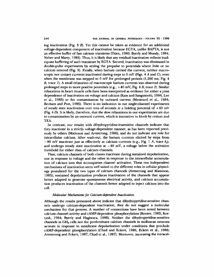
These experiments demonstrate that the calcium current through one class of chan- nels, which are sensitive to dihydropyridines and cAMP-dependent protein phos- phorylation, can be studied in virtual isolation by holding the membrane at - 4 0 mV. These channels resemble those called HVA, L, or FD, on the basis of their high-activation threshold, large conductance, and fast-deactivation kinetics, respec- tively, in many vertebrate cell types including GHs (Carbone and Lux, 1984; Bean, 1985; Fedulova et al., 1985; Nilius et al., 1985; Nowycky et al., 1985; Matteson and Armstrong, 1986). At - 4 0 mV, other voltage-activated calcium channels, which resemble those designated LVA, T, or SD, inactivate almost completely (Fig. 1). In previous studies on GH3 cells, tall-current kinetics recorded with patch pipettes filled with EGTA-containing solutions were used to estimate the relative contribu- tion of the two types of calcium channels to the current elicited from different hold- ing potentials (Matteson and Armstrong, 1986; Cohen and McCarthy, 1987). How- ever, when we omitted calcium buffers from the pipette solution in order to investi- gate the calcium-dependence of inactivation, the tail currents were often contami- nated by calcium-activated channels that were insensitive to block by cesium and TEA (Fig. 2 C). Therefore, we estimated the degree of separation at - 4 0 mV by measuring how much current washed out. By those criteria, 92% of the current, on average, elicited by steps from - 4 0 mV, passed through dihydropyridine-sensitive channels (Fig. 1 A; see also single-channel recordings in Fig. 1 B).
Inactivation Is Calcium-dependent
Several observations show that inactivation of dihydropyridine-sensitive channels is calcium dependent and only depends on voltage insofar as calcium entry does. First, inactivation was eliminated by buffering intracellular calcium to ~ 10 -8 M with 5 mM BAPTA (Fig. 3 C). Like others (Brown et al., 1981; Matteson and Armstrong, 1984; Bean, 1985), we found that EGTA was less effective than BAPTA at suppress-
5 4 4 THE JOURNAL OF GENERAL PHYSIOLOGY �9 VOLUME 92 �9 1 9 8 8
ing inactivation (Fig. 3 B). Yet this cannot be taken as evidence for an additional voltage-dependent component of inactivation because EGTA, unlike BAPTA, is not an effective buffer of fast calcium transients (Tsien, 1980; Byerly and Moody, 1984; Neher and Marty, 1985). Thus, it is likely that any residual inactivation reflects inad- equate buffering of such transients by EGTA. Second, inactivation was eliminated in double-pulse experiments by setting the prepulse to potentials where little or no calcium entered (Fig. 5). Finally, when barium carried the current, neither macro- scopic nor unitary currents inactivated during steps to 0 mV (Figs. 4 A and C), even when the membrane was stepped to 0 mV for prolonged periods (1,200 ms; Fig. 4 B, trace 1). A small relaxation of macroscopic barium currents was observed during prolonged steps to more positive potentials (e.g., + 40 mV; Fig. 4 B, trace 2). Similar relaxations in heart muscle cells have been interpreted as evidence for either a joint dependence of inactivation on voltage and calcium (Kass and Sanguinetti, 1984, Lee et al., 1985) or for contamination by outward current (Mentrard et al., 1984; Becham and Pott, 1985). There is no indication in our single-channel experiments of steady state inactivation over tens of seconds at a holding potential of +40 mV (Fig. 4 D). It is likely, therefore, that the slow relaxations in our experiments are due to contamination by an outward current, which is insensitive to block by cesium and TEA.
In contrast, our results with dihydropyridine-insensitive channels indicate that they inactivate in a strictly voltage-dependent manner, as has been reported previ- ously by others (Matteson and Armstrong, 1986), and do not indicate any role for intracellular calcium. After wash-out, the barium currents elicited by steps from - 8 0 mV inactivate just as effectively as calcium currents (e.g., Fig. 1 A, trace b2), and undergo steady state inactivation at - 4 0 mV, a voltage below the activation threshold for either class of calcium channels.
Thus, calcium channels of both classes inactivate during sustained depolarization, one in response to voltage and the other in response to the intracellular accumula- tion of calcium ions that accompanies channel activation. These two independent mechanisms of inactivation seem well suited to the different roles in cellular physiol- ogy postulated for the two types of calcium channels (Armstrong and Matteson, 1985); sustained depolarization produces inactivation of the channels that appear better adapted to generate spontaneous electrical activity, and calcium accumula- tion produces inactivation of the channels better adapted to inject calcium into the cell.
Molecular Mechanisms for Calcium-dependent Inactivation
Although the results presented above indicate that dihydropyridine-sensitive chan- nels undergo calcium-dependent inactivation, they do not suggest a molecular mechanism for that process. A number of connections have been noted between calcium channel activity and cAMP-dependent phosphorylation (Reuter, 1983; Kos- tyuk, 1984; Byerly and Hagiwara, 1988). Neither the dihydropyridine-sensitive channels in GHs cells nor the predominant calcium channels in molluscan neurons activate in response to membrane depolarization under conditions that preclude cAMP-dependent phosphorylation (Chad and Eckert, 1986; Eckert et al., 1986; Armstrong and Eckert, 1987; Chad et al., 1987). Moreover, increasing the intracel-

KALM~ gr AL. Calcium-dependent Inactivation 545
lular concent ra t ion o f cAMP has been repor ted to slow calcium channel inactivation in several cell types (Doroshenko et al., 1982; Bean et al., 1984; Bean, 1985; Chad and Eckert, 1986; Eckert et al., 1986; Chad et al., 1987). Here , we also f o u n d that raising cAMP levels r educed the effectiveness o f calcium in p roduc ing inactivation (Fig. 6). Together , these observations suggest that ca lc ium-dependent inactivation may be a general p roper ty o f calcium channels regulated by cAMP-dependen t phos- phorylation.
Eckert and Chad (1984) p roposed that inactivation results f rom dephosphoryla- tion o f the channel o r a closely associated molecule in the m e m b r a n e by an endoge- nous ca lc ium-dependent phosphatase. They f o u n d that perfus ing molluscan neu- rons internally with calcineurin, a calcium- and ca lmodul in-dependent phosphatase purif ied f rom mammalian brain (Klee et al., 1979; Stewart et al., 1982), accelerates inactivation in a ca lc ium-dependent m a n n e r (Chad and Eckert, 1986). While GHs cells contain calcineurin (Farber et al., 1987), it remains to be de te rmined whether it o r ano the r phosphatase participates in the inactivation process. Alternatively, inacti- vation might result f rom calcium binding directly to the channel. In this view, dephosphoryla t ion at some o ther site might alter allosterically the efficacy o f cal- c ium action. Elucidating the precise role o f calcium and calcineurin, o r o ther endogenous phosphatases, in calcium channel gat ing will require the measuremen t o f channel activitity u n d e r condit ions in which phosphatase activity is blocked directly.
We are indebted to Francisco Bezanilla and Vladimir Brezina for many helpful discussions while the experiments were in progress, and to them and to George Augustine, Paul Brehm, and Carol Vandenberg for commenting on an earlier draft of the manuscript. We would also like to thank Angus Nairn for providing us with the affinity-purified catalytic subunit of the cAMP-dependent protein kinase, A. Scriabine (Miles Laboratories) for the gift of nimodipine, and Taras Momdjian for technical assistance.
This work was supported by U.S. Public Health Service grant NS-08364 to Roger Eckert. D. Kal- man was a Predoctoral Fellow on the Cell and Molecular Biology Training Grant (U.S. Public Health Service grant GM-07185) at University of California, Los Angeles, and D. Armstrong was a Senior Investigator of the Greater Los Angeles Affiliate of the American Heart Association.
Original version received 4 September 1987 and accepted version received 14 June 1988.
R E F E R E N C E S
Armstrong, C. M., and D. R. Matteson. 1985. Two distinct populations of calcium channels in a clonal line of pituitary cells. &/once. 227:65-67.
Armstrong, D., and R. Eckert. 1987. Voltage-activated calcium channels that must be phosphoryl- ated to respond to membrane depolarization. Proceedings of the Natimml Academy of Sciences USA. 84:2518-2522.
Armstrong, D., and D. Kalman. 1988. The role of protein phosphorylation in the response of dihydropyridine-sensitive calcium channels to membrane depolarization in mammalian pituitary tumor cells. In Calcium and Ion Channel Modulation. A. Grinnell, D. Armstrong, and M. Jack- son, editors. Plenum Publishing Corp., New York. 215-288.
Bean, P. B. 1985. Two kinds of calcium channels in canine atrial cells.Journal of General Physiology. 86:1-30.
546 THE JOURNAL OF GENERAL PHYSIOLOGY �9 VOLUME 92 �9 1988
Bean, P. B., M. C. Nowycky, and R. W. Tsien. 1984. Beta-adrenergic modulation of calcium chan- nels in frog ventricular heart cells. Nature. 307:371-375.
Becham, M., and L. Pott. 1985. Removal of Ca current inactivation in dialysed guinea-pig atrial
cardioballs by Ca chelators. Pfliigers Archiv European Journal of Physiology. 404:10-20. Brown, A. M., K. Morimoto, Y. Tsuda, and D. L. Wilson. 1981. Calcium current dependent and
voltage dependent inactivation of calcium channels in Helix aspersa. Journal of Physiology. 320:193-218.
Byerly, L., and S. Hagiwara, 1982. Calcium currents in internally perfused nerve cell bodies of Lymnaea stagnalis. Journal of Physiology. 322:503-528.
Byerly, L., and S. Hagiwara, 1988. Calcium channel diversity. In Calcium and Ion Channel Modu- lation. A. Grinnell, D. Armstrong, and M. Jackson, editors. Plenum Publishing Corp., New York.
3-18. Byerly, L., and W. Moody. 1984. Intracellular calcium ions and calcium currents in perfused neu-
rons of the snail, Lymnaea stagnalis. Journal of Physiology. 352:637-652. Carbone, E., and H. D. Lux. 1984. A low voltage-activated fully inactivating Ca channel in verte-
brate sensory neurons. Nature. 310:501-502. Cavalie, A., R. Ochi, D. Pelzer, and W. Trautwein. 1983. Elementary currents through Ca ~+ chan-
nels in guinea pig myocytes. PflV~gers Archly European Journal of Physiology. 398:284-297. Chad, J. E., and R. Eckert. 1986. An enzymatic mechanism for calcium current inactivation in
dialyzed Helix neurons.Journal of Physiology. 378:31-51. Chad, J. E., R. Eckert, and D. Ewald. 1984. Kinetics of Ca-dependent inactivation of calcium cur-
rent in neurones of Aplysia californica. Journal of Physiology. 347:279-300. Chad, J. E., D. Kalman, and D. Armstrong, 1987. The role of cyclic AMP-dependent phosphoryla-
tion in the maintenance and modulation of voltage-activated calcium channels. In Cell Calcium and the Control of Membrane Transport. Voi. 42. D. C. Eaton and L. J. Mandel, editors. The Rockefeller University Press, New York. 167-186.
Cohen, C. J., and R. T. McCarthy. 1987. Nimodipine block of calcium channels in rat anterior pituitary cells. Journal of Physiology. 387:195-225.
Cota, G., and C. M. Armstrong. 1988. Potassium channel "inactivation" induced by soft glass patch pipettes. Biophysical Journal. 53:107-109.
Deitmer, J. 1984. Evidence for two voltage-dependent calcium currents in the membrane of the ciliate Stylonychia mytilus. Journal of Physiology. 355:137-159.
Deitmer, J. 1986. Voltage-dependence of two inward currents carded by calcium and barium in the ciliate Stylonychia raytilus. Journal of Physiology. 380:551-574.
Doroshenko, P. A., P. G. Kostyuk, and A. I. Martynyuk. 1982. Intracellular metabolism of adeno- sine 3'-5'-cyclic monophosphate and calcium inward current in perfused neurons of Helix poma- tia. Neuroscience. 7:2125-2134.
Doroshenko, P. A., P. G. Kostyuk, A. I. Martynyuk, M. D. Kursky, and Z. D. Vorobetz. 1984. Intracellular protein kinase and calcium inward currents in perfused neurons of the snail Helix pomatia. Neuroscience. 11:263.
Drust, D. S., C. A. Sutton, and T. F .J . Martin. 1982. Thyrotropin-releasing hormone and cyclic AMP activate distinctive pathways of protein phosphorylation in GH pituitary cells. Journal of Biological Chemistry. 257:3306-3312.
Dubinsky, J. M., and G. S. Oxford. 1984. Ionic currents in two strains of rat anterior pituitary tumor cells. Journal of General Physiology. 83:309-339.
Eckert, R., andJ. E. Chad. 1984. Inactivation of calcium channels. Progress In Biophysics and Molec- ular Biology. 44:215-267.
Eckert, R., J. E. Chad, and D. Kalman. 1986. Enzymatic regulation of the calcium current in dia- lyzed and intact molluscan neurones of Aplysia californica. Journal de Physiologie. 81:318-324.

gr AL. C~2dum-depend~ Inactivation 547
Eckert, R., and D. Tillotson. 1981. Calcium-mediated inactivation of the calcium conductance in caesium-loaded giant neurones of Aplysia californica. Journal of Physiology. 314:265-280.
Farber, L., and D. J. Wolff. 1987. Calmodulin dependent phosphatases of PC12 and C6 cells: physical, kinetic, and immunochemical properties. Journal of Neurochemistr 3. 49:404-414.
Fedulova, S. A., P. G. Kostyuk, and N. S. Veselovsky. 1985. Two types of calcium channels in the somatic membrane of new-born rat dorsal root ganglion neurones. Journal of Physiology. 359:431-446.
Fenwick, E. M., A. Marry, and E. Neber. 1982. Sodium and calcium channels in bovine chromaffm cells. Journal of Physiology. 331:599-635.
Gourdji D., D. Bataille, N. Vauclin, D. Grouselle, D. Rosselin, and A. Tixier-Vidal. 1979. Vasoac- tire intestinal peptide stimulates prolactin release and cAMP production in a rat pituitary cell line. FEBS LaUrs. 104:165-168.
Hagiwara, S., and H. Ohmori. 1982. Studies of calcium channels in rat clonal pituitary cells with patch electrode voltage clamp. Journal of Physiology. 331:231-252.
Hamill, O. P., A. Marty, E. Neher, B. Sakmann, and F. J. Sigworth. 1981. Improved patch clamp techniques for high-resolution current recording from cells and cell-free membrane patches. Pfliigers Archiv European Journal of Physiology. 398:284--297.
Hess, P., J. B. Lansman, and R. W. Tsien. 1984. Different modes of calcium channel gating behav- iour favoured by dihydrnpyridine Ca agonists and antagonism. Nature. 311:538-544.
Kalrnan, D., C. Erxleben, and D. Armstrong. 1987a. Inactivation of the dihydropyridine-sensitive calcium current in GH3 cells is a calcium-dependent process. Biophysical Journal. 51:432a. (Abstr.)
Kalman, D., P. H. O'Lague, and D. Armstrong. 1987b. Increasing the intracellular concentration of cAMP reduces Ca-dependent inactivation of Ca channels. Neuroscience Abstracta. 13:104. (Abstr.)
Kass, R. S., and M. C. Sanguinetti. 1984. Inactivation of calcium channel current in the calf cardiac Purkinje fiber. Journal of General Physiology. 84:705-726.
Klee, C. B., T. H. Crouch, and M. H. Krinks. 1979. Calcineurin: a calcium- and calmodulin-binding protein of the nervous system. Proceedings of the National Academy of Sciences USA. 76:6270- 6273.
Korn, S. J., and F. F. Weight, 1987. Patch clamp study of the calcium-dependent chloride current in AtT-20 pituitary cells. Journad of Neurophysiology. 58:1431-1450.
Kostyuk, P. G. 1984. Metabolic control of ionic channels in the neuronal membrane. Neuroscience. 13:983-989.
Lee, K. S., E. Marban, and R. W. Tsien. 1985. Inactivation of calcium channels in mammalian heart cells: joint dependence on membrane potential and intracellular calcium. Journal of Physi- ology. 364:395-411.
Matteson, D. R., and C. M. Armstrong. 1984. Na and Ca channels in a transformed line of anterior pituitary cells.Journal of General Physiology. 83:371-394.
Matteson, D. R., and C. M. Armstrong. 1986. Properties of two types of calcium channels in clonal pituitary cells.Journal of General Physiology. 87:161-182.
Mentrard, D., G. Vassort, and R. Fischmeister. 1984. Calcium-mediated inactivation of the calcium conductance in cesium-loaded frog heart cells.Journal of General Physiology. 83:105-131.
Neher, E., and A. Marty. 1985. BAPTA, unlike EGTA, efficiently suppresses Ca transients in chro- maffin cells. Biophysical Journal. 47:278a. (Abstr.)
Nilius, B., P. Hess, J. B. Lansman, and R. W. Tsien. 1985. A novel type of cardiac calcium channel in ventricular cells. Nature. 316:443-446.

548 THE JOVRNAL OF GENERAL PHYSIOLOGY �9 VOLUME 92 �9 1988
Nowycky, M. C., A. P. Fox, and R. W. Tsien. 1985. Three types of neuronal calcium channel with different calcium agonist sensitivity. Nature. 316:440-443.
Reuter, H. 1983. Calcium channel modulation by neurotransmitters, enzymes and drugs. Nature. 301:569-574.
Ritchie, A. K. 1987. Two distinct calcium-activated potassium currents in a rat anterior pituitary cell line. Journal of Physiology. 385:591-609.
Seamon, K. B., W. Padgett, andJ . W. Daly. 1981. Forskolin: a unique diterpene activator of ade- nylate cyclase in membranes and in intact cells. Proceedings of the National Academy of Sciences (IVY). 78:3363-3367.
Sobel, A., and A. H. Tashjian, Jr. 1983. Distinct patterns of cytoplasmic protein phosphorylation related to regulation of synthesis and release of prolactin by GH cells.Journal of Biological Chem- istry. 258:10312-10324.
Stewart, A. A., T. S. Inghretsen, A. Manalan, C. B. Klee, and P. Cohen. 1982. Discovery of a Ca- and calmodulin-dependent protein phosphatase: probably identity with calcineurin (C, aM-BPs0). FEBS Letters. 137:80-84.
Tashjian, A. H. 1979. Cional strains of hormone-producing cells. Methods in Enzymology. 58:527- 535.
Tsien, R. Y. 1980. New calcium indicators and buffers with high selectivity against magnesium and protons: design, synthesis, and properties of prototype structures. Biochemistry. 19:2396-2404.
YeUen, G. 1982. Single Ca~+-activated non-selective cation channels in neuroblastoma. Nature. 296:357-359.