Embed Size (px)
Citation preview

Approach to Patient with Headache
Mohammed Al Arabi 431104498Abdullah Al Turki 431101446

Question 1: Which of the following is true about headache?
a) Headache is overdiagnosed and over-treated.
b) Headache is more commonly a symptom of another systemic
disease.
c) All headache cases are referred to specialists.
d) Headache is under-recognized and under-treated.
Question 2: Which of the following investigations is indicated
in a patient with headache, nausea/vomiting, and
papilledema once an intracranial mass lesion is
ruled out?
a) Electroencephalogram.
b) Blood culture.
c) Cerebrospinal Fluid analysis.
d) PET scan.
Question 3: Which of the following features presenting
with headache does NOT indicate the need
of neuroimaging?
a) History of cancer.
b) Altered level of consciousness.
c) Rythmical recurrence.
d) Immunocompromised state

Objectives: Common types of headache “Migraine, Tension headache, Cluster
headache”.
How to approach a patient with headache.
Red Flags and indications for further investigations like CT brain, MRI.
Brief comment on Migraine, Tension Headache, Cluster headache, benign
intracranial hypertension, temporal arteritis, space occupying headaches.
What is the role of primary health care physician in management “Drug
treatment and Prophylaxis”.
What investigations could be requested if needed
When to refer to specialist.
Definition and Epidemiology:
Headache is pain in any part of the head and
neck, including the scalp, face (including the
orbitotemporal area), and interior of the head.
Headache disorders are among the most common
disorders of the nervous system.
It has been estimated that 47% of the adult
population have headache at least once within
last year in general.

Definition and Epidemiology:
Headache disorders are associated with personal and societal burdens of pain, disability, damaged quality of life and financial cost.
A minority of people with headache disorders worldwide are diagnosed appropriately by a health-care provider.
Headache has been underestimated, under-recognized and under-treated throughout the world.
[WHO, http://www.who.int/mediacentre/factsheets/fs277/en/]

Etiology: Primary Causes:
o Tension Headacheo Cluster Headacheo Migraine Headache

Etiology: Secondary Causes:
Cause Examples
Extracranial disorders Glaucoma, Sinusitis, …
Intracranial disorders Brain tumors and other masses, Hemorrhage, Idiopathic intracranial hypertension, Infections, Vascular disorders,…
Systemic disorders Acute severe hypertension, Fever, Giant cell arteritis,…
Drugs and toxins Analgesic overuse,Caffeine withdrawal,…

Aim of Approach: Determining whether a secondary headache is
present and checking for symptoms that suggest a serious cause.
If no cause or serious symptoms are identified, evaluation focuses on diagnosing primary headache disorders.
[http://www.merckmanuals.com/professional/neurologic_disorders/headache/approach_to_the_patient_with_headache.html]

History:History of present illness: Site.
Onset (eg, sudden, gradual) and duration
Character (eg, throbbing, constant, intermittent, pressure-like).
Radiation.
Associated symptoms.
Timing of the day/week/month/year.
Exacerbating & relieving factors (eg, position, light, activity, odors, chewing).
Severity
Recurrence (Age at onset, previous diagnosis, whether current headache is similar, frequency of episodes, temporal pattern, response to treatments)

History: Associated Symptoms 1 :
Vomiting: Migraine or increased intracranial pressure
Fever: Infection (eg, encephalitis, meningitis, sinusitis)
Red eye and/or visual symptoms (halos, blurring): Acute
angle-closure glaucoma
Visual field deficits, diplopia, or blurring vision: Ocular
migraine, brain mass lesion, or idiopathic intracranial
hypertension
Lacrimation and facial flushing: Cluster headache
Rhinorrhea: Sinusitis
History: Associated Symptoms 2 :
Pulsatile tinnitus: Idiopathic intracranial hypertension
Preceding aura: Migraine
Focal neurologic deficit: Encephalitis, meningitis,
intracerebral hemorrhage, subdural hematoma, tumor, or
other mass lesion
Seizures: Encephalitis, tumor, or other mass lesion
Syncope at headache onset: Subarachnoid hemorrhage
Myalgias and/or vision changes (in people > 50 yr): Giant
cell arteritis

History:Past medical history :• Drugs.• Substances (particularly caffeine).• Toxins.• Recent lumbar puncture.• Immunosuppressive disorders or IV drug use. • Hypertension.• Cancer. • Dementia, trauma, coagulopathy, or use of
anticoagulants or ethanol.
Family and social history.

Physical ExaminationA general examination, with a focus on the head
and neck, and a full neurologic examination are done:
General appearance: Whether restless or calm in a
dark room.
Vital signs.
Head: Swelling or tenderness on scalp, palpable
temporal artery, tenderness and crepitance in
temporomandibular joints.

Physical Examination Eyes: Lacrimation, conjunctiva, pupillary size, light
responses, extraocular movements, visual fields,
fundus examination (eg, papilledema).
Nose and Mouth: Discharge, swellings, and
tenderness.
Neck: Stiffness, palpable or tender cervical spine.
Full neurological examination: Motor or sensory
deficits, and cognitive impairment.

Red Flags• Neurologic symptoms or signs
(altered mental status, weakness,
diplopia, papilledema, focal
neurologic deficits).
• Immunosuppression or cancer.
• Meningism.
• Onset of headache after age 50.
• Thunderclap headache (severe
headache that peaks within a few
seconds).
• Vomiting without other obvious
cause.
• Symptoms of giant cell arteritis
(eg, visual disturbances, jaw
claudication, fever, weight loss,
temporal artery tenderness,
proximal myalgias).
• Systemic symptoms (eg, fever,
weight loss).
• Progressively worsening headache.
• Red eye and halos around lights.
• Recent head traumahttps://www.nice.org.uk/guidance/cg150/resources/guidance-headaches-pdf
http://www.merckmanuals.com/professional/neurologic_disorders/headache/approach_to_the_patient_with_headache.html

Suggestive Findings CausesNeurologic symptoms or signs. Subdural hematoma, subarachnoid or
intracerebral hemorrhage, intracranial mass, increased intracranial pressure
Immunosuppression or cancer CNS infection, metastases
Meningismus Meningitis, subarachnoid hemorrhage.
Onset of headache after age 50 Risk of a serious cause (tumor, giant cell arteritis)
Thunderclap headache Subarachnoid hemorrhage
Fever, weight loss, visual disturbances, jaw claudication, temporal artery tenderness.
Giant cell arteritis
Systemic symptoms (eg, fever, weight loss)
Sepsis, hyperthyroidism, cancer
Progressively worsening headache Secondary headache
Red eye and halos around lights Acute angle-closure glaucoma
Red Flags

Tests:Most patients can be diagnosed without testing. However, some serious disorders may require urgent or immediate testing.
Non-imaging investigations:
Tonometry: if findings suggest acute narrow-angle glaucoma.
ESR: if symptoms suggesting giant cell arteritis.
Lumbar puncture and CSF analysis: if suspecting meningitis, subarachnoid hemorrhage, idiopathic intracranial hypertension.
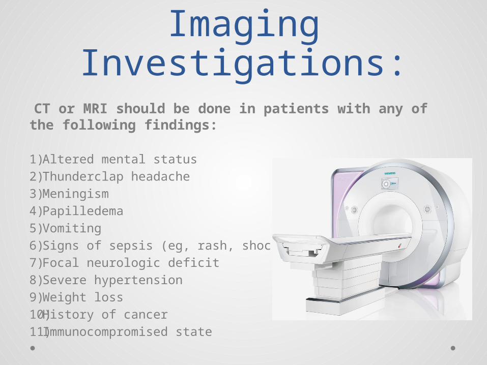
Imaging Investigations:
CT or MRI should be done in patients with any of the following findings:
1) Altered mental status2) Thunderclap headache3) Meningism4) Papilledema5) Vomiting6) Signs of sepsis (eg, rash, shock)7) Focal neurologic deficit8) Severe hypertension 9) Weight loss10) History of cancer11) Immunocompromised state

Case 1:• A 27 year old woman presented
to the PHC with longstanding episodic headache that interferes with her work.
• How would you approach this patient?

Case 1: History:• Pain is usually on one side of the head, starts suddenly
and intensifies over a period of an hour, then lasts for
about 2 days, it is throbbing, associated with seeing
lights flickering (lasting about 30 minutes) and high
sensitivity to noise, exacerbated by movement and
noise, relieved by sitting in a dark room alone, and has
an intensity of 8/10.
• Last episode was two weeks ago, and similar episodes
have occurred at least 7 times.

Case 1: History:• Past medical history is insignificant.
• No allergies or drugs, except failed attempts to
relieve headache with paracetamol.
• She is single, works as a teacher in a primary
school, and her episodic headache significantly
affects her job.

Case 1: Physical Examination:
• General inspection:The patient is sitting comfortably on the chair and doesn't seem to be in any distress.• Vital Signs: HR:80, RR:18, BP:119/70, Temp:37, and her BMI was 23 kg/m2.• Head & Neck:There doesn't seem to be any tenderness or swellings.• Ear, Nose, and Mouth:Nothing significant• Eye:Nothing significant• Neurological examination:A full Neurological examination was carried out but with no significant findings, and her visual symptoms (lights flickering) and phonophobia were abscent.

Case 1: Investigations:• No investigations were done.• Neuroimaging is not necessary in patients with a
history of recurrent migraine headaches and a normal neurologic examination.

Case 1: Diagnosis:• Most likely diagnosis:
Migraine Headache with Aura
• How would you manage this patient?

Case 1: Management:1. Using a “headache diary” .
2. Oral triptan (almotriptan 6.25 mg) combined with an NSAID
(Ibuprofen 400 mg) to abort the headache episode. (Acute
Treatment)
3. Oral Topiramate (dose of 25 mg once at night, and gradually
increased over the course of 4 weeks to 50 mg once at night and
once in the morning) to prevent episodes of migraine.
(Prophylactice Treatment)
4. Educating patient about the nature of the disease and the
adverse effects of drugs used, specially that topiramate is
associated with a risk of fetal malformations and can impair the
effectiveness of hormonal contraceptives

Migraine:• A complex disorder characterized by recurrent episodes of
headache, often unilateral and may be associated with visual
or sensory symptoms (aura).
• Most common in women and has a strong genetic component.
• The diagnosis of migraine is based on patient history. Patients
must have headache attacks that last 4-72 hours and are
characterized by unilateral location, pulsating quality,
moderate or severe pain intensity.
• Patients’ headache is also commonly aggravated by or causes
avoidance of routine physical activity.
Migraine cont.:• Other symptoms include:
o Unusual sensitivity to light and/or sound or nausea and/or vomiting
o Aura: Symptoms can occur with or without headache and are fully reversible, develop
over at least 5 minutes, last 5−60 minutes. Typical aura symptoms include visual
symptoms such as flickering lights, spots or lines and/or partial loss of vision; sensory
symptoms such as numbness and/or pins and needles; and/or speech disturbance.
• Management plan includes:
o Headache diary
o Acute Treatment: oral triptan and an NSAID, or an oral triptan and paracetamol
o Prophylactic Treatment: topiramate, propranolol, or amitriptyline.
o Education and Support.
o Follow-up.
Case 2:• A 32 year old woman presented to the PHC with
headache for the past 3 months.
• How would you approach this patient?

Case 2: History:• Pain is all around the head, was gradual in onset, dull in
character, comes and goes irregularly, slightly relieved
by taking paracetamol, and its severity is reported to be
6/10.
• She also reported having frequent pulsatile tinnitus,
nausea and vomiting.
• Past medical history is insigificant.
• She takes oral contraceptive pills.
• Mother to three children and works in a bank.

Case 2: Physical Examination:
• General inspection:The patient is sitting comfortably on the chair and doesn't seem to be in any distress.• Vital Signs:
HR:65, RR:17, BP:126/75, Temp:36.4, and her BMI was 32 kg/m2.• Head & Neck:There doesn't seem to be any tenderness or swellings.• Ear, Nose, and Mouth:Nothing significant• Eye:
Horizontal diplopia on ocular motility test, decrease peripheral vision on confrontation test, and papilledema on Fundoscopy.• Neurological examination:A full Neurological examination was carried out but with no significant findings.

Case 2: Investigations:• An MRI was done.
• Lumbar Puncture:
o Pressure: 27 CmH2O
o Clear in apperance
o Protein: 0.35 g/L
o Glucose: 3 mmol/L
o Glucose CSF/Serum Ratio: 0.6
o WBC: <3
Case 2: Other routine investigations:
• Complete blood count (CBC)
• Erythrocyte sedimentation rate (ESR)
• Serum iron and iron-binding capacity
• Antinuclear antigen (ANA) profile (eg, anti-dsDNA
and anti-ssDNA)
• Full procoagulant profile

Case 2: Diagnosis :• Most likely diagnosis:
Ideopathic Intracranial Hypertension,
A.K.A Pseudotumor Cerebri or Benign Intracranial Hypertension
• How would you manage this patient?

Case 2: Management:1. Referral to neurologist
2. Educate patient regarding:
• Nature of the disease
• Avoiding possible causative drug
• Weight loss benefit
• Ophthalmic complications

Ideopathic Intracranial Hypertension:
• Common in obese women of childbearing age.
• Patients usually present with symptoms related to increased ICP and papilledema.
• Symptoms & Signs may include: o Headaches, typically nonspecific and varying in type, location,
and frequency,
o Diplopia, usually horizontal but rarely vertical,
o Pulsatile tinnitus,
o Transient visual obscurations, Progressive loss of peripheral vision in one or both eyes, Blurring and distortion of central vision, Sudden visual loss,
o Other symptoms like dizziness, nausea, vomiting, photopsias, and retrobulbar pain.

Ideopathic Intracranial Hypertension cont.:
• MRI is study of choice, and when a mass is ruled out LP is
indicated. Other routine test and procoagulant profile is
recommended.
• Patients should be referred to Neurologist for management and
follow-up
• Patients should be educated about:
o Causative agents (e.g Drugs, Systemic Diseases)
o The role of weight loss in managing the disease
o Ophthalmic complication (i.e. irreversible optic neuropathy with accompanying constriction of
the visual field and loss of color vision, and even involvement of central visual acuity in end-
stage papilledema.

Case 3:• A 65 year old man came to PHC clinic for his
usual check-up, he asked the doctor to prescribe a good medication for a headache that started five months ago.
• How would you approach this patient?

Case 3: History:• He is 65 year old Saudi man.
• His pain is usually felt on the side of his head, it
was gradual in onset, throbbing, associated with
fatigue, no relieving or exacerbating factors, and
it has an intensity of 4/10.
• He also reported having muscle aches, and
notices recent discomfort on chewing firm food.

Case 3: History:• He is hypertensive controlled on maximum dose
of diuretic and ACE inhibitor and regularly visits
his doctor.
• Past medical history includes one past admissions
for anal fistula surgery.
• He is retired and lives with his wife. He smokes
half a pack of cigarettes every day, and has been
doing so for the past 20 years.

Case 3: Physical Examination:
• General inspection:The patient is sitting comfortably on the chair and doesn't seem to be in any distress.• Vital Signs:
HR:70, RR:18, BP:140/85, Temp:38.4, and his BMI was 26 kg/m2. • Head & Neck:
Tenderess on scalp and over the temporal artery, with palpable temporal artery. No tenderness on neck examination.• Ear, Nose, and Mouth:Nothing significant.• Eye:
Mild hypertensive retinopathy on fundoscopy and no sign of arteritic anterior ischemic optic neuropathy.• Neurological examination:A full Neurological examination was carried out but with no significant findings.• Cardiovascular examination:Normally positioned PMI, and no murmurs.

Case 3: Investigations:• CBC:
• ESR: 70 mm/h• C-reactive protein : 2.5 mg/dl• LFT:
o AST: 250o ALP:200
• Autoantibodies:o ANCA +
CBC:
Hb: 11.5 g/dL
WBC: 10 k/uL
RBC: 4.5 m/uL
MCV: 85 fL
MCH: 30 pg
PLT: 600 k/uL

Case 3: Investigations:• Histology:
o Superficial temporal artery biopsy shows vasculitis with giant cell infiltration
• Duplex ultrasonography:o Halo sign: hypoechoic region around the lumen of the artery.
• CT:o No Large vessel aneurysms.

Case 3: Diagnosis:• Most likely diagnosis:
Giant Cell (temporal) Arteritis
• How would you manage this patient?

Case 3: Management:1. Referral to Rheumatologist
2. Patient Education:
• Nature and seriousness of the disease.
• Disease complications, specially ophthalmic (sudden
painless vision loss)
• Therapy adverse effects (high dose corticosteroids)

Giant Cell (Temporal) Arteritis:
• Giant cell arteritis (temporal arteritis) is a
systemic vasculitis categorized as a medium/large vessel vasculitis.
• GCA is the most common form of systemic vasculitis in adults, and
may lead to blindness if not treated.
• Symptoms & Signs reflect the involvement of the temporal artery and
other medium-sized arteries of the head and the neck and include:
o Headache,
o Visual disturbances,
o Jaw claudication,
o Neck pain,
o Scalp tenderness.

Giant Cell (Temporal) Arteritis cont.:
• Constitutional manifestations, such as fatigue, malaise, and
fever, may also be present.
• Investigations (including ESR, Duplex US, and temporal artery
biopsy) play an important role in diagnosis.
• Patients should be referred to Rheumatologist for treatment
(high dose corticosteroid) and follow-up.
• Patients should also be educated regarding the nature of the
disease and its complications, the importance of adhering to
steroid therapy and its possible side effects.

Case 4:• 35 year old male came to your clinic complaining
of a headache.
• How would you approach this patient?

Case 4 : History:• Ahmed is a 35 year old teacher who has been complaining
of headaches for the past 2 months, he says the pain is felt
around his head in a band like fashion. The character of the
pain was described as something tightening around his
head. The headaches happen around 2 time per week and
last anywhere from 30 min to 10 hours. He doesn't report
anything that makes the headaches worst but says the pain
is relieved slightly when he lays down. He rates the pain as
a 5/10.

Case 4 : History:• He is not taking any medications.
• Past medical history is insignificant.
• He is married, and is happy with his job. He
doesn’t smoke or drink alcohol.

Case 4: Physical Examination:
• General inspection:The patient is sitting comfortable on the chair and doesn't seem to be in any distress.• Vital Signs: BP 110/80Heart Rate 72 bpmTemperature 37.2°C• Head & Neck:There doesn't seem to be any tenderness or swellings around the scalp.• Ear, Nose, and Mouth:Nothing significant• Eye:Nothing significant• Neurological examination:A full Neurological examination was carried out but with no significant findings that would suggest neurological issues.

Case 4: Investigations:• In the case of this patient no investigations were
ordered as the history and physical examination did not raise any red flags.
Case 4: Diagnosis:• Most likely diagnosis:
Episodic Tension headache
• How would you manage this patient?

Case 4: Management:1. Using a “headache diary”.
2. Paracetamol prescription for the acute treatment of
tension type headache.
3. Educating the patient about the commonness of the
condition.

Tension headache• Tension headaches are the most common type of
headaches among adults.
• An episodic tension headache may be described
as a mild to moderate constant band-like pain,
tightness, or pressure around the forehead or
back of the head and neck.
• Tension headaches usually don't keep a person
from performing daily tasks.
Tension headache cont.:
• A tension headache may appear periodically ("episodic," <15
days per month) or daily ("chronic," >15 days per month).
• Management plan:
o Acute treatment: Consider aspirin, paracetamol or an NSAID for the acute treatment of
tension-type headache, taking into account the person's preference, comorbidities and
risk of adverse events.
o Do not offer opioids for the acute treatment of tension-type headache.
o Prophylactic treatment: Consider a course of up to 10 sessions of acupuncture over 5–8
weeks for the prophylactic treatment of chronic tension-type headache.

Case 5• A 46-year-old man comes to your office with a 5-
week history of recurrent headaches.
• How would you approach this patient.

Case 5: History:• Abdullah is a 46-year-old male who came to your office with
a 5-week history of recurrent headaches that wake him up
in the middle of the night. The headaches have been
occurring twice daily and have been lasting approximately
1 hour. The headaches are described as a deep burning
sensation centered behind the left eye. The headaches are
excruciating (he rates them as a 12 on a 10-point scale)
and are associated with watery left eye. Before the onset of
these headaches 5 weeks ago, the patient describes no
more than occasional tension headache.

Case 5: History:• Past medical history is insignificant.
• He is not using any drugs.
• The patient describes no recent life changes and
no major life stresses. He is happily married, has
three children, and has a secure job that he
enjoys.

Case 5: Physical Examination:
• General inspection:The patient appears to be in some distress and seems to be restless.• Vital Signs: BP 120/70Heart Rate 82 bpmTemperature 36.6°C• Head & Neck:
There doesn't seem to be any tenderness or swellings.• Ear, Nose, and Mouth:Nothing significant• Eye:Tearing coming from Left eye, along with nasal discharge. • Neurological examination:A full Neurological examination was carried out but with no significant findings that would suggest neurological issues.

Case 5: Investigations:• No neuroimaging was done. • It was discussed with the patient that if other
symptoms appear it is preferred to do certain tests to exclude other diseases.

Case 5: Diagnosis:• Most likely diagnosis:
Cluster headache (Episodic cluster headache)
• How would you manage this patient?

Case 5: Management:1. Using a “headache diary”.
2. Nasal triptan for acute treatment.
3. Oral verapamil for prophylaxis.
4. Educating patient about the nature of the
disease, the inefficasy of using paracetamol
or NSAIDs for acute treatment, and the
adverse effects of the drugs prescribed to
him.

Cluster Headache:• Cluster headache is a primary neurovascular primary headache disorder,
and is far less common than migraine headache or tension headache.
• Cluster headaches begin usually around the eye and along the side of the
head/face. The headache exhibits a clustering of painful attacks over a
period of many weeks.
• The pain of a cluster headache peaks in about 5 minutes and lasts 15 to
180 minutes. It is usually severe and associated with the following
symptoms on the same side:
o red and/or watery eye
o nasal congestion and/or runny nose
o swollen eyelid
o forehead and facial sweating
o constricted pupil and/or drooping eyelid

Cluster Headache cont.:
• Someone with a cluster headache may get several headaches a day for weeks at
a time - perhaps months - usually interrupted by a pain-free period of variable
length.
Management Plan:
o Acute treatment:
• Consider neuroimaging for people with a first bout of cluster headache..
• Offer oxygen and/or a subcutaneous or nasal triptan for the acute
treatment of cluster headache.
• Do not offer paracetamol, NSAIDS, opioids, ergots or oral triptans for the
acute treatment of cluster headache.
o Prophylactic treatment
• Consider verapamil for prophylactic treatment during a bout of cluster
headache. If unfamiliar with its use for cluster headache, seek specialist
advice before starting verapamil, including advice on electrocardiogram
monitoring.

Case 6• 56 year old male came to your clinic complaining
of headache for the past 8 months.
• How would you approach this patient?

Case 6: History:• Ali is a male, 56-year-old university professor who
has experienced constant headache for the past 8
months. Location of the headache cannot be
identified, it started gradually, and is dull in
character, associated with nausea and vomiting,
constant and wakes him from sleep at times, not
relieved by anything, and its severity is reported
as 4/10.

Case 6: History:• He also reported difficulty in walking and numbness
in the left leg.
• Past medical history is insignificant, except for one
admission for appendectomy 30 years ago,
• He is diabetic controlled on oral hypoglycemic drugs.
• He is married with four children, works in a university
as a lecturer. No history of smoking or alcohol abuse.

Case 6: Physical Examination:
• General inspection:The patient appears to be well, sitting comfortably on the chair.• Vital Signs: BP 137/80Heart Rate 82 bpmTemperature 36.8°C• Head & Neck:There doesn't seem to be any tenderness or swellings.• Ear, Nose, and Mouth:Nothing significant• Eye:Fundoscopy shows papilledema.• Neurological examination:A full Neurological examination was carried out, showing weakness in the left leg (Power 4), and hyperreflexia (+3), decrease sensation, and positive Babinski sign.

Case 6: Investigations:• MRI was done:

Case 6: Diagnosis:• Most likely diagnosis:
Headache due to space occupying lesion.
• How would you manage this patient?

Case 6: Management:1. Referral to neurosurgery.
2. Education about the nature of the disease and its
seriousness.
3. Provide information and support.

Space Occupying Lesion Headache:
• Space occupying headaches occur in patients
with cerebral masses. A space-occupying lesion of
the brain is usually due to malignancy but it can
be caused by other pathology such as an abscess
or a hematoma.
• Almost half of intracerebral tumors are primary
but the rest have originated outside the CNS and
are metastases.

Space Occupying Lesion Headache cont.:• Symptoms & Signs usually include:
o Headache, Nausea & Vomiting, and Papilledema
o Focal Neurological defects
o Seizures
o Behavioral or mental changes
• Imaging is imperative for diagnosis or etiology, MRI is
preferable to CT.
• Management plan includes:
o Referral to specialists.
o Education
o Support

Role of Primary Health Care Physician
• Assess patient complaining of headache.
• Establish a differential diagnosis.
• Manage headaches which can be managed by
PHC clinics.
• Referring cases to appropriate specialists.
• Educate patients regarding their headache, what
affects it, and how to manage it.
• Follow-up.
Treat or Refer?• PHC Physicians usually treat primary headaches
(Tension, Migraine, and Cluster Headaches)
o Acute Treatment
o Prophylaxis.
• Patients who need further assessment or have any
red flags (e.g. vomiting, history of cancer, impaired
level of consciousness) are referred to specialists.

Question 1:• Which of the following is true about headache?
a) Headache is overdiagnosed and over-treated.
b) Headache is more commonly a symptom of another systemic
disease.
c) All headache cases are referred to specialists.
d) Headache is under-recognized and under-treated.

Question 2:• Which of the following investigations is indicated
in a patient with headache, nausea/vomiting, and
papilledema once an intracranial mass lesion is
ruled out?
a) Electroencephalogram.
b) Blood culture.
c) Cerebrospinal Fluid analysis.
d) PET scan.

Question 3:• Which of the following features presenting with
headache does not indicate need of
neuroimaging?
a) History of cancer.
b) Altered level of consciousness.
c) Rythmical recurrence.
d) Immunocompromised state

References:1. [http://www.nice.org.uk/guidance/cg150/chapter/guidance#assessment]
2. [http://emedicine.medscape.com]
3. [http://www.merckmanuals.com/professional/neurologic_disorders/
headache/approach_to_the_patient_with_headache.html]
4. [WHO, http://www.who.int/mediacentre/factsheets/fs277/en/]

THANK YOU!