Embed Size (px)
Citation preview

Application of 2D frameless angiography in planning
Gamma Knife radiosurgery for AVMs
A.V. Dalechina1, A.V. Golanov1,2, V.V. Kostjuchenko1
1Moscow Gamma Knife Center2Burdenko Neurosurgical Institute
Moscow Gamma Knife Center
Burdenko Neurosurgical Institute

No conflict of interest disclosure in presentation

3 main questions of this presentation
•How do we apply this method?
• Are we sure that this method is correct ?
•What is the advantage of using this method?

How do we apply this method?

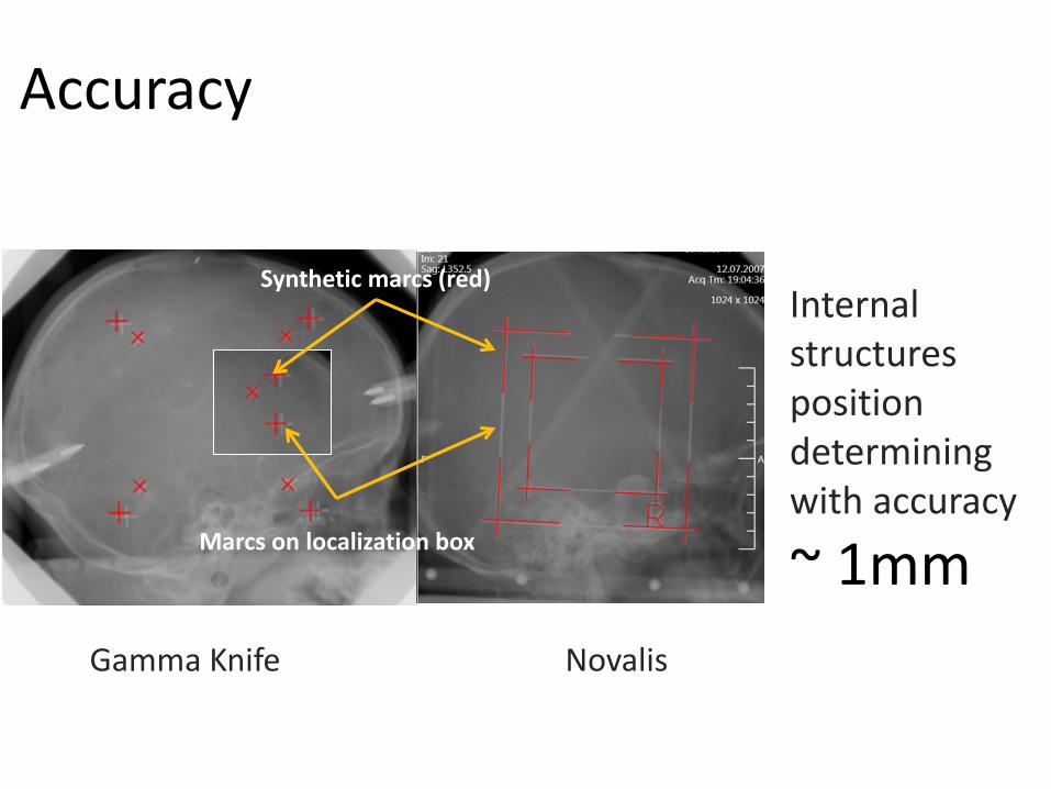
2DSA
STANDARD WAY

2DSA

Draft manual adjustment
Autofusion and verification
CT
XA+
Contouring
any treatment planning
system, which supports import
DICOM / DICOM
RTSTRUCT
iPlan
GammaPlan
multiPlan

1. Contouring on one projection
2. Continue contouring on second projection
3. Volumes, contoured on the angiography are projected to the CT and volumes, contoured on the CT are projected to XA . Adjust contours using agreement between planes and observing different angio frames
4. Draw contours on the CT inside crossection of two XA projections
5. Observe back projection from the CT to XA
6. Finish. Export DICOM CT and RTSTRUCT data

Localization
ImageFusion
CT
XA+
Choose frameSynthetic marcsDefine in LGP

Math behind
Where
1.15
1.16
1.17
1.18
1.19
-5 -4 -3 -2 -1 0 1 2 3 4 5
Ox Oy Oz Ga Ka Ca Sdd R
Pattern Intensity measure function graphs in thevicinity of global solution. One unit correspondsto 0.4 mm or 0.4 degrees.
1. Pattern Intensity measure function
2. Simplex optimization method
Vary 8 parameters (6 position and rotation, one source-detector distance, and one distortion correction factor if necessary)

Are we sure that this method is correct ?
Accuracy
NovalisGamma Knife
Internal structures position determining with accuracy
~ 1mmMarcs on localization box
Synthetic marcs (red)

Verification60 patients with frame-based SRS3 independent usersiPlan RTImage 4.1
Results:
No significant difference in AVM volume. The mean difference between stereotactic coordinates:
AP: 0.6±0.5 mmLAT: 0.9±0.7 mmCC : 0.7±0.6 mm
“This frameless approach assures accurate target localization and can be used in a clinical setting”.


What is the advantage of using this method?

Clinic
• XNAV is routinely used at Burdenko Neurosurgical Institute during last 5 years after 2011
524 patients
• XNAV is routinely used at Moscow Gamma Knife Center during last 4 years after 2012
90 patients
75%
25%
Cyber KnifeXNav without XNav
36%
64%
Gamma KnifeXNav without XNav
2008 – 2012 y.
2012 – 2016 y.
2011 – 2016 y.
2009– 2011 y.

Wait time before treatment
0
1
2
3
4
5
6
7
8
9
10
aver
age
wai
ting
time,
hour
s
2008 2009 2010 2011 2012 2012 (after july) 2013 2014 2015
XNav

Conclusion
• Frameless approach assures accurate AVM localization
• Avoid discomfort and risks associated with additional invasive angiography
• In perspective 2D frameless angiography – powerful tool for effectively use of Gamma Knife Icon for AVM irradiation

Thank you for listening!


![A.v.-la Filolakia [Bibliotecacatolica.wordpress.com]](https://img.pdfslide.us/doc/110x75/577ce3691a28abf1038c1047/av-la-filolakia-bibliotecacatolicawordpresscom.jpg)