Embed Size (px)
Citation preview

907K. Benirschke et al., Pathology of the Human Placenta, DOI 10.1007/978-3-642-23941-0, © Springer-Verlag Berlin Heidelberg 2012
Normative Values and Tables
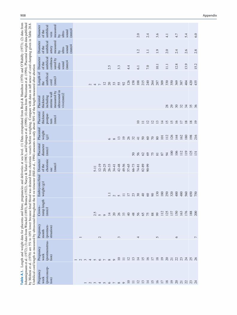
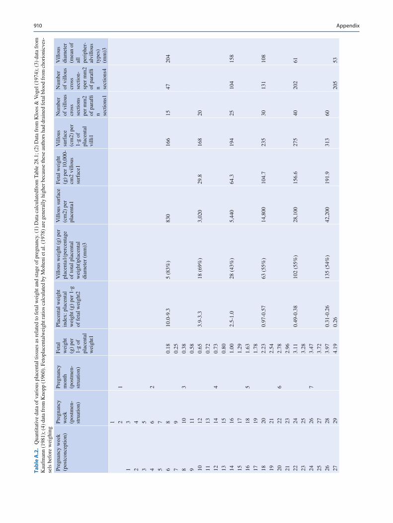
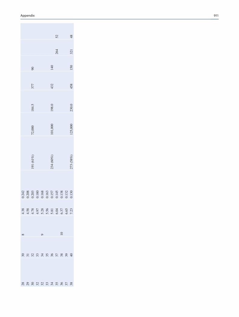
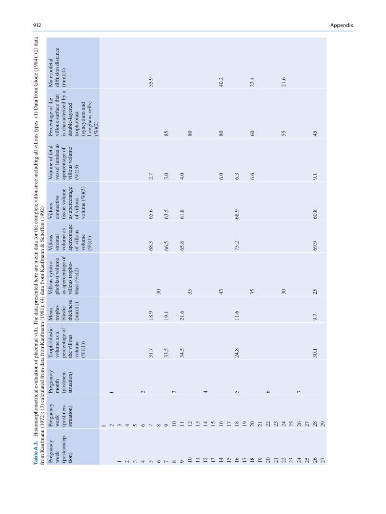
Some quantitative structural and biochemical data concern-ing pregnancy, placental development, and composition of the term placenta are given in Tables A.1, A.2, A.3, A.4, A.5, A.6, A.7, A.8, A.9, and A.10 .
When examining the tables on placental morphometry and comparing the results from different authors, it is impor-tant to note that quantitative structural data are heavily in fl uenced by the mode of sampling and by the preparation of the material. Because of the high degree of maternal and fetal vascularization, the placenta reacts immediately to changes in intravascular pressure. Thus, the mode of birth, the time elapsing from cessation of maternal and fetal blood fl ows to tissue fi xation (see Tables A.6, A.7, A.8, A.9, and A.10 ), and the nature of cord clamping (see Table A.8) directly in fl uence the volumetric relations of villi and inter-villous space. In particular, parameters such as the width of fetal vessels, degree of fetal vascularization, maternal-fetal diffusion distance, and trophoblastic thickness are easily affected. Moreover, the composition of the fi xative and its osmolarity (see Table A.10 ), as well as the mode of fi xation (immersion versus perfusion fi xation) are of importance. Normally, immersion fi xation of the entire placenta or of small pieces is used. The more advanced methods, such as perfusion fi xation (Burton et al. 1987) or puncture biopsy of the still maternally perfused placenta during Cesarean sec-tion (Schweikhart and Kaufmann 1977; Voigt et al. 1978; Sen et al. 1979), are very time-consuming. When studying immersion fi xed material, however, one should keep in mind that this material differs quantitatively and qualitatively
from the in vivo conditions (see Tables A.6 and A.9 ). It is impossible to include the results of numerous other valuable contributions to placental morphometry into these tables. For further information on special issues, we refer to the fol-lowing publications: placental growth development in rela-tion to birth weight (Bouw et al. 1978; Molteni et al. 1978); relationship of placental weight to body size at 7 years of age, and to abnormalities in children (Naeye 1987); fetal and placental weights in relation to maternal weight (Auinger and Bauer 1974); ultrasonographic measurements of volu-metric growth of the placenta (Bleker et al. 1977); weight development of placenta and membranes in early pregnancy (Abramovich 1969); ratio of gestational sac volume to crown-rump length in early pregnancy (Goldstein et al. 1986); villous surface area and villous volume densities in various placental regions and along different levels of the chorial basal axis (Teasdale 1978; Boyd et al. 1980; Cabezon et al. 1985; Bacon et al. 1986); local variations of villous surface, fetal vascularization, and amount of vasculo-syncy-tial membranes in the placentone (maternal-fetal circulatory unit) (Schuhmann et al. 1986); total villous surface in rela-tion to fetal weight, in normal and various pathological cases (Clavero-Nunez and Botella-Llusia 1961, 1963); morpho-metric data affecting placental oxygen diffusion (Mayhew et al. 1984, 1986); computer measurement of the mass of syncytiotrophoblast (Boyd et al. 1983); ultrastructural mor-phometric analysis of the villous syncytiotrophoblast (Sala et al. 1983); microvillous surface enlargement of the villous surface (Teasdale and Jean-Jacques 1985); comparison of villous structure following immersion and perfusion fi xation (Burton et al. 1987).
Appendix
908 Appendix
Tab
le A
.1.
Len
gth
and
wei
ght
data
for
pla
cent
a an
d fe
tus;
pre
gnan
cies
and
del
iver
ies
at s
ea l
evel
. (1
) D
ata
extr
apol
ated
fro
m B
oyd
& H
amilt
on (
1970
) an
d O
’Rah
illy
(197
3);
(2)
data
fro
m
Joha
nnig
man
n et
al.
(197
2); (
3) d
ata
from
Win
ckel
(189
3), B
ranc
a (1
922)
, Nae
ye &
Taf
ari (
1983
), a
nd F
ujin
aga
et a
l. (1
990)
; (4)
dat
a fr
om W
eiss
man
et a
l. (1
994)
. Pla
cent
al w
eigh
t dat
a pu
blis
hed
by M
olte
ni e
t al
. (19
78)
are
5% t
o 10
% l
ower
bec
ause
fet
al b
lood
was
dra
ined
fro
m c
hori
onic
ves
sels
bef
ore
wei
ghin
g. C
ompa
re w
ith d
ata
on i
n fl u
ence
of
cord
cla
mpi
ng g
iven
in
Tabl
e 28
.8.
Um
bilic
al c
ord
leng
th a
s m
easu
red
by u
ltras
ound
thro
ugho
ut th
e fi
rst t
rim
este
r (H
ill e
t al.,
199
4) a
mou
nts
to o
nly
abou
t 50%
of
the
leng
th m
easu
red
afte
r ab
ortio
n
Preg
nanc
y w
eek
(pos
tcon
cep-
tion)
Preg
nanc
y w
eek
(pos
tmen
stru
a-tio
n)
Preg
nanc
y m
onth
(p
ostm
en-
stru
atio
n)
Cro
wn–
rum
p le
ngth
(m
m) 1
Em
bryo
nic/
feta
l w
eigh
t (g)
1 D
iam
eter
of
the
chor
ioni
c sa
c (m
m) 1
Plac
enta
l di
amet
er
(mm
) 1
Plac
enta
l w
eigh
t (g
) 1
Plac
enta
l th
ickn
ess
post
par-
tum
(m
m) 1
Plac
enta
l th
ickn
ess
incl
udin
g ut
erin
e w
all
mea
sure
d by
ul
tras
ound
in
vivo
(mm
) 2
Len
gth
of
the
umbi
lical
co
rd
(mm
) 3
Dia
met
er
of th
e um
bilic
al
cord
mea
-su
red
by
ultr
a-so
und
(mm
) 4
Dia
met
er
of th
e um
bilic
al
arte
ry
mea
sure
d by
ul
tra-
soun
d (m
m) 4
Dia
met
er
of th
e um
bilic
al
vein
m
easu
red
by
ultr
a-so
und
(mm
) 4
1 2 1
1 3
2 4
2
3 5
2.5
5-11
4
4 6
2 5
12-1
9 7
5 7
9 20
-25
12
6 8
14
1.1
26-3
3 6
20
2.5
7 9
20
2 34
-41
8 33
8 10
3
26
5 42
-48
13
55
3.3
9 11
33
11
49
-56
19
92
10
12
40
17
57-6
5 26
12
6 4.
4
11
13
48
23
66-7
3 50
32
15
8
12
14
4 56
30
74
-81
56
41
10
188
6.1
1.2
2.0
13
15
65
40
82-8
9 62
50
11
21
5
14
16
75
60
90-9
9 69
60
12
24
0 7.
0 1.
1 2.
4
15
17
88
90
75
70
12
264
16
18
5 99
13
0 81
80
13
28
7 10
.1
1.9
3.6
17
19
112
180
87
101
14
309
18
20
125
250
94
112
15
28
330
11.1
2.
0 4.
1
19
21
137
320
100
126
15
29
350
20
22
6 15
0 40
0 10
6 14
4 16
30
36
9 12
.8
2.4
4.7
21
23
163
480
112
162
17
32
387
22
24
176
560
119
180
18
34
404
13.9
2.
6 5.
4
23
25
188
650
125
198
18
35
420
24
26
7 20
0 75
0 13
1 21
6 19
36
43
5 15
.2
2.8
6.0

909Appendix
25
27
213
870
137
234
19
37
450
26
28
226
1,00
0 14
4 25
2 20
38
46
4 15
.9
3.1
6.6
27
29
236
1,13
0 15
0 27
0 20
39
47
7
28
30
8 25
0 1,
260
156
288
21
40
490
16.3
3.
4 7.
3
29
31
263
1,40
0 16
2 30
6 21
41
50
2
30
32
276
1,55
0 16
9 32
4 22
42
52
0 17
.6
3.6
7.7
31
33
289
1,70
0 17
5 34
2 22
42
53
0
32
34
9 30
2 1,
900
181
360
23
43
540
17.4
3.
3 7.
4
33
35
315
2,10
0 18
7 37
8 23
43
54
9
34
36
328
2,30
0 19
4 39
6 24
44
55
7 17
.4
3.7
7.6
35
37
341
2,50
0 20
0 41
4 24
44
56
5
36
38
10
354
2,75
0 20
6 43
2 24
45
57
2 18
.0
4.2
8.2
37
39
367
3,00
0 21
3 45
1 25
45
57
9
38
40
380
3,40
0 22
0 47
0 25
45
58
5 17
.0
3.9
7.8
910 Appendix
Tab
le A
.2.
Qua
ntita
tive
data
of
vari
ous
plac
enta
l tis
sues
as
rela
ted
to f
etal
wei
ght a
nd s
tage
of
preg
nanc
y. (
1) D
ata
calc
ulat
edfr
om T
able
28.
1; (
2) D
ata
from
Klo
os &
Vog
el (
1974
); (
3) d
ata
from
K
aufm
ann
(198
1); (
4) d
ata
from
Kno
pp (1
960)
. Fet
opla
cent
alw
eigh
t rat
ios
calc
ulat
ed b
y M
olte
ni e
t al.
(197
8) a
re g
ener
ally
hig
her b
ecau
se th
ese
auth
ors
had
drai
ned
feta
l blo
od fr
om c
hori
onic
ves-
sels
bef
ore
wei
ghin
g
Preg
nanc
y w
eek
(pos
tcon
cept
ion)
Pr
egna
ncy
wee
k (p
ostm
en-
stru
atio
n)
Preg
nanc
y m
onth
(p
ostm
en-
stru
atio
n)
Feta
l w
eigh
t (g
) pe
r 1-
g of
pl
acen
tal
wei
ght 1
Plac
enta
l wei
ght
inde
x: p
lace
ntal
w
eigh
t (g)
per
1-g
of
fet
al w
eigh
t 2
Vill
ous
wei
ght (
g) p
er
plac
enta
1(pe
rcen
tage
of
tota
l pla
cent
al
wei
ght)
plac
enta
l di
amet
er (
mm
) 3
Vill
ous
surf
ace
(cm
2 ) p
er
plac
enta
1
Feta
l wei
ght
(g)
per
10,0
00-
cm 2
villo
us
surf
ace 1
Vill
ous
surf
ace
(cm
2 ) p
er
1-g
of
plac
enta
l vi
lli 1
Num
ber
of v
illou
s cr
oss
sect
ions
pe
r m
m 2
of p
araf
fi n se
ctio
ns 1
Num
ber
of v
illou
s cr
oss
sect
ion-
sper
mm
2 of
par
af fi
n sect
ions
4
Vill
ous
diam
eter
(m
ean
of
all
peri
pher
-al
villo
us
type
s)
( m m
) 3
1
2 1
1 3
2 4
3 5
4 6
2
5 7
6 8
0.18
10
.0-9
.3
5 (8
3%)
830
166
15
47
204
7 9
0.25
8 10
3
0.38
9 11
0.
58
10
12
0.65
3.
9-3.
3 18
(69
%)
3,02
0 29
.8
168
20
11
13
0.72
12
14
4 0.
73
13
15
0.80
14
16
1.00
2.
5-1.
0 28
(43
%)
5,44
0 64
.3
194
25
104
158
15
17
1.29
16
18
5 1.
63
17
19
1.78
18
20
2.23
0.
97-0
.57
63 (
55%
) 14
,800
10
4.7
235
30
131
108
19
21
2.54
20
22
6 2.
78
21
23
2.96
22
24
3.11
0.
49-0
.38
102
(55%
) 28
,100
15
6.6
275
40
202
61
23
25
3.28
24
26
7 3.
47
25
27
3.72
26
28
3.97
0.
31-0
.26
135
(54%
) 42
,200
19
1.9
313
60
27
29
4.19
0.
26
205
53
911Appendix
28
30
8 4.
38
0.24
2
29
31
4.58
0.
208
30
32
4.78
0.
203
191
(61%
) 72
,000
18
4.5
377
90
32
33
4.97
0.
180
32
34
9 5.
28
0.16
8
33
35
5.56
0.
163
34
36
5.81
0.
157
234
(60%
) 10
1,00
0 19
8.0
432
140
35
37
6.04
0.
145
264
52
36
38
10
6.37
0.
138
37
39
6.65
0.
132
38
40
7.23
0.
130
273
(58%
) 12
5,00
0 23
0.0
458
150
321
48
912 Appendix
Tab
le A
.3.
His
tom
orph
omet
rica
l eva
luat
ion
of p
lace
ntal
vill
i. T
he d
ata
pres
ente
d he
re a
re m
ean
data
for
the
com
plet
e vi
llous
tree
incl
udin
g al
l vill
ous
type
s. (
1) D
ata
from
Glö
de (
1984
); (
2) d
ata
from
Kau
fman
n (1
972)
; (3)
cal
cula
ted
from
dat
a fr
omK
aufm
ann
(198
1); (
4) d
ata
from
Kau
fman
n &
Sch
effe
n (1
992)
Preg
nanc
y w
eek
(pos
tcon
cep-
tion)
Preg
nanc
y w
eek
(pos
tmen
-st
ruat
ion)
Preg
nanc
y m
onth
(p
ostm
en-
stru
atio
n)
Tro
phob
last
ic
volu
me
as a
pe
rcen
tage
of
the
villo
us
volu
me
(%) (
1))
Mea
n tr
opho
-bl
astic
th
ickn
ess
( m m
) (1)
Vill
ous
cyto
tro-
phob
last
vol
ume
as a
perc
enta
ge o
f vi
llous
trop
ho-
blas
t (%
) (2)
Vill
ous
stro
mal
vo
lum
e as
ap
erce
ntag
e of
vill
ous
volu
me
(%) (
1)
Vill
ous
conn
ectiv
e tis
sue
volu
me
as a
perc
enta
ge
of v
illou
s vo
lum
e (%
) (3)
Vol
ume
of f
etal
ve
ssel
lum
ina
as
aper
cent
age
of
villo
us v
olum
e (%
) (3)
Perc
enta
ge o
f th
e vi
llous
sur
face
that
is
cha
ract
eriz
ed b
y a
doub
le-l
ayer
ed
trop
hobl
ast
(syn
cytiu
m a
nd
Lan
ghan
s ce
lls)
(%) (
2)
Mat
erno
feta
l di
ffus
ion
dist
ance
( m
m) (
4)
l
2 l
1 3
2 4
3 5
4 6
2
5 7
31.7
18
.9
68.3
65
.6
2.7
55.9
6 8
30
7 9
33.5
19
.1
66.5
63
.5
3.0
85
8 10
3
9 11
34
.5
21.6
65
.8
61.8
4.
0
10
12
35
80
11
13
12
14
4
13
15
14
16
43
6.0
80
40.2
15
17
16
18
5 24
.8
11.6
75
.2
68.9
6.
3
17
19
18
20
35
6.6
60
22.4
19
21
20
22
6
21
23
22
24
30
55
21.6
23
25
24
26
7
25
27
26
28
30.1
9.
7 25
69
.9
60.8
9.
1 45
27
29

913Appendix
28
30
8
29
31
30
32
20
35
20.6
31
33
32
34
9
33
35
34
36
30.8
5.
2 15
69
.2
47.9
21
.3
25
11.7
35
37
36
38
10
37
39
38
40
32.9
4.
1 14
67
.1
38.7
28
.4
23
4.8
914 Appendix
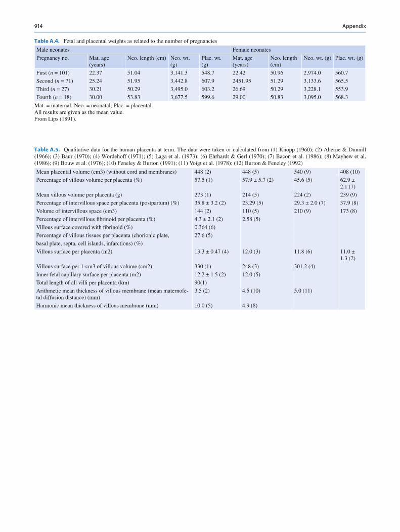
Table A.4. Fetal and placental weights as related to the number of pregnancies
Male neonates Female neonates
Pregnancy no. Mat. age (years)
Neo. length (cm) Neo. wt. (g)
Plac. wt. (g)
Mat. age (years)
Neo. length (cm)
Neo. wt. (g) Plac. wt. (g)
First ( n = 101) 22.37 51.04 3,141.3 548.7 22.42 50.96 2,974.0 560.7
Second ( n = 71) 25.24 51.95 3,442.8 607.9 2451.95 51.29 3,133.6 565.5
Third ( n = 27) 30.21 50.29 3,495.0 603.2 26.69 50.29 3,228.1 553.9
Fourth ( n = 18) 30.00 53.83 3,677.5 599.6 29.00 50.83 3,095.0 568.3
Mat. = maternal; Neo. = neonatal; Plac. = placental. All results are given as the mean value. From Lips (1891).
Table A.5. Qualitative data for the human placenta at term. The data were taken or calculated from (1) Knopp (1960); (2) Aherne & Dunnill (1966); (3) Baur (1970); (4) Wördehoff (1971); (5) Laga et al. (1973); (6) Ehrhardt & Gerl (1970); (7) Bacon et al. (1986); (8) Mayhew et al. (1986); (9) Bouw et al. (1976); (10) Feneley & Burton (1991); (11) Voigt et al. (1978); (12) Burton & Feneley (1992)
Mean placental volume (cm 3 ) (without cord and membranes) 448 (2) 448 (5) 540 (9) 408 (10)
Percentage of villous volume per placenta (%) 57.5 (1) 57.9 ± 5.7 (2) 45.6 (5) 62.9 ± 2.1 (7)
Mean villous volume per placenta (g) 273 (1) 214 (5) 224 (2) 239 (9)
Percentage of intervillous space per placenta (postpartum) (%) 35.8 ± 3.2 (2) 23.29 (5) 29.3 ± 2.0 (7) 37.9 (8)
Volume of intervillous space (cm 3 ) 144 (2) 110 (5) 210 (9) 173 (8)
Percentage of intervillous fi brinoid per placenta (%) 4.3 ± 2.1 (2) 2.58 (5)
Villous surface covered with fi brinoid (%) 0.364 (6)
Percentage of villous tissues per placenta (chorionic plate, 27.6 (5)
basal plate, septa, cell islands, infarctions) (%)
Villous surface per placenta (m 2 ) 13.3 ± 0.47 (4) 12.0 (3) 11.8 (6) 11.0 ± 1.3 (2)
Villous surface per 1-cm 3 of villous volume (cm 2 ) 330 (1) 248 (3) 301.2 (4)
Inner fetal capillary surface per placenta (m 2 ) 12.2 ± 1.5 (2) 12.0 (5)
Total length of all villi per placenta (km) 90(1)
Arithmetic mean thickness of villous membrane (mean maternofe-tal diffusion distance) (mm)
3.5 (2) 4.5 (10) 5.0 (11)
Harmonic mean thickness of villous membrane (mm) 10.0 (5) 4.9 (8)
915Appendix
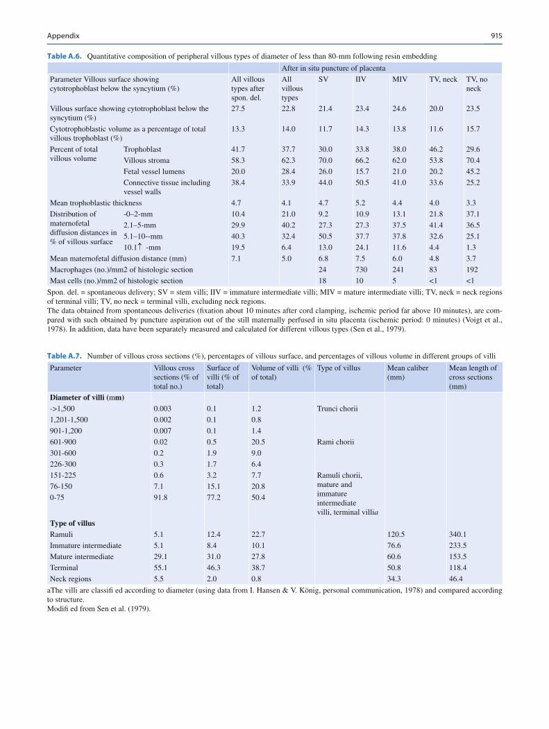
Table A.7. Number of villous cross sections (%), percentages of villous surface, and percentages of villous volume in different groups of villi
Parameter Villous cross sections (% of total no.)
Surface of villi (% of total)
Volume of villi (% of total)
Type of villus Mean caliber ( m m)
Mean length of cross sections ( m m)
Diameter of villi ( m m) ->1,500 0.003 0.1 1.2 Trunci chorii
1,201-1,500 0.002 0.1 0.8
901-1,200 0.007 0.1 1.4
601-900 0.02 0.5 20.5 Rami chorii
301-600 0.2 1.9 9.0
226-300 0.3 1.7 6.4
151-225 0.6 3.2 7.7 Ramuli chorii, mature and immature intermediate villi, terminal villi a
76-150 7.1 15.1 20.8
0-75 91.8 77.2 50.4
Type of villus
Ramuli 5.1 12.4 22.7 120.5 340.1
Immature intermediate 5.1 8.4 10.1 76.6 233.5
Mature intermediate 29.1 31.0 27.8 60.6 153.5
Terminal 55.1 46.3 38.7 50.8 118.4
Neck regions 5.5 2.0 0.8 34.3 46.4
aThe villi are classi fi ed according to diameter (using data from I. Hansen & V. König, personal communication, 1978) and compared according to structure. Modi fi ed from Sen et al. (1979).
Table A.6. Quantitative composition of peripheral villous types of diameter of less than 80- m m following resin embedding
After in situ puncture of placenta
Parameter Villous surface showing cytotrophoblast below the syncytium (%)
All villous types after spon. del.
All villous types
SV IIV MIV TV, neck TV, no neck
Villous surface showing cytotrophoblast below the syncytium (%)
27.5 22.8 21.4 23.4 24.6 20.0 23.5
Cytotrophoblastic volume as a percentage of total villous trophoblast (%)
13.3 14.0 11.7 14.3 13.8 11.6 15.7
Percent of total villous volume
Trophoblast 41.7 37.7 30.0 33.8 38.0 46.2 29.6
Villous stroma 58.3 62.3 70.0 66.2 62.0 53.8 70.4
Fetal vessel lumens 20.0 28.4 26.0 15.7 21.0 20.2 45.2
Connective tissue including vessel walls
38.4 33.9 44.0 50.5 41.0 33.6 25.2
Mean trophoblastic thickness 4.7 4.1 4.7 5.2 4.4 4.0 3.3
Distribution of maternofetal diffusion distances in % of villous surface
-0–2- m m 10.4 21.0 9.2 10.9 13.1 21.8 37.1
2.1–5- m m 29.9 40.2 27.3 27.3 37.5 41.4 36.5
5.1–10-- m m 40.3 32.4 50.5 37.7 37.8 32.6 25.1
10.1� - m m 19.5 6.4 13.0 24.1 11.6 4.4 1.3
Mean maternofetal diffusion distance ( m m) 7.1 5.0 6.8 7.5 6.0 4.8 3.7
Macrophages (no.)/mm 2 of histologic section 24 730 241 83 192
Mast cells (no.)/mm 2 of histologic section 18 10 5 <1 <1
Spon. del. = spontaneous delivery; SV = stem villi; IIV = immature intermediate villi; MIV = mature intermediate villi; TV, neck = neck regions of terminal villi; TV, no neck = terminal villi, excluding neck regions. The data obtained from spontaneous deliveries ( fi xation about 10 minutes after cord clamping, ischemic period far above 10 minutes), are com-pared with such obtained by puncture aspiration out of the still maternally perfused in situ placenta (ischemic period: 0 minutes) (Voigt et al., 1978). In addition, data have been separately measured and calculated for different villous types (Sen et al., 1979).
916 Appendix
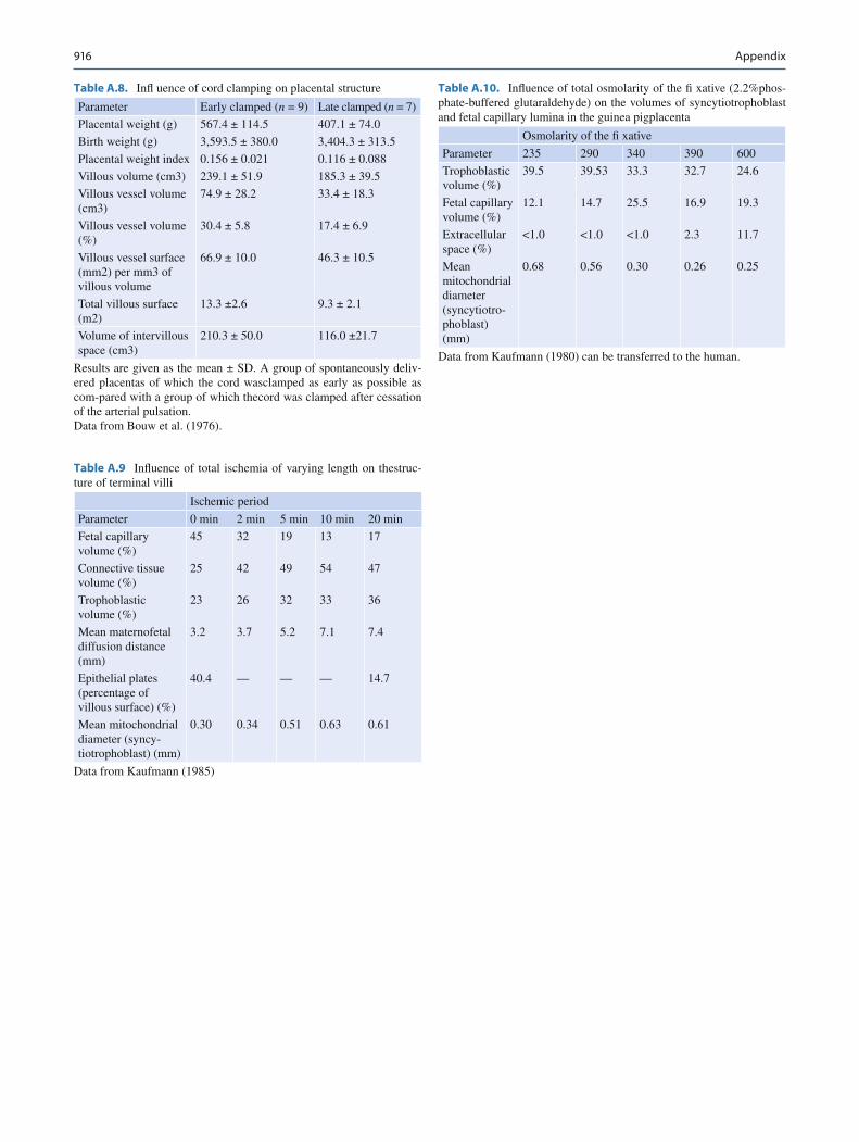
Table A.8. In fl uence of cord clamping on placental structure
Parameter Early clamped ( n = 9) Late clamped ( n = 7)
Placental weight (g) 567.4 ± 114.5 407.1 ± 74.0
Birth weight (g) 3,593.5 ± 380.0 3,404.3 ± 313.5
Placental weight index 0.156 ± 0.021 0.116 ± 0.088
Villous volume (cm 3 ) 239.1 ± 51.9 185.3 ± 39.5
Villous vessel volume (cm 3 )
74.9 ± 28.2 33.4 ± 18.3
Villous vessel volume (%)
30.4 ± 5.8 17.4 ± 6.9
Villous vessel surface (mm 2 ) per mm 3 of villous volume
66.9 ± 10.0 46.3 ± 10.5
Total villous surface (m 2 )
13.3 ±2.6 9.3 ± 2.1
Volume of intervillous space (cm 3 )
210.3 ± 50.0 116.0 ±21.7
Results are given as the mean ± SD. A group of spontaneously deliv-ered placentas of which the cord wasclamped as early as possible as com-pared with a group of which thecord was clamped after cessation of the arterial pulsation. Data from Bouw et al. (1976).
Table A.9 In fl uence of total ischemia of varying length on thestruc-ture of terminal villi
Ischemic period
Parameter 0 min 2 min 5 min 10 min 20 min
Fetal capillary volume (%)
45 32 19 13 17
Connective tissue volume (%)
25 42 49 54 47
Trophoblastic volume (%)
23 26 32 33 36
Mean maternofetal diffusion distance ( m m)
3.2 3.7 5.2 7.1 7.4
Epithelial plates (percentage of villous surface) (%)
40.4 — — — 14.7
Mean mitochondrial diameter (syncy-tiotrophoblast) ( m m)
0.30 0.34 0.51 0.63 0.61
Data from Kaufmann (1985)
Table A.10. In fl uence of total osmolarity of the fi xative (2.2%phos-phate-buffered glutaraldehyde) on the volumes of syncytiotrophoblast and fetal capillary lumina in the guinea pigplacenta
Osmolarity of the fi xative
Parameter 235 290 340 390 600
Trophoblastic volume (%)
39.5 39.53 33.3 32.7 24.6
Fetal capillary volume (%)
12.1 14.7 25.5 16.9 19.3
Extracellular space (%)
<1.0 <1.0 <1.0 2.3 11.7
Mean mitochondrial diameter (syncytiotro-phoblast) ( m m)
0.68 0.56 0.30 0.26 0.25
Data from Kaufmann (1980) can be transferred to the human.
917References
References
Abramovich DR (1969) The weight of placenta and membranes in early pregnancy. Obstet Gynaecol Br Commonw 76:523–526
Aherne W, Dunnill MS (1966) Morphometry of the human placenta. Br Med Bull 22:5–8
Auinger W, Bauer P (1974) Zum Zusammenhang zwischen Kindsgewicht, Placentagewicht, Muttergewicht und Muttergröße. Arch Gynäkol 217:69–83
Bacon BJ, Gilbert RD, Longo LD (1986) Regional anatomy of the term human placenta. Placenta 7:233–241
Baur R (1970) Über die Relation zwischen Zottenober fl äche der Geburtsplacenta und Gewicht des Neugeborenen bei verschiedenen Säugetieren. Z Anat Entwicklungsgesch 131:31–38
Bleker OP, Kloosterman GJ, Breur W, Mieras DJ (1977) The volumetric growth of the human placenta: a longitudinal ultrasonic study. Am J Obstet Gynecol 127:657–661
Bouw GM, Stolte LAM, Baak JPA, Oort J (1976) Quantitative morphology of the placenta. 1. Standardization of sampling. Eur J Obstet Gynecol Reprod Biol 6:325–331
Bouw GM, Stolte LAM, Baak JPA, Oort J (1978) Quantitative morphology of the placenta. 3. The growth of the placenta and its relationship to birth weight. Eur J Obstet Gynecol Reprod Biol 8:73–76
Boyd JD, Hamilton WJ (1970) The human placenta. Heffer & Sons, Cambridge
Boyd PA, Brown RA, Stewart WJ (1980) Quantitative structural differ-ences within the normal term human placenta: a pilot study. Placenta 1:337–344
Boyd PA, Brown RA, Coghill GR, Slidders W, Stewart WJ (1983) Measurement of the mass of syncytiotrophoblast in a range of human placentae using an image analysing computer. Placenta 4:255–262
Branca A (1922) Precis D’ Embryologie. Librairie J.-B. Bailliere et Fils, Paris
Burton GJ, Feneley MR (1992) Capillary volume fraction is the princi-pal determinant of villous membrane thickness in the normal human placenta at term. J Dev Physiol 17:39–45
Burton GJ, Ingram SC, Palmer ME (1987) The in fl uence of mode of fi xation on morphometrical data derived from terminal villi in the human placenta at term: a comparison of immersion and perfusion fi xation. Placenta 8:37–51
Cabezon C, De la Fuente F, Jurado M, Lopez G (1985) Histometry of the placental structures involved in the respiratory interchange. Acta Obstet Gynecol Scand 64:411–416
Clavero-Nunez JA, Botella-Llusia J (1961) Measurement of the villus surface in normal and pathologic placentas. Am J Obstet Gynecol 86:234–240
Clavero-Nunez JA, Botella-Llusia J (1963) Ergebnisse von Messungen der Gesamtober fl äche normaler und krankhafter Placenten. Arch Gynäkol 198:56–60
Ehrhardt G, Gerl D (1970) Eine einfache Methode zur Bestimmung der Zottenober fl äche von Placenten mit Hilfe der Flächenintegration nach der “Nadelmethode”. Zentralbl Gynäkol 92:728–731
Feneley MR, Burton GJ (1991) Villous composition and membrane thickness in the human placenta at term: a stereological study using unbiased estimators and optimal fi xation techniques. Placenta 12:131–142
Fujinaga M, Chinn A, Shepard TH (1990) Umbilical cord growth in human and rat fetuses: evidence against the “stretch hypothesis”. Teratology 41:333–339
Glöde B (1984) Morphometrische Untersuchungen zur Reifung men-schlicher Placentazotten. Medical thesis, University of Hamburg
Goldstein SR, Subramanyam BR, Snyder JR (1986) Ratio of gestational sac volume to crown-rump length in early pregnancy. Hum Pathol 31:320–321
Hill LM, DiNofrio DM, Guzick D (1994) Sonographic determination of fi rst trimester umbilical cord length. J Clin Ultrasound 22:435–438
Johannigmann J, Zahn V, Thieme V (1972) Einführung in die Ultraschalluntersuchung mit dem Vidoson. Elektromedica 2:1–11
Kaufmann P (1972) Untersuchungen über die Langhanszellen in der menschlichen Placenta. Z Zellforsch 128:283–302
Kaufmann P (1980) Der osmotische Effekt der Fixation auf die Placentastruktur. Verh Anat Ges 74:351–352
Kaufmann P (1981) Entwicklung der Plazenta. In: Becker V, Schiebler TH, Kubli F (eds) Die Plazenta des Menschen. Thieme, Stuttgart
Kaufmann P (1985) In fl uence of ischemia and arti fi cial perfusion on placental ultrastructure and morphometry. Contrib Gynecol Obstet 13:18–26
Kaufmann P, Scheffen I (1992) Placental development. In: Polin R, Fox W (eds) Neonatal and fetal medicine – physiology and pathophysi-ology. Saunders, Orlando, pp 47–55
Kloos K, Vogel M (1974) Pathologie der Perinatalperiode. Grundlage, Methodik und erste Ergebnisse einer Kyematopathologie. Thieme, Stuttgart, pp 1–361
Knopp J (1960) Das Wachstum der Chorionzotten vom 2. bis 10. Monat Z Anat Entwicklungsgesch 122:42–59
Laga EM, Driscoll SG, Munro HN (1973) Quantitative studies of human placenta. 1. Morphometry. Biol Neonate 23:231–259
Lips F (1891) Über die Gewichtsverhaeltnisse der neugeborenen Kinder zu ihren Placenten. Medical thesis, University of Erlangen
Mayhew TM, Joy CF, Haas JD (1984) Structure-function correlation in the human placenta: the morphometric diffusing capacity for oxy-gen at full term. J Anat 139:691–708
Mayhew TM, Jackson MR, Haas JD (1986) Microscopical morphology of the human placenta and its effects on oxygen diffusion: a mor-phometric model. Placenta 7:121–131
Molteni RA, Stys SJ, Battaglia FC (1978) Relationship of fetal and pla-cental weight in human beings: fetal/placental weight ratios at vari-ous gestational ages and birth weight distributions. J Reprod Med 21:327–334
Naeye RL (1987) Do placental weights have clinical signi fi cance? Hum Pathol 18:387–391
Naeye RL, Tafari N (1983) Noninfectious disorders of the placenta, fetal membranes and umbilical cord. In: Risk factors in pregnancy and disease of the fetus and newborn. Williams & Wilkins, Baltimore, pp. 145–172
O’Rahilly R (1973) Developmental stages in human embryos. Part A, Publ. 631. Carnegie Institute, Washington, D.C.
Sala MA, Valeri V, Matheus M (1983) Stereological analysis of syncy-tiotrophoblast from human mature placenta. Arch Anat Microsc 72:99–106
Schweikhart G, Kaufmann P (1977) Zur Abgrenzung normaler, arte fi zieller und pathologischer Strukturen in reifen menschlichen Plazentazotten. I. Ultrastruktur des Syncytiotrophoblasten. Arch Gynäkol 222:213–230
Sen DK, Kaufmann P, Schweikhart G (1979) Classi fi cation of human placental villi. II. Morphometry. Cell Tissue Res. 200:425–434
Teasdale F (1978) Functional signi fi cance of the zonal morphologic dif-ferences in the normal human placenta. Am J Obstet Gynecol 130:773–781
Teasdale F, Jean-Jacques G (1985) Morphometric evaluation of the microvillous surface enlargement factor in the human placenta from mid-gestation to term. Placenta 6:375–381
Voigt S, Kaufmann P, Schweikhart G (1978) Zur Abgrenzung normaler, arte fi zieller und pathologischer Strukturen in reifen menschlichen

918 Appendix
Plazentazotten. II. Morphometrische Untersuchungen zum Ein fl uss des Fixationsmodus. Arch Gynäkol 226:347–362
Weissman A, Jakobi P, Bronshtein M, Goldstein I (1994) Sonographic measurements of the umbilical cord and vessels during normal pregnancies. J Ultrasound Med 13:11–14
Winckel FKLW (1893) Lehrbuch der Geburtshilfe, 2nd edn. Veit, Leipzig
Wördehoff B (1971) Zur Bestimmung der Zottenober fl äche der men-schlichen Plazenta. Medical thesis, Würzburg
919K. Benirschke et al., Pathology of the Human Placenta, DOI 10.1007/978-3-642-23941-0, © Springer-Verlag Berlin Heidelberg 2012
Glossary
This glossary is intended to trace the roots of the often confusing terms used in placental pathology and perinatal development. The accents are placed for pronunciation.
Gk . = Greek; L . = Latin; Fr . = French; OE = Old English; ME = Middle English.
Acárdius Malformed twin without heart, invariably one of monozygotic twins [Gk. a = without, not + kardia = heart]
Abrúptio (placéntae) Detachment of placenta [L. abrumpere = to break away]
Adventítia Outer layer of vessel wall [L. adve-nire = to add]
Allántoïs (allantóic) Designation of one type of placenta, because of its roots in other mammals; thin membrane between amnion and chorion [Gk. allantos = sausage]
Ámnion Thin membrane surrounding the fetus; lamb’s caul [Gk. amnos = lamb]. Note : Because we use chorionic and not choriotic, it is here preferred to speak of amnionic, rather than amniotic. Hyrtl (1880) explored the various ter-minologies used to describe placental structures. He concluded that amnios and amnion were both correct. It was fi rst used by Galen, refer-ring to skin. The reference to lamb comes from Vesalius.
Androgénesis Development of male gender [Gk. andros = man + gennan = produce]
Anídian monster A hideous fetus with peculiar features; a form of acardiac twin [Gk. an = not + idios = peculiar + L. monstrum ]
Artiodáctyla Order of mammals, the even-hoofed animals, e.g ., cow, deer [Gk. artios = even + daktylos = toe]
Báttledore (placenta) Marginal insertion of cord [OE = fl at, wooden paddle used in the game of battledore to hit the shuttlecock]
Blástocyst Early germinative vesicle [Gk. blaste = germ + kystis = vesicle]
Bosselátion Surface granulation of placenta [Fr. bosseler = to ornament with bosses; from bosse = knob]
Capillary Hair- fi ne blood vessel [L. capilla = hair]
Céllular Belonging to the cell [L. cellula = small cabin, cell]
Extracellular Outside the cell Intercellular Between cells Intracellular Within cells Transcellular Across cells
Chiméra (Chímerism) The composite of several genotypes [Gk. chimaira = a monstrous beast. In Greek mythology, a monster made of the head of a lion, body of a goat, and tail of a dragon.]
Chirálity The quality of being chiral [Gk. chiro = the hand; a three-dimensional form, as a mole-cule, that cannot be superimposed on its mirror image. Used in designating the twist of the umbilical cord. Its fi rst usage was discussed by McManus (2002).]
Chorioangiópagus parasíticus A fetus, connected by blood vessels to another fetus [Gk. chorion =
920 Glossary
“little gut” = outer membranes around embryo + angeios = vessel + pagos = something set or fi xed; para = next to + sitos = food]
Chórion Outer membrane around embryo [Gk. chorion = “Little gut”. According to Hyrtl (1880) the term was also used by Galen as the outer shell of the membranes.]
Chórion frondósum The placenta proper [L. frondosus = richly covered with leaves, as in tree]
Chórion laéve The membranous portion of the chorionic sac [L. levis = smooth, without villi]
Chorionepithelióma Malignant tumor of trophoblast = chorioepithelioma or choriocarcinoma [Gk. chorion = “little gut” (outer membrane enclosing an embryo) + epi = on + thele = nipple + oma = tumor]
Circumvállate (placenta) An abnormal form of placenta with circumferential, old hemorrhages [L. circum + vallare = to wall around]
Cirsoid Aneurysmal dilatation of vessel [Gk. kirsos = enlarged vein]
Cotylédon Originally name for the single spots of placental tissue in the ruminants; lobe of the human placenta [Gk. kotyle = cup]
Cytotrophoblast Cellular type of the trophoblast [Gk. kytos = cell + trephein = to nourish + blaste = germ]
Decídua The endometrium at end of the luteal phase [L. decidere = fall, die]
Basalis Basal portion of placenta [Gk. basis = base]
Capsuláris Outer portion of membranes [L. capsula = little box]
Parietális Endometrium of pregnancy, covering wall portion of uterus [L. paries = wall; parieta-lis = pertaining to wall]
Véra Uterine decidua, contrasting it to pseudo-decidua, outside of uterus, as in endometriosis [L. verus = true]
Désmosome Intercellular junction [Gk. desmos = Ligament + soma = body]
Dizygótic Twins of two ova, “fraternal twins” [Gk. dis = twice, two + zygon = yoke]
Eclámpsia Coma and convulsive seizure in preg-nancy [Gk. eklampsis = shining forth; or ek = out + lampein = to shine]
Émbryoblast Embryo-forming cells of the blastocyst [Gk. embryon = unborn child + blaste = germ]
Endométrium Innermost layer of the uterus [Gk. endon = inside + metra = uterus]
Endothélium Innermost layer of blood vessels [Gk. endon = inside + thele = mamilla]
Endoplásmic reticulum A net-like, membrane-lined cell organelle [Gk. endon = inside + plasma = juice; L. reticulum = small net]
Epithelium Super fi cial cellular layer [Gk. epi = upon + thele = mamilla]
Epígnathus Tumorous mass in mouth, af fi xed to jaw and possibly a twin [Gk. epi = upon, at, over + gnathos = jaw]
Fétus papyráceus Paper-like, macerated, compressed (“compressus”) fetus [L. papyrus = plant from which paper is made]
Fíbrin Blood clot product [L. fi bra = fi ber]
Fíbrinoid A substance similar to but not identical with fi brin [Gk: … eides = looking like]
Fíbroblast Connective tissue cell [Gk. blaste = germ]
Freemártin The female of fraternal cattle twins, sterilized in utero by the male co-twin with whose placental vessels she is joined. The term presumably comes from the St. Martin’s feast in England, when these animals were consumed.
Fúnis (Funículus) Umbilical cord [L. funis = rope, cord]
Funiculópagous (twins) Twins joined at umbilical cord [L. funis = rope, cord + Gk. pagos = fi xed]
921Glossary
Funisítis In fl ammation of umbilical cord [L. funis = rope]
Fúrcate (cord) (“insértio funículi furcáta”) Forked insertion of the little rope [L. furca = fork]
Glycocálix Super fi cial layer of polysaccharides, covering the cell surface [Gk. glycos = sweet + kalyx = goblet]
Granulomatósis infantiséptica Neonatal disseminated listeriosis [L. granulum = little grain + Gk. oma = tumor]
Gynogénesis Female development [Gk. gyne = woman + gennan = produce]
Hemiacárdius Acardiac monster in which remnants of heart may be found [L. hemi = a half]
Holoacárdius Completely heartless monster (twin) [Gk. holos = whole]
Hydatídiform (mole) Severely hydropic placenta with bulbous, villous enlargement [Gk. hydatis = watery vesicle + L. forma = shape; L. mola = false conception; mass]
Hydrámnios Excessive amount of amnionic fl uid [Gk. hydor = water ( hydros = sweat; some early authors believed that amnionic fl uid was fetal sweat) + amnion = lamb’s caul]
Implantátion Establishing of intimate fetomaternal contact in the uterus [L. implan-tare = to embed]
Intervíllous Between the placental villi, i.e ., in the maternal blood space [L. inter = between]
Intravíllous Within a villus [L. intra = in]
Lacúna [L. = hole, gap]
Lithopédion Stone-like fetus [Gk. lithos = stone + paidion = child]
Lóchia (pl.) Uterine discharges after birth [Gk. lochia ]
Mácrophage Phagocytotic and paracrine cell type [Gk. macros = large + phagein = to eat]
Mágma reticuláre Jelly-like fl uid in original embryonic sac [Gk. magma = suspension of fi nely divided material in small amount of water + L. reticulum = little net (network)]
Mármoset Family of small South American pri-mates that always produce fraternal twins [Old Fr. marmouset = grotesque fi gure]
Mecónium Fetal intestinal content [Gk. mekon-ion = poppy juice]
Mésenchyme Undifferentiated connective tissue [Gk. mesos = in the middle of + chein = to pour (something poured in between)]
Mésoderm The middle germ layer [Gk. derma = skin]
Mésothelium Connective tissue–derived epithelial layer [Gk. thele = mamilla]
Microvíllus Finger-like extension of the cell surface
Mitochóndrion Rod-shaped cell organelle [Gk. mitos = thread + chondros = grain]
Mole (hydatidiform, Breus’, etc.) Vesicular mass of placenta [L. mola = false conception]
Monozygótic Single-egg-derived twins (“identical twins”) [Gk. monos = single + zygo-tos = yoked]
Nódus spúrius vasculósus (gelatínus) False knot in umbilical cord of vascular genesis [L. nodus = knot + spurius = not genuine + gelatina = gelatin]
Núchal (cord) Umbilical cord entwined around neck [L. nucha = nape of the neck]
Óctoploid Having eight sets of chromosomes [Gk. okto = eight + ploos = fold + eidos = form]
Oligohydrámnios Too little, or no amnionic fl uid [Gk. oligos = little + hydor = water ( hydros = Sweat) + amnion = lamb caul]
Ómphalo-(mesentéric) Umbilicus [Gk. ompha-los = Navel]
922 Glossary
Páraplacénta Those parts of the chorionic sac, not belonging to the placenta, e.g ., membranes [Gk. para = beside]
Périvillous Around the placental villi [Gk. peri = around]
Placénta [L. fl at cake] According to Hyrtl (1880) this term, with an originally Greek root, was introduced in 1559 by Realdus Columbus. Others referred to it as “secundines.”
Accréta Unusually adherent placenta that fails to detach [L. accrescere = to grow together, adhere]
Incréta Placenta that has grown into the myo-metrium [L. increscere = to grow into]
Membranácea Very thin placental membrane, the entire outside of which is covered with villi [L. membrana = from parchment, membrane]
Percréta Placenta that has grown through the uterine wall [L. percrebescere = to crowd everywhere]
Prévia Placenta that is located in the lower uterus and is in the way before the fetus can be delivered [L. praevius = in front of, before, lead-ing the way]
Plasmódium Multinucleate mass of protoplasm [Gk. plasma = a thing formed (juice) + eidos = to form]
Pólyploid Having many sets of chromosomes [Gk. polys = much, many + ploos = fold + eidos = form]
Pólypoid Protrusion of tissue, polyp [Gk. polys = many + pous = foot + oid = like]
Pycnósis (pyknosis) Degenerative condensation of cells or nuclei [Gk. pyknos = dense]
Secúndines Synonymous with afterbirth [L. secundus = following]
Schístocytes Broken red cells in disseminated intravascular coagulation [Gk. schistos = split]
Sínusoid Enlarged capillary [L. sinus = bight + Gk. eides = looking like]
Siréniform (fétus) Malformed infant with fused legs [Gk. seiren = mermaid]
Sirenomélia Malformed infant with fused legs [Gk. seiren = mermaid + melos = limb]
Stróma Connective tissue core of a organ [L. stroma = cushion]
Subchórial Under the chorionic plate [L. sub = below + Gk. chorion = leather, embryonic membrane]
Succentúriate (lobe) Accessory lobe of placenta [L. succenturiare = to substitute]
Superfecundátion Fertilization of two or more ova at different times during the same menstrual period [L. super = beyond, excessively + fecun-dus = fertile]
Superfetátion Pregnancy on top of already exist-ing pregnancy [L. superfetare = to bring forth while already pregnant; L. fetus = fruit, offspring]
Sympus Fetus with fused legs [Gk. syn = together + pous = foot (same as siren)]
Syncytium Multinuclear mass, derived from cell fusion [Gk. syn = together + kytos = cell]
Syncytiotrophoblast Syncytial type of trophoblast
Synéchia (pl. synéchiae) Adhesion of parts, here in the uterus [Gk. synecheia = continuity]
Tessellátion Irregular surface of placenta [L. tessella = little square stone (From Gk. tessares = four)]
Tétraploid Having four sets of chromosomes [Gk. tettares = four + ploos = fold]
Thalass é mias Hemoglobin disorders [from Gk. thalassa = the sea; generally referring to the Mediterranean]
Thixotrópic (Thyxo-) gel Gel that lique fi es when shaken, generally the extraembryonic fl uid [Gk. thixis = touching + trope = turn]
Thoracopágus Twins, conjoined at chest [Gk. thorax = chest + pagos = fi x]
Trabécula Small septum [L. small beam]
Tróphoblast The epithelium that covers the placenta [Gk. trophe = nourishment + blastos = germ]
923Glossary
Trophótropism The “wandering” of the placenta to the site of best nourishment [Gk. trophe = nourishment + trope = turn]
Urachus Connection of bladder to allantoic sac [Gk. ourachos (“that which has a tail”) = cord that extends from bladder to navel]
Vas prévium (pl. Vása prévia) Blood vessels within membranes that present before fetal parts during delivery [L. vas = vessel(s) that is (are) ahead (“previous”) of fetal part]
Vas vasórum (Vása vasórum) Blood vessels that nourish the vessels [L. vas = vessel]
Velaméntous (cord insertion) Membranous insertion of umbilical cord [L. velamen = veil + velamentum = cover]
Vérnix caseósa Sebum hair and other skin secre-tions from fetus [L. vernix = varnish + caseus = cheese]
Víllus (pl. vílli) Rami fi cations of placenta with fetal vessels that are the “business end” of the placenta, covered with trophoblast [L. villus = tuft of hair]
Vitélline Belonging to the yolk sac [L. vitellus = yolk of an egg]
Acknowledgment We are grateful to Professor E.N. Genovese, San Diego State University, for correcting this text.
References
Hyrtl J (1880) Onomatologia anatomica. W. Braumüller, Vienna McManus C (2002) Right hand left hand. The origins of asymmetry in
brains, bodies, atoms, and cultures. Harvard University Press, Cambridge
Bibliography
Haubrich WS (1984) Medical meanings. A glossary of word origins. Harcourt, Brace, Jovanovich, San Diego
Thomas CL (1985) Taber’s cyclopedic medical dictionary, 15th edn. Davis, Philadelphia
Webster D (1983) Webster’s unabridged dictionary. Dorset & Baber, Cleveland
925K. Benirschke et al., Pathology of the Human Placenta, DOI 10.1007/978-3-642-23941-0, © Springer-Verlag Berlin Heidelberg 2012
Index
A Abdominal pregnancy , 201, 208, 210, 212–214,
902. See also Ectopic pregnancy Abortion
chromosome studies in , 662–670 criminal , 559, 666, 670 erythroblastosis , 431, 436, 446, 449 habitual and lupus anticoagulant , 679 incomplete , 670–671 induced , 383, 477, 658, 661, 662, 666–670, 701 recurrent and lupus anticoagulant , 679 septic , 574, 669, 670, 672 spontaneous , 287, 288, 328, 333, 343, 436, 449, 463, 501,
534, 537, 586, 589, 599, 617, 627, 628, 657–660, 662, 663, 667, 671–673, 676–679, 695, 729, 802, 847, 850
studies of , 660 therapeutic , 202, 286, 291, 328, 446, 463, 475, 496, 500, 537, 602,
611, 660, 667–669, 676, 698, 706, 738, 739, 798, 804 Abruptio placentae
chorangioma and , 466 fetal effects , 526 hemorrhage and , 526 hemosiderin and , 890 infarction and , 523 infection and , 524 legal issues and , 882 preeclampsia and , 523, 524 scleroderma and , 495 toxemia and , 524–526 trauma and , 524
Abscesses, villous , 565, 583, 591, 597 Acardiac twins
animals and , 832 karyotypes and , 833
Accidental hemorrhage , 500, 522 Accreta placenta , 193, 204–208, 210–213, 244, 377, 379, 382,
384–386, 400, 509, 512, 622, 671, 851 Achordia umbilical cord , 326 Acquired immunode fi ciency syndrome (AIDS) , 598, 613,
614, 886. See also Human immunode fi ciency virus (HIV) Actinomycin D , 707, 708, 739, 740 ADAM-complex , 291. See also Amnionic band syndrome Adhesions amnionic , 287, 290, 292, 325, 676 AFP . See Alpha-fetoprotein (AFP) AIDS . See Acquired immunode fi ciency syndrome Alastrim , 608–609 Alcohol abuse , 497, 498 Allantochorial placentas , 309 Allantois , 27, 28, 121, 188, 309, 310, 317–318 Alpha-fetoprotein (AFP) , 50, 69, 204, 209, 219, 220, 362, 436, 448,
467, 469, 628, 667, 676, 730, 804, 810, 843 Alpha-Thalassemia and hydrops , 431, 437, 439
Altitude studies . See High altitude Aminopterin , 500 Amniocentesis , 3, 222, 258, 338, 378, 433, 462, 490, 523,
557, 659, 765. See also Genetic studies amnionic band syndrome and , 292 chorioamniotic separation , 272 hemorrhage and , 222 meconium and , 276 placental injury and , 462 tenting and , 268
Amnion amnionic sac infection syndrome , 280, 351, 407, 557, 560,
561, 565, 569, 572, 574, 592, 888 amniopatch , 825, 826 barrier function of , 256 burns and , 263 carbonic anhydrase , 257 chorionic plate and , 188 chromosomal determinations , 259, 260 clinical applications of , 253 cysts , 269–271 development of , 50, 51 examinations of ( see Amniocentesis) fl uid of ( see Amnionic fl uid) functions of , 50, 51 gastroschisis and , 258 keratinization , 188 mesoderm , 188 metaplasia of , 188 plica in twins , 388, 389, 788, 789 prostaglandins and , 178 re fl ected , 253, 264 research applications , 263 squamous metaplasia , 4, 5, 23, 188
Amnionic adhesions , 287, 290, 292, 325, 676 Amnionic band syndrome
collagen, hereditary defects and , 292 etiology of , 290 hydramnios and , 290 twins and , 290
Amnionic epithelium membranes , 254 umbilical cord , 311
Amnionic fl uid cells of , 259 composition , 258 cord length and , 259 embolism , 272–274 loss of , 257 pH-regulation , 257 sources of , 257 volume of , 258
926 Index
Amnionic lipids , 255 Amnionic sheet , 291, 293 Amnion nodosum , 4, 260, 261, 270, 285–287, 292, 294, 295,
356, 358, 666, 790, 799, 804, 815, 827, 841 Amniorrhea, extramembranous pregnancy and , 291, 293–294 Amniotropism of leukocytes , 561 Amputations . See Amnionic band syndrome Anastomoses in multiple pregnancy . See Vascular anastomoses Anchoring villi , 17, 22–25, 45, 82, 83, 101, 104, 157, 159,
160, 162, 166, 169, 174, 191–193, 197, 201, 202, 204, 211, 622, 624
Androgenetic cause of molar pregnancy , 396 Anemia
fetal , 421 hemorrhage and , 440 maternal , 420–421 neonatal , 329, 382, 388, 438, 466, 470–472, 729, 890 parvovirus infection and , 447, 449 sickle cell , 446 thalassemia , 451
Aneuploid cells, abortions and , 260 Aneurysms
chorionic surface vessels , 351–353 cirsoid , 352 surface placental , 351 umbilical cord , 351, 352
Angioarchitecture of villi , 113–117. See also Fetoplacental circulation
Angiomas , 271, 361, 362, 399, 401, 441, 442, 747–754, 756, 889. See also Chorangiomas
hydrops and , 750 umbilical cord , 751
Angiomatosis , 396 Angiotensin , 88, 129, 314, 315, 321, 822 Animal studies
comparative , 37 choriocarcinoma and , 740 toxemia (preeclampsia) and , 529
Anomalies congenital, hydrops and , 443–444 Antibodies
HLA type , 504, 700 lupus and , 532 moles and , 700 preeclampsia and , 514, 517
Anticoagulants , 222, 536–538 Antigenic studies
choriocarcinoma and , 735 HLA type , 735
Aplasia cutis in twins , 792, 794, 804, 810, 811, 816 Apoptosis , 79 Apt-Downey test for fetal hemoglobin , 470 Armadillo and polyembryony , 771 Arrhythmia fetal and hydrops , 445 ART (assisted reproductive technology) , 839, 851 Arteriopathy of decidua , 515–519, 533–535, 539, 659
in preeclampsia (PIH) , 515 Asphyxia fetal , 574. See also Fetal distress Aspirin therapy preeclampsia and lupus , 538 Asynchronous villous immaturity , 408 Atherosis decidua , 220, 504
preeclampsia (PIH) , 220 Atriopeptin in twins , 813 Attachment site of placenta , 531 Automobile accidents , 463, 464 A-V anastomosis , 7, 8, 847 Avascular villi . See Villi avascular
B Babesiosis , 619–620 Bacterial vaginosis , 564, 570, 572, 575, 589, 619 Bacteroides fragilis , 563, 580 Barberpole cord , 594 Barr body , 268, 690, 712, 740 Barrier, maternofetal , 28, 30, 161 Bart’s hemoglobin , 437 Basal plate
development , 191 extravillous trophoblast , 191 fi brinoid ( see Fibrinoids) junctional zone , 193 layers of , 191 separation zone , 193 uteroplacental vessels ( see Uteroplacental vessels)
Basement membrane endothelial , 499 histopathological relevance , 399 trophoblastic , 80–81
Battledore placenta , 330 Beckwith-Wiedemann syndrome (BWS) , 35, 450, 451, 752, 756, 842, 901 Bed Rest and twins , 854 Benign tumors , 747–756. See also speci fi c types Bicornuate uterus , 42, 381, 851 Bidiscoidal placenta , 34 Bilirubin, fetal , 277 Blastocyst , 13, 27, 28, 30, 41, 42, 44, 45, 50, 145, 167, 175, 176,
187, 250, 251, 253, 267, 334, 377, 381, 389, 672, 772, 777, 784, 811, 846, 849, 898–900
Blebs, syncytiotrophoblastic , 69 Blighted ovum , 659, 663, 803 Blood
ABO incompatibility and , 429 blood chimeras , 767, 781, 843, 901 clotting and fi brinoids , 24, 89, 403 erythroblastosis and ( see Erythroblastosis fetalis) transfusion syndrome ( see Transfusion syndrome)
Blood fl ow interrelations, maternofetal , 31–32 Blood vessels
chorionic ( see Chorionic surface vessels) fetal capillaries ( see Capillaries) fetal circulation and ( see Fetoplacental circulation) single umbilical artery ( see Single umbilical artery) umbilical cord ( see Umbilical artery; Umbilical veins) uteroplacental vessels ( see Uteroplacental vessels) villous trees and ( see Fetoplacental circulation) vitelline vessels , 319
Borrelia infection , 595 Bosselations , 13 Bouin’s fi xative , 697 Braxton-Hicks contractions , 268 Breast carcinoma metastases , 511 Breus’ mole, abortions and , 190, 214–218, 271, 400, 444,
475, 660, 664–666 Brucella abortus , 581 Burns amnion use , 263 BWS . See Beckwith-Wiedemann syndrome (BWS)
C Calci fi cation
basal lamina and , 185 placental grading and , 185 umbilical cord and , 339, 341, 356, 357, 592 villi and , 185

927Index
Calcium concentration of placenta , 185 Campylobacter infection , 564, 595 Cancer, metastases to placenta , 510 Candida albicans etc. infection , 565, 569, 595, 596 Capillaries , 24, 29, 45, 55, 103, 145, 182, 247, 264, 336,
397, 415, 431, 470, 487, 501, 574, 670, 697, 748, 778, 892. See also Fetoplacental circulation
congestion , 108, 397, 399 development of , 88 histopathological relevance , 397, 399 hypoxia and , 87 paravascular ( see Paravascular capillaries) terminal villi and , 87
Capped placenta , 381 Capsular decidua . See Decidua capsularis Carbonic anhydrase
amnionic , 257 villous , 259
Carcinomas. See Speci fi c types Carnegie development stages , 145–147 Caudal regression syndrome and diabetes , 344, 507 Cell columns , 313, 615, 740 Cell culture , 313, 615, 740 Cell islands, cysts , 25 Cerebral palsy
hemorrhage causing , 356 legal issues and , 881–884 twins and , 898
Cervix Carcinoma , 251, 501, 510 CMV infection , 599 infection , 588
Cesarean section hemorrhage in , 462–466, 471–474 placenta accreta and , 205, 206
Chagas’ disease , 446, 618–619, 624 Chemotherapy. See Speci fi c drugs Chickenpox , 607–608 Chimerism , 7, 55, 478, 672, 673, 694, 767, 782, 784, 832,
842–847, 901 Chirality spirals of umbilical cord , 321 Chlamydia , 566, 572, 574, 586–588, 590 Cholestasis of pregnancy , 353, 497 Cholesterol ester storage disease , 492 Chorangiocarcinoma , 730, 749, 754 Chorangiomas
and hydrops , 430, 436, 441 and transplacental hemorrhage , 466, 476
Chorangiomatosis , 396, 399, 450, 752–756 Chorangiosis, legal issues and , 882, 883 Chorioadenoma destruens , 687, 688, 704, 706, 707, 723, 727,
737, 740 Chorioamnionitis
diagnosis of , 273, 293, 342, 569, 572, 574, 577, 584, 594, 847, 885
gonococcal , 575, 586, 587 meconium and , 3, 280, 397, 498, 499, 560, 563, 575, 576, 579,
584, 597, 606, 882, 884, 888, 889 membranes and , 3, 256, 269, 280, 397, 399, 404, 498, 500, 557,
558, 560–564, 566–581, 583, 586–588, 593, 596, 597, 606, 672, 847, 853, 886
mycoplasma and , 572, 573, 586, 587, 589 streptococcus and , 563, 564, 572, 575, 576, 578 thrombosis and , 342, 351, 357, 399, 404, 433, 434, 525, 564, 566,
567, 587, 592, 882, 886, 889, 893 Chorioangiomas . See Chorangiomas
Choriocarcinoma animals and , 688, 728, 740 cell lines , 134, 160, 173, 700, 735, 740 ectopic , 160, 687, 688, 708, 709, 723–728, 738–739 endocrine aspects , 737–738 epidemiology of , 688, 736–737 fallopian tubes and , 738 genetic studies , 740 immune response , 705 in situ and fetal hemorrhage , 466, 729 mediastinal , 738 metastases , 699, 705, 706, 708, 723–730, 732, 733, 737–740 moles and , 687–690, 692, 695, 697, 699, 700, 703–706, 708,
709, 712, 723–728, 735–737, 739, 740 pregnancy , 134, 160, 264, 466, 688, 689, 695, 697, 699, 700,
703–706, 708–710, 712, 723–734, 738–740, 754 regression spontaneous , 705, 739 teratoma and , 728, 754 therapy , 687, 692, 695, 703–706, 708, 709, 723, 726, 729,
732, 736–740 Chorion
biopsy of ( see Chorionic villous biopsy or sampling (CVB or CVS))
blood vessels ( see Chorionic surface vessels) carcinoma of ( see Choriocarcinoma) frondosum , 46, 48, 134, 147, 249, 250, 252, 265, 292, 310, 387 irregular fusion in twins , 800, 849 mesoderm , 17, 18, 20, 22, 23, 148, 188, 189, 253, 264–265, 309
Chorionepitheliosis interna , 729 Chorionic plate. See also Extravillous trophoblast aneurysms , 351, 352 decidua and , 15, 146, 188, 190, 258 development of , 45, 187–188 extravillous trophoblast , 17, 22, 131, 189–190, 202, 265, 733 vasculature , 86, 696
Chorionic sac diameter , 134 Chorionic surface vessels
distribution patterns , 330 doppler studies and , 329 hemorrhages , 329 thrombosis , 329 wall thickness , 337
Chorionic villous biopsy or sampling (CVB or CVS) , 222, 259, 264, 293, 436, 438, 463, 466, 487, 488, 490–492, 615, 658, 671–674, 901. See also Villous trees
amnionic band syndrome and , 293 hemorrhage and , 222
Chorion laeve. See also Extravillous trophoblast decidua and , 48, 49, 157, 182, 252, 258, 292, 501 development , 48, 250–253 extravillous trophoblast , 124, 157, 252 immunological considerations , 661 meconium staining , 257 permeability of , 266
Chromosomal studies abortions and , 260 acardiac twins and , 834 amnion and , 259, 260 Barr bodies and , 695 conjoined twins and , 836 karyotypic analysis , 840 lyonization and , 260 molar pregnancies and , 688, 690 mosaicism and , 843 nonimmune hydrops , 437 twins and , 833
928 Index
Circulating lupus anticoagulant syndrome (CLAS) , 536–539 Circumvallate placentas , 3, 13, 190, 215, 271, 277, 282, 283, 294,
377, 381, 387–390, 445, 593, 606, 728, 755, 888 CLAS . See Circulating lupus anticoagulant syndrome (CLAS) Cleft lip in twins , 841 Cleft palate in twins , 828 Clomiphene citrate , 849 Closing ring, subchorial , 13, 190 Clostridial infections , 669 Coagulation disorders
amnionic fl uid and , 273, 274 thrombosis and , 359 twins and , 794
Cocaine abuse , 498, 524 Coccidioides immitis , 597 Coiling index, umbilical cord , 819 Coitus and labor , 575 Collagen, hereditary defects , 292 Colliquation necrosis , 217 Comparative anatomy , 309 Complete hydatidiform mole (CHM) , 35, 398, 662, 687, 689,
690, 692, 696–700, 704, 723, 724, 728, 738 Concurrent fl ow , 32 Con fi ned placental mosaicism (CPM) , 259, 672, 673, 693, 701,
729, 901 Congenital diseases , 615, 616. See also Speci fi c diseases
amnionic bands and , 287 candidiasis and , 595, 596 cytomegalovirus and , 599, 600 enzyme de fi ciencies and , 492 heart disease and , 444–445 hydrops and , 443–444 nephrosis and , 445, 446 neuroblastoma and , 440, 441 syphilis and , 565, 589–594 tuberculosis and , 582
Conjoined twins , 762, 765, 772–774, 795, 796, 820, 835–838, 841 Connective tissue
membranes and , 271 umbilical cord and , 269 villous trees and , 81–83
Consumption coagulopathy , 273, 355, 464 Cordocentesis , 333, 338, 351, 430, 433, 434, 449, 471, 474, 504,
615, 813, 816, 890, 891 Cord, umbilical . See Umbilical cord Cor pulmonale , 726, 821 Corynebacterium kutscheri , 579 Cotyledonary placenta , 28, 29 Cotyledons , 6, 7, 14, 15, 135, 137, 205, 280, 336, 435, 528, 581, 618,
778, 781, 813, 819, 820, 823, 824. See also Placentones Countercurrent fl ow , 32, 138 Couvelaire uterus , 514, 526 Coxiella burnetii , 621 Coxsackie virus infection , 451, 557, 609, 610, 886 CPM . See Con fi ned placental mosaicism (CPM) Crack cocaine abuse , 881 CRL . See Crown-rump length (CRL) Crosscurrent fl ow , 32 Crown-rump length (CRL) , 145–151, 384, 657 Cryptococcosis , 598 C-type particles , 614 Cushing’s disease , 506 Cushion, vascular , 346, 398, 886 Cutis aplasia in twins , 810, 811 Cystic degeneration of villi , 214 Cystic fi brosis , 274, 275, 501 Cystic hygroma , 444
Cystinosis , 502 Cysts
amnionic , 271 breus mole and , 214–218 cell islands and , 204 membranes and , 217 septal , 202, 216, 218–220 subchorionic thrombi and , 214 umbilical cord and , 318
Cytogenetic studies, abortions and , 260, 659 Cytokines and infection , 573 Cytomegalovirus (CMV)
villous structure and , 398 Cytotrophoblast
early development of , 73 extravillous ( see Extravillous trophoblast) formation of , 78 fusion , 73 necrosis , 72 plugs , 23, 47, 164 shell , 44, 162 villous ( see Langhans’ cells)
D Dead fetus syndrome , 274, 793 Decidua
arteriopathy and , 515–519 chorionic plate and , 188 degeneration of , 222–223 extracellular matrix , 181–182 granular cell decidua ( see Endometrial large granular lymphocytes) hemorrhages of , 525, 558, 660, 886 herpes and , 605–606 human placental lactogen and , 266 infection , 179, 221 lupus and , 180 membranes and , 176, 178 necrosis and , 180 preeclampsia and , 178–180
Decidua basalis , 6, 23, 46, 157, 162, 165, 175, 179, 180, 211, 241, 243, 250, 267, 385, 401, 407, 465, 501, 518, 533, 535, 536, 539, 583, 591, 624, 628, 660, 670
Decidua capsularis , 3, 4, 48, 104, 175, 178, 222, 223, 249–252, 256, 265, 269, 292, 401, 502, 515, 516, 518, 526, 533, 534, 557, 568, 575, 605, 790, 831, 883
Decidual cells function , 176 structure , 176
Decidua parietalis (vera) , 179, 180, 982 Deciduitis , 241, 498, 533, 557–559, 568, 571, 573, 579, 597, 605, 606,
608, 616, 623, 886, 902 Delivery
membrane rupture and , 885 placenta after , 204
Deportation of villi of trophoblast , 705, 706
Dermatitis , 502 Dermatomyositis , 496 Development stages of the placenta , 145–154 Diabetes mellitus of the mother
growth restriction in , 507 intravillous fi brinoid and , 89 nucleated red blood cells in , 338 placenta in , 506 thrombotic lesions and , 504 villous structure and , 506, 508, 509
929Index
Diamnionic dichorionic (DiDi) twin placentas , 7, 335, 435, 564, 596, 764–768, 774, 777, 779, 781–783, 789, 791, 792, 795, 799–802, 805, 826, 834, 842, 844, 845, 848, 850, 852
Diamnionic monochorionic (DiMo) twin placentas , 7, 335, 577, 765, 901
Diandry , 664, 702 DIC . See Disseminated intravascular coagulation (DIC) Dichorionic placentas , 764 DiDi twin placentas . See Diamnionic dichorionic (DiDi) twin
placentas Diffusion distances maternofetal , 31, 113, 422 DiMo twin placentas . See Diamnionic monochorionic (DiMo) twin
placentas Diphenylhydantoin , 500 Diphtheroids , 579, 583 Diplococcus pneumoniae , 578 Discoidal placenta , 34 Discoid lupus , 533 Dispermy , 693, 702 Disseminated intravascular coagulation (DIC) , 244, 273, 355,
358, 362, 363, 464, 526, 527, 529, 565, 589, 668–670, 705, 706, 751, 763, 790–793, 795, 808, 810, 831, 835, 836
Dizygotic (DZ) twins , 7, 290, 293, 352, 397, 431, 435, 530, 599, 615, 694, 703, 709, 710, 712, 753, 761–764, 767–771, 773, 774, 776, 777, 781, 782, 784, 805, 810, 822, 826, 832, 837, 840–844, 846, 853, 885, 899, 901
Doppler studies chorionic surface vessels and , 329 fetoplacental circulation , 315 intrauterine growth restriction , 819 stem vessels and , 404
Drew-Smythe catheter , 472 Duplex placentas , 381 Dynamic placentation , 335, 377, 378 Dysmaturity villous , 395, 892 Dysmorphism acardiac , 826 Dysplasia mesenchymal , 352, 753, 756 DZ twins . See Dizygotic (DZ) twins
E Echinococcus granulosus , 621 ECHO virus , 609 Eclampsia . See Preeclampsia Ectopic pregnancy
abdominal , 213, 385 choriocarcinoma , 709, 723, 738 erythroblastosis , 436 fallopian tubes, 160, 163, 688, 709, 738 ( see also Tubal
pregnancies) moles , 436, 670, 709, 723, 739 umbilical cord , 212–214, 325, 676
Edema umbilical cord , 2, 214, 269, 339, 351, 431, 441, 442, 488, 507,
611, 751, 827, 849, 891 villous , 105, 214, 280, 399, 407–408, 418, 432, 491, 503, 507,
508, 576, 577, 580, 587, 604, 660, 664, 751, 891–893 Electrophoresis of hemoglobin , 438, 470 Embolization , 273, 274, 333, 357, 436, 527, 669, 727, 740, 790,
791. See also Amnionic fl uid embolism Embryoblast , 251, 309 Embryonic staging , 145 Embryonic weight development , 147 Encephaloclastic lesions , 451, 464 Encephalomalacia , 794, 815 Endarteritis obliterans , 360, 405 Endocervix infection, mucous plug , 573
Endocrine disorders. See Speci fi c disorders Endometrial glands , 17, 18, 22, 23, 36, 45, 46, 62, 65, 71, 82, 106,
110, 145–148, 162, 173, 174, 178–181, 191, 195, 201, 242, 243, 599, 733
Endometrial large granular lymphocytes , 176, 178, 679 Endometrial stromal cells , 22, 23, 45, 176–177, 192, 207, 250 Endometritis
herpetic , 604 postpartum , 245, 604 streptococcus , 565 syncytial , 705, 723, 731, 733
Endometrium CMV infection , 599 decidua , 134, 176, 250, 385 de fi ciency of , 205, 206, 385 implantation , 44, 45, 145, 146, 241, 247, 250, 384 infection of , 179, 243, 244, 573, 599, 605, 616
Endothelial cells, fetal , 421 edema of , 405, 499 fetomaternal transfer and , 25, 626
Endothelial cells, maternal , 22, 25, 31, 124, 129, 152, 191, 192, 197 Endothelins , 74, 88, 178, 257, 315, 337, 405, 499, 813 Endotheliochorial placenta , 30, 31, 34, 44 Endoplasmic reticulum stress , 130 Entanglement, in cord , 273, 326–330, 348, 765, 785, 790, 823, 827,
848, 889 Enterobius vermicularis , 622 Enterovirus , 609–610 Epidermoid cysts of membranes , 269 Epidermolysis bullosa , 284–285, 407 Epignathus , 838–840 Epithelial plates , 66, 107, 154, 401. See also Vasculosyncytial
membranes Epitheliochorial placenta , 30, 31, 33, 34, 196 Epithelioid trophoblastic tumor , 687, 690, 724, 731, 733 EPO . See Erythropoietin (EPO) Errors in outline, placental forms , 79 Erythema infectiosum , 446 Erythroblastosis fetalis (EF)
ectopic pregnancy and , 436 hydrops and , 429–451 meconium and , 451 placental pathology , 430–439 twins and , 431, 435, 438, 442–444, 450 villous structure in , 403, 430, 431
Erythropoietin (EPO) , 128, 277, 405, 430, 467, 499, 794, 821, 891 Escherichia coli , 2, 563–565, 569, 574–576, 578 Estrogens, effects on villous development , 71 Ethnic studies
molar pregnancies , 689 multiple births , 771 thalassemia , 437
Evans syndrome , 504 Exaggerated placental site , 245, 687, 690, 704, 705, 723, 724,
731–740 Examination of the placenta, protocol , 2, 7 Exocoel , 253, 264 Exsanguination , 5, 333, 338, 352, 383, 439, 462–465, 471, 473, 474,
504, 748, 752, 783, 785, 791–793, 807, 810, 817, 831 Extraamnionic , 268, 286 Extracellular matrix
nonvillous parts of the placenta , 157, 161, 163, 165, 167, 168, 176, 178, 181–182, 185, 187, 199, 204
villous trees , 81, 83, 84, 133 Extrachorial placenta , 387–390 Extraembryonic coelom , 50 Extraembryonic mesenchyme , 45, 188, 264
930 Index
Extramembranous pregnancy , 291, 293–295, 387, 389 Extravillous trophoblast
adhesion molecules , 160 basal plate and , 22, 25, 89, 157, 174, 175, 191, 192, 217 cell islands and , 22, 24, 89, 157, 158, 183, 203, 217 endocrine activities , 175 fi brinoid and , 18, 22, 24, 157, 181, 182, 188, 190–193,
203, 731 growth factors and receptors , 167 history , 206, 385, 732, 739 homogeneity of population , 737 invasion of uteroplacental vessels ( see Intraarterial trophoblast) invasive properties , 47, 160, 162–164, 166–168, 171, 180, 190,
191, 193, 902 major basic protein , 902 maternofetal immune interactions , 134, 193 membranes and , 6, 22, 75, 111, 157–160, 163, 185, 189, 217,
492, 700, 733 nomenclature , 207, 214, 750 proliferation patterns , 22, 24, 134, 157, 166, 167, 174, 200,
201, 218, 221
F Fallopian tubes , 160, 163, 166, 242, 575, 586, 688, 709,
738, 897. See also Tubal pregnancies choriocarcinoma in , 160, 709, 738–739
False knot, of cord . See Umbilical cord Fatty liver , 497 Fetal death and nuchal cord , 327, 329 Fetal weight development , 5 Fetectomy, monkey , 247 Fetoplacental circulation
blood fl ow impedance , 126 blood vessels , 5, 766 capillary diameters , 130 congestion , 399, 403, 755 development of , 45 hemorrhage and , 461, 477, 729, 890 hemorrhagic endovasculitis (HEV) , 395 Hofbauer cells and , 616 immature intermediate villi , 113 maternal fl ow and , 137–138 obliterative changes , 515 oxygen and , 48, 126, 130–131, 415, 505 paravascular capillaries , 113 sinusoids , 55, 87, 108, 116, 135, 150 stem villi and , 86, 114, 152 terminal villi and , 46, 113, 415, 421 thrombosis , 395 vasculogenesis , 117–121 vasoregulation , 106
Fetus blood circulation and ( see Fetoplacental circulation) distress syndrome ( see Fetal distress) extramembranous ( see Extramembranous pregnancy) fetal storage disorders and , 487–492, 674 fetectomy and , 247 giant pigmented nevi and , 511 hydrops of ( see Hydrops fetalis) hypoxia of ( see High altitude; Hypoxia; Oxygen) immature ( see Immature fetus) mole and , 712 papyraceous , 215, 272, 404, 450, 781, 784, 791, 792, 794,
797, 801, 802, 804–811, 814, 815, 823, 829, 839, 848, 850, 885
tumors of , 440–443
Fibrin , 5, 6, 9, 24, 25, 44, 70, 79, 80, 84, 89, 146, 157, 158, 160, 165, 182–187, 190, 192, 193, 201, 203–205, 210, 214, 218–221, 241, 247, 259, 265, 266, 271, 284, 346, 387–389, 398–401, 403, 467, 474, 475, 495, 499, 503, 504, 515, 517, 520, 526, 527, 529, 533, 534, 539, 564, 565, 578, 582, 585, 597, 598, 608, 610, 613, 618, 621–623, 627–629, 668–671, 674, 678, 699, 726, 765, 793, 795, 802, 805, 882, 890, 892
Fibrinoids blood clotting and , 24, 89, 266, 403 calci fi cation and , 533, 601 cellular adhesiveness , 185 composition of , 89, 182, 192 fi brin-type , 24, 25, 80, 89, 158, 182–184, 190, 192, 193, 201, 204 functions , 183–185 immunologic signi fi cance of , 509 intramural , 182 intravillous , 24, 89, 148, 182, 403 matrix-type , 20, 22, 24, 25, 89, 158, 181–185, 188, 190, 192, 193,
201, 204, 265, 266 membranes and , 23, 80, 182, 249 Nitabuch’s , 20, 24, 25, 146, 182, 184, 191, 193 paratrophoblastic transport and , 184 perivillous , 20, 24, 68, 89, 102, 182–184, 200, 403 regulation of intervillous circulation and , 89, 184 Rohr’s , 20, 24, 25, 146, 191, 192 trophoblastic invasion and , 14, 18, 22, 24, 25, 89, 102, 151, 152,
181–183, 188, 190–193, 200, 201, 265, 266, 403, 474, 509, 731 uteroplacental ( see Fibrinoids, Nitabuch’s)
Fibroblasts, villous trees and , 22, 71, 84, 103, 108, 109 Fibroma , 747 Fibromuscular sclerosis of vessels , 404 Fibronectin, oncofetal , 181–183, 265 Fibrosis, villous stromal
de fi ciency of , 357, 406, 417, 420–422, 424, 492 histopathological relevance , 22, 137, 395–408, 886 oxygen and , 66, 83, 126–137
Finnish nephrosis , 408, 446 Fixation of the placenta, villous structure and , 395, 401, 506, 509,
755, 820 Flow cytometry , 8, 450, 467, 468, 471, 527, 659, 660, 662, 674,
692–694, 698, 701, 703, 709, 710, 723 Folate de fi ciency , 505 Folded type of placenta , 29, 30 Follicle stimulating hormone (FSH) , 258, 769–771 Francisella tularensis , 581 Fraternal twins . See Dizygotic (DZ) twins Freemartinism , 784, 832, 844 FSH . See Follicle stimulating hormone (FSH) Fungus infections , 595–599 Funiculopagus , 831 Funisitis
chronic , 566, 594, 604 necrotizing , 557, 560, 566, 568, 591–595, 607
Furcate cord insertion , 215, 330, 331 Fusobacterium , 564, 566, 573, 579, 580
G Ganglion cells, ectopic intestinal , 319, 490 Gangliosidosis , 488, 490, 491 Gardnerella , 581, 589 Gastrin and meconium , 3, 4, 257, 267, 268, 272, 274–284, 312, 322,
326, 340, 341, 353, 354, 361, 396–398, 451, 463, 472, 473, 497–499, 501, 505, 506, 513, 515, 560, 563, 575, 576, 579, 584, 597, 599, 605, 606, 609–611, 625, 749, 769, 799, 827, 882–884, 887–890
931Index
Gastroschisis , 258, 278, 281, 283–284, 293, 326, 331, 792, 809, 842 Gaucher’s disease , 450, 487, 488, 490, 491, 502, 673 G-cells , 275 Genetic studies
amniocentesis ( see Amniocentesis) choriocarcinomas and , 695, 740 hCG and , 658, 676, 692 multiple births and , 770, 826 spontaneous abortions and , 343, 660, 662, 663, 676
German measles , 612 Gestational trophoblastic neoplasia (GTN) , 687, 704, 735–740 Giant cells
molar pregnancies and , 704, 706 multinucleated, trophoblastic , 45, 163, 164, 269, 518, 582,
607, 706 placental site , 175, 242, 243, 245, 247, 384, 671, 704
Gitterinfarct , 496 Glial cell missing-1 factor (GCM1) , 75 Glossary , 911 Glycogen storage disease , 488, 492 Gonorrheal infection , 560 Gordon’s syndrome , 501 Granular cells . See Endometrial large granular lymphocytes Granulomatosis infantiseptica , 583, 584 Green placenta , 887–888 Grosser classi fi cation , 31 Group B streptococcus infection , 563, 568, 572 Growth factors
decidua and , 70, 110, 134, 167, 173, 177, 516 extravillous trophoblast and , 70, 111, 134, 166–168, 173, 174 vasculogenesis and , 124, 125 villous development and , 126
GTN . See Gestational trophoblastic neoplasia (GTN)
H Habitual abortion , 538, 615, 617, 677–680, 710 Haemangioendothelioblastomas , 750 Haemophilus in fl uenzae , 578 Hamartoma , 361, 442, 730, 750 Handedness , 322, 825, 841 hCG . See Human chorionic gonadotropin (hCG) Heart , 127, 214, 274, 315, 328, 330, 334, 335, 349, 403, 420,
430, 436, 439–445, 464, 465, 468, 469, 472, 473, 492, 498, 499, 501, 507, 533, 658, 678, 700, 751, 765, 783, 784, 786, 793, 795, 813, 814, 816–819, 821–823, 826–828, 830, 831, 837, 838, 844, 849, 881, 882, 884, 887, 892
acardiac twins and , 826 arrhythmias and , 445 block in lupus , 533, 678
Heart failure, maternal and villous structure , 403 Helices, umbilical cord . See Umbilical cord, spiraling of Hellin’s rule , 846 Hemangioblastic cells , 84 Hemangiomas, umbilical cord , 361 Hematomas
membranes and , 222, 271, 463 subchorionic , 214, 215, 271, 660 umbilical cord and , 271, 889
Hematopoiesis , 28, 51, 318, 384, 430, 431, 438–440, 667, 754, 821
Hemicardiac twins , 826 Hemochorial placenta , 31, 32, 55 Hemochromatosis , 451 Hemoglobin, maternal abnormalities , 430, 466–468, 505 Hemolytic diseases. See Speci fi c types
Hemorrhage accidental , 500, 522 amnion and , 222, 271, 320 amnionic band syndrome and , 290, 292 chorionic surface vessels , 330 decidua and , 3, 222, 241, 525, 660, 665, 886 fetal to maternal , 338, 467, 473 fetus and , 210, 219, 320, 362, 432, 439–443, 462, 463, 465–469,
471–474, 477, 729, 890 Kline’s type , 474 maternal-fetal , 439, 461, 467, 472 postpartum , 204, 241, 244, 245, 344, 382, 390, 503, 506, 852 prenatal , 294, 335, 471, 472 subamnionic , 271, 351, 496 transplacental , 405, 439, 440, 461–479, 729 trauma and , 351, 461–466, 468, 473 umbilical cord and , 466
Hemorrhagic endovasculitis (HEV) , 360, 361, 399, 404, 436, 490, 515, 625
Hemosiderin differential diagnosis from , 275, 438 erythroblastosis and , 434, 438, 520 extramembranous pregnancy and , 294 meconium , 3, 267, 275, 277, 280, 282, 283, 312, 888
Hemoxygenase , 88 Hepatic angioma , 442, 443, 753 Hepatitis , 488, 600, 610–611, 886, 890 Hepatocellular adenoma , 754 Heroin abuse , 499 Herpes
gestations , 593, 606, 607, 624 simplex virus , 222, 603–607, 610 virus hominis , 603, 605
Heterokaryotypic twins , 842–843 Heterotopic pregnancies , 851 Heuser’s membrane , 253 HEV . See Hemorrhagic endovasculitis (HEV) High altitude. 67 , 125–131, 136, 174, 217, 399,
402, 403, 412, 420–422, 505, 747, 755, 892 See also Hypoxia; Oxygen
chorangiosis and , 399 trophoblast proliferation and , 131, 173, 174 vasculogenesis and , 125 villous structure and , 755
Histiotrophic nutrition , 46 HIV . See Human immunode fi ciency virus infection (HIV) HLA antibodies , 504, 670 HLA antigens , 85, 157, 171, 193, 530, 679, 735, 778 Hoboken valves (nodes) , 315–317 Hodgkin’s disease , 500–501, 512
Hofbauer cells. See also Macrophages de fi ciency of , 406 development of , 121, 133, 149–151 development of fetal circulation and , 616, 618 erythroblastosis and , 431, 433, 438 fetal storage disorders and , 488–492 functions , 85 histopathological relevance , 399, 406, 408 history of , 431, 628 immature intermediate villi and , 22, 84, 150,
406, 408 immunological aspects , 84 in fl uence on villous development , 84 morphology of , 84, 491, 666, 677 origin of , 84 persisting immaturity and , 406
932 Index
Hormones , 9, 35, 55, 59, 60, 62, 65, 67, 71, 78, 88, 105, 128, 130, 166–169, 173, 176, 177, 204, 222, 223, 255–258, 267, 275–277, 398, 405, 506, 508, 676, 700, 701, 704, 732, 737, 752, 769, 770, 813, 844, 846, 848–850, 853. See also Speci fi c hormones
extravillous trophoblast and , 166–169, 204, 700 ovulation and , 769, 770, 773, 777, 846 twins and , 769, 770, 851, 855 villous development and , 128
Hospitals, litigation and , 881 hPL . See Human placental lactogen (hPL) Human chorionic gonadotropin (hCG)
abortions and , 658, 676, 699 amnion and , 256–258, 508 chorioangioma and , 752 choriocarcinomas and , 703, 704, 730, 732, 737 diabetes and , 508 effects on decidua , 658 extravillous trophoblast and , 71, 168, 175, 243 hydrops and , 430, 431, 441 Langhans’ cells and , 430 molar pregnancies and , 692, 694, 696 syncytiotrophoblast and , 71, 431, 477, 614, 699 twin pregnancy and , 855
Human immunode fi ciency virus (HIV) fetal endothelium and , 314, 405, 614 Hofbauer cells and , 613, 614
Human immunode fi ciency virus (HIV) infection , 59, 85, 314, 405, 598, 599, 610, 613–614, 618, 620
Human placental lactogen (hPL) decidua and , 266 extravillous trophoblast and , 732–734 Langhans’ cells and , 430
Human umbilical vein endothelial cells (HUVECs) , 313 Hurler’s syndrome , 489, 490 HUVECs . See Human umbilical vein endothelial cells (HUVECs) Hyaline membrane disease and streptococcus infection , 219, 569, 577,
578, 815, 847, 849, 883 Hyaline plaques of implantation , 242 Hydatid disease , 621 Hydatidiform mole
antigenic studies , 735 Breus’ mole and , 214, 215, 660 chemotherapy and , 692, 693, 703–705, 709, 710, 712,
728, 739 deportation , 705–713, 738, 740 diagnosis, prenatal , 671, 700 DNA fi ngerprinting , 694, 698, 701 edematous villi and , 105, 408, 687 embryo in , 35, 436 erythroblastosis and , 408, 430, 432, 467, 671 ethnic distribution , 689 fetus and , 35, 430, 432, 513, 687, 688, 696, 700–703, 705,
706, 710, 711 fl ow cytometry , 527, 659, 662, 692–694, 698, 701, 703, 709,
710, 723 frequency of , 474, 660, 688, 690, 692, 695, 710, 723, 736 genetics , 398, 662, 671, 687, 688, 690–695, 728 Hofbauer cells and , 406, 698 imprinting , 35, 673, 692, 695 invasive , 687, 688, 690, 692, 697, 703–708, 728, 735, 740 morphology , 671, 687, 693, 694, 698, 701, 756 partial (PHM)
morphology of PHM , 701 ovary in PHM , 688, 709, 738
recurrence , 719, 737
therapy , 687, 695, 704, 706–709 transitional , 698, 699 twin and , 711–713
Hydramnios amnionic band syndrome , 290 calci fi cation , 187, 242, 340, 341, 444, 445, 491, 751, 822 chorioangiomas , 747–753, 755 diabetes , 341, 439, 505, 508, 755 membranes , 187, 255, 277, 282, 330, 597, 813, 814, 823,
837, 849, 853 single umbilical artery , 287, 341, 390, 837, 849, 885 teratomas , 361, 441, 750, 830, 839 transfusion syndrome , 8, 336, 755, 813–815, 817, 819,
821–823, 840, 852, 885 Hydrops fetalis
chorangioma , 436, 441, 505, 749, 751 congenital anomalies , 443–445 etiology of , 440, 442 hemorrhage , 432, 439–443, 445 idiopathic , 436, 437, 444, 450 parvovirus infection , 449, 450 unknown etiology , 450–451, 599
Hydrops placenta , 2, 330, 431, 442, 692 Hygroma , 201, 444, 450, 666, 667 Hypercapillarization . See Hypervascularization Hyperemesis gravidarum , 497–499 Hyperlipemia , 501 Hypermaturity of villi , 397 Hyperparathyroidism , 187 Hypertension in pregnancy
trophoblastic proliferation , 658, 702, 738 Hyperthyroidism , 276, 505, 506, 703 Hypobetalipoproteinemia , 501 Hypoxia
fi brinoid deposition , 398, 403 in fl ammation , 398, 560, 562, 567, 580, 886 litigation in , 466, 794, 884 meconium , 277 preplacental , 126 postplacental , 126 trophoblast proliferation , 71, 131, 133, 173,
174, 421 uteroplacental , 126 vasculogenesis , 109, 125 vasculosyncytial membranes , 80, 87, 109, 126, 403 villous trees , 64, 71, 75, 80, 83
Hypoxic conditions, maternal villous structure , 401, 506, 508, 509, 755
Hyrtl’s anastomosis , 5, 315, 316, 342
I I-cell disease (mucolipidosis II) , 451, 489–491, 503 Ichthyosis congenita , 261 Identical twins . See Monozygotic twins Idiopathic hydrops , 437, 444, 450 Idiopathic thrombocytopenia (ITP) , 503–504 Immature fetus, meconium , 4, 276 Immature intermediate villi. See also Villous trees
blood vessel , 82, 117 development of , 22, 24 diagnostic problems , 106, 407, 414 histopathological relevance , 886
Immaturity persisting villous ( see Persisting villous immaturity) synchronous villous ( see Synchronous villous immaturity)
933Index
Immune interactions , 172, 626, 735 with abortions , 343, 508, 586, 659, 660, 663, 678, 679 with choriocarcinoma , 728, 730, 735–737, 739, 740 with chronic villitis , 210, 607, 627 with fi brinoid , 516 with trophoblast , 18, 177, 193, 202, 529, 614, 707, 727–729, 734
Immune response and preeclampsia , 530 Immunode fi ciency syndromes
AIDS , 598, 886 listeriosis , 583
Immunoglobulins (Igs) absorption to fi brinoid , 705 maternofetal transfer , 87, 451
Immunohistochemical markers collagen IV , 406 cytokeratin , 9, 64, 192, 199, 257 desmin , 81, 111, 121, 311 factor VIII , 200 fi brin , 9, 84, 89, 182, 192, 193, 266, 608, 610 Ki-69 , 110 laminin , 81, 87, 89, 181, 182, 262, 265, 266, 311 MIB-1 , 190 oncofetal fi bronectin , 181, 265 PCNA , 110, 190 a -sm-actin , 121 sm-myosin , 111, 121 vimentin , 9, 81, 87, 111, 121, 257
Impetigo herpetiformis , 501 Implantation, placental
hyaline plaques , 242 implantation site , 42, 48, 145, 146, 164, 168, 175, 181, 195, 198,
205, 213, 214, 222, 241–244, 247, 309, 515, 516, 518, 622, 670, 671, 704, 705, 733, 768, 772
placenta previa , 3, 205, 206, 208, 334, 346, 378, 379, 381, 382, 384, 900
Imprinting , 35, 36, 260, 672, 673, 692, 695, 756, 763, 842, 901 Inborn errors of metabolism , 492 Incompetent cervix , 384, 573, 574 Increta, placenta , 706 Infarction
fi brinoid ( see Fibrinoids, perivillous) maternal fl oor ( see Maternal fl oor infarction) placental , 1, 89, 198, 214, 217, 218, 221, 247, 337, 352, 355, 357,
381, 398–401, 403, 404, 421, 422, 463, 464, 471, 475, 479, 495, 498, 514, 519–522, 529, 533, 536, 538, 539, 565, 607, 623, 751, 787, 791, 890, 902
preeclampsia , 59, 89, 198, 217, 220, 399, 400, 403, 421–424, 464, 475, 495, 498, 514, 519–521, 526, 529, 533, 538, 539, 890
succenturiate lobes , 381 Infectious diseases. See Speci fi c diseases In fl ammatory lesions , 1, 8, 568, 573, 619, 882 In fl uenza , 502, 578, 610 Inhibin, Langhan’s cells , 17, 22, 70–78, 158, 159, 401–403, 440, 614 Insuf fi ciency, placental , 220, 397–398, 534, 537, 611, 622, 701,
888, 891 Insulin , 35, 60, 62, 65, 71, 87, 167, 177–178, 257, 258, 275, 423, 430,
506–509, 516. See also Diabetes vasculogenesis , 87, 431
Integrins , 69, 81, 120, 185, 513 Intelligence quotient (IQ) values, umbilical cord length , 325 Intercotyledonary septa . See Septa, placental Interferon , 85, 168, 180, 198, 586, 614, 679 Interleukins , 168, 180, 256, 567 Intermediate fi lament proteins . See Immunohistochemical markers Intermediate layer . See Spongy layer
Intermediate trophoblast , 671, 700, 723, 731, 733, 738. See also Extravillous trophoblast
Interposition, cord insertion , 3, 330–331, 334 Interstitial cytotrophoblast . See Extravillous trophoblast Intervillositis , 564, 588, 590, 610, 621, 623, 624, 627–629, 659,
670, 678 Intervillous blood fl ow , 104, 183, 378, 400, 432, 515, 534, 539 Intervillous space. See also Villous trees
circulation , 13, 14, 22, 23, 36, 45–49, 68, 83, 103, 126, 133–134, 146, 147, 172–174, 184, 185, 195, 196, 247, 401, 472, 474, 510, 528, 537, 618, 621, 662
fi brinoid ( see Fibrinoids, perivillous; Maternal fl oor infarction (MFI))
infarction , 6, 198, 247, 401, 421 pressure within , 117, 196 thrombi in ( see Intervillous thrombosis) volume of , 46, 103, 110, 131, 136, 152, 164, 422 width of , 17, 23, 104, 765
Intervillous thrombosis , 422, 467 Intestinal obstruction , 277, 320 Intraarterial trophoblast , 198 Intracytoplasmic sperm injection (ICSI) , 777, 898–901 Intramural fi brinoid . See Fibrinoids, intramural Intrauterine devices (IUDs) , 5, 222, 334, 349, 677 Intrauterine growth restriction (IUGR)
absent end-diastolic umbilical fl ow , 419 single umbilical artery (SUA) , 342 villous structure , 395
Intravascular trophoblast . See Intraarterial trophoblast Intravillous fi brinoid . See Fibrinoids, intravillous Invasive mole , 687, 688, 690, 692, 697, 704–708, 723, 724, 727,
728, 735, 737, 740 Involution, uterus , 241–245, 247, 739 Irregular chorionic fusion in twins , 800, 801, 849 Ischemia and villous structure , 88, 412, 529, 623 Islands . See Cell islands Islands of Langerhans, fetal , 320, 430, 439, 508 IUD . See Intrauterine devices IUGR . See Intrauterine growth restriction IVF (in vitro fertilization) , 41, 292, 564, 627, 664, 679, 695, 710,
772, 777, 782, 784, 802, 804, 825, 836, 838, 844, 846, 848, 850, 851, 853, 898–902
J Junctional zone , 14, 18, 22, 24, 32, 36, 185, 193, 253
K Karyotypic analysis
in abortions , 841 in acardiac twins , 833, 834 in twins , 833, 840
K-cell , 176, 178. See also Endometrial large granular lymphocytes Keratinization of amnion , 260, 261, 270 Kidneys diseases and placenta , 446, 582, 795 Kleihauer-Betke stains, technique for , 436 Kline’s hemorrhages , 474 Knots
false umbilical , 67, 69, 315–317, 324, 346, 347, 411 trophoblastic , 24, 67, 108, 183, 315, 402, 411, 412, 415, 505,
527, 699 umbilical cord , 4, 317, 324, 326, 328, 330, 331, 345–347, 357,
397, 399, 431, 676, 785, 889 Knotting . See Knots, trophoblastic Körnchenzellen , 178
934 Index
L Labyrinthine type of placenta , 29, 30, 34, 36, 509 Lactogen . See Human placental lactogen Lacunar stage, placental development , 43–45 Lacunar system , 45, 145, 191 Lamellar placenta , 29, 30, 34, 81, 89 Langhans’ cells
development of , 187 diabetes and , 403 endocrine activities , 72 histopathological relevance , 17, 20, 23, 24, 402–403 proliferation , 17, 22, 70, 158, 159 syncytial fusion , 49, 412 volumes , 152, 421
Langhans’ fi brinoid layer , 24, 188, 190. See also Fibrinoids Lead contamination , 187 Legal considerations , 881–893 Leiomyomas , 208, 385, 509, 750 Leprosy , 559, 581–582 Leptospirosis , 595 Leukemia , 47, 74, 168, 173, 440–442, 477, 510, 512, 513 Leukocyte migration , 562, 568 Leukotaxins , 351, 561 Leukotrienes , 256, 274, 531 LH . See Luteinizing hormone (LH) Limb-body wall complex , 287, 288, 290, 291, 293 Limulus test , 570 Lipids, amnionic , 255 Listeria monocytogenes , 2, 70, 559, 583 Listeriosis , 161, 407, 446, 564, 566, 583–586, 816 Lithopedion , 809 Litigations , 2, 244, 329, 334, 466, 794, 881–884, 887, 892, 893 Lobes, placental . See Cotyledons LSD . See Lysergic acid diethylamide (LSD) Lungs . See Pulmonary conditions Lupus anticoagulant , 2, 222, 495–539, 679, 889, 890 Lupus erythematosus , 180, 495, 517, 532–539, 598, 614, 678, 680 Luteinizing hormone (LH) , 258, 704, 769, 770 Lyme disease , 595 Lymphangiectasia , 444 Lyonization, chromosome studies , 260, 261, 762, 763 Lysergic acid diethylamide (LSD) , 292, 499
M Macrophages
basal plate and , 179 meconium , 4, 279–282, 312, 611, 888 membranes and , 611 villous ( see Hofbauer cells)
Macroscopic examination of the placenta , 1, 336 Major basic protein (MBP) , 9, 169, 170, 202, 217, 219, 243, 266,
671, 734, 737, 738, 902 Malaria , 58, 59, 557, 620–621, 628, 629 Maldevelopment, villous . See Villous maldevelopment Malignant melanoma metastases , 510 Malpractice , 881, 887 Marginal sinus , 190–191, 250, 251, 383, 524 Marginal zone , 15, 32, 137, 190–192, 200, 403 Masculinization, chimerism and , 843 Mast cells , 86, 88, 274, 311, 339 Maternal conditions. See also Speci fi c conditions
abortion and , 335, 390, 463, 477, 495–504, 506, 532, 579, 581, 584, 600
age and twinning , 762, 771, 773, 776, 842 diabetes and ( see Diabetes mellitus)
erythroblastosis and ( see Erythroblastosis fetalis) pregnancy and ( see Pregnancy)
Maternal endothelial cells , 22, 25, 31, 129, 152, 191, 192, 197 Maternal fl oor infarction (MFI) , 6, 214, 217–222, 400, 403, 495,
496, 607, 628, 673, 678, 890 herpes and , 222
Maternofetal barrier . See Barrier, maternofetal Maternofetal blood fl ow interrelations , 28, 31–34 Maternofetal transport. See also Syncytiotrophoblast
cell traf fi c , 790, 844 paratrophoblastic transport and fi brinoids , 184 transtrophoblastic channels , 70
Matrix-type fi brinoid , 20, 22, 24, 25, 89, 158, 181–185, 188, 190, 192, 193, 201, 204, 265, 266
Mature intermediate villi , 24, 57, 101, 104, 106, 107, 111–113, 115, 116, 118, 123, 135, 149–154, 402, 405, 415–418, 420, 422
Maturitas precox placentae, villous structure and , 402 MBP . See Major basic protein (MBP) Measles infection , 612–613 Meckel’s diverticulum , 319, 320 Meconium
aspiration syndrome , 275, 276, 278, 281, 888 chorioamnionitis and , 280, 397, 499, 576, 579, 584, 884 hemosiderin, differential diagnosis from meconium , 275 immature fetus and , 4, 276 legal consideration , 882–884, 887–890 toxic effects , 278, 340, 354, 397
Medicolegal considerations , 882 Melanin , 187, 260, 510 Melanoma metastasis , 510 Melena, test for , 470 Membranacea, placenta , 13, 28, 206, 208, 213, 386–387, 582 Membranes, placental
amnion ( see Amnion) chorion laeve ( see Chorion laeve) color , 4, 5, 281, 890 de fi nition , 249 development of , 250–253 hemorrhages, subamnionic , 271 in fl ammation , 250, 260, 280, 282, 284, 292 meconium staining , 257, 275–277, 280–282 physical properties of , 267 premature rupture of (PROM) , 268–269, 277, 292 rupture of , 249, 258, 259, 262, 265 teratomas , 269, 271 trophoblast layer , 265–266, 282
Mesenchymal cells , 45, 50, 71, 81, 82, 84, 111, 118, 120, 124, 125, 193, 257, 265, 311, 489, 752
Mesenchymal dysplasia , 352, 753, 756 Mesenchymal villi , 22, 49, 81, 83, 84, 101, 104, 106, 109–113,
117, 118, 121, 123, 135, 147–150, 153, 154, 407, 415, 416, 418
Mesoderm amnionic , 20, 23, 148, 188, 253, 261–263 chorionic , 17, 18, 20, 22, 23, 148, 188, 189, 253, 264–265, 309
Metaplasia, amnionic , 4, 5, 23, 188, 260 Metastases to placenta , 440, 510, 511, 838 Methotrexate , 205, 213, 385, 496, 500, 703, 706–710, 738–740 MFI . See Maternal fl oor infarction (MFI) Microscopic survey of the placenta , 17–25 Microvilli, syncytiotrophoblastic , 55–61 Middle ear , 562, 578 Mineral deposits, placental , 187 Miscarriages, amnionic bands and , 292 Missed abortion , 207, 215, 216, 657, 661, 678, 700 Molar pregnancies , 687–713
935Index
MoMo placenta . See Monoamnionic, monochorionic (MoMo) placenta
Monoamnionic, monochorionic (MoMo) placenta , 7, 348, 762, 765, 781, 784–798, 826, 836, 838, 843, 885
Monochorial twins , 336, 764, 771, 791, 810, 811, 851 Monochorionic placentas , 7, 577, 763, 764, 767, 768, 777, 782,
784, 785, 789, 813, 819, 831, 883, 885, 901 Monocytes , 47, 76, 80, 84, 278, 441, 620, 621, 623 Monosomy X , 201, 216, 444, 658, 664–667 Monozygotic (MZ) twins , 7, 32, 290, 292, 323–325, 342, 361, 444,
450, 530, 577, 599, 613, 615, 623, 695, 710, 711, 713, 761–778, 781, 782, 784, 792–796, 798–800, 802, 807, 810, 811, 814, 822–826, 828, 830, 835–837, 840–844, 846, 848–850, 852, 853, 898–900
Morquio’s disease , 488, 491 Mortality
maternal in abortion , 666, 667, 669 perinatal , 126, 322, 323, 328, 329, 384, 396, 399, 472, 497,
499, 503, 567, 583, 765, 784, 785, 804, 840, 846, 853–855, 882, 885
twins , 348, 764, 765, 773, 853, 854 Mosaicism
con fi ned placental , 259, 672, 673, 693, 701, 729, 901 twins and , 694, 842
Motilin, and meconium transport , 275, 276 Mucoid degeneration of cord cysts , 339 Mucolipidosis , 451, 489–491 Multiple births , 6–8, 334, 342, 343, 382, 761, 764, 765, 769–774,
776, 820, 826, 846–851, 881, 884, 898 Multiple myeloma , 512 Multiple pregnancies . See Twins Multivillous fl ow , 33, 138 Mumps , 610 Mural thrombosis of fetal vessels , 566, 612 Mushroom poisons , 497 Mycobacterium leprae , 581, 582 Mycoplasma , 572, 573, 586, 587 Myocarditis , 446–451, 537, 608, 610, 618 Myo fi broblasts , 81–83, 86, 103, 104, 109, 111, 176, 189, 195, 264,
311–313 Myositis ossi fi cans , 497 MZ twins . See Monozygotic (MZ) twins
N Necrosis, decidual , 222, 475, 515, 516, 524, 525, 532, 535,
591, 669, 671 Necrotizing funisitis , 557, 560, 566, 591–595, 607 Neisseria gonorrhoeae , 575 Nematosomes , 72 Neoplasms, malignant , 706, 723, 730
metastases to placenta , 510 Nephropathy , 502, 507, 514, 520 Nephrotic syndrome , 445–447, 732 Netzinfarkt , 218 Neuroblastoma, congenital , 440, 441 Neuropeptide Y (NPY) and Langhans’ cells , 315 Nevus, skin lesions , 511 Newborns, hemolytic diseases , 429, 438 Nidation . See Implantation, placental Nitabuch’s fi brinoid . See Fibrinoids Nitric oxide (NO) synthase , 65, 86–88, 167, 169, 315, 529 Nodus spurious
gelatinosus , 346 vasculosus , 346
Non-immune hydrops . See Hydrops fetalis
Noonan’s syndrome , 444 Normative tables , 430 NO synthase . See Nitric oxide (NO) synthase NRBCs . See Nucleated red blood cells Nuchal cord
fetal distress and , 328, 330 hypovolemic shock and , 329
Nucleated red blood cells (NRBCs) histopathological relevance , 398, 404, 405 legal issues , 891–893 in maternal circulation , 469, 470, 478, 891 trauma and , 465, 466, 470, 473
O Obliterative endarteritis , 360, 404, 422, 508 Obliterative vascular changes of fetoplacental circulation , 360 Obstetricians, litigation against , 881 Oligohydramnios , 4, 220, 261, 282, 285–287, 293, 325–327, 341,
357, 390, 506, 524, 813, 815, 888 Omphalocele , 283, 284, 286, 320, 330, 363, 450, 607, 670, 826,
828, 830, 833 Omphalomesenteric duct , 271, 309, 318–321 Oncogenes , 700 Ophthalmia neonatorum , 560, 575, 588 Otitis, and chorioamnionitis , 562 Ovarian lutein cysts , 430 Oxidative stress , 48, 65, 78, 87, 126, 134, 174, 196, 198 Oxygen. See also Fibrosis, villous stromal; High altitude
high altitude and , 126, 127, 130, 131, 420, 505 intervillous circulation and , 134, 135 Langhans’ cell proliferation and , 71 onset of the maternal arterial circulation and , 48, 173 partial pressure in intervillous space , 173, 402, 406 partial pressure in villi , 133 sensing , 128 stromal fi brosis and , 406 transfer of , 32, 505 trophoblast and , 131 trophoblast invasion and , 172 vasculogenesis and , 130 villous stroma and , 133
Oxytocin amnion and , 256 umbilical vessels and , 314, 315
P Papular dermatitis , 502 Parabiotic circulation , 820 Paraplacenta , 28 Parasitic infections. See Speci fi c types Parathyroid adenoma, calci fi cations in , 185 Paravascular capillaries , 24, 103, 104, 113–115, 117, 121, 123 Parietal decidua . See Decidua parietalis (vera) Partial hydatidiform mole (PHM) , 352, 398, 408, 513, 662, 664,
673, 687, 688, 690, 692–694, 697, 700–705, 709–713, 728, 730, 754, 756
Parvovirus infection and hydrops , 447, 449, 450 Percreta, placenta , 166, 206–213, 292, 385, 400, 570, 622, 708,
848, 852 Periarteritis nodosa , 496 Pericytes , 87 Perinatal medicine, litigations , 882
cord complications , 882 Perivascular capillaries . See Paravascular capillaries
936 Index
Perivillous fi brinoid . See Fibrinoids, perivillous Persisting villous immaturity , 23, 401, 417 Phenylketonuria , 501 Pheochromocytoma , 502, 506, 525 Phlebitis , 354, 559, 562, 565, 568, 591, 893 PHM . See Partial hydatidiform mole (PHM) Phospholipase , 256, 275, 562, 571 Phylogeny, placenta and , 33 Placenta
abdominal pregnancy and , 213–214 abruptio and ( see Abruptio placentae) accreta , 193, 204–208, 210–213, 244, 377, 379, 382, 384–386,
400, 509, 512, 622, 671, 852 chorioallantoic , 27, 28, 36, 110, 128, 250, 307 amnion and ( see Amnion) annular type , 381, 382 battledore type , 330 bed , 14, 15, 22, 47, 162–164, 167–169, 171, 172, 174, 175, 179,
181, 193, 195, 196, 243, 245, 272, 384, 385, 389, 421, 513–519, 522, 529, 533, 733, 779
benign tumors and , 747–756 bidiscoidal type , 34 bilobate type , 13, 378, 379, 534 circumvallate type , 3, 13, 190, 215, 271, 277, 282, 283, 294, 377,
381, 387–390, 445, 593, 606, 728, 755, 888 classi fi cation systems , 34 cotyledonary type , 6, 28, 29, 34, 36, 509 cotyledons , 6, 14, 280, 336, 581, 823 deciduochorial , 36 developmental stages , 145–154 diffusa type , 386 discoidal type , 34, 36 duplex type , 13, 380, 381 dynamic placentation , 335, 377, 378 ectopic pregnancy , 203, 213, 852 endotheliochorial type , 30, 31, 33, 34, 44 epitheliochorial type , 30, 31, 33, 34 evolution , 33 examination of , 1–10 extrachorial , 387–390 fenestrata type , 381 folded type , 29, 30, 34 functions , 76, 129, 136, 396, 397, 537, 622, 891 grading of , 185, 187 hemochorial type , 31, 33, 34, 36, 48, 55 hydrops and , 2, 429–451, 557 immature ( see Persisting villous immaturity; Synchronous
villous immaturity) implantation of ( see Implantation, placental) increta , 206–210, 385, 386, 688, 703, 706, 852 insuf fi ciency of , 220, 397–398, 534, 537, 611, 622, 701,
888, 891 interspecies differences , 27 intrauterine growth restriction (IUGR) , 9, 49, 126, 130, 172, 402,
405, 421–424, 662 labyrinthine type , 29, 30, 34, 36 lamellar type , 29, 30, 34 lobes , 3, 315, 379–381 localization of , 377, 378, 462, 614 macroscopic features , 13–15, 487 marginata , 13, 190, 389, 390 meconium staining and , 275, 276, 281, 282, 497, 499, 883, 887, 890 membranacea , 13, 28, 206, 208, 213, 386–387, 582 membranes of ( see Membranes, placental) MoMo type , 7, 335, 765, 781, 782, 789, 790, 826, 838, 843 multiple births and , 6–8, 764, 769
percreta , 166, 206–213, 292, 377, 385, 400, 570, 622, 708, 848, 852
polyp , 204, 241, 244–247, 380, 671 previa , 3, 205–208, 272, 273, 326, 333–335, 346, 377–386, 389,
445, 466, 470, 471, 495, 498, 500, 502, 523, 526, 586, 751, 752, 815, 853, 897, 900
residual , 345 separation of , 14, 193, 241, 335, 386, 503,
523, 890 shape aberrations of , 377–390 shapes , 1, 3, 13, 28–29, 34, 335, 782 succenturiate lobe and , 3, 212, 213, 380, 381, 386, 534 surface vessels ( see Chorionic surface vessels) trabecular type , 29, 30, 34 transplacental hemorrhage , 439, 461–479 types , 27–37, 768, 812, 821 uterine involution and , 242 villous type , 29, 102, 147, 152, 167, 337, 382, 383, 395, 445, 501,
532, 595, 747, 754, 755 weight of , 5, 8, 32–34, 145, 148–151, 247, 324, 341, 349, 382,
397, 439, 497, 499, 501, 505, 506, 591, 662, 674, 676, 755, 764, 782, 819, 899
zonary , 28, 29, 34, 379 Placental barrier . See Barrier, maternofetal Placental diameter , 145, 148–151, 379 Placental mosaicism , 259, 672, 673, 693, 701, 729, 901 Placental polyp , 204, 241, 244–247, 380, 671 Placental septa . See Septa, placental Placental site
giant cells , 242, 243, 245, 247, 704 involution , 241–243 nodules, plaques , 245, 687, 690, 709, 724, 731–740 tumor , 732, 733
Placental thickness , 145, 148–151, 441 Placental transfusion , 344–345 Placental transporters , 31, 35, 59 Placental volume , 29, 344, 379, 398, 820 Placental weight , 5, 8, 32–34, 145, 148–151, 247, 324, 341, 349, 382,
397, 439, 497, 499, 501, 505, 506, 591, 662, 674, 676, 755, 764, 782, 819, 899
Placental weight index , 755 Placentones , 14, 134–137, 336, 395 Plakopathia diabetica , 508 Plaque
placental site, nodule , 245, 687, 690, 709, 724, 731–740 Plasma cells , 86, 176, 180, 210, 220, 243, 245, 407, 451, 533, 558,
561–563, 589, 591–593, 595, 596, 599–603, 605, 606, 608, 611, 616, 623–625, 677, 727, 887
Plasmalemmas, syncytiotrophoblastic , 59–61 Plasma protrusions, syncytiotrophoblastic , 57, 115 Plasmodia, infection , 620, 621 Plethoric twins , 793, 816, 818, 821 Pneumonia , 558, 561–564, 569, 575, 578, 579, 588, 596, 597, 607,
608, 619, 885 Polar bodies, twins and , 774, 841, 843, 845, 898 Polyembryony , 771, 772 Polymorphonuclear leukocytes in villitis , 241, 406 Polyploidy , 163, 164, 259, 664, 692, 710, 734 Polyps, placental , 204, 241, 244–247, 380, 671 Pompe’s disease , 488, 492 Porphyria , 504, 600 Postmaturity , 185, 186, 277, 362, 396, 419 Post mortem Cesarean section , 219 Postpartum hemorrhage , 204, 241, 244, 245, 344, 382, 390, 503,
506, 853 Potter’s syndrome , 261, 294, 796, 841, 851
937Index
Preeclampsia (PIH) , 2, 23, 58, 119, 162, 245, 267, 315, 377, 397, 411, 439, 462, 495–539, 557, 664, 705, 747, 815, 883
abruptio and , 495, 497–500, 502–506, 509, 514–516, 521–526, 528, 529, 533, 534, 536, 537, 539
animal models , 514 chorangioma and , 498, 505 etiology of , 495, 502, 524, 529–532 hypoxia and , 498, 499, 503, 505, 526–528 infarcts and , 495, 496, 500, 501, 503–507, 514, 515, 519–523, 525,
526, 528, 530, 532–538 Langhans’ cells and , 509 molar pregnancies and , 705, 713 prostaglandins and , 531 scleroderma and , 495–496, 532 syncytial knots in , 505, 514, 527, 528, 533 Tenney-Parker changes , 503, 505, 514, 520, 527, 528, 530,
533–535, 538 umbilical cord and , 496, 498, 500, 502, 503, 507, 510, 512,
534, 539 villous structure in , 501, 506, 508, 509
Pregnancy extramembranous , 291, 293–295, 387, 389 extrauterine , 175, 193, 213, 582, 852 hypertension and ( see Preeclampsia (PIH)) placenta previa and , 207, 208, 335, 377, 378, 382–384, 386, 495,
498, 502, 815 prolonged , 220, 351, 362, 402, 408, 419–420, 883, 887 stages of, histological characterization , 22, 23, 28, 45, 81, 101,
188, 202, 253, 258, 319, 341, 406, 419, 497 Pregnancy-induced hypertensions (PIH) . See Preeclampsia Prelacunar stage, placental development , 41–44, 145 Premature deliveries
capillaries in , 405 chorioamnionitis and , 560, 567, 572–575 legal issues , 885, 886, 892 membranes rupture and (PROM) , 567, 572, 574, 575 villous structure in , 397
Prenatal infections , 449, 557–560, 565, 569, 570, 572, 578, 579, 587, 595, 600, 602–604, 609, 799, 887
Preterm villous maturation , 419 Previa, placenta
accreta and , 205–207, 272, 273, 379, 382, 384–386 chorangiomas and , 466, 752 localization studies , 377–379
Primary villous stage , 43, 147 Progesterone
effects on decidua , 176, 178 effects on Hofbauer cells , 614 effects on villous development , 62
Prolactin , 47, 177–178, 258, 704 Prolapse, umbilical cord and , 309, 314, 318, 320, 326, 328, 329,
334, 399, 404, 853 Prolonged pregnancy, villous structure , 419–420 Propranolol , 501 Prostacyclin , 315, 500, 531, 538, 539 Prostaglandins
amnionic fl uid and , 178, 255, 256, 571, 572 decidua and , 176, 178, 573 E2 type , 85, 176, 178, 180, 315 extravillous trophoblast and , 256 infection and , 256, 571, 573, 574 preeclampsia and , 531 umbilical cord and , 314, 315
Proteases placental development and , 259, 269 trophoblastic invasion and , 165, 168, 178
Protein C & S de fi ciency , 355–356, 504, 680 Pruritus , 501–502 Pseudochorangiosis , 396 Pseudotumor , 706, 731 Puerperal fever , 245, 577 Pulmonary conditions
amnion and , 258, 273, 278, 451 embolism and , 245, 273, 274, 668, 669 hypertension in choriocarcinoma , 726 hypoplasia and extramembranous pregnancy , 294 hypoplasia and oligohydramnios , 286, 287 meconium and , 278, 281 molar tissue and , 705, 710, 726 trophoblast embolism , 436, 527
Pulmonary hypertension amnionic fl uid embolism , 273, 274 choriocarcinoma and , 726
Pump twin , 822, 830, 831 Purpura , 333, 356, 357, 360 Pyelonephritis , 317, 495 Pygopagous twins , 848
Q Q-fever , 621 Quadrichorial placenta , 848 Quantitative data of the placenta , 17, 174 Quintuplets , 828, 829, 846, 848–852, 855
R Rabies , 610 Racial studies
in hydatidiform moles , 688, 689, 700 in multiple pregnancy , 768, 770, 771, 776
Radioangiographic studies , 14 Rami chorii, villous types , 101, 114 Ramuli chorii, villous types , 101, 102, 114 Reduction, selective , 849, 854 Re fl ected chorion . See Chorion laeve Rejection, as immune reaction , 626 Relaxin, decidua and , 176, 178, 267 Renal conditions , 497, 513 Reticulum cells, villous trees and , 106, 108, 109, 111 Retromembranous hematoma , 222, 223, 463, 559, 672, 888 Retroplacental hematomas , 401, 464, 466, 472, 477, 523, 524, 557,
629, 890 Rhesus incompatibility. See also Erythroblastosis fetalis
villous structure , 23, 105, 417 Rheumatoid arthritis , 180, 477, 496–497, 532 Rohr’s stria . See Fibrinoids Rubella virus infection , 360, 407, 612, 623, 625, 821 Rubeola , 612–613 Rupture, of umbilical cord , 286, 288, 294, 327, 330, 348–349
S Sacrococcygeal teratoma , 358, 441, 750, 838–840 Saline infusion , 670 Salmonella typhi , 579–580 Sandhoff’s disease , 488, 491 Sarcoidosis , 502, 534 Scalloping of trophoblast , 664 Schistosoma haematobium , 622 Scleroderma , 70, 495–496, 532 Seatbelts, uterine injury , 464
938 Index
Secondary villous stage , 43, 148 Sectional artifacts of villi , 67 Selective reduction of twins , 849, 854 Separation of the cord, premature , 329 Separation of the placenta
premature , 522 Septal cysts , 202, 216, 218–220 Septa, placental , 14, 22, 25, 150, 157, 182, 191, 201, 214, 217, 218 Septicemia , 407, 564, 578, 579, 585, 597 Serotonin , 60, 314, 321, 337, 513 Sex chromosome studies. See also Chromosomal studies
abortions and , 658, 659, 662–670 chromatin and moles , 262, 690
Shigella sonnei , 579 Shock, hypovolemic
nuchal cord and , 329 short cord syndrome and , 329
Shwartzman phenomenon , 361 Sialidosis , 488, 491 Siamese twins , 791 Single umbilical artery (SUA)
acardiac , 828, 830, 831 circumvallate placenta and , 390 diabetes and , 507 hydramnios and , 341 renal abnormalities and , 843 smoking and , 315, 500 twin placentas , 799
Sinusoids , 24, 45, 55, 84, 87, 101, 107–109, 115–117, 130, 131, 272, 421, 668, 669. See also Fetoplacental circulation
Sireniform fetuses and SUA , 343 Sirenomelia , 285, 319, 343, 796, 841 Situs inversus , 837 Smallpox , 608–609 Smoking
placental calci fi cation and , 186 placental effects and , 186, 314, 315 thrombosis and , 65 umbilical vessels and , 315
Smooth chorion . See Chorion laeve Snake bite , 497 Somatostatin and Langhans’ cells , 17, 22, 55, 70–71, 158, 159,
401–403, 440, 614 Spider bite , 497 Spiral arteries , 18, 23, 43, 46–49, 83, 104, 126, 127, 130, 133, 134,
136, 148, 157, 162, 164, 165, 172, 174, 176, 178, 179, 191, 192, 194–201, 207, 244, 267, 385, 401, 421, 498, 513–515, 519, 522, 525, 528–530, 535, 539, 703. See also Uteroplacental artery
Spiraling of umbilical cord, coiling index , 322 Spirochetes , 565, 589–592, 594, 595 Spongy layer
of the chorionic plate , 20, 23, 188 of the membranes , 20, 23, 189
Spontaneous abortion , 287, 288, 327, 333, 343, 436, 449, 463, 501, 534, 537, 586, 589, 599, 617, 627, 628, 657–660, 662, 663, 667, 671–673, 676–679, 695, 729, 802, 847, 850
SP-1, pregnancy speci fi c b 1 -glycoprotein , 704
Sprouts, villous trees and. See also Knots histopathological relevance , 137
Squamous metaplasia, amnion and , 5, 260 Stem vessels . See Fetoplacental circulation Stem villi
development of , 22 stromal fi brosis ( see Fibrosis, villous stromal) vascularization, pathohistological fi ndings , 101
Steroid hormones metabolism , 128, 222 villous development and , 128, 222
Stillbirths calci fi cation and , 186 meconium and , 276, 277 villous stromal fi brosis and , 889
Storage disorders (inborn errors), fetal , 487–492 Storage of the placenta , 584 Streeter’s bands , 293 Streptobacillus moniliformis , 581 Streptococcal infections , 568, 575–578 Stricture, of umbilical cord , 346–348 Stromal channels in immature intermediate villi , 125 Stuck twin , 285, 811, 813, 823, 827 SUA . See Single umbilical artery Subamnionic hemorrhages , 271, 351 Subchorial closing ring , 13, 190 Subchorionic cysts . See Thrombi, subchorionic Subinvolution
placental site , 244 uterus , 244
Succenturiate lobe, placental , 212, 380, 381, 386, 534 Sulfasalazine therapy and hydrops , 440 Superfecundation , 778 Superfetation , 778 Symmelia , 344, 841 Synchronous villous immaturity , 397, 416–418 Syncytial. See also Knots
bridges , 70, 415 embolism , 527 endometritis , 705, 723, 731, 733 knots ( see Knots) sprouts , 18, 22, 48, 67–69, 109, 110, 131, 195, 417, 422
Syncytin , 74 Syncytiogenesis
extravillous trophoblast and , 505 villous trophoblast and , 505
Syncytiotrophoblast. See also Vasculosyncytial membranes blebs , 69 cytoskeleton , 64 embolism , 527, 706 enzymes , 64 epithelial plates , 66 glycocalyx , 58, 59 histopathological relevance , 64 intercellular junctions , 87 plasmalemmas and , 59, 60 receptors , 59 specializations of , 67
Syphilis , 105, 351, 360, 407, 408, 450, 557, 559, 565, 589–594, 627, 671, 886 Systemic lupus erythematosus (SLE) . See Lupus anticoagulant; Lupus
erythematosus toxemia and , 532
T Tables of normative data
crown-rump length and weight , 149, 150 developmental stages , 145–154 placental proteins , 63, 175, 430, 704 umbilical cord insertion , 260 umbilical cord length , 150–152 weight , 149–153
Takayasu’s arteritis , 497 Tay Sachs’ disease , 488, 491
939Index
Teratogens , 48, 612, 773, 837, 839. See also Speci fi c agents Teratoma
amnion and , 838, 840 choriocarcinoma and , 728 membranes and , 838 sacrococcygeal , 389, 441, 750, 838–840 umbilical cord , 838
Terminal villi blood vessels in , 109, 117, 125 capillary growth in , 123 de fi ciency , 113, 396 development of , 112–113 sinusoids and , 101, 115–118
Tertiary villous stage , 43 Tesselations , 5, 212 Tetraploid mole , 692–694 Thalassemia
calci fi cation and , 187 a -thalassemia , 437
Theca lutein cysts , 439, 440, 696, 705, 737 Therapeutic abortion , 202, 286, 291, 328, 446, 463, 475, 496, 500,
537, 589, 602, 611, 661, 667–669, 676, 698, 706, 738, 739, 798, 804
Thin cord syndrome , 341, 348 Third circulation in twins , 779 Thoracopagus, twins , 836 Thrombi, subchorionic
cystic degeneration , 214 Thrombocytopenia , 351, 352, 354, 430, 442, 502–504, 532, 533, 536,
626, 749, 751, 752, 755, 790, 847 Thrombohematoma, subchorionic , 271 Thrombosis
chorionic surface vessels , 330, 626 diabetes and , 356, 357, 360 infections and , 360 intervillous , 360 legal issues in , 886 umbilical vessels and , 354–357 velamentous vessels in , 354, 357 villous vessels , 359, 360
Thrombotic thrombocytopenic purpura (TTP) , 504 Thromboxane , 85, 88, 315, 531, 539 Thyroid diseases , 505–506 Thyroid hormones and villous development , 169 Tobacco usage and placenta , 499, 881 Torsion, umbilical cord , 326, 345 Torulopsis glabrata , 595, 597 Toxemia of pregnancy . See Preeclampsia Toxoplasmosis , 105, 408, 602, 607, 614–618, 624, 810, 889 Trabecular type of placenta , 29 Transfusion, placental , 344–345 Transfusion syndrome
erythroblastosis and , 282 legal issues , 886 twins and
diagnosis of , 811–813, 816, 818, 822 frequency of , 817, 819, 820 treatment for , 822, 823
Transplacental hemorrhage , 405, 439, 440, 461–479 Transtrophoblastic channels , 70 TRAP sequence , 826, 827, 834, 836 Trauma
abdominal , 461, 466 abortion and , 463, 464 abruptio and , 461–466, 468, 471, 474 amniocentesis and , 462, 463, 471, 472
hemorrhage and , 461–463, 466–468 pregnancy and , 461, 466, 473
Treponema pallidum , 589 Trichomoniasis , 619–620 Triplets , 6, 711, 762, 763, 765, 770, 772, 776, 777, 782, 785, 786, 792,
796, 797, 804, 805, 807, 809, 812, 820, 823, 828–830, 834, 836–838, 840–842, 846–852, 854, 898–900. See also Multiple births
Triploidy , 398, 408, 430, 436, 658, 659, 662, 664, 673, 675, 690, 693, 694, 700–703, 710, 711, 728, 770, 813
Trisomy , 74, 78, 253, 318, 325, 331, 350, 357, 441, 444, 450, 477, 487, 513, 662–664, 673–676, 701, 755, 842, 843, 845
Trophoblast. See also Langhans’ cells; Syncytiotrophoblast apoptosis , 79 cell islands ( see Cell islands) degeneration in stem villi , 214 development , 166 differentiation of , 41 extravillous ( see Extravillous trophoblast) immune interactions , 172 intermediate ( see Extravillous trophoblast) intraarterial ( see Intraarterial trophoblast) invasion ( see Extravillous trophoblast) pseudotumors of , 706, 731 villous , 157, 167, 169, 175, 184, 221 volume , 164
Trophoblastic shell , 45, 148, 250, 265, 309, 672, 697 Trophotropism, cord insertion and , 334 Trunci chorii, villous types , 135 Trypanosoma cruzi , 618 T-strains , 586, 587 Tubal choriocarcinoma , 738 Tubal pregnancies , 175, 198, 207, 211–213, 320, 384, 385, 708,
738. See also Ectopic pregnancy Tuberculosis , 502, 559, 582–583 Tularemia , 581 Tumors
hydrops and , 495, 505, 513, 533 metastasis to placenta , 510 umbilical cord and , 496, 498, 500, 502, 503, 507, 510, 512,
534, 539 Turner syndrome , 216, 440, 444, 754, 830, 842 Twins
abortion and , 841, 847, 850–852 acardiac , 826–835 anastomoses, vascular , 765, 778, 784, 789, 794–796, 799, 805,
806, 810, 838, 841, 849 anomalies and , 763, 772, 782, 785, 786, 795, 809, 822, 826, 831,
832, 834, 836, 837, 840–842 discordant , 763–765, 774, 778, 782, 795, 814, 816, 820,
841–843 bed rest and twins , 854 binovular, and calci fi cation , 186 binovular, chorionic surface vessels , 778 birth interval, delayed , 852 causes of twinning , 846 congenital anomalies and , 840–842 conjoined , 835–838 cord spiraling and , 323, 404 dichorionic , 800–801 discordance , 761, 763, 764, 782, 795, 811, 814, 841, 842 ethnic studies , 771, 776 extrachorial placenta and , 389 fetal elimination , 795 fetus papyraceus , 805–810
940 Index
Twins ( cont. ) frequency , 761, 762, 768, 770, 771, 776, 781, 783, 784, 800, 802,
807, 820, 851 genotyping , 764 hormones and , 854 infection and , 794, 821, 844, 852 laser therapy of transfusion syndrome , 811, 815 legal issues in , 793 likeness studies , 762 molar pregnancy and , 761 monoamnionic (MoMo) , 784–798 monochorionic , 784–800 monozygotic , 763–765, 771, 794, 795, 798, 810, 811, 822,
841–843 morbidity and mortality of , 852–854 placenta and , 761, 763–769, 778–802, 804, 811–813, 815, 820,
821, 823, 826, 836, 844, 845, 851, 852 plethora and , 793, 795, 816–818 polar bodies and , 774, 841 pygopagous , 848 sacrococcygeal teratoma and , 838–840 Siamese , 791, 835 sirenomelia and , 796, 841 sonography , 765, 786, 795, 816, 838 stuck , 811, 813, 823, 827 teratomas and , 804 third type of , 774–775 thoracopagus , 836, 838, 851 transfusion syndrome , 778, 790, 793, 795, 799, 816–818 umbilical cord insertion , 782, 818, 829, 837, 848 uterine anomalies and delayed births , 851 vanishing twin phenomenon , 801–805 Weinberg formula , 774 zygosity studies , 764–769
Twin-to-twin transfusion syndrome (TTTS) , 811–825
U Umbilical artery. See also Single umbilical artery
thrombosis , 752 vasoregulation , 85, 106
Umbilical cord achordia , 326 alcoholism and , 315, 326 allantoic remnant , 317, 320 amnionic band syndrome and , 322 amnionic epithelium , 311 aneurysms , 351–354 angiomas , 362 angiomyxoma , 363 barberpole type , 594 calci fi cation , 356, 357 clamping of , 320 cysts , 317, 339–341 development , 309–311 edema of , 339–341 examination of , 321, 357 false knots , 315–317, 346 fetal compression and , 319, 326 forked in twins , 795, 796 furcate insertion , 331 giant umbilical cord , 317 helices , 321–323 hemangioma , 320, 362 hematoma , 349–351 hematoma and in fl ammation , 351
hemorrhage , 320, 329, 341, 351 implantation theories , 334 insertion , 310, 330–336 insertion in twins , 332, 335 knots of , 345–346 legal issues in , 360 length and CNS problems , 325 length of , 324–325 multiple gestation and , 329, 332 nerve supply , 314 nuchal ( see Nuchal cord) polarity theory , 334 premature separation of , 329 prolapse of , 309, 320, 329, 334 prostaglandins , 314 rupture of , 348–349 single umbilical artery and ( see Single umbilical artery) smoking and , 314, 315 spiraling of , 323, 326, 328 strictures of , 346–348 tensile properties , 312 teratoma , 359, 362, 363 thin cord syndrome , 340, 348 torsion of , 330, 346, 348 trophotropism , 334 tumors , 362–363 varices , 351–354 velamentous insertion , 332, 333, 354 vitelline remnants , 319 Wharton’s jelly , 311–312
Umbilical veins, thrombosis , 331, 352, 358, 363 Urachus , 317, 318 Ureaplasma urealyticum , 573, 581, 586–587, 589 Uterine Natural Killer cells , 178 Uteroplacental artery , 23, 104, 158, 182, 196–200, 244,
421, 514, 658 Uteroplacental blood fl ow , 200, 892 Uteroplacental vessels
atherosis , 198 development , 195, 196, 202 functional aspects , 196 intervillous space and , 195–198, 200, 201 number of , 196 position of openings , 201 structure of arteries , 198–200 structure of veins , 200 trophoblast invasion
Uterus anomalies of , 291, 852 bicornuate type , 42, 208, 381, 523, 852 intrauterine devices ( see Intrauterine devices) involution of , 242 leiomyomas of , 750
V Vaccinia , 608–609 Vagina
bleeding and , 222, 271, 333, 334, 378, 379, 383, 388, 440, 463, 466, 498, 523, 558, 596, 696, 706, 755, 783, 802
infection of , 572, 574, 575, 595, 596 Vaginosis , 564, 570, 572, 575, 589, 619 Vanishing twin phenomenon , 801–805 Varicella , 607–608 Varices, umbilical cord , 351–354 Vasa previa, aberrantia of umbilical cord , 331, 334
941Index
Vascular anastomoses acardiac twins and , 841 twin placentas and , 7, 778–784, 796, 838, 901 umbilical arteries and , 849
Vasculitis, immune , 518 Vasculogenesis, fetal , 121. See also Fetoplacental circulation Vasculosyncytial membranes
formation , 66 histopathological relevance , 108
Vasoregulation . See Fetoplacental circulation Veins
umbilical cord ( see Umbilical veins) uteroplacental ( see Uteroplacental vessels)
Velamentous cord insertion , 42, 212, 213, 331–337, 348, 357, 382, 397, 769, 782–784, 817, 820, 847, 848, 852, 899, 900
Velocimetry, cord length and , 215 Vernix caseosa , 3, 241, 255, 272, 273, 292, 357, 560 Vernix granulomas . See Amnion nodosum Version, fetal, and transplacental hemorrhage , 405, 466, 473 Vessels
chorionic ( see Chorionic surface vessels) fetoplacental ( see Fetoplacental circulation; Umbilical cord)
Villi. See also Fetoplacental circulation abscesses of , 565, 583 angioarchitecture of , 113–117 architecture of , 101–138 avascular , 109 basic structure , 55–89 biopsy of ( see Chorionic villous biopsy or sampling
(CVB or CVS)) calci fi cation , 403–404, 490 capillarization ( see Capillaries; Fetoplacental circulation) classi fi cation of , 101–110 control of development , 112–113, 126–137 cytotrophoblast of ( see Langhans’ cells) developmental stages of , 88, 113, 117 differentiation of , 110–113 dysmaturity , 395, 623, 892 edema , 105, 106, 214, 280, 399, 407–408, 418, 432,
491, 503, 507, 508, 576, 577, 580, 587, 604, 660, 664, 751, 891–893
evaluation of abnormal structure , 402, 419 evaluation of normal structure , 697 extracellular matrix , 81, 84 fi brinoid and ( see Fibrinoids) fi brosis ( see Fibrosis, villous stromal) Hofbauer cells and ( see Hofbauer cells) hydropic in abortion , 659, 692 hypermaturity of ( see Hypermaturity of villi) immature ( see Immature intermediate villi) immunohistochemical characterization , 59, 111, 121 intervillous space ( see Intervillous space) maldevelopment of ( see Villous maldevelopment) mature intermediate ( see Mature intermediate villi) mesenchymal ( see Mesenchymal villi) oxygen and , 131–134 parvovirus and , 450 rami chorii and ( see Rami chorii) ramulus chorii ( see Ramuli chorii) reticulum cells and ( see Reticulum cells) sectional artifacts , 69 stem villi ( see Stem villi) structure of , 55–89 terminal ( see Terminal villi) trunci chorii ( see Trunci chorii) vascular system , 113 volume relations , 103, 109
Villitis acute , 407, 578, 581, 670 CMV , 399 of unknown etiology (VUE) , 622–629
Villous diameter , 49, 145, 147, 149 Villous edema . See Edema, villous Villous maldevelopment , 395, 411–424. See also Speci fi c types Villous maturation
arrested , 396 asynchronous postterm , 396 delayed , 396, 417 dissociated , 396 normal , 397 premature , 396 retarded , 417 synchronous , 397, 417
Villous surface area , 49, 61, 395 Villous Trees , 13–15, 17, 22–24, 29, 32, 45, 48, 49, 55–89,
101–138, 148, 150–152, 154, 157, 169, 183, 190, 196, 197, 200, 202, 250, 326, 357, 397, 401, 403, 405, 408, 414–417, 419, 422, 424, 505, 889
Villous types , 24, 29, 30, 36, 49, 84, 101–113, 115, 135, 150, 151, 153, 154, 396, 397, 402, 406, 408, 414, 415, 417. See also Speci fi c types
Viruses. See Speci fi c types Vitamin C , 223, 268 Vitamin D , 187, 445 Vitelline vessels , 312, 319, 321, 344 VUE . See Villitis of unknown etiology (VUE)
W Water, transplacental transfer , 451 Weight of
embryo , 147 fetus , 31 placenta , 5, 31
Weinberg formula, twins and , 774, 841 Wharton’s jelly
calci fi cation of , 359, 496 de fi ciency of , 347, 348, 351
White infarct , 336, 520, 521, 525, 597 Whole body chimerism , 843, 845–846 Williams syndrome , 445, 490 Wilson’s disease , 501 Windei , 659, 663 Wolman’s disease , 451, 491 Worms, transplacental , 622
X X-cell cysts . See Cysts X-cells . See Extravillous trophoblast
Y Yolk sac , 3, 4, 27, 28, 33, 42, 47, 49–51, 117, 145, 254, 270–272,
309, 310, 312, 317, 319–321, 323, 330, 450, 666–668, 696, 754, 769, 773, 778, 783, 784, 797–799, 802, 815, 825, 829, 835, 836
Z Zebra bodies , 490, 491 Zinc levels in smokers , 499 Zygosity determination of twins , 771, 853