Embed Size (px)
Citation preview
Neurobiology of Disease
A Peptide c-Jun N-Terminal Kinase (JNK) Inhibitor BlocksMechanical Allodynia after Spinal Nerve Ligation: RespectiveRoles of JNK Activation in Primary Sensory Neurons andSpinal Astrocytes for Neuropathic Pain Developmentand Maintenance
Zhi-Ye Zhuang,1* Yeong-Ray Wen,1,2* De-Ren Zhang,1 Tiziana Borsello,3 Christophe Bonny,3,4 Gary R. Strichartz,1
Isabelle Decosterd,3,5 and Ru-Rong Ji1
1Department of Anesthesiology, Pain Research Center, Brigham and Women’s Hospital and Harvard Medical School, Boston, Massachusetts 02115,2Department of Anesthesiology, Shin-Kong Wu Ho-Su Memorial Hospital, Taipei 110, Taiwan, 3Department of Cell Biology and Morphology, Faculty ofBiology and Medicine, Lausanne University, CH-1005 Lausanne, Switzerland, 4Unit of Medical Genetics, Lausanne University, CH-1003, Lausanne,Switzerland, and 5Department of Anesthesiology, Anesthesiology Pain Research Group, Lausanne University Hospital, CH-1011 Lausanne, Switzerland
Optimal management of neuropathic pain is a major clinical challenge. We investigated the involvement of c-Jun N-terminal kinase (JNK)in neuropathic pain produced by spinal nerve ligation (SNL) (L5). SNL induced a slow (�3 d) and persistent (�21 d) activation of JNK,in particular JNK1, in GFAP-expressing astrocytes in the spinal cord. In contrast, p38 mitogen-activated protein kinase activation wasfound in spinal microglia after SNL, which had fallen to near basal level by 21 d. Intrathecal infusion of a JNK peptide inhibitor, D-JNKI-1,did not affect normal pain responses but potently prevented and reversed SNL-induced mechanical allodynia, a major symptom ofneuropathic pain. Intrathecal D-JNKI-1 also suppressed SNL-induced phosphorylation of the JNK substrate, c-Jun, in spinal astrocytes.However, SNL-induced upregulation of GFAP was not attenuated by spinal D-JNKI-1 infusion. Furthermore, SNL induced a rapid(�12 h) but transient activation of JNK in the L5 (injured) but not L4 (intact) DRG. JNK activation in the DRG was mainly found insmall-sized C-fiber neurons. Infusion of D-JNKI-1 into the L5 DRG prevented but did not reverse SNL-induced mechanical allodynia.Finally, intrathecal administration of an astroglial toxin, L-�-aminoadipate, reversed mechanical allodynia. Our data suggest that JNKactivation in the DRG and spinal cord play distinct roles in regulating the development and maintenance of neuropathic pain, respec-tively, and that spinal astrocytes contribute importantly to the persistence of mechanical allodynia. Targeting the JNK pathway in spinalastroglia may present a new and efficient way to treat neuropathic pain symptoms.
Key words: glia; neuropathy; nerve injury; dorsal root ganglion; MAP kinase; spinal cord
IntroductionDamage to peripheral nerve very often results in neuropathicpain. Current treatments for this pain are only partially effective,and additional development is hindered by our incompleteknowledge of how neuropathic pain is induced and maintained.Increasing evidence shows that mitogen-activated protein ki-nases (MAPKs) play important roles in the induction and main-tenance of chronic pain (for review, see Ji and Woolf, 2001; Ji and
Strichartz, 2004; Obata and Noguchi, 2004). The MAPK familyhas three major members, extracellular signal-regulated kinase(ERK), p38, and c-Jun N-terminal kinase (JNK), representingthree different signal transduction pathways. Compared withwell documented roles of p38 and ERK in neuropathic pain, thedistinct role of JNK in dorsal root ganglion (DRG) and spinalcord has not been investigated.
JNK contributes importantly to stress-induced apoptosis invarious cell types (Xia et al., 1995; Borsello et al., 2003). Forexample, cerebral ischemia activates JNK, leading to excitotoxicneuronal death (Borsello et al., 2003). Recently, a cell-penetratingand proteinase-resistant peptide inhibitor of JNK (D-JNKI-1),derived from the JNK binding domain of JNK-interactingprotein-1, was developed to competitively inhibit JNK binding toits substrates. D-JNKI-1 is an extremely effective neuroprotectantin vitro against excitotoxicity of cortical neurons and in vivoagainst two models of focal cerebral ischemia (Borsello et al.,
Received Aug. 30, 2005; revised Jan. 18, 2006; accepted Jan. 29, 2006.This work was supported by National Institutes of Health Grants NS40698 and TW7180 (R.-R.J.), by Swiss
National Science Foundation Grants 3232-066354 and 3200-066355, and by the Pierre Mercier Science Foundation(I.D.).
*Z.-Y.Z. and Y.-R.W. contributed equally to this work.Correspondence should be addressed to Ru-Rong Ji, Department of Anesthesiology, Brigham and Women’s
Hospital, 75 Francis Street, Medical Research Building, Room 604, Boston, MA 02115. E-mail:[email protected].
DOI:10.1523/JNEUROSCI.5290-05.2006Copyright © 2006 Society for Neuroscience 0270-6474/06/263551-10$15.00/0
The Journal of Neuroscience, March 29, 2006 • 26(13):3551–3560 • 3551
2003; Borsello and Bonny, 2004), as well as in other neurodegen-erative paradigms (Wang et al., 2003; Tezel et al., 2004).
Studies on pain research focus primarily on neuronal cells; lessis known about the importance of glial cells. However, accumu-lating evidence indicates that spinal glia contribute to the patho-genesis of pain (DeLeo and Yezierski, 2001; Watkins et al., 2001;Tsuda et al., 2005). Both microglia and astrocytes are activated inthe spinal cord after nerve injury (Jin et al., 2003; Raghavendra etal., 2003). Glial cells not only express various receptors for neu-rotransmitters but, on activation, also produce multiple inflam-matory mediators [e.g., interleukin-1� (IL-1�), IL-6, tumor ne-crosis factor-� (TNF-�), and prostaglandin E2 (PGE2)] that areimplicated in pain sensitization (Watkins et al., 2001; Watkinsand Maier, 2003; DeLeo et al., 2004; Ji and Strichartz, 2004).Increasing evidence suggests that spinal microglia are essential tothe initial development of neuropathic pain (Abbadie et al., 2003;Jin et al., 2003; Raghavendra et al., 2003; Tsuda et al., 2003, 2005;Coull et al., 2005; Tanga et al., 2005). Although astroglia arepersistently activated in the spinal cord after nerve injury(Honore et al., 2000), the participation of this glial subtype inpain sensitization remains essentially unknown.
Here, we demonstrate another powerful but different role ofJNK for controlling neuropathic pain. We report that JNK isactivated in both DRG and spinal cord in a neuropathic paincondition produced by ligation of the L5 spinal nerve. Whereastransient activation of JNK in DRG neurons is involved in theinduction of neuropathic pain, persistent activation of JNK inspinal astrocytes appears critical for the maintenance of neuro-pathic pain. In particular, D-JNKI-1 can effectively block me-chanical allodynia, a characteristic behavioral response for neu-ropathic pain. Furthermore, an astroglial toxin administered inspinal fluid potently attenuates established neuropathic pain.
Materials and MethodsAnimals and surgery. Male adult Sprague Dawley rats (220 –260 g) wereused under Harvard Medical School Animal Care institutional guide-lines. To produce a spinal nerve ligation (SNL), under pentobarbitalanesthesia (40 –50 mg/kg, i.p.), the L5 transverse process was removed toexpose the L4 and L5 spinal nerves. The L5 spinal nerve was then isolatedand tightly ligated with 6-0 silk thread (Kim and Chung, 1992).
Drug administration. The peptide JNK inhibitor was synthesized bylinking the 10 amino acid human immunodeficiency virus Tat (48 –57)transporter sequence to a 20 amino acid JNK-binding motif (JBD20) ofJNK-interacting protein-1/islet-brain 1. The peptide was also synthe-sized as protease-resistant D-form to expand its half-life in vivo (Borselloet al., 2003). A peptide with Tat sequence but lacking JBD20 was used asthe control. The peptide inhibitor is highly selective and does not affectthe activity of 40 other protein kinases (Borsello et al., 2003). The smallchemical JNK inhibitor SP600125 (anthra[1,9-cd]pyrazol-6(2 H)-one)and the astroglial toxin L-�-aminoadipate (L-�-AA) were purchasedfrom Calbiochem (La Jolla, CA) and Sigma (St. Louis, MO), respectively.
For sustained drug infusion, laminectomy was performed at thespinal level, 2 cm below the lumbar enlargement, and a polyethylene(PE5) catheter was implanted into the intrathecal space of the spinalcord, ending at spinal L4 –L5 level. The catheter was connected to anosmotic pump (Alzet, Cupertino, CA) for drug infusion. The drug-filled pumps were soaked in saline for 3 h before implantation andplaced subcutaneously on the back of rats (Ji et al., 2002a,b). For druginfusion into the DRG, a PE5 catheter was inserted into the L5 spinalnerve and threaded toward to the L5 DRG either 30 min before or 10 dafter the ligation of the same spinal nerve (L5). For single intrathecalinjection, spinal cord puncture was made under brief sevofluoraneanesthesia with a 27 gauge needle between the L5 and L6 level todeliver the reagents (20 �l) to the CSF (Zhuang et al., 2005). Imme-diately after the needle entry into subarachnoid space (change inresistance), a brisk tail flick could be observed.
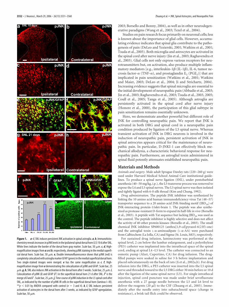
Figure 1. a–l, SNL induces persistent JNK activation in spinal astroglia. a, b, Immunohisto-chemistry reveals increases in pJNK levels in the ipsilateral spinal dorsal horn (L5) 10 d after SNL.White lines indicate the border of the dorsal horn gray matter. Scale bar, 50 �m. c, d, High-magnification images from a and b, respectively, showing pJNK staining in the medial superfi-cial dorsal horn. Scale bar, 50 �m. e, Double immunofluorescence shows that pJNK (red) iscompletely colocalized with astroglia marker GFAP (green) in the medial superficial dorsal horn.Two single-stained images were merged. e has the same magnification as c. f, High-magnification image from e demonstrating the colocalization of pJNK and GFAP. Scale bar, 25�m. g, h, SNL also induces JNK activation in the dorsal horn after 3 weeks. Scale bar, 25 �m. i,Colocalization of pJNK (i) and GFAP (i�) in the superficial dorsal horn 21 d after SNL. i� is themerge of i and i�. Scale bar, 25 �m. j, Time course of pJNK induction in the L5 spinal cord afterSNL, as indicated by the number of pJNK-IR cells in the superficial dorsal horn (laminas I–III).**p � 0.01 by ANOVA compared with control (n � 3 and 4). k, l, SNL induces persistentactivation of astrocytes in the dorsal horn after 3 weeks, as indicated by GFAP upregulation.Scale bar, 50 �m.
3552 • J. Neurosci., March 29, 2006 • 26(13):3551–3560 Zhuang et al. • JNK, Spinal Astrocytes, and Neuropathic Pain
Western blots. As described previously (Zhuang et al., 2005), animalswere rapidly killed, and the L5 spinal segments (dorsal part) were quicklyremoved and homogenized in a SDS sample buffer containing a mixtureof proteinase and phosphatase inhibitors (Sigma). Protein samples (30�g) were separated on SDS-PAGE gel and transferred to polyvinylidenedifluoride blots. The blots were blocked with 5% milk and incubatedovernight at 4°C with antibody against phosphorylated JNK (pJNK) orJNK (anti-rabbit, 1:1000; Cell Signaling Technology, Beverly, MA).These blots were further incubated with HRP-conjugated secondary an-tibody, developed in ECL solution, and exposed onto Hyperfilm (Amer-sham Biosciences, Arlington Heights, IL).
Immunohistochemistry. Animals were terminally anesthetized withisoflurane and perfused through the ascending aorta with saline followedby 4% paraformaldehyde/1.5% picric acid. After the perfusion, the spinalcord segments (L5) and the DRGs (L4, L5) were removed and postfixedin the same fixative overnight. Spinal sections (transverse, free-floating,30 �m) and DRG sections (15 �m) were cut in a cryostat and processedfor immunofluorescence (Ji et al., 1995; Jin et al., 2003). All of the sec-tions were blocked with 2% goat serum in 0.3% Triton X-100 for 1 h atroom temperature (RT) and incubated overnight at 4°C with anti-pJNKantibody (anti-rabbit, 1:1000; Cell Signaling Technology). The sectionswere then incubated for 1 h at RT with cyanine 3-conjugated secondary
antibody (1:400; Jackson ImmunoResearch,West Grove, PA). Other primary antibodiesused in this study were polyclonal antibodiesfor JNK1 (rabbit, 1:2000; Santa Cruz Biotech-nology, Santa Cruz, CA), pERK, p-p38, p-c-Jun(rabbit, 1:500, phosphorylated forms; Cell Sig-naling Technology), and monoclonal antibod-ies for neuronal-specific nuclear protein(NeuN) (mouse, 1:5000; Chemicon, Temecula,CA), glial fibrillary acidic protein (GFAP)(mouse, 1:5000; Chemicon), and neurofila-ment 200 kDa (NF-200) (mouse, 1:5000;Sigma). For double immunofluorescence, sec-tions were incubated with a mixture of two pri-mary antibodies (monoclonal and polyclonal),followed by a mixture of the two respective sec-ondary antibodies. The specificity of the stain-ing was tested by omission of primary antibod-ies or absorption with peptide antigens. Stainedsections were examined with a Nikon (Tokyo,Japan(fluorescence microscope, and images
were captured with a CCD Spot camera.Behavioral analysis. Animals were habituated to the testing environ-
ment daily for 3–7 d before baseline testing. For testing mechanical sen-sitivity, animals were put under inverted plastic boxes (11 � 13 � 24 cm)on an elevated mesh floor and allowed 30 min for habituation, before thethreshold testing. Mechanical allodynia was tested using von Frey hairs ina blinded manner. The paw was pressed with one of a series of von Freyhairs with logarithmically incrementing stiffness (0.6, 1, 1.4, 2, 4, 6, 8, 10,15, and 26 g) (Stoelting, Kiel, WI), presented perpendicular to the plantarsurface (5– 6 s for each hair). The 50% withdrawal threshold was deter-mined using Dixon’s up– down method (Chaplan et al., 1994).
Quantification and statistics. To quantify positive cell profiles in thespinal cord, five to eight sections from the L5 spinal cord segments wererandomly selected. An image in a square (450 � 338 �m) on the medialtwo-thirds of the superficial dorsal horn (laminas I–III), as describedpreviously by Molander et al. (1984), was captured under 20� objective,and all of the positively stained cells in the area were counted by aninvestigator who did not know the treatment. Those cells with distinctcontrast to the background were scored as positive. Background levelswere obtained from sections incubated without primary antibody.Roughly, the densities of these positive cells should exhibit a signal/back-ground ratio �2. This counting does not determine the total numbers ofcells (Coggeshall and Lekan, 1996). Instead, it provides a comparison toevaluate differences between control and treated animals. To quantifypJNK expression in the DRG, the percentages of pJNK-positive neuronsin the L5 and L4 DRG from four nonadjacent sections were determinedas described previously (Ji et al., 1996). Data were presented as mean �SEM. Differences between groups were compared using Student’s t testor ANOVA, followed by Fisher’s PLSD test. The criterion for statisticalsignificance was p � 0.05.
ResultsActivation of JNK in the spinal astrocytes after nerve injurySNL produced rapidly appearing (�1 d) and persistent (�3weeks) neuropathic pain (Kim and Chung, 1992). SNL also in-duced a marked activation of JNK in the spinal cord. Many morepJNK (active form of JNK)-immunoreactive (IR) cells werefound in the ipsilateral than the contralateral spinal cord, pre-dominantly in the superficial dorsal horn (laminas I–III) on day10 (Fig. 1a– d) and day 21 (Fig. 1g,h). Double immunofluores-cence indicated that pJNK was completely colocalized withGFAP, a marker for astrocytes, in the spinal cord at differenttimes (days 3–21) of neuropathic pain development (Fig. 1e,f, i).
To determine the percentage of cells that showed pJNK/GFAPdouble labeling, we quantified the number of pJNK and GFAPsingle-stained cells, as well as pJNK/GFAP double-stained cells inthe medial laminas I–III (see Materials and Methods) in six spinal
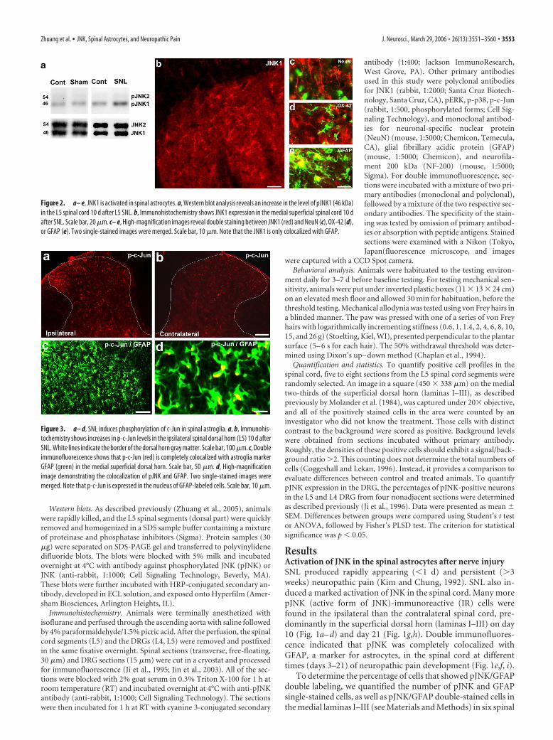
Figure 2. a– e, JNK1 is activated in spinal astrocytes. a, Western blot analysis reveals an increase in the level of pJNK1 (46 kDa)in the L5 spinal cord 10 d after L5 SNL. b, Immunohistochemistry shows JNK1 expression in the medial superficial spinal cord 10 dafter SNL. Scale bar, 20 �m. c– e, High-magnification images reveal double staining between JNK1 (red) and NeuN (c), OX-42 (d),or GFAP (e). Two single-stained images were merged. Scale bar, 10 �m. Note that the JNK1 is only colocalized with GFAP.
Figure 3. a– d, SNL induces phosphorylation of c-Jun in spinal astroglia. a, b, Immunohis-tochemistry shows increases in p-c-Jun levels in the ipsilateral spinal dorsal horn (L5) 10 d afterSNL. White lines indicate the border of the dorsal horn gray matter. Scale bar, 100 �m. c, Doubleimmunofluorescence shows that p-c-Jun (red) is completely colocalized with astroglia markerGFAP (green) in the medial superficial dorsal horn. Scale bar, 50 �m. d, High-magnificationimage demonstrating the colocalization of pJNK and GFAP. Two single-stained images weremerged. Note that p-c-Jun is expressed in the nucleus of GFAP-labeled cells. Scale bar, 10 �m.
Zhuang et al. • JNK, Spinal Astrocytes, and Neuropathic Pain J. Neurosci., March 29, 2006 • 26(13):3551–3560 • 3553
sections from three different rats perfused10 d after SNL. Approximately 99% ofpJNK-IR cells (358 of 363) were alsoGFAP-IR, suggesting that pJNK is almostexclusively expressed in astrocytes. How-ever, only 33% of GFAP-IR cells (358 of1080) were pJNK-IR, indicating that JNKis only activated in a fraction of spinalastrocytes.
A time course study showed that pJNKelevation was not evident at day 1, whensymptoms of neuropathic pain began todevelop (Fig. 1j). The rise in pJNK levelswas significant on day 3, reached a peak onday 10, and remained at high levels on day21 after SNL (Fig. 1j). Consistently, SNLinduced a persistent upregulation of GFAPin the dorsal horn. GFAP expression re-mained at high level on day 21 (Fig. 1k,l).
Activation of JNK1 and c-Jun in spinalastrocytes after nerve injuryWestern blot analysis confirmed an in-crease in spinal pJNK after SNL. Althoughboth JNK1 (46 kDa) and JNK2 (54 kDa)were constitutively expressed in the spinalcord, only JNK1 was phosphorylated inthe spinal cord (Fig. 2a). There was no sig-nificant change in pJNK expression after sham surgery (Fig. 2a).Double immunofluorescence showed that JNK1 was colocalizedwith GFAP but not with NeuN (neuronal marker) or with OX-42(an antibody for the microglial marker CD11b) in the spinal cordafter SNL (Fig. 2b– e). Therefore, JNK1 appears to be the isoformthat is phosphorylated, exclusively in spinal astrocytes.
The transcription factor c-Jun is the best known substrate ofJNK. JNK is known to activate p-c-Jun. SNL also induced anupregulation of p-c-Jun in the ipsilateral spinal cord (Fig. 3).Interestingly, p-c-Jun was also localized in GFAP-expressing as-trocytes in the dorsal horn, with predominant localization in thenucleus (Fig. 3).
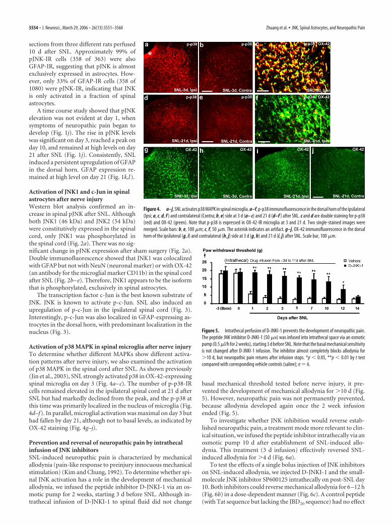
Activation of p38 MAPK in spinal microglia after nerve injuryTo determine whether different MAPKs show different activa-tion patterns after nerve injury, we also examined the activationof p38 MAPK in the spinal cord after SNL. As shown previously(Jin et al., 2003), SNL strongly activated p38 in OX-42-expressingspinal microglia on day 3 (Fig. 4a– c). The number of p-p38-IRcells remained elevated in the ipsilateral spinal cord at 21 d afterSNL but had markedly declined from the peak, and the p-p38 atthis time was primarily localized in the nucleus of microglia (Fig.4d–f). In parallel, microglial activation was maximal on day 3 buthad fallen by day 21, although not to basal levels, as indicated byOX-42 staining (Fig. 4g–j).
Prevention and reversal of neuropathic pain by intrathecalinfusion of JNK inhibitorsSNL-induced neuropathic pain is characterized by mechanicalallodynia (pain-like response to preinjury innocuous mechanicalstimulation) (Kim and Chung, 1992). To determine whether spi-nal JNK activation has a role in the development of mechanicalallodynia, we infused the peptide inhibitor D-JNKI-1 via an os-motic pump for 2 weeks, starting 3 d before SNL. Although in-trathecal infusion of D-JNKI-1 to spinal fluid did not change
basal mechanical threshold tested before nerve injury, it pre-vented the development of mechanical allodynia for �10 d (Fig.5). However, neuropathic pain was not permanently prevented,because allodynia developed again once the 2 week infusionended (Fig. 5).
To investigate whether JNK inhibition would reverse estab-lished neuropathic pain, a treatment mode more relevant to clin-ical situation, we infused the peptide inhibitor intrathecally via anosmotic pump 10 d after establishment of SNL-induced allo-dynia. This treatment (3 d infusion) effectively reversed SNL-induced allodynia for �4 d (Fig. 6a).
To test the effects of a single bolus injection of JNK inhibitorson SNL-induced allodynia, we injected D-JNKI-1 and the small-molecule JNK inhibitor SP600125 intrathecally on post-SNL day10. Both inhibitors could reverse mechanical allodynia for 6 –12 h(Fig. 6b) in a dose-dependent manner (Fig. 6c). A control peptide(with Tat sequence but lacking the JBD20 sequence) had no effect
Figure 4. a–j, SNL activates p38 MAPK in spinal microglia. a–f, p-p38 immunofluorescence in the dorsal horn of the ipsilateral(Ipsi; a, c, d, f ) and contralateral (Contra; b, e) side at 3 d (a– c) and 21 d (d–f ) after SNL. c and d are double staining for p-p38(red) and OX-42 (green). Note that p-p38 is expressed in OX-42-IR microglia at 3 and 21 d. Two single-stained images weremerged. Scale bars: b, e, 100 �m; c, f, 50 �m. The asterisk indicates an artifact. g–j, OX-42 immunofluorescence in the dorsalhorn of the ipsilateral (g, i) and contralateral (h, j) side at 3 d (g, h) and 21 d (i, j) after SNL. Scale bar, 100 �m.
Figure 5. Intrathecal perfusion of D-JNKI-1 prevents the development of neuropathic pain.The peptide JNK inhibitor D-JNKI-1 (50 �M) was infused into intrathecal space via an osmoticpump (0.5 �l/h for 2 weeks), starting 3 d before SNL. Note that the basal mechanical sensitivityis not changed after D-JNKI-1 infusion. The inhibitor almost completely blocks allodynia for�10 d, but neuropathic pain returns after infusion stops. *p � 0.05, **p � 0.01 by t testcompared with corresponding vehicle controls (saline); n � 6.
3554 • J. Neurosci., March 29, 2006 • 26(13):3551–3560 Zhuang et al. • JNK, Spinal Astrocytes, and Neuropathic Pain
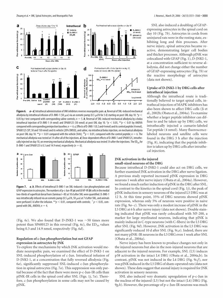
(Fig. 6c). We also found that D-JNKI-1 was �50 times morepotent than SP600125 in this reversal (Fig. 6c), the ED50 valuesbeing 0.3 and 14.9 nmol, respectively (Fig. 6d).
Regulation of c-Jun phosphorylation but not GFAPexpression in astrocytes by JNKTo explore the mechanisms by which JNK activation would me-diate neuropathic pain, we examined the effect of D-JNKI-1 onSNL-induced phosphorylation of c-Jun. Intrathecal infusion ofD-JNKI-1, at a concentration that fully reversed allodynia (Fig.6a), significantly suppressed SNL-induced c-Jun phosphoryla-tion in spinal astrocytes (Fig. 7a). This suppression was only par-tial because of the fact that there were more p-c-Jun-IR cells thanpJNK-IR cells in the spinal cord after SNL (Figs. 1j, 7a). There-fore, c-Jun phosphorylation in some cells may not be caused bypJNK.
SNL also induced a doubling of GFAP-expressing astrocytes in the spinal cord onday 10 (Fig. 7b). Astrocytes in cords fromuninjured rats were in the resting state, ex-hibiting long and thin processes. Afternerve injury, spinal astrocytes became re-active, demonstrating larger cell bodiesand thicker processes. Although pJNK wascolocalized with GFAP (Fig. 1), D-JNKI-1,at a concentration sufficient to reverse al-lodynia, did not change either the numberof GFAP-expressing astrocytes (Fig. 7b) orthe reactive morphology of astrocytes(data not shown).
Uptake of D-JNKI-1 by DRG cells afterintrathecal injectionAlthough the intrathecal route is tradi-tionally believed to target spinal cells, in-trathecal injection of MAPK inhibitors hasalso been shown to affect DRG cells (Ji etal., 2002b; Obata et al., 2004a). To examinewhether a larger peptide inhibitor can dif-fuse to and be taken up by DRG cells, weintrathecally injected a cumarin-labeledTat peptide (4 nmol). Many fluorescence-labeled neurons and satellite cells werefound in the DRG 3 h after the injection(Fig. 8), indicating that the peptide inhib-itor is taken up by DRG cells after intrathe-cal injection.
JNK activation in the injuredsmall-sized neurons of the DRGBecause intrathecal D-JNKI-1 could also act on DRG cells, wefurther examined JNK activation in the DRG after nerve ligation.A previous study reported increased pJNK expression in DRGneurons 1 week after nerve injury (Obata et al., 2004a). However,we found a much earlier induction of pJNK in the DRG after SNL.In contrast to the kinetics in the spinal cord (Fig. 1j), the peak ofpJNK induction in sensory neurons of the injured L5 DRG was at12 h. At this time point, 35% of DRG neurons showed pJNKexpression, whereas only 5% of neurons were positive in naiverats (Fig. 9a– c). There was only a modest increase of pJNK in theL5 DRG at 6 h after nerve injury (data not shown). Double stain-ing indicated that pJNK was rarely colocalized with NF-200, amarker for large myelinated neurons, indicating that pJNK ismainly induced in C-type neurons (with C-fibers) in the L5 DRGafter SNL (Fig. 9d). However, JNK activation in the L5 DRG wassignificantly reduced 10 d after SNL (Fig. 9c,e). Indeed, there arenot many pJNK-IR neurons in the L5 DRG even 1 week after SNL(Obata et al., 2004a).
Nerve injury has been known to produce changes not only inthe injured neurons but also in the non-injured neurons that areadjacent to the injured neurons. For example, SNL (L5) inducesp38 activation in the intact L4 DRG (Obata et al., 2004a,b). Incontrast, pJNK was not induced in the L4 DRG (Fig. 9c,f), norwas pJNK induced in the L5 DRG of sham-operated rats (data notshown). These data suggest that axonal injury is required for JNKactivation in sensory neurons.
SNL also induced very dramatic upregulation of p-c-Jun inthe nucleus of the injured (L5) but not the intact (L4) DRG (Fig.9g,h). However, the percentage of p-c-Jun-IR neurons was much
Figure 6. a– d, Intrathecal administration of JNK inhibitors reverses neuropathic pain. a, Reversal of SNL-induced mechanicalallodynia by intrathecal infusion of D-JNKI-1 (50 �M) via an osmotic pump (0.5 �l/h for 3 d) starting on post-SNL day 10. *p �0.05 by t test compared with corresponding saline controls; n � 5. b, Reversal of SNL-induced mechanical allodynia by a bolusintrathecal injection of D-JNKI-1 (4 nmol) and SP600125 (50 nmol) at post-SNL day 10. *p � 0.05, **p � 0.01 by ANOVAcompared with corresponding preinjection baseline; n�6. c, Effects of D-JNKI-1 (0.2 and 4 nmol) and its control peptide (4 nmol),SP600125 (SP; 50 and 150 nmol) and its vehicle (20% DMSO), and saline, via intrathecal bolus injection, on mechanical allodyniaon post-SNL day 10. **p � 0.01 compared with the vehicle (Veh); ##p � 0.01, compared with the control peptide; n � 6. Themechanical allodynia was tested at 3 h after all of the injections. d, Dose-dependent effects of D-JNKI-1 and SP600125, intrathe-cally injected on day 10, on reversing mechanical allodynia. Mechanical allodynia was tested 3 h after the injections. The ED50 forD-JNKI-1 and SP600125 is 0.3 and 14.9 nmol, respectively (n � 6).
Figure 7. a, b, Effects of intrathecal D-JNKI-1 on SNL-induced c-Jun phosphorylation andGFAP expression in astrocytes. The numbers of p-c-Jun-IR (a) and GFAP-IR (b) cells in the medialtwo-thirds of superficial dorsal horn (laminas I–III) at 10 d after SNL were quantified. D-JNKI-1was intrathecally infused via an osmotic pump (0.5 �l/h, 50 �M) at 7 d after SNL, and animalswere perfused 3 d after the infusion. **p � 0.01, compared with controls; �p � 0.05, com-pared with SNL, ANOVA; n � 4.
Zhuang et al. • JNK, Spinal Astrocytes, and Neuropathic Pain J. Neurosci., March 29, 2006 • 26(13):3551–3560 • 3555
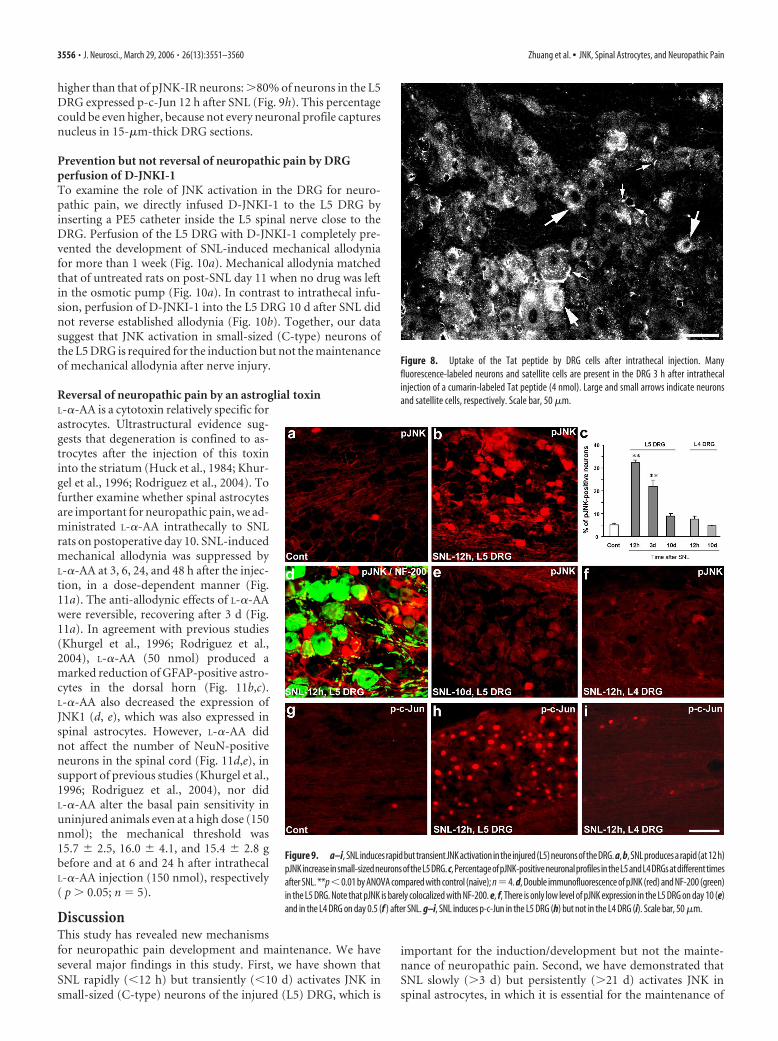
higher than that of pJNK-IR neurons: �80% of neurons in the L5DRG expressed p-c-Jun 12 h after SNL (Fig. 9h). This percentagecould be even higher, because not every neuronal profile capturesnucleus in 15-�m-thick DRG sections.
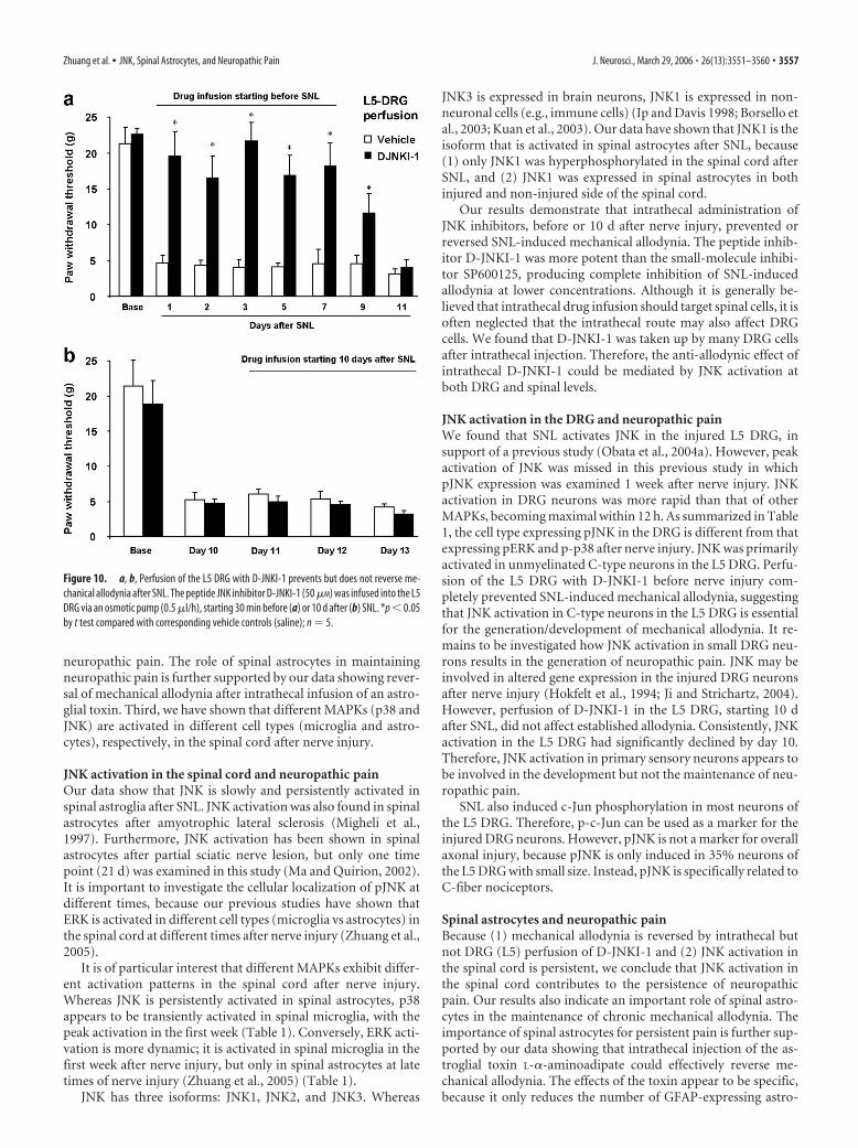
Prevention but not reversal of neuropathic pain by DRGperfusion of D-JNKI-1To examine the role of JNK activation in the DRG for neuro-pathic pain, we directly infused D-JNKI-1 to the L5 DRG byinserting a PE5 catheter inside the L5 spinal nerve close to theDRG. Perfusion of the L5 DRG with D-JNKI-1 completely pre-vented the development of SNL-induced mechanical allodyniafor more than 1 week (Fig. 10a). Mechanical allodynia matchedthat of untreated rats on post-SNL day 11 when no drug was leftin the osmotic pump (Fig. 10a). In contrast to intrathecal infu-sion, perfusion of D-JNKI-1 into the L5 DRG 10 d after SNL didnot reverse established allodynia (Fig. 10b). Together, our datasuggest that JNK activation in small-sized (C-type) neurons ofthe L5 DRG is required for the induction but not the maintenanceof mechanical allodynia after nerve injury.
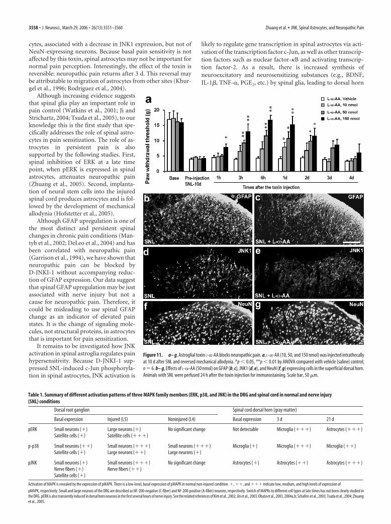
Reversal of neuropathic pain by an astroglial toxinL-�-AA is a cytotoxin relatively specific forastrocytes. Ultrastructural evidence sug-gests that degeneration is confined to as-trocytes after the injection of this toxininto the striatum (Huck et al., 1984; Khur-gel et al., 1996; Rodriguez et al., 2004). Tofurther examine whether spinal astrocytesare important for neuropathic pain, we ad-ministrated L-�-AA intrathecally to SNLrats on postoperative day 10. SNL-inducedmechanical allodynia was suppressed byL-�-AA at 3, 6, 24, and 48 h after the injec-tion, in a dose-dependent manner (Fig.11a). The anti-allodynic effects of L-�-AAwere reversible, recovering after 3 d (Fig.11a). In agreement with previous studies(Khurgel et al., 1996; Rodriguez et al.,2004), L-�-AA (50 nmol) produced amarked reduction of GFAP-positive astro-cytes in the dorsal horn (Fig. 11b,c).L-�-AA also decreased the expression ofJNK1 (d, e), which was also expressed inspinal astrocytes. However, L-�-AA didnot affect the number of NeuN-positiveneurons in the spinal cord (Fig. 11d,e), insupport of previous studies (Khurgel et al.,1996; Rodriguez et al., 2004), nor didL-�-AA alter the basal pain sensitivity inuninjured animals even at a high dose (150nmol); the mechanical threshold was15.7 � 2.5, 16.0 � 4.1, and 15.4 � 2.8 gbefore and at 6 and 24 h after intrathecalL-�-AA injection (150 nmol), respectively( p � 0.05; n � 5).
DiscussionThis study has revealed new mechanismsfor neuropathic pain development and maintenance. We haveseveral major findings in this study. First, we have shown thatSNL rapidly (�12 h) but transiently (�10 d) activates JNK insmall-sized (C-type) neurons of the injured (L5) DRG, which is
important for the induction/development but not the mainte-nance of neuropathic pain. Second, we have demonstrated thatSNL slowly (�3 d) but persistently (�21 d) activates JNK inspinal astrocytes, in which it is essential for the maintenance of
Figure 8. Uptake of the Tat peptide by DRG cells after intrathecal injection. Manyfluorescence-labeled neurons and satellite cells are present in the DRG 3 h after intrathecalinjection of a cumarin-labeled Tat peptide (4 nmol). Large and small arrows indicate neuronsand satellite cells, respectively. Scale bar, 50 �m.
Figure 9. a–i, SNLinducesrapidbuttransientJNKactivationintheinjured(L5)neuronsoftheDRG. a, b,SNLproducesarapid(at12h)pJNKincreaseinsmall-sizedneuronsoftheL5DRG.c,PercentageofpJNK-positiveneuronalprofiles intheL5andL4DRGsatdifferenttimesafter SNL. **p�0.01 by ANOVA compared with control (naive); n�4. d, Double immunofluorescence of pJNK (red) and NF-200 (green)in the L5 DRG. Note that pJNK is barely colocalized with NF-200. e, f, There is only low level of pJNK expression in the L5 DRG on day 10 (e)and in the L4 DRG on day 0.5 (f ) after SNL. g–i, SNL induces p-c-Jun in the L5 DRG (h) but not in the L4 DRG (i). Scale bar, 50 �m.
3556 • J. Neurosci., March 29, 2006 • 26(13):3551–3560 Zhuang et al. • JNK, Spinal Astrocytes, and Neuropathic Pain
neuropathic pain. The role of spinal astrocytes in maintainingneuropathic pain is further supported by our data showing rever-sal of mechanical allodynia after intrathecal infusion of an astro-glial toxin. Third, we have shown that different MAPKs (p38 andJNK) are activated in different cell types (microglia and astro-cytes), respectively, in the spinal cord after nerve injury.
JNK activation in the spinal cord and neuropathic painOur data show that JNK is slowly and persistently activated inspinal astroglia after SNL. JNK activation was also found in spinalastrocytes after amyotrophic lateral sclerosis (Migheli et al.,1997). Furthermore, JNK activation has been shown in spinalastrocytes after partial sciatic nerve lesion, but only one timepoint (21 d) was examined in this study (Ma and Quirion, 2002).It is important to investigate the cellular localization of pJNK atdifferent times, because our previous studies have shown thatERK is activated in different cell types (microglia vs astrocytes) inthe spinal cord at different times after nerve injury (Zhuang et al.,2005).
It is of particular interest that different MAPKs exhibit differ-ent activation patterns in the spinal cord after nerve injury.Whereas JNK is persistently activated in spinal astrocytes, p38appears to be transiently activated in spinal microglia, with thepeak activation in the first week (Table 1). Conversely, ERK acti-vation is more dynamic; it is activated in spinal microglia in thefirst week after nerve injury, but only in spinal astrocytes at latetimes of nerve injury (Zhuang et al., 2005) (Table 1).
JNK has three isoforms: JNK1, JNK2, and JNK3. Whereas
JNK3 is expressed in brain neurons, JNK1 is expressed in non-neuronal cells (e.g., immune cells) (Ip and Davis 1998; Borsello etal., 2003; Kuan et al., 2003). Our data have shown that JNK1 is theisoform that is activated in spinal astrocytes after SNL, because(1) only JNK1 was hyperphosphorylated in the spinal cord afterSNL, and (2) JNK1 was expressed in spinal astrocytes in bothinjured and non-injured side of the spinal cord.
Our results demonstrate that intrathecal administration ofJNK inhibitors, before or 10 d after nerve injury, prevented orreversed SNL-induced mechanical allodynia. The peptide inhib-itor D-JNKI-1 was more potent than the small-molecule inhibi-tor SP600125, producing complete inhibition of SNL-inducedallodynia at lower concentrations. Although it is generally be-lieved that intrathecal drug infusion should target spinal cells, it isoften neglected that the intrathecal route may also affect DRGcells. We found that D-JNKI-1 was taken up by many DRG cellsafter intrathecal injection. Therefore, the anti-allodynic effect ofintrathecal D-JNKI-1 could be mediated by JNK activation atboth DRG and spinal levels.
JNK activation in the DRG and neuropathic painWe found that SNL activates JNK in the injured L5 DRG, insupport of a previous study (Obata et al., 2004a). However, peakactivation of JNK was missed in this previous study in whichpJNK expression was examined 1 week after nerve injury. JNKactivation in DRG neurons was more rapid than that of otherMAPKs, becoming maximal within 12 h. As summarized in Table1, the cell type expressing pJNK in the DRG is different from thatexpressing pERK and p-p38 after nerve injury. JNK was primarilyactivated in unmyelinated C-type neurons in the L5 DRG. Perfu-sion of the L5 DRG with D-JNKI-1 before nerve injury com-pletely prevented SNL-induced mechanical allodynia, suggestingthat JNK activation in C-type neurons in the L5 DRG is essentialfor the generation/development of mechanical allodynia. It re-mains to be investigated how JNK activation in small DRG neu-rons results in the generation of neuropathic pain. JNK may beinvolved in altered gene expression in the injured DRG neuronsafter nerve injury (Hokfelt et al., 1994; Ji and Strichartz, 2004).However, perfusion of D-JNKI-1 in the L5 DRG, starting 10 dafter SNL, did not affect established allodynia. Consistently, JNKactivation in the L5 DRG had significantly declined by day 10.Therefore, JNK activation in primary sensory neurons appears tobe involved in the development but not the maintenance of neu-ropathic pain.
SNL also induced c-Jun phosphorylation in most neurons ofthe L5 DRG. Therefore, p-c-Jun can be used as a marker for theinjured DRG neurons. However, pJNK is not a marker for overallaxonal injury, because pJNK is only induced in 35% neurons ofthe L5 DRG with small size. Instead, pJNK is specifically related toC-fiber nociceptors.
Spinal astrocytes and neuropathic painBecause (1) mechanical allodynia is reversed by intrathecal butnot DRG (L5) perfusion of D-JNKI-1 and (2) JNK activation inthe spinal cord is persistent, we conclude that JNK activation inthe spinal cord contributes to the persistence of neuropathicpain. Our results also indicate an important role of spinal astro-cytes in the maintenance of chronic mechanical allodynia. Theimportance of spinal astrocytes for persistent pain is further sup-ported by our data showing that intrathecal injection of the as-troglial toxin L-�-aminoadipate could effectively reverse me-chanical allodynia. The effects of the toxin appear to be specific,because it only reduces the number of GFAP-expressing astro-
Figure 10. a, b, Perfusion of the L5 DRG with D-JNKI-1 prevents but does not reverse me-chanical allodynia after SNL. The peptide JNK inhibitor D-JNKI-1 (50 �M) was infused into the L5DRG via an osmotic pump (0.5 �l/h), starting 30 min before (a) or 10 d after (b) SNL. *p � 0.05by t test compared with corresponding vehicle controls (saline); n � 5.
Zhuang et al. • JNK, Spinal Astrocytes, and Neuropathic Pain J. Neurosci., March 29, 2006 • 26(13):3551–3560 • 3557
cytes, associated with a decrease in JNK1 expression, but not ofNeuN-expressing neurons. Because basal pain sensitivity is notaffected by this toxin, spinal astrocytes may not be important fornormal pain perception. Interestingly, the effect of the toxin isreversible: neuropathic pain returns after 3 d. This reversal maybe attributable to migration of astrocytes from other sites (Khur-gel et al., 1996; Rodriguez et al., 2004).
Although increasing evidence suggeststhat spinal glia play an important role inpain control (Watkins et al., 2001; Ji andStrichartz, 2004; Tsuda et al., 2005), to ourknowledge this is the first study that spe-cifically addresses the role of spinal astro-cytes in pain sensitization. The role of as-trocytes in persistent pain is alsosupported by the following studies. First,spinal inhibition of ERK at a late timepoint, when pERK is expressed in spinalastrocytes, attenuates neuropathic pain(Zhuang et al., 2005). Second, implanta-tion of neural stem cells into the injuredspinal cord produces astrocytes and is fol-lowed by the development of mechanicalallodynia (Hofstetter et al., 2005).
Although GFAP upregulation is one ofthe most distinct and persistent spinalchanges in chronic pain conditions (Man-tyh et al., 2002; DeLeo et al., 2004) and hasbeen correlated with neuropathic pain(Garrison et al., 1994), we have shown thatneuropathic pain can be blocked byD-JNKI-1 without accompanying reduc-tion of GFAP expression. Our data suggestthat spinal GFAP upregulation may be justassociated with nerve injury but not acause for neuropathic pain. Therefore, itcould be misleading to use spinal GFAPchange as an indicator of elevated painstates. It is the change of signaling mole-cules, not structural proteins, in astrocytesthat is important for pain sensitization.
It remains to be investigated how JNKactivation in spinal astroglia regulates painhypersensitivity. Because D-JNKI-1 sup-pressed SNL-induced c-Jun phosphoryla-tion in spinal astrocytes, JNK activation is
likely to regulate gene transcription in spinal astrocytes via acti-vation of the transcription factor c-Jun, as well as other transcrip-tion factors such as nuclear factor-�B and activating transcrip-tion factor-2. As a result, there is increased synthesis ofneuroexcitatory and neurosensitizing substances (e.g., BDNF,IL-1�, TNF-�, PGE2, etc.) by spinal glia, leading to dorsal horn
Figure 11. a– g, Astroglial toxin L-�-AA blocks neuropathic pain. a, L-�-AA (10, 50, and 150 nmol) was injected intrathecallyat 10 d after SNL and reversed mechanical allodynia. *p � 0.05, **p � 0.01 by ANOVA compared with vehicle (saline) control;n � 6. b– g, Effects of L-�-AA (50 nmol) on GFAP (b, c), JNK1 (d, e), and NeuN (f, g) expressing cells in the superficial dorsal horn.Animals with SNL were perfused 24 h after the toxin injection for immunostaining. Scale bar, 50 �m.
Table 1. Summary of different activation patterns of three MAPK family members (ERK, p38, and JNK) in the DRG and spinal cord in normal and nerve injury(SNL) conditions
Dorsal root ganglion Spinal cord dorsal horn (gray matter)
Basal expression Injured (L5) Noninjured (L4) Basal expression 3 d 21 d
pERK Small neurons (�) Large neurons (�) No significant change Not detectable Microglia (���) Astrocytes (���)Satellite cells (�) Satellite cells (���)
p-p38 Small neurons (��) Small neurons (���) Small neurons (���) Microglia (�) Microglia (���) Microglia (��)Satellite cells (�) Large neurons (��) Large neurons (�)
pJNK Small neurons (�) Small neurons (���) No significant change Astrocytes (�) Astrocytes (��) Astrocytes (���)Nerve fibers (�) Nerve fibers (��)Satellite cells (�)
Activation of MAPK is revealed by the expression of pMAPK. There is a low-level, basal expression of pMAPK in normal non-injured condition. �, ��, and ��� indicate low, medium, and high levels of expression of
pMAPK, respectively. Small and large neurons of the DRG are described as NF-200-negative (C-fiber) and NF-200-positive (A-fiber) neurons, respectively. Switch of MAPKs to different cell types at late times has not been clearly studied inthe DRG. pERK is also transiently induced in dorsal horn neurons in the first several hours of nerve injury. See the related references of Kim et al., 2002; Jin et al., 2003; Obata et al., 2003, 2004a,b; Schafers et al., 2003; Tsuda et al., 2004; Zhuanget al., 2005.
3558 • J. Neurosci., March 29, 2006 • 26(13):3551–3560 Zhuang et al. • JNK, Spinal Astrocytes, and Neuropathic Pain
neuron sensitization (Samad et al., 2001; Watkins et al., 2001;DeLeo et al., 2004; Ji and Strichartz, 2004; Coull et al., 2005;Tsuda et al., 2005).
Concluding remarksThis study has identified the JNK cascade as a critical signalingpathway for neuropathic pain development and maintenance viadistinct mechanisms in the DRG and spinal cord. In particular,we have shown that spinal astrocytes play an important role inmaintaining persistent mechanical allodynia, although the un-derlying mechanisms remain to be investigated.
Many patients in the pain clinic suffer from neuropathic paincaused by injury to the peripheral nervous system or the CNS,and one of the most distinct symptoms is mechanical allodynia.At present, few drugs are effective in treating neuropathic pain,and their efficacy is only demonstrated in �30% of patients(Woolf and Mannion, 1999; Gardell et al., 2003). However, allcurrent drugs were developed or considered to act against molec-ular targets in neurons. Targeting the JNK pathway in spinal gliaand sensory neurons may offer a new option to treat intractableneuropathic pain.
ReferencesAbbadie C, Lindia JA, Cumiskey AM, Peterson LB, Mudgett JS, Bayne EK,
DeMartino JA, MacIntyre DE, Forrest MJ (2003) Impaired neuropathicpain responses in mice lacking the chemokine receptor CCR2. Proc NatlAcad Sci USA 100:7947–7952.
Borsello T, Bonny C (2004) Use of cell-permeable peptides to prevent neu-ronal degeneration. Trends Mol Med 10:239 –244.
Borsello T, Clarke PG, Hirt L, Vercelli A, Repici M, Schorderet DF, Bogous-slavsky J, Bonny C (2003) A peptide inhibitor of c-Jun N-terminal ki-nase protects against excitotoxicity and cerebral ischemia. Nat Med9:1180 –1186.
Chaplan SR, Bach FW, Pogrel JW, Chung JM, Yaksh TL (1994) Quantitativeassessment of tactile allodynia in the rat paw. J Neurosci Methods53:55– 63.
Coggeshall RE, Lekan HA (1996) Methods for determining numbers of cellsand synapses: a case for more uniform standards of review. J Comp Neu-rol 364:6 –15.
Coull JA, Beggs S, Boudreau D, Boivin D, Tsuda M, Inoue K, Gravel C, SalterMW, De Koninck Y (2005) BDNF from microglia causes the shift inneuronal anion gradient underlying neuropathic pain. Nature438:1017–1021.
DeLeo JA, Yezierski RP (2001) The role of neuroinflammation and neuro-immune activation in persistent pain. Pain 90:1– 6.
DeLeo JA, Tanga FY, Tawfik VL (2004) Neuroimmune activation and neu-roinflammation in chronic pain and opioid tolerance/hyperalgesia. TheNeuroscientist 10:40 –52.
Gardell LR, Wang R, Ehrenfels C, Ossipov MH, Rossomando AJ, Miller S,Buckley C, Cai AK, Tse A, Foley SF, Gong B, Walus L, Carmillo P, WorleyD, Huang C, Engber T, Pepinsky B, Cate RL, Vanderah TW, Lai J, et al.(2003) Multiple actions of systemic artemin in experimental neuropathy.Nat Med 9:1383–1389.
Garrison CJ, Dougherty PM, Carlton SM (1994) GFAP expression in lum-bar spinal cord of naive and neuropathic rats treated with MK-801. ExpNeurol 129:237–243.
Hofstetter CP, Holmstrom NA, Lilja JA, Schweinhardt P, Hao J, Spenger C,Wiesenfeld-Hallin Z, Kurpad SN, Frisen J, Olson L (2005) Allodynialimits the usefulness of intraspinal neural stem cell grafts; directed differ-entiation improves outcome. Nat Neurosci 8:346 –353.
Hokfelt T, Zhang X, Wiesenfeld-Hallin Z (1994) Messenger plasticity inprimary sensory neurons following axotomy and its functional implica-tions. Trends Neurosci 17:22–30.
Honore P, Rogers SD, Schwei MJ, Salak-Johnson JL, Luger NM, Sabino MC,Clohisy DR, Mantyh PW (2000) Murine models of inflammatory, neu-ropathic and cancer pain each generates a unique set of neurochemicalchanges in the spinal cord and sensory neurons. Neuroscience98:585–598.
Huck S, Grass F, Hortnagl H (1984) The glutamate analogue �-aminoadipic
acid is taken up by astrocytes before exerting its gliotoxic effect in vitro.J Neurosci 4:2650 –2657.
Ip YT, Davis RJ (1998) Signal transduction by the c-Jun N-terminal kinase(JNK): from inflammation to development. Curr Opin Cell Biol10:205–219.
Ji RR, Strichartz G (2004) Cell signaling and the genesis of neuropathic pain.Sci STKE 2004:reE14.
Ji RR, Woolf CJ (2001) Neuronal plasticity and signal transduction in noci-ceptive neurons: implications for the initiation and maintenance ofpathological pain. Neurobiol Dis 8:1–10.
Ji RR, Zhang Q, Law PY, Low HH, Elde R, Hokfelt T (1995) Expression of�-, �-, and �-opioid receptor-like immunoreactivities in rat dorsal rootganglia after carrageenan-induced inflammation. J Neurosci15:8156 – 8166.
Ji RR, Zhang Q, Pettersson RF, Hokfelt T (1996) aFGF, bFGF and NGFdifferentially regulate neuropeptide expression in dorsal root ganglia afteraxotomy and induce autotomy. Regul Pept 66:179 –189.
Ji RR, Befort K, Brenner GJ, Woolf CJ (2002a) ERK MAP kinase activationin superficial spinal cord neurons induces prodynorphin and NK-1 up-regulation and contributes to persistent inflammatory pain hypersensi-tivity. J Neurosci 22:478 – 485.
Ji RR, Samad TA, Jin SX, Schmoll R, Woolf CJ (2002b) p38 MAPK activa-tion by NGF in primary sensory neurons after inflammation increasesTRPV1 levels and maintains heat hyperalgesia. Neuron 36:57– 68.
Jin SX, Zhuang ZY, Woolf CJ, Ji RR (2003) p38 mitogen-activated proteinkinase is activated after a spinal nerve ligation in spinal cord microglia anddorsal root ganglion neurons and contributes to the generation of neuro-pathic pain. J Neurosci 23:4017– 4022.
Khurgel M, Koo AC, Ivy GO (1996) Selective ablation of astrocytes by in-tracerebral injections of alpha-aminoadipate. Glia 16:351–358.
Kim SH, Chung JM (1992) An experimental model for peripheral neurop-athy produced by segmental spinal nerve ligation in the rat. Pain50:355–363.
Kim SY, Bae JC, Kim JY, Lee HL, Lee KM, Kim DS, Cho HJ (2002) Activa-tion of p38 MAP kinase in the rat dorsal root ganglia and spinal cordfollowing peripheral inflammation and nerve injury. NeuroReport13:2483–2486.
Kuan CY, Whitmarsh AJ, Yang DD, Liao G, Schloemer AJ, Dong C, Bao J,Banasiak KJ, Haddad GG, Flavell RA, Davis RJ, Rakic P (2003) A criticalrole of neural-specific JNK3 for ischemic apoptosis. Proc Natl Acad SciUSA 100:15184 –15189.
Ma W, Quirion R (2002) Partial sciatic nerve ligation induces increase in thephosphorylation of extracellular signal-regulated kinase (ERK) and c-JunN-terminal kinase (JNK) in astrocytes in the lumbar spinal dorsal hornand the gracile nucleus. Pain 99:175–184.
Mantyh PW, Clohisy DR, Koltzenburg M, Hunt SP (2002) Molecular mech-anisms of cancer pain. Nat Rev Cancer 2:201–209.
Migheli A, Piva R, Atzori C, Troost D, Schiffer D (1997) c-Jun, JNK/SAPKkinases and transcription factor NF-kappa B are selectively activated inastrocytes, but not motor neurons, in amyotrophic lateral sclerosis. J Neu-ropathol Exp Neurol 56:1314 –1322.
Molander C, Xu Q, Grant G (1984) The cytoarchitectonic organization ofthe spinal cord in the rat. I. The lower thoracic and lumbosacral cord.J Comp Neurol 230:133–141.
Obata K, Noguchi K (2004) MAPK activation in nociceptive neurons andpain hypersensitivity. Life Sci 74:2643–2653.
Obata K, Yamanaka H, Dai Y, Tachibana T, Fukuoka T, Tokunaga A, Yo-shikawa H, Noguchi K (2003) Differential activation of extracellularsignal-regulated protein kinase in primary afferent neurons regulatesbrain-derived neurotrophic factor expression after peripheral inflamma-tion and nerve injury. J Neurosci 23:4117– 4126.
Obata K, Yamanaka H, Kobayashi K, Dai Y, Mizushima T, Katsura H,Fukuoka T, Tokunaga A, Noguchi K (2004a) Role of mitogen-activatedprotein kinase activation in injured and intact primary afferent neuronsfor mechanical and heat hypersensitivity after spinal nerve ligation. J Neu-rosci 24:10211–10222.
Obata K, Yamanaka H, Dai Y, Mizushima T, Fukuoka T, Tokunaga A, Nogu-chi K (2004b) Differential activation of MAPK in injured and uninjuredDRG neurons following chronic constriction injury of the sciatic nerve inrats. Eur J Neurosci 20:2881–2895.
Raghavendra V, Tanga F, DeLeo JA (2003) Inhibition of microglial activa-
Zhuang et al. • JNK, Spinal Astrocytes, and Neuropathic Pain J. Neurosci., March 29, 2006 • 26(13):3551–3560 • 3559
tion attenuates the development but not existing hypersensitivity in a ratmodel of neuropathy. J Pharmacol Exp Ther 306:624 – 630.
Rodriguez MJ, Martinez-Sanchez M, Bernal F, Mahy N (2004) Heterogene-ity between hippocampal and septal astroglia as a contributing factor todifferential in vivo AMPA excitotoxicity. J Neurosci Res 77:344 –353.
Samad TA, Moore KA, Sapirstein A, Billet S, Allchorne A, Poole S, BonventreJV, Woolf CJ (2001) Interleukin-1beta-mediated induction of Cox-2 inthe CNS contributes to inflammatory pain hypersensitivity. Nature410:471– 475.
Schafers M, Svensson CI, Sommer C, Sorkin LS (2003) Tumor necrosisfactor-� induces mechanical allodynia after spinal nerve ligation by acti-vation of p38 MAPK in primary sensory neurons. J Neurosci23:2517–2521.
Tanga FY, Nutile-McMenemy N, DeLeo JA (2005) The CNS role of Toll-like receptor 4 in innate neuroimmunity and painful neuropathy. ProcNatl Acad Sci USA 102:5856 –5861.
Tezel G, Yang X, Yang J, Wax MB (2004) Role of tumor necrosis factorreceptor-1 in the death of retinal ganglion cells following optic nervecrush injury in mice. Brain Res 996:202–212.
Tsuda M, Shigemoto-Mogami Y, Koizumi S, Mizokoshi A, Kohsaka S, SalterMW, Inoue K (2003) P2X4 receptors induced in spinal microglia gatetactile allodynia after nerve injury. Nature 424:778 –783.
Tsuda M, Mizokoshi A, Shigemoto-Mogami Y, Koizumi S, Inoue K (2004)
Activation of p38 mitogen-activated protein kinase in spinal hyperactivemicroglia contributes to pain hypersensitivity following peripheral nerveinjury. Glia 45:89 –95.
Tsuda M, Inoue K, Salter MW (2005) Neuropathic pain and spinal micro-glia: a big problem from molecules in “small” glia. Trends Neurosci28:101–107.
Wang J, Van De Water TR, Bonny C, de Ribaupierre F, Puel JL, Zine A (2003)A peptide inhibitor of c-Jun N-terminal kinase protects against both ami-noglycoside and acoustic trauma-induced auditory hair cell death andhearing loss. J Neurosci 23:8596 – 8607.
Watkins LR, Maier SF (2003) Glia: a novel drug discovery target for clinicalpain. Nat Rev Drug Discov 2:973–985.
Watkins LR, Milligan ED, Maier SF (2001) Glial activation: a driving forcefor pathological pain. Trends Neurosci 24:450 – 455.
Woolf CJ, Mannion RJ (1999) Neuropathic pain: aetiology, symptoms,mechanisms, and management. Lancet 353:1959 –1964.
Xia Z, Dickens M, Raingeaud J, Davis RJ, Greenberg ME (1995) Opposingeffects of ERK and JNK-p38 MAP kinases on apoptosis. Science270:1326 –1331.
Zhuang ZY, Gerner P, Woolf CJ, Ji RR (2005) ERK is sequentially activatedin neurons, microglia, and astrocytes by spinal nerve ligation and contrib-utes to mechanical allodynia in this neuropathic pain model. Pain114:149 –159.
3560 • J. Neurosci., March 29, 2006 • 26(13):3551–3560 Zhuang et al. • JNK, Spinal Astrocytes, and Neuropathic Pain