Embed Size (px)
Citation preview
NERVOUS SYSTEM - ORGANIZATION 2. Choose the key responses that best correspond to the descriptions provided in the following statements. Insert
the appropriate letter or term in the answer blanks.
Key Choices
A. Autonomic nervous system C. Peripheral nervous system (PNS)
B. Central nervous system (CNS) D. Somatic nervous system
_______________ 1. Nervous system subdivision that is composed of the brain and spinal cord
_______________ 2. Subdivision of the PNS that controls voluntary activities such as the activation of skeletal muscles
_______________ 3. Nervous system subdivision that is composed of the cranial and spinal nerves and ganglia
_______________ 4. Subdivision of the PNS that regulates the activity of the heart and smooth muscle, and of glands; it is also called the involuntary nervous system
_______________ 5. A major subdivision of the nervous system that interprets incoming information and issues orders
_______________ 6. A major subdivision of the nervous system that serves as communication lines, linking all parts of the body to the CNS
NERVOUS TISSUE�STRUCTURE AND FUNCTION 3. This exercise emphasizes the difference between neurons and neuroglia. Indicate which cell type is identified
by the following descriptions. Insert the appropriate letter or term in the answer blanks.
Key Choices
A. Neurons B. Neuroglia
_______________ 1. Support, insulate, and protect cells
_______________ 2. Demonstrate irritability and conductivity, and thus transmit electrical messages from one area of the body to another area
_______________ 3. Release neurotransmitters
_______________ 4. Are amitotic
_______________ 5. Able to divide; therefore are responsible for most brain neoplasms
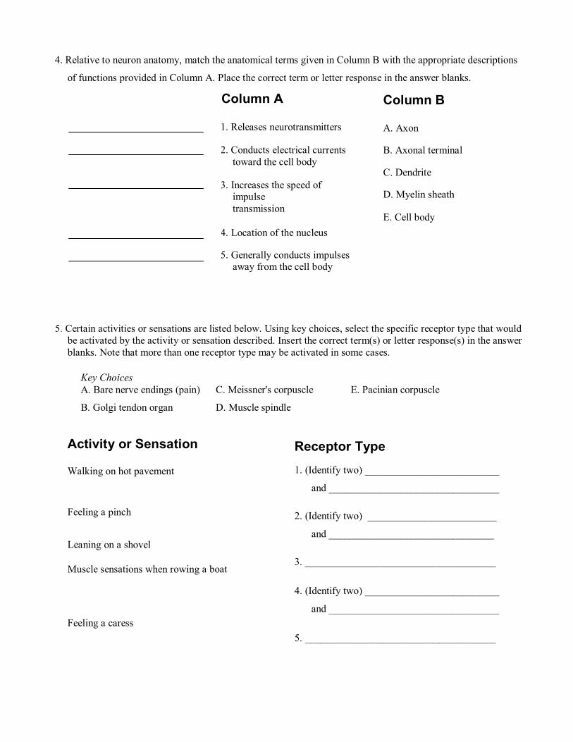
4. Relative to neuron anatomy, match the anatomical terms given in Column B with the appropriate descriptions
of functions provided in Column A. Place the correct term or letter response in the answer blanks.
5. Certain activities or sensations are listed below. Using key choices, select the specific receptor type that would be activated by the activity or sensation described. Insert the correct term(s) or letter response(s) in the answer blanks. Note that more than one receptor type may be activated in some cases.
Activity or Sensation
Walking on hot pavement
Feeling a pinch
Leaning on a shovel
Muscle sensations when rowing a boat
Feeling a caress
Column B
A. Axon
B. Axonal terminal
C. Dendrite
D. Myelin sheath
E. Cell body
Column A
1. Releases neurotransmitters
2. Conducts electrical currents toward the cell body
3. Increases the speed of impulse transmission
4. Location of the nucleus
5. Generally conducts impulsesaway from the cell body
Receptor Type1. (Identify two) __________________________
and _________________________________
2. (Identify two) _________________________
and ________________________________
3. _____________________________________
4. (Identify two) __________________________
and _________________________________
5. _____________________________________
Key Choices A. Bare nerve endings (pain) C. Meissner's corpuscle E. Pacinian corpuscle
B. Golgi tendon organ D. Muscle spindle
6. Using key choices, select the terms identified in the following descriptions by inserting the appropriate letter or term in the spaces provided.
Key Choices A. Afferent neuron F. Neuroglia K. Proprioceptors
B. Association neuron G. Neurotransmitters L. Schwann cells
C. Cutaneous sense organs H. Nerve M. Synapse
D. Efferent neuron I. Nodes of Ranvier N. Stimuli
E. Ganglion J. Nuclei 0. Tract
_______________ 1. Sensory receptors found in the skin, which are specialized to detect temperature, pressure changes, and pain
_______________ 2. Specialized cells that myelinate the fibers of neurons found in the PNS
_______________ 3. Junction or point of close contact between neurons
_______________ 4. Bundle of nerve processes inside the CNS
_______________ 5. Neuron, serving as part of the conduction pathway between sensory and motor neurons
_______________ 6. Gaps in a myelin sheath
_______________ 7. Collection of nerve cell bodies found outside the CNS
_______________ 8. Neuron that conducts impulses away from the CNS to muscles and glands
_______________ 9. Sensory receptors found in muscle and tendons that detect their degree of stretch
______________ 10. Changes, occurring within or outside the body, that affect nervous system functioning
______________ 11. Neuron that conducts impulses toward the CNS from the body periphery
______________ 12. Chemicals released by neurons that stimulate other neurons, muscles, or glands
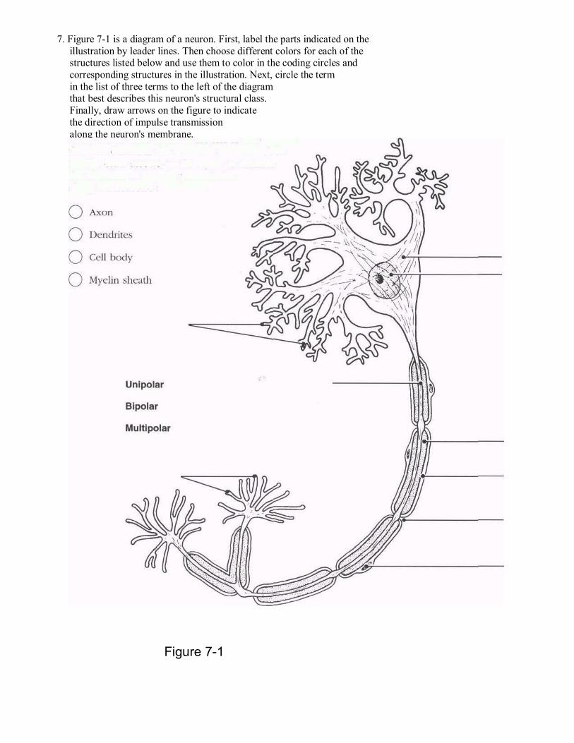
Figure 7-1
7. Figure 7-1 is a diagram of a neuron. First, label the parts indicated on the illustration by leader lines. Then choose different colors for each of the structures listed below and use them to color in the coding circles and corresponding structures in the illustration. Next, circle the term in the list of three terms to the left of the diagram that best describes this neuron's structural class. Finally, draw arrows on the figure to indicate the direction of impulse transmission along the neuron's membrane.
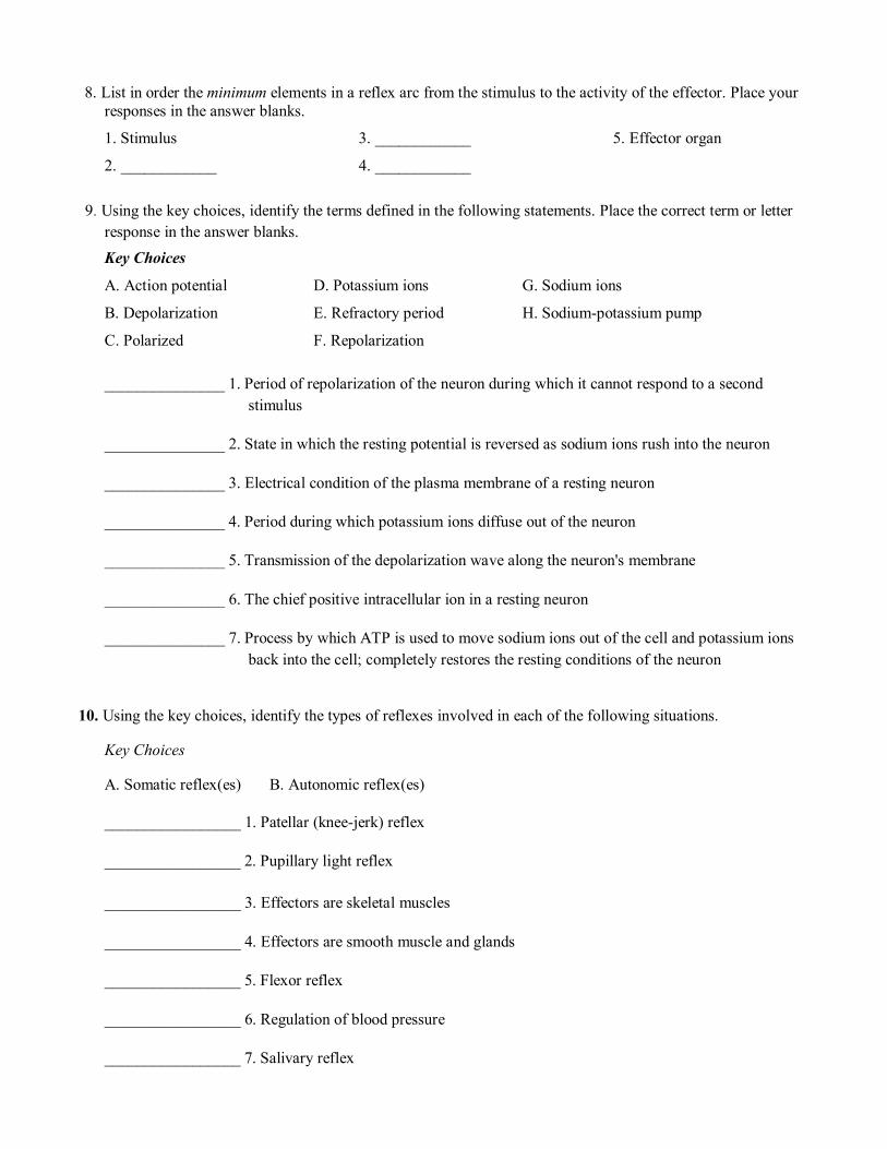
8. List in order the minimum elements in a reflex arc from the stimulus to the activity of the effector. Place your responses in the answer blanks.
1. Stimulus
2. ____________
3. ____________
4. ____________
5. Effector organ
9. Using the key choices, identify the terms defined in the following statements. Place the correct term or letter response in the answer blanks. Key Choices
A. Action potential D. Potassium ions G. Sodium ions
B. Depolarization E. Refractory period H. Sodium-potassium pump
C. Polarized F. Repolarization
_______________ 1. Period of repolarization of the neuron during which it cannot respond to a second stimulus
_______________ 2. State in which the resting potential is reversed as sodium ions rush into the neuron
_______________ 3. Electrical condition of the plasma membrane of a resting neuron
_______________ 4. Period during which potassium ions diffuse out of the neuron
_______________ 5. Transmission of the depolarization wave along the neuron's membrane
_______________ 6. The chief positive intracellular ion in a resting neuron
_______________ 7. Process by which ATP is used to move sodium ions out of the cell and potassium ions back into the cell; completely restores the resting conditions of the neuron
10. Using the key choices, identify the types of reflexes involved in each of the following situations.
Key Choices
A. Somatic reflex(es) B. Autonomic reflex(es)
_________________ 1. Patellar (knee-jerk) reflex
_________________ 2. Pupillary light reflex
_________________ 3. Effectors are skeletal muscles
_________________ 4. Effectors are smooth muscle and glands
_________________ 5. Flexor reflex
_________________ 6. Regulation of blood pressure
_________________ 7. Salivary reflex
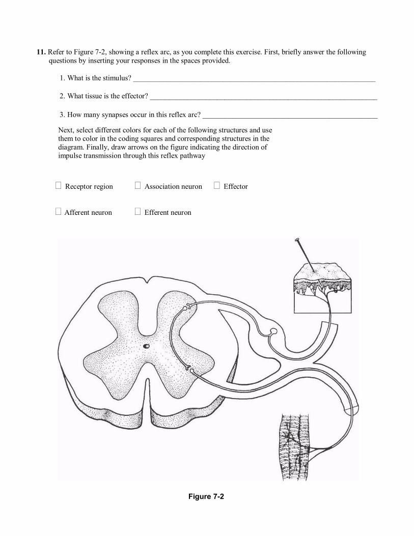
11. Refer to Figure 7-2, showing a reflex arc, as you complete this exercise. First, briefly answer the following questions by inserting your responses in the spaces provided.
1. What is the stimulus? _________________________________________________________________
2. What tissue is the effector? _____________________________________________________________
3. How many synapses occur in this reflex arc? _______________________________________________
Next, select different colors for each of the following structures and use them to color in the coding squares and corresponding structures in the diagram. Finally, draw arrows on the figure indicating the direction of impulse transmission through this reflex pathway
Receptor region Association neuron Effector
Afferent neuron Efferent neuron
Figure 7-2
12. Circle the term that does not belong in each of the following groupings.
1. Astrocytes Neurons Oligodendrocytes Microglia
2. K+ enters the cell K+ leaves the cell Repolarization Refractory period
3. Nodes of Ranvier Myelin sheath Unmyelinated Saltatory conduction
4. Predictable response Voluntary act Involuntary act Reflex
5. Oligodendrocytes Schwann cells Myelin Microglia
6. Cutaneous receptors Free dendritic endings Stretch Pain and touch
7. Cell interior High Na+ Low Na+ High K+
CENTRAL NERVOUS SYSTEM
Brain
13. Complete the following statements by inserting your answers in the answer blanks.
_______________ 1. The largest part of the human brain is the (paired) (1) . The other major subdivisions of the brain are the (2) and the _______________ 2. (3) . The cavities found in the brain are called (4) . They contain (5) . _______________ 3.
_______________ 4.
_______________ 5.
14. Circle the terms indicating structures that are not part of the brain stem.
Cerebral hemispheres Midbrain Medulla
Pons Cerebellum Diencephalon
15. Complete the following statements by inserting your answers in the answer blanks.
_______________ 1. A (1) is an elevated ridge of cerebral cortex tissue. The convulsions seen in the cerebrum are important because they increase the (2) . _______________ 2. Gray matter is composed of (3) . White matter is composed of (4) . which provide for communication between different parts of the brain as _______________ 3. well as with lower CNS centers. The lentiform nucleus, the caudate, and other nuclei are collectively called the (5) . _______________ 4. _______________ 5.
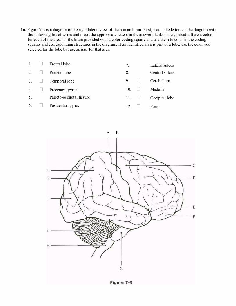
16. Figure 7-3 is a diagram of the right lateral view of the human brain. First, match the letters on the diagram with the following list of terms and insert the appropriate letters in the answer blanks. Then, select different colors for each of the areas of the brain provided with a color-coding square and use them to color in the coding squares and corresponding structures in the diagram. If an identified area is part of a lobe, use the color you selected for the lobe but use stripes for that area.
1. Frontal lobe
2. Parietal lobe
3. Temporal lobe
4. Precentral gyrus
5. Parieto-occipital fissure
6. Postcentral gyrus
7. Lateral sulcus
8. Central sulcus
9. Cerebellum
10. Medulla
11. Occipital lobe
12. Pons
A B
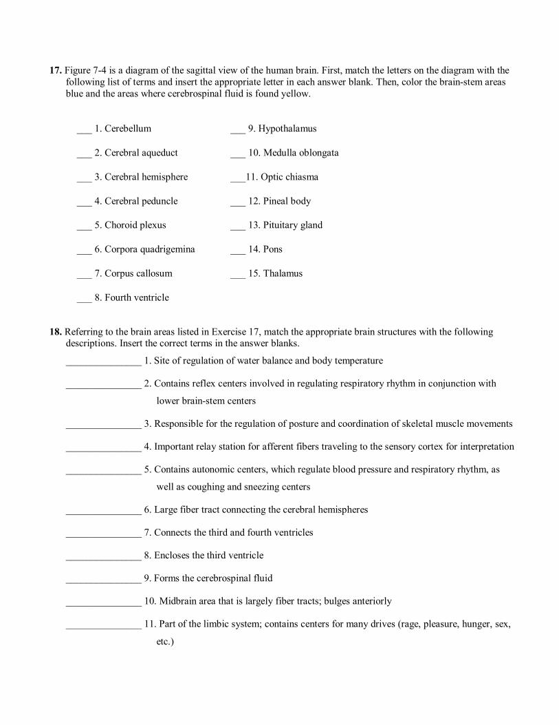
17. Figure 7-4 is a diagram of the sagittal view of the human brain. First, match the letters on the diagram with the following list of terms and insert the appropriate letter in each answer blank. Then, color the brain-stem areas blue and the areas where cerebrospinal fluid is found yellow.
___ 1. Cerebellum ___ 9. Hypothalamus
___ 2. Cerebral aqueduct ___ 10. Medulla oblongata
___ 3. Cerebral hemisphere ___11. Optic chiasma
___ 4. Cerebral peduncle ___ 12. Pineal body
___ 5. Choroid plexus ___ 13. Pituitary gland
___ 6. Corpora quadrigemina ___ 14. Pons
___ 7. Corpus callosum ___ 15. Thalamus
___ 8. Fourth ventricle
18. Referring to the brain areas listed in Exercise 17, match the appropriate brain structures with the following descriptions. Insert the correct terms in the answer blanks.
_______________ 1. Site of regulation of water balance and body temperature
_______________ 2. Contains reflex centers involved in regulating respiratory rhythm in conjunction with
lower brain-stem centers
_______________ 3. Responsible for the regulation of posture and coordination of skeletal muscle movements
_______________ 4. Important relay station for afferent fibers traveling to the sensory cortex for interpretation
_______________ 5. Contains autonomic centers, which regulate blood pressure and respiratory rhythm, as
well as coughing and sneezing centers
_______________ 6. Large fiber tract connecting the cerebral hemispheres
_______________ 7. Connects the third and fourth ventricles
_______________ 8. Encloses the third ventricle
_______________ 9. Forms the cerebrospinal fluid
_______________ 10. Midbrain area that is largely fiber tracts; bulges anteriorly
_______________ 11. Part of the limbic system; contains centers for many drives (rage, pleasure, hunger, sex,
etc.)
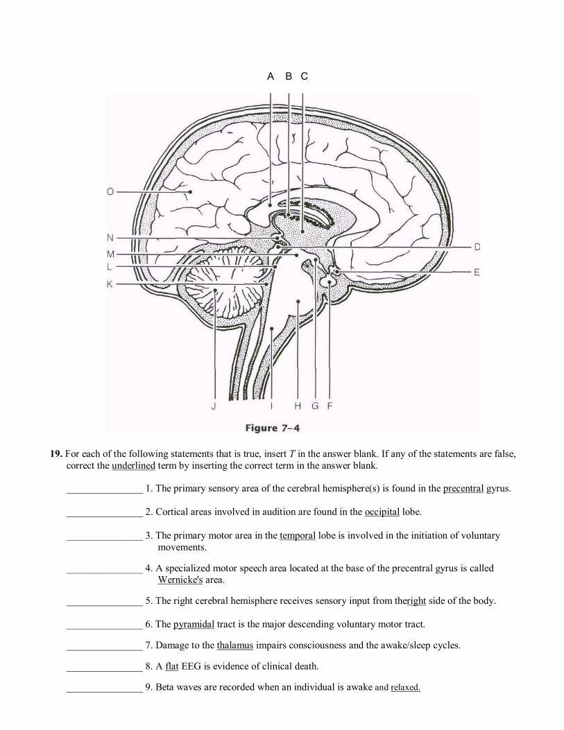
A B C
19. For each of the following statements that is true, insert T in the answer blank. If any of the statements are false, correct the underlined term by inserting the correct term in the answer blank.
_______________ 1. The primary sensory area of the cerebral hemisphere(s) is found in the precentral gyrus.
_______________ 2. Cortical areas involved in audition are found in the occipital lobe.
_______________ 3. The primary motor area in the temporal lobe is involved in the initiation of voluntary movements.
_______________ 4. A specialized motor speech area located at the base of the precentral gyrus is called Wernicke's area.
_______________ 5. The right cerebral hemisphere receives sensory input from theright side of the body.
_______________ 6. The pyramidal tract is the major descending voluntary motor tract.
_______________ 7. Damage to the thalamus impairs consciousness and the awake/sleep cycles.
_______________ 8. A flat EEG is evidence of clinical death.
_______________ 9. Beta waves are recorded when an individual is awake and relaxed.
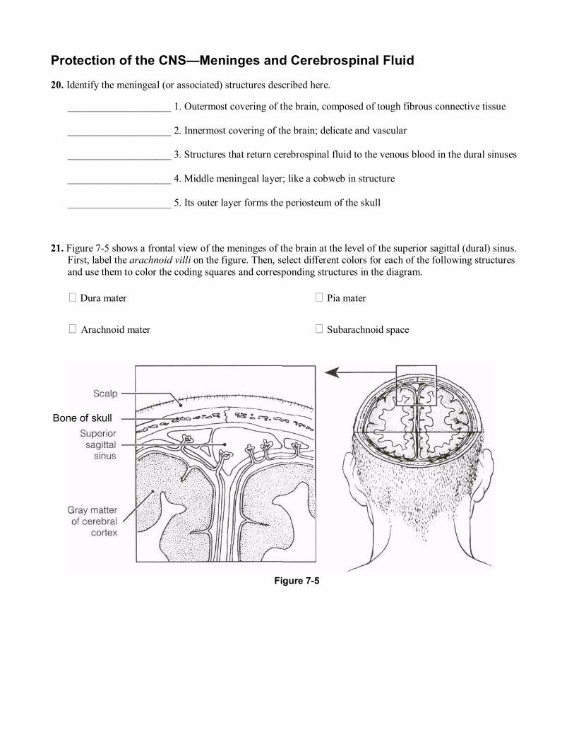
Protection of the CNS�Meninges and Cerebrospinal Fluid
20. Identify the meningeal (or associated) structures described here.
____________________ 1. Outermost covering of the brain, composed of tough fibrous connective tissue
____________________ 2. Innermost covering of the brain; delicate and vascular
____________________ 3. Structures that return cerebrospinal fluid to the venous blood in the dural sinuses
____________________ 4. Middle meningeal layer; like a cobweb in structure
____________________ 5. Its outer layer forms the periosteum of the skull
21. Figure 7-5 shows a frontal view of the meninges of the brain at the level of the superior sagittal (dural) sinus. First, label the arachnoid villi on the figure. Then, select different colors for each of the following structures and use them to color the coding squares and corresponding structures in the diagram.
Dura mater
Arachnoid mater
Pia mater
Subarachnoid space
Figure 7-5
Bone of skull
22. Complete the following statements by inserting your answers in the answer blanks.
_______________ 1.
_______________ 2.
_______________ 3.
_______________ 4.
_______________ 5.
_______________ 6.
_______________ 7.
Brain Dysfunctions 23. Match the brain disorders listed in Column B with the conditions described in Column A. Place the correct
answers in the answer blanks.
Column A
_______________ 1. Slight and transient brain injury
_______________ 2. Traumatic injury that destroys brain tissue
_______________ 3. Total nonresponsiveness to stimulation
_______________ 4. May cause medulla oblongata to be wedged into foramen magnum by pressure of blood
_______________ 5. After head injury, retention of water by brain
_______________ 6. Results when a brain region is deprived of blood or exposed to prolonged ischemia
_______________ 7. Progressive degeneration of the brain with abnormal protein deposits
_______________ 8. Autoimmune disorder with extensive demyelination
_______________ 9. A mini-stroke; fleeting symptoms of a CVA
Cerebrospinal fluid is formed by capillary knots called (1) . which hang into the (2) of the brain. Ordinarily, cerebro- spinal fluid flows from the lateral ventricles to the third ventricle and then through the (3) to the fourth ventricle. Some of the fluid continues down the (4) of the spinal cord, but most of it circulates into the (5) by passing through three tiny openings in the walls of the (6) . As a rule, cerebrospinal fluid is formed and drained back into the venous blood at the same rate. If its drainage is blocked, a condition called (7) occurs, which results in increased pressure on the brain.
Column B
A. Alzheimer's disease
B. Cerebral edema
C. Cerebrovascular accident (CVA)
D. Coma
E. Concussion
F. Contusion
G. Intracranial hemorrhage
H. Multiple sclerosis
I. Transient ischemic attack (TIA)
Spinal Cord 24. Complete the following statements by inserting your responses in the answer blanks.
_______________ 1.
_______________ 2.
_______________ 3.
_______________ 4.
_______________ 5.
_______________ 6.
_______________ 7.
_______________ 8.
_______________ 9.
25. Using key choices, select the appropriate terms to respond to the following descriptions referring to spinal cord anatomy. Place the correct term or letter in the answer blanks. Key Choices
A. Afferent (sensory)
B. Efferent (motor)
C. Both afferent and efferent
D. Association neurons (interneurons)
_______________ 1. Neuron type found in the dorsal horn
_______________ 2. Neuron type found in the ventral horn
_______________ 3. Neuron type in a dorsal root ganglion
_______________ 4. Fiber type in the ventral root
_______________ 5. Fiber type in the dorsal root
_______________ 6. Fiber type in a spinal nerve
The spinal cord extends from the (1) of the skull to the
(2) region of the vertebral column. The meninges, which
cover the spinal cord, extend more inferiorly to form a sac
from which cerebrospinal fluid can be withdrawn without
damage to the spinal cord. This procedure is called a (3) .
(4) pairs of spinal nerves arise from the cord. Of these,
(5) pairs are cervical nerves, (6) pairs are thoracic
nerves, (7) pairs are lumbar nerves, and (8) pairs are
sacral nerves. The tail-like collection of spinal nerves at the
inferior end of the spinal cord is called the (9) .
26. Figure 7-6 is a cross-sectional view of the spinal cord. First, select different colors to identify the following structures and use them to color the coding squares and corresponding structures in the figure.
� Pia mater � Dura mater � Arachnoid
Then, identify the areas listed in the key choices by inserting the correct choices/letter next to the appropriate leader line on the figure. Color the butterfly-shaped gray matter of the cord gray and the spinal roots and nerves yellow.
Key Choices A. Central canal D. Dorsal root G. Ventral horn
B. Column of white matter E. Dorsal root ganglion H. Ventral root
C. Dorsal horn F. Spinal nerve
Figure 7-6
27. Using choices from Column B, indicate what would happen if the structures in Column A were damaged or transected. Place the correct letter in the answer blanks. Column A
_____ 1. Dorsal root of a spinal nerve
_____ 2. Ventral root of a spinal nerve
_____ 3. Anterior ramus of a spinal nerve
Column B
A. Loss of motor function
B. Loss of sensory function
C. Loss of both motor and sensory function
Another name for a bundle of nerve fibers is (1) . Nerves
carrying both sensory and motor fibers are called (2)
nerves, whereas those carrying just sensory fibers are referred
to as sensory, or (3) nerves.
PERIPHERAL NERVOUS SYSTEM
Structure of a Nerve
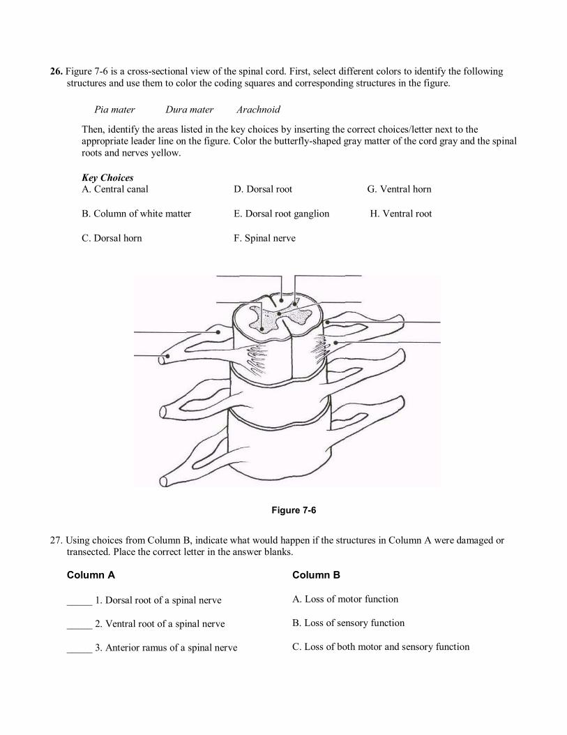
28. Figure 7-7 is a diagrammatic view of a nerve wrapped in its connective tissue coverings. Select different colors to identify the following structures and use them to color the coding circles and corresponding structures in the figure. Then, label each of the sheaths indicated by leader lines on the figure.
Endoneurium Perineurium Epineurium
Figure 7-7
29. Complete the following statements by inserting your responses in the answer blanks.
_______________ 1.
_______________ 2.
_______________ 3.
Myelin sheath
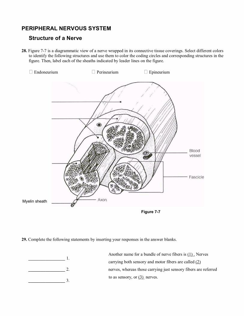
30. Figure 7-8 is an anterior view of the principal nerves arising from the brachial plexus. Select five different colors and color the coding circles and the nerves listed below. Also, label each nerve by inserting its name at the appropriate leader line.
Figure 7-8
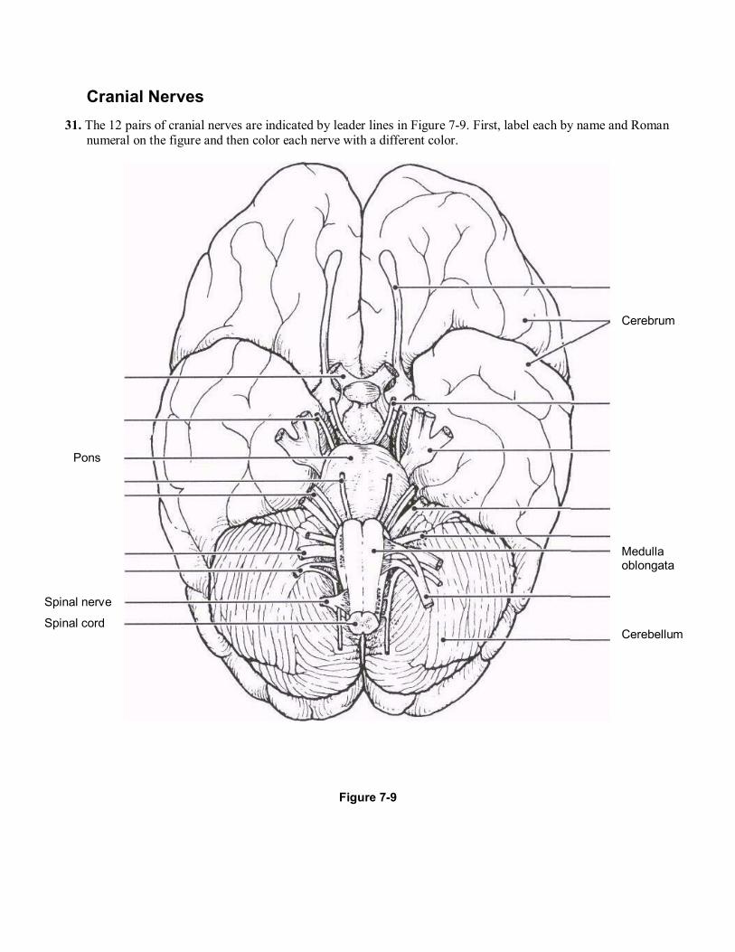
Cranial Nerves 31. The 12 pairs of cranial nerves are indicated by leader lines in Figure 7-9. First, label each by name and Roman
numeral on the figure and then color each nerve with a different color.
Figure 7-9
Pons
Spinal nerve
Spinal cord
Cerebrum
Medulla oblongata Cerebellum
The ventral rami of spinal nerves C1 through T1 and L1 throughS4 take part in forming (1), which serve the (2)_ of the body. The ventral rami of T1 through T12 run between the ribs to serve the (3). The posterior rami of the spinal nerves serve the (4) .
32. Provide the name and number of the cranial nerves involved in each of the following activities, sensations, or disorders. Insert your response in the answer blanks.
_______________ 1. Shrugging the shoulders
_______________ 2. Smelling a flower
_______________ 3. Raising the eyelids and focusing the lens of the eye for accommodation; constriction of the eye pupils
_______________ 4. Slows the heart; increases the mobility of the digestive tract
_______________ 5. Involved in smiling
_______________ 6. Involved in chewing food
_______________ 7. Listening to music; seasickness
_______________ 8. Secretion of saliva; tasting well-seasoned food
_______________ 9. Involved in "rolling" the eyes (three nerves�provide numbers only)
_______________ 10. Feeling a toothache
_______________ 11. Reading Tennis magazine or this study guide
_______________ 12. Purely sensory (three nerves�provide numbers only)
Spinal Nerves and Nerve Plexuses 33. Complete the following statements by inserting your responses in the answer blanks.
_______________ 1.
_______________ 2.
_______________ 3.
_______________ 4.
34. Name the major nerves that serve the following body areas. Insert your responses in the answer blanks.
_______________ 1. Head, neck, shoulders (name plexus only)
_______________ 2. Diaphragm
_______________ 3. Posterior thigh
_______________ 4. Leg and foot (name two)
_______________ 5. Most anterior forearm muscles
_______________ 6. Arm muscles
_______________ 7. Abdominal wall (name plexus only)
_______________ 8. Anterior thigh
_______________ 9. Medial side of the hand
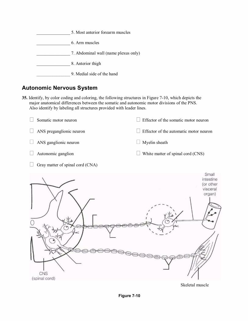
Autonomic Nervous System 35. Identify, by color coding and coloring, the following structures in Figure 7-10, which depicts the
major anatomical differences between the somatic and autonomic motor divisions of the PNS. Also identify by labeling all structures provided with leader lines.
Somatic motor neuron
ANS preganglionic neuron
ANS ganglionic neuron
Autonomic ganglion
Gray matter of spinal cord (CNA)
Effector of the somatic motor neuron
Effector of the automatic motor neuron
Myelin sheath
White matter of spinal cord (CNS)
Figure 7-10
Skeletal muscle
36. The following table indicates a number of conditions. Use a check (!) to show which division of the autonomic nervous system is involved in each condition.
Condition Sympathetic Parasympathetic
1. Postganglionic axons secrete norepinephrine; adrenergic fibers
2. Postganglionic axons secrete acetylcholine; cholinergic fibers
3. Long preganglionic axon, short postganglionic axon
4. Short preganglionic axon, long postganglionic axon
5. Arises from cranial and sacral nerves
6. Arises from spinal nerves T^ to La
7. Normally in control
8. Fight-or-flight system
9. Has more specific control
10. Causes a dry mouth, dilates bronchioles
11. Constricts eye pupils, decreases heart rate
37. You are alone in your home late in the evening, and you hear an unfamiliar sound in your backyard. In the spaces provided, list four physiologic events promoted by the sympathetic nervous system that would help you to cope with this rather frightening situation.
1. ___________________________________________________________________________________
2. ___________________________________________________________________________
3. ___________________________________________________________________________
4. ___________________________________________________________________________