Embed Size (px)
Citation preview

Chapter 11 Worksheet
Autonomic Nervous System
What regulates the ANS? (p 271)
What are the two major branches?
Draw the flowchart of the nervous system:
What do autonomic sensory neurons detect?
What are some structures that autonomic motor neurons effect?
What are some autonomic responses?
What does “dual innervation” mean?
What neurotransmitters are present in the sympathetic nervous system?
What neurotransmitters are present in the parasympathetic nervous system?
What is mostly secreted from the sympathetic postganglionic neurons?
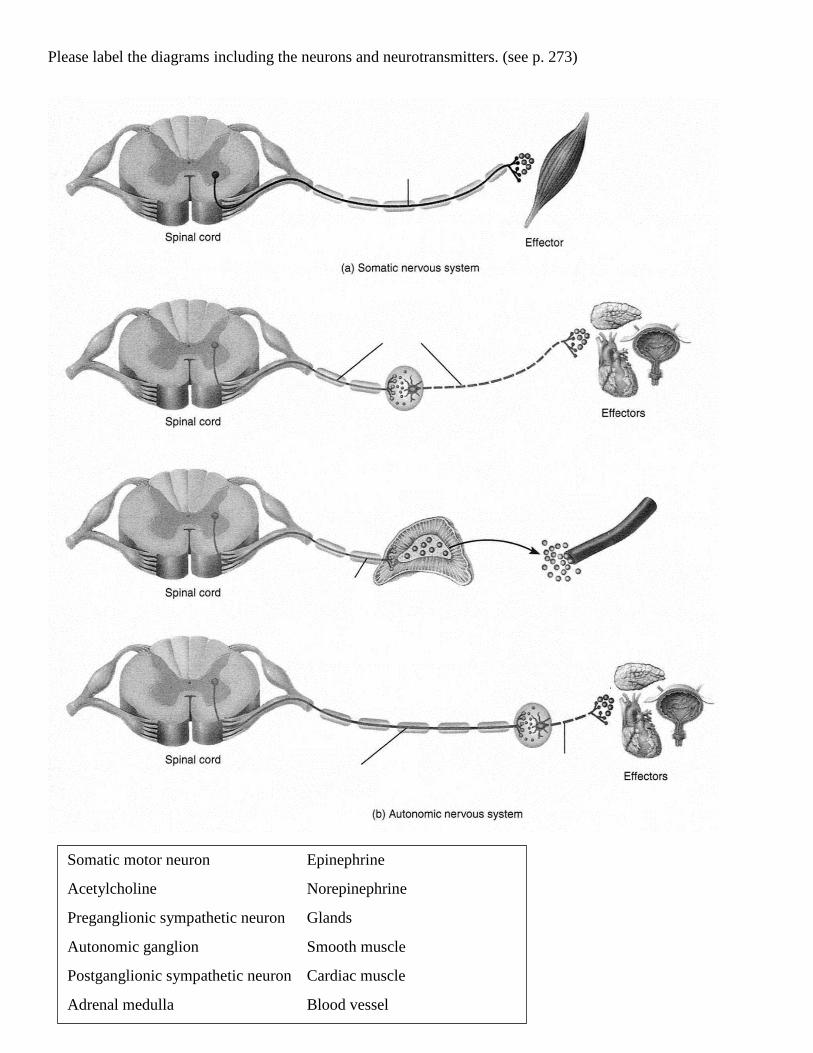
Please label the diagrams including the neurons and neurotransmitters. (see p. 273)
Somatic motor neuron Epinephrine
Acetylcholine Norepinephrine
Preganglionic sympathetic neuron Glands
Autonomic ganglion Smooth muscle
Postganglionic sympathetic neuron Cardiac muscle
Adrenal medulla Blood vessel

Please read over the structures of the sympathetic division of the autonomic nervous system and say them
aloud. Which neurons synapse in a sympathetic trunk ganglion?

Please read over the structures of the sympathetic division of the autonomic nervous system and say them
aloud. Which division, sympathetic or parasympathetic, has longer preganglionic axons?

What are the activities of the sympathetic nervous system? (p. 277: 1-8)
Which division of the autonomic nervous system is “fight-or-flight”?
Which division of the autonomic nervous system is “rest-and-digest”?
Frontal lobe (color red) – motor strip location, impulsivity, short term memory, emotion, voluntary
movement, social functioning, creativity, expressive language.
Parietal lobe (color blue) - sensory strip location, perception, touch/ pain, ability to draw, reading and
writing, calculations.
Temporal lobe (color yellow) – hearing, long term memory, verbal and written recognition memory,
receptive memory, music, initiation of verbal.
Occipital lobe (color green) – perception, vision.
Cerebellum (not shown) – coordination, balance, ability to judge distance, muscle tone, including muscles
required for speech.
Brain stem (not shown) – connects the brain with the spinal cord, consists of midbrain, pons, medulla
oblongata.
Diencephalon (not shown) – consists of thalamus, hypothalamus, pineal gland.