Embed Size (px)
Citation preview
40 EXPEDITION Volume 61 Number 1
A JOURNEY INTO THE HUMAN BODY studying mummies
to understandancient diseaseby michael r. zimmerman
41EXPEDITION Spring 2019
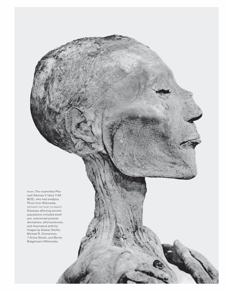
RIGHT: The mummified Pha-raoh Ramses V (died 1145 BCE), who had smallpox. Photo from Wikimedia. OPPOSITE TOP (LEFT TO RIGHT): Diseases affecting ancient populations included small-pox, subcorneal pustular dermatosis, atherosclerosis, and rheumatoid arthritis. Images by Gaetan Stoffel, Michael R. Zimmerman, 7 Active Studio, and Bernd Brägelmann/Wikimedia.
42 EXPEDITION Volume 61 Number 1
UNDERSTANDING ANCIENT DISEASE
AS AN ANTHROPOLOGIST AND RETIRED PATHOLOGIST, DR. MICHAEL ZIMMERMAN’S RESEARCH FOCUSES ON MUMMY PALEOPATHOLOGY. HE DETAILS WHAT CAN BE ACCOMPLISHED IN STUDYING ANCIENT DISEASE AND REFLECTS ON WHAT REMAINS TO BE DISCOVERED USING 21ST CENTURY MEDICAL TECHNIQUES.
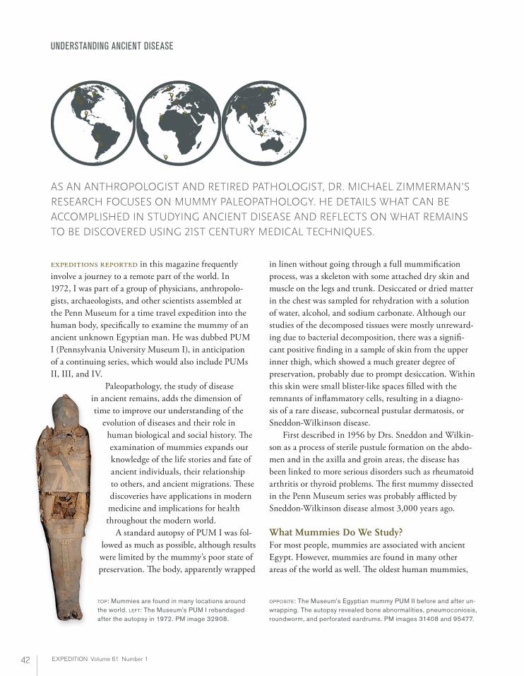
expeditions reported in this magazine frequently involve a journey to a remote part of the world. In 1972, I was part of a group of physicians, anthropolo-gists, archaeologists, and other scientists assembled at the Penn Museum for a time travel expedition into the human body, specifically to examine the mummy of an ancient unknown Egyptian man. He was dubbed PUM I (Pennsylvania University Museum I), in anticipation of a continuing series, which would also include PUMs II, III, and IV.
Paleopathology, the study of disease in ancient remains, adds the dimension of time to improve our understanding of the
evolution of diseases and their role in human biological and social history. The examination of mummies expands our knowledge of the life stories and fate of ancient individuals, their relationship to others, and ancient migrations. These discoveries have applications in modern medicine and implications for health throughout the modern world. A standard autopsy of PUM I was fol-
lowed as much as possible, although results were limited by the mummy’s poor state of preservation. The body, apparently wrapped
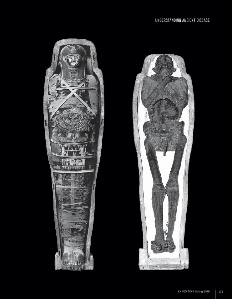
OPPOSITE: The Museum’s Egyptian mummy PUM II before and after un-wrapping. The autopsy revealed bone abnormalities, pneumoconiosis, roundworm, and perforated eardrums. PM images 31408 and 95477.
in linen without going through a full mummification process, was a skeleton with some attached dry skin and muscle on the legs and trunk. Desiccated or dried matter in the chest was sampled for rehydration with a solution of water, alcohol, and sodium carbonate. Although our studies of the decomposed tissues were mostly unreward-ing due to bacterial decomposition, there was a signifi-cant positive finding in a sample of skin from the upper inner thigh, which showed a much greater degree of preservation, probably due to prompt desiccation. Within this skin were small blister-like spaces filled with the remnants of inflammatory cells, resulting in a diagno-sis of a rare disease, subcorneal pustular dermatosis, or Sneddon-Wilkinson disease. First described in 1956 by Drs. Sneddon and Wilkin-son as a process of sterile pustule formation on the abdo-men and in the axilla and groin areas, the disease has been linked to more serious disorders such as rheumatoid arthritis or thyroid problems. The first mummy dissected in the Penn Museum series was probably afflicted by Sneddon-Wilkinson disease almost 3,000 years ago.
What Mummies Do We Study? For most people, mummies are associated with ancient Egypt. However, mummies are found in many other areas of the world as well. The oldest human mummies,
TOP: Mummies are found in many locations around the world. LEFT: The Museum’s PUM I rebandaged after the autopsy in 1972. PM image 32908.
43EXPEDITION Spring 2019 43EXPEDITION Spring 2019
UNDERSTANDING ANCIENT DISEASE
44 EXPEDITION Volume 61 Number 1
UNDERSTANDING ANCIENT DISEASE
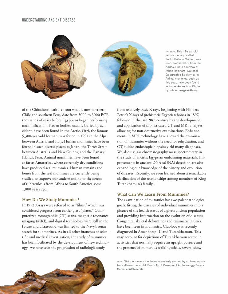
of the Chinchorro culture from what is now northern Chile and southern Peru, date from 5000 to 3000 BCE, thousands of years before Egyptians began performing mummification. Frozen bodies, usually buried by ac-cident, have been found in the Arctic. Ötzi, the famous 5,300-year-old Iceman, was found in 1991 in the Alps between Austria and Italy. Human mummies have been found in such diverse places as Japan, the Torres Strait between Australia and New Guinea, and the Canary Islands, Peru. Animal mummies have been found as far as Antarctica, where extremely dry conditions have produced seal mummies. Human remains and bones from the seal mummies are currently being studied to improve our understanding of the spread of tuberculosis from Africa to South America some 1,000 years ago.
How Do We Study Mummies?In 1972 X-rays were referred to as “films,” which was considered progress from earlier glass “plates.” Com-puterized tomographic (CT) scans, magnetic resonance imaging (MRI), and digital technology were still in the future and ultrasound was limited to the Navy’s sonar search for submarines. As in all other branches of scien-tific and medical investigation, the study of mummies has been facilitated by the development of new technol-ogy. We have seen the progression of radiologic study
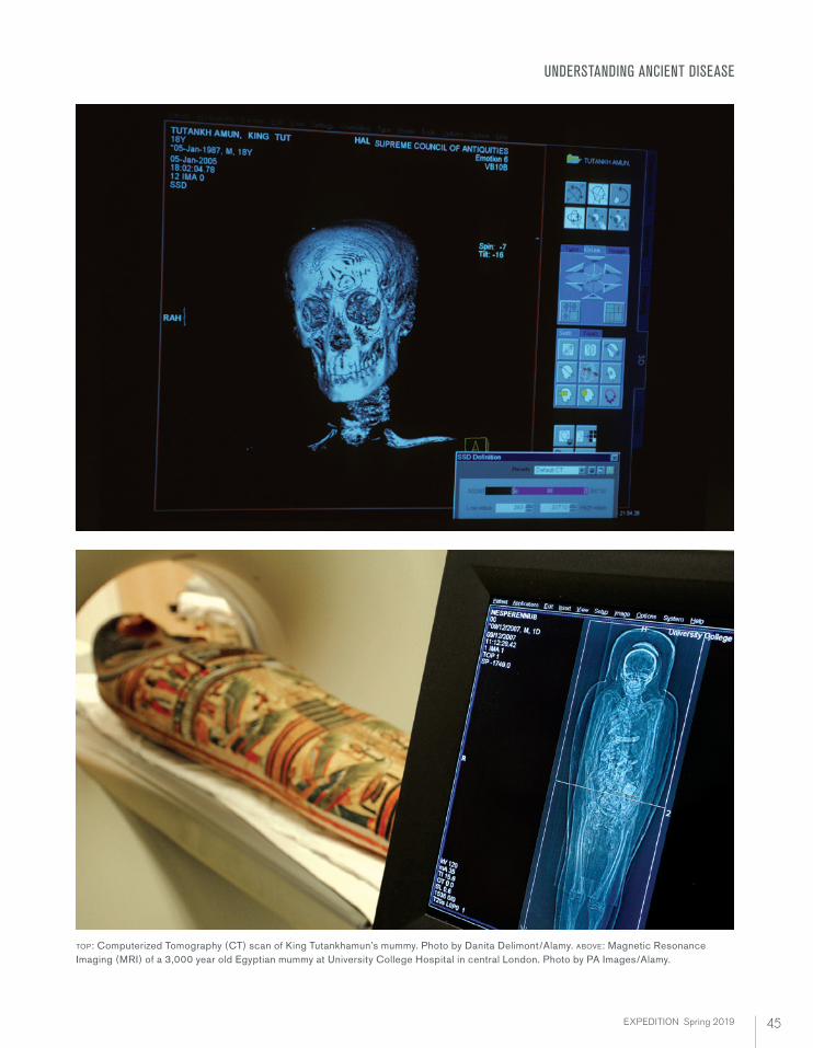
from relatively basic X-rays, beginning with Flinders Petrie’s X-rays of prehistoric Egyptian bones in 1897, followed in the late 20th century by the development and application of sophisticated CT and MRI analyses, allowing for non-destructive examinations. Enhance-ments in MRI technology have allowed the examina-tion of mummies without the need for rehydration, and CT-guided endoscopic biopsies yield many diagnoses. We also use gas chromatography mass spectrometry in the study of ancient Egyptian embalming materials. Im-provements in ancient DNA (aDNA) detection are also expanding our knowledge of the history and evolution of diseases. Recently, we even learned about a remarkable clarification of the relationships among members of King Tutankhamun’s family.
What Can We Learn From Mummies?The examination of mummies has two paleopathological goals: fitting the diseases of individual mummies into a picture of the health status of a given ancient population and providing information on the evolution of diseases. Congenital skeletal deformities and traumatic injuries have been seen in mummies. Clubfoot was recently diagnosed in Amenhotep III and Tutankhamun. This may account for depictions of Tutankhamun seated in activities that normally require an upright posture and the presence of numerous walking sticks, several show-
LEFT: Ötzi the Iceman has been intensively studied by archaeologists from all over the world. South Tyrol Museum of Archaeology/Eurac/Samadelli/Staschitz.
FAR LEFT: This 13-year-old female mummy, called the Llullaillaco Maiden, was recovered in 1999 from the Andes. Photo courtesy of Johan Reinhard, National Geographic Society. LEFT: Animal mummies, such as this seal, have been found as far as Antarctica. Photo by Johner Images/Alamy.
45EXPEDITION Spring 2019
UNDERSTANDING ANCIENT DISEASE
TOP: Computerized Tomography (CT) scan of King Tutankhamun’s mummy. Photo by Danita Delimont/Alamy. ABOVE: Magnetic Resonance Imaging (MRI) of a 3,000 year old Egyptian mummy at University College Hospital in central London. Photo by PA Images/Alamy.
46 EXPEDITION Volume 61 Number 1
UNDERSTANDING ANCIENT DISEASE
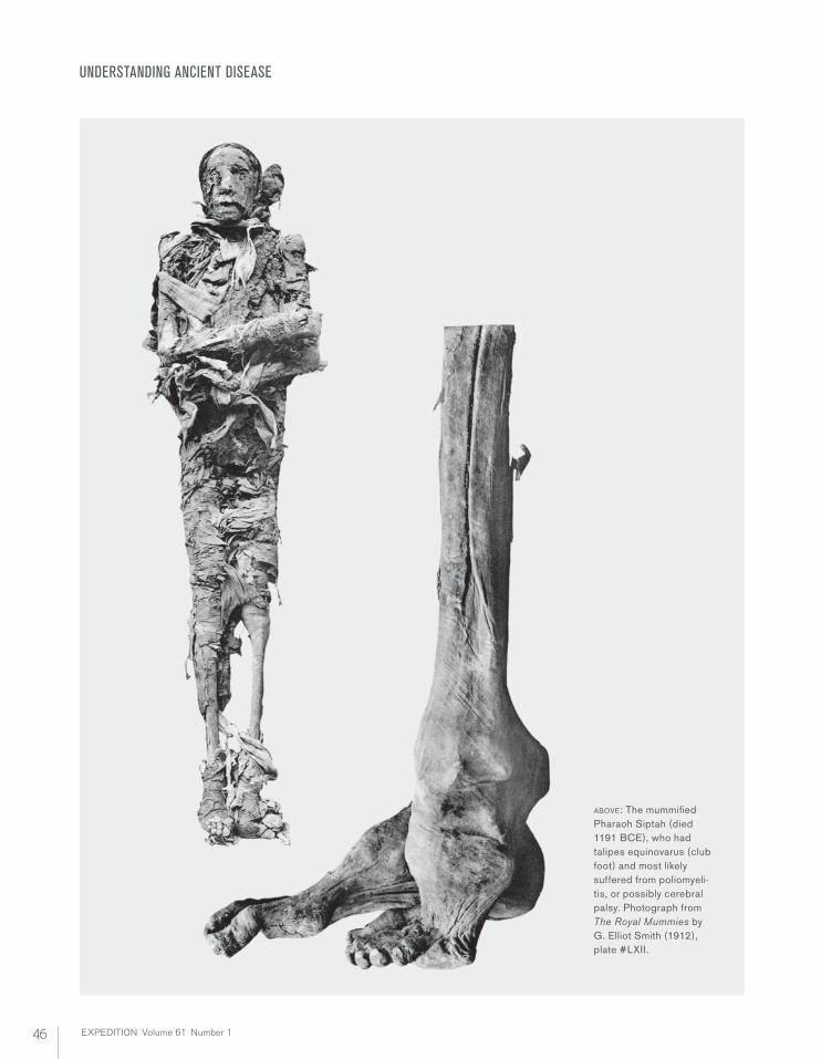
ABOVE: The mummified Pharaoh Siptah (died 1191 BCE), who had talipes equinovarus (club foot) and most likely suffered from poliomyeli-tis, or possibly cerebral palsy. Photograph from The Royal Mummies by G. Elliot Smith (1912), plate #LXII.
47EXPEDITION Spring 2019
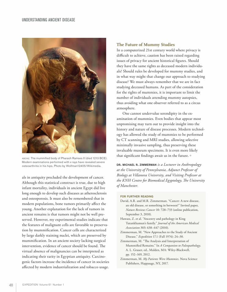
ing wear, in Tutankhamun’s tomb. Another Egyptian mummy suffered from a club foot and his liver showed the scarring of cirrhosis, perhaps due to self-medication by an excess of wine during his life. One of the most notable traumatic injuries was diagnosed in the Iceman by CT scanning, an arrowhead in the shoulder region, resulting in a fatal hemorrhage. There have been many reports of infectious and inflammatory disease found in mummies. Examination of the Pharaoh Siptah using x-ray technology showed an overall shortening of the entire right leg and atrophy of the soft tissues, diagnosed as characteristic of poliomyeli-tis, or possibly cerebral palsy. Smallpox has been diag-nosed in the mummy of Ramses V. Pneumonia, one of the most common bacterial infections and a major cause of death in the pre-antibiotic era, has also been diag-nosed in Egyptian mummies. Tuberculosis has been well documented in ancient Egypt and pre-Columbian South America. Of interest is the absence of evidence of tuber-culosis in pre-Dynastic Nubian skeletons and mummies, suggesting the Dynastic period (from ca. 3000 BCE) for the onset of human tuberculosis in the Nile Valley, but recent molecular evidence indicates a much older date for the evolution of human tuberculosis. Studies of aDNA have determined that Tutankhamun suffered from falciparum malaria, the most serious form of the disease. Parasitic worms remain well preserved for millennia, and the characteristic ova of Ascaris lumbricoi-des, Schistosoma hematobium, and Taenia solium have all been reported in Egyptian mummies. The Dakhleh Oasis is far from the Nile but schistosome ova were discovered in a mummy from that area; this may be evidence of a trade route or the movement of people via oases in the western desert. Dental and middle ear disease have also long been part of the human condition. Periodontal disease, dental wear, and caries (cavities) have been noted in pharaohs and fellahs (Arabic for commoners). These conditions can lead to infection of the middle ear and mastoid sinuses, and, in fact, perforated eardrums were seen in PUM II. The degenerative process most common in mummies is osteoarthritis, often seen in Egyptian mummies, where its presence in the hot dry climate of Egypt and Nubia belies the folk attribution of the disease to damp climates. X-rays of Ramses II have revealed severe osteoarthritis in his hips.
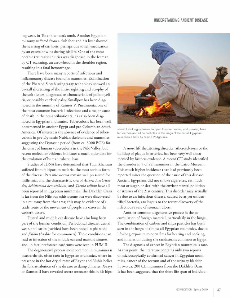
A more life threatening disorder, atherosclerosis or the buildup of plaque in arteries, has been very well docu-mented by historic evidence. A recent CT study identified the disorder in 9 of 22 mummies in the Cairo Museum. This much higher incidence than had previously been reported raises the question of the cause of this disease. Ancient Egyptians did not smoke cigarettes, eat much meat or sugar, or deal with the environmental pollution or stresses of the 21st century. This disorder may actually be due to an infectious disease, caused by as yet uniden-tified bacteria, analogous to the recent discovery of the infectious cause of stomach ulcers. Another common degenerative process is the ac-cumulation of foreign material, particularly in the lungs. The combination of carbon and silica particles has been seen in the lungs of almost all Egyptian mummies, due to life-long exposure to open fires for heating and cooking, and inhalation during the sandstorms common to Egypt. The diagnosis of cancer in Egyptian mummies is rare. At this point, the literature contains only two reports of microscopically confirmed cancer in Egyptian mum-mies, cancer of the rectum and of the urinary bladder in two ca. 200 CE mummies from the Dakhleh Oasis. It has been suggested that the short life span of individu-
UNDERSTANDING ANCIENT DISEASE
ABOVE: Life-long exposure to open fires for heating and cooking have left carbon and silica particles in the lungs of almost all Egyptian mummies. Photo by Simon Podgorsek.
48 EXPEDITION Volume 61 Number 1
UNDERSTANDING ANCIENT DISEASE
als in antiquity precluded the development of cancer. Although this statistical construct is true, due to high infant mortality, individuals in ancient Egypt did live long enough to develop such diseases as atherosclerosis and osteoporosis. It must also be remembered that in modern populations, bone tumors primarily affect the young. Another explanation for the lack of tumors in ancient remains is that tumors might not be well pre-served. However, my experimental studies indicate that the features of malignant cells are favorable to preserva-tion by mummification. Cancer cells are characterized by large darkly staining nuclei, which are preserved by mummification. In an ancient society lacking surgical intervention, evidence of cancer should be found. The virtual absence of malignancies can be interpreted as indicating their rarity in Egyptian antiquity. Carcino-genic factors increase the incidence of cancer in societies affected by modern industrialization and tobacco usage.
The Future of Mummy StudiesIn a computerized 21st century world where privacy is difficult to achieve, caution has been raised regarding issues of privacy for ancient historical figures. Should they have the same rights as deceased modern individu-als? Should rules be developed for mummy studies, and in what way might that change our approach to studying disease? We must always remember that we are in fact studying deceased humans. As part of the consideration for the rights of mummies, it is important to limit the number of individuals attending mummy autopsies, thus avoiding what one observer referred to as a circus atmosphere. One cannot undervalue serendipity in the ex-amination of mummies. Even bodies that appear most unpromising may turn out to provide insight into the history and nature of disease processes. Modern technol-ogy has allowed the study of mummies to be performed by CT scanning and MRI studies, allowing selective minimally invasive sampling, thus preserving these invaluable museum specimens. It is even more likely that significant findings await us in the future. Ä
DR. MICHAEL R. ZIMMERMAN is a Lecturer in Anthropology at the University of Pennsylvania, Adjunct Professor of Biology at Villanova University, and Visiting Professor at the KNH Centre for Biomedical Egyptology, The University of Manchester.
FOR FURTHER READING
David, A.R. and M.R. Zimmerman. “Cancer: A new disease, an old disease, or something in between?” Invited paper, Nature Reviews Cancer 10: 728–733 (online publication, September 3, 2010).
Hawass, Z. et al. “Ancestry and pathology in King Tutankhamun’s family.” Journal of the American Medical Association 303: 638–647 (2010).
Zimmerman, M. “New Approaches to the Study of Ancient Disease.” Expedition 17.1 (Fall 1974): 24–30.
Zimmerman, M. “The Analysis and Interpretation of Mummified Remains.” In A Companion to Paleopathology. A. L. Grauer, ed., Malden, MA: Wiley-Blackwell, pp. 152–169, 2012.
Zimmerman, M. My Patients Were Mummies. Nova Science Publishers, Happauge, NY, 2017.
ABOVE: The mummified body of Pharaoh Ramses II (died 1213 BCE). Modern examinations performed with x-rays have revealed severe osteoarthritis in his hips. Photo by Wolfman12405/Wikimedia.