Embed Size (px)
Citation preview

1
Five things everyone needsto know about
echocardiography
Lynn Cronin, MDKarthik Ananthsubramaniam, MD
Felix Rogers, DO
Our Five Topics
1. Approach to low gradient, severe aortic stenosis(including patients with normal EF)
2. The right ventricle: PE, RVMI & RV failure3. Patient prosthesis mismatch4. Practical approach to a mitral regurgitation
severity score5. Artifacts: how to see through them
Aortic Aortic StenosisStenosis Low Flow Low Gradient With Normal EFLow Flow Low Gradient With Normal EF
Can We Look Beyond GradientsCan We Look Beyond Gradients
Karthik Karthik AnanthasubramaniamAnanthasubramaniam, MD FACC FASE FASNC, MD FACC FASE FASNCAssociate Professor of Medicine, Wayne State UniversityAssociate Professor of Medicine, Wayne State University
Director, Echocardiography and Nuclear Cardiology LaboratoryDirector, Echocardiography and Nuclear Cardiology LaboratoryHeart and Vascular InstituteHeart and Vascular InstituteHenry Ford Hospital, DetroitHenry Ford Hospital, Detroit
CaseCase•• 80yr M.80yr M.
•• New progressive worsening of SOB for 3 weeks. AssociatedNew progressive worsening of SOB for 3 weeks. Associatedwith LE edema.with LE edema.
•• PMHxPMHx: HTN, DM, COPD, CAD S/P CABG x2 in 2002,: HTN, DM, COPD, CAD S/P CABG x2 in 2002,•• Moderate AS; 1 yr ago ( EF 55%,peak/ mean gradients 56.2/Moderate AS; 1 yr ago ( EF 55%,peak/ mean gradients 56.2/
22.2 mmHg. AVA 1.3 cm22.2 mmHg. AVA 1.3 cm22))
•• BP 163/63, HR 75, 95% on 3 BP 163/63, HR 75, 95% on 3 litreslitres; scattered wheezing with; scattered wheezing withbilateral crackles, Normal S1, soft S2, 2/6 systolic murmur;bilateral crackles, Normal S1, soft S2, 2/6 systolic murmur;2+ bilateral pitting edema.2+ bilateral pitting edema.
•• Laboratory findings and CXR consistent with ADHF and lungLaboratory findings and CXR consistent with ADHF and lungnodulenodule
•• Echo Echo evaleval
Low Flow Preserved EF SevereLow Flow Preserved EF SevereASAS
Stroke volume = 69 ccStroke volume index =69/2.03= 33 ml/m2
Highest velocityrecorded from rt parasternaland suprasternalviews

2
AS/ What We KnowAS/ What We Know
•• Severe AS:Severe AS:–– AVA <1 cmAVA <1 cm22 (0.6 cm (0.6 cm22/m/m22).).–– Peak velocity >4 Peak velocity >4 m/secm/sec..–– Mean gradient >40 Mean gradient >40 mHgmHg..
•• But, in some patients, But, in some patients, ““numbers do not add upnumbers do not add up”” even evenin the presence of in the presence of ““normalnormal”” functioning LV (normal functioning LV (normalEF):EF):–– Our groups of interest: AVA <1 cmOur groups of interest: AVA <1 cm2 2 & mean gradient <40& mean gradient <40
mmHgmmHg
With normalcardiac output
Most decisions1.Gradient2.AVA
Take a Take a ““ Time Out Time Out””Correct technical errorsCorrect technical errors•• LVOT measurements errors LVOT measurements errors ********
2. LVOT flow errors( sample volume not in LVOT 2. LVOT flow errors( sample volume not in LVOTwhere measurement done, LVOT where measurement done, LVOT vel vel > 1.5 > 1.5 mm/sec/secbut wrong but wrong Benoulli Benoulli equation appliedequation applied
3. Heart rhythm issues not factored in 3. Heart rhythm issues not factored in•• CW errorsCW errors
Wrong jet measuredWrong jet measuredMax velocity not capturedMax velocity not capturedAveraging of velocities were appliedAveraging of velocities were appliedPressure recovery/ aorta size not accounted forPressure recovery/ aorta size not accounted for
Low gradient/normal EFLow gradient/normal EF
•• Hypothesized to be due to markedHypothesized to be due to markedconcentric remodeling and hypertrophyconcentric remodeling and hypertrophyleading to small LV cavity withleading to small LV cavity withdecreased stroke volume being fromdecreased stroke volume being fromdeficient filling rather than emptying.deficient filling rather than emptying.
How Common?How Common?•• In a study, over 6 years,In a study, over 6 years,
patients with severe AS,patients with severe AS,defined by indexed AVA,defined by indexed AVA,with EF with EF ≥≥50% were50% wereretrospectively reviewed.retrospectively reviewed.–– 35% had inconsistent AV peak35% had inconsistent AV peak
velocities and velocities and transvalvulartransvalvularmean gradients:mean gradients:
•• Peak velocity: 3.5±0.9 Peak velocity: 3.5±0.9 m/secm/sec..•• Mean gradient: 32±17 mmHg.Mean gradient: 32±17 mmHg.
–– 55% of patients had mean55% of patients had meangradient <30 mmHg.gradient <30 mmHg.
Circulation. 2007 Jun 5;115(22):2856-64
•• ELI = ( AVA X AELI = ( AVA X AAA ) / ( A ) / ( AAA –– AVA ) AVA )–– AAAA: Aortic cross sectional area.: Aortic cross sectional area.
•• ELI ELI ≤≤ 0.52 cm 0.52 cm22/m/m22 was the best was the bestpredictor of adverse outcomes (deathpredictor of adverse outcomes (deathand AV replacement).and AV replacement).–– Positive predictive value of 67%.Positive predictive value of 67%.
Energy Loss IndexEnergy Loss Index
Circulation. 2000 Feb 22;101(7):765-71
•• Valvulo-arterial impedenceValvulo-arterial impedence::–– ZZVAVA = ( SAP + = ( SAP + GradientGradientmeanmean ) / SVI) / SVI
•• SAP: Systolic Arterial Pressure.SAP: Systolic Arterial Pressure.•• GradientGradientmeanmean: mean : mean transvalvular transvalvular pressure gradient.pressure gradient.•• SVI: Stroke Volume Index.SVI: Stroke Volume Index.
–– It represents the It represents the valvular valvular and arterial factors that oppose LVand arterial factors that oppose LVejection by absorption the mechanical energy generated byejection by absorption the mechanical energy generated bythe LV.the LV.
–– A value of A value of ≥≥5.5 mmHg/mL/m5.5 mmHg/mL/m2 2 was the best predictor ofwas the best predictor ofadverse outcomesadverse outcomes
The flow-independent indicesThe flow-independent indices
Circulation. 2007 Jun 5;115(22):2856-64

3
Back to our patientBack to our patient……•• AVA= 0.94 (VTI), 0.93 (AVA= 0.94 (VTI), 0.93 (VmaxVmax))
•• Dimensionless velocity index: =0.29Dimensionless velocity index: =0.29
•• Energy loss index (Energy loss index (≤≤ 0.52): 0.42 0.52): 0.42
•• Valvulo-arterial Valvulo-arterial impedance (impedance (≥≥5.5): 5.95.5): 5.9
•• Cath Cath planned when stable for graft assessment/ planned when stable for graft assessment/ hemodynamicshemodynamicssuspecting moderate-severe AS which was symptomatic.suspecting moderate-severe AS which was symptomatic.
•• Patient diagnosed with lung cancer the next month and died onPatient diagnosed with lung cancer the next month and died on chemotherapy chemotherapy
ConclusionsConclusions•• Severe AS with low velocity and gradients in theSevere AS with low velocity and gradients in the
presence of normal EF is a common condition.presence of normal EF is a common condition.
•• Mostly itMostly it’’s secondary to increased vascular resistances secondary to increased vascular resistanceand decreased arterial compliance which adds moreand decreased arterial compliance which adds moreworkload on the LV.workload on the LV.
•• If suspected:If suspected:–– Make sure the measurements are accurate with good qualityMake sure the measurements are accurate with good quality
study. Calculate indexed stroke volume ( N= study. Calculate indexed stroke volume ( N= > > 35 ml/m2 )35 ml/m2 )–– Record BP at the time of getting Doppler measurements toRecord BP at the time of getting Doppler measurements to evaluate more evaluate more hemodynamic hemodynamic indices. Measure AAindices. Measure AA
CSA/diameterCSA/diameter
ConclusionsConclusions•• If still suspected, use other less flow-dependentIf still suspected, use other less flow-dependent
indices:indices:–– AVA (<1.0) Direct AVA (<1.0) Direct planimetry planimetry ; TEE, MRI or CT; TEE, MRI or CT
–– Dimensionless velocity index (<0.25)Dimensionless velocity index (<0.25)
–– Energy loss index (Energy loss index (≤≤ 0.52) 0.52)
–– Valvulo-arterial Valvulo-arterial impedance (impedance (≥≥5.5)5.5)
•• Patients with severe AS and low flow (inconsistentPatients with severe AS and low flow (inconsistentgrading of AS) have at least similar prognosis asgrading of AS) have at least similar prognosis asthose with normal flow and consistent grading of ASthose with normal flow and consistent grading of ASseverity, and they may benefit from surgicalseverity, and they may benefit from surgicaltreatment.treatment.
•• It seems reasonable to refer such patients to AVR, ifIt seems reasonable to refer such patients to AVR, ifclinically indicated, despite inconsistent grading ofclinically indicated, despite inconsistent grading ofAS severity.AS severity.
•• More prospective randomized trials are needed toMore prospective randomized trials are needed toknow how to better manage those patients.know how to better manage those patients.
ConclusionsConclusions
The Right Ventricle
Lynn Cronin, MD FACCWest Michigan Heart

4
Right Ventricular Infarction
Pulmonary Embolism
Pacer Wires
Pulmonary Hypertension
Right Ventricular Infarction
Clinical signs: Inferior ST elevation/RV ST ↑ Hypotension Elevated JVP +/- clear lung fields
Hemodynamic criteria: Mean RAP > 10mmHg RAP (mean):PCWP > 0.8
(with normal PCWP)
Right Ventricular Infarction: regional wall motion analysisPulmonary Embolism
Of course the “Holy Grail” is finding a clot floppingaround in the RV, but don’t forget the other signs and
clues . . .
Pulmonary EmbolismAcute RV dilation with sparing of the apex in the
setting of PTE—”McConnell’s Sign”
Pacer Wires

5
Pulmonary Hypertension
Acute: pulmonary embolism
Chronic: cor pulmonale (various causes)
Cor pulmonale: progression of RV pathology andhemodynamics by echo
Cor pulmonale: progression of RV pathology andhemodynamics by echo
Septal motion in RV Pressure Overload, VolumeOverload, and Pressure-Volume Overload
RV pressure overload:
Paradoxical/anterior septal motion, essentially throughout the cardiac cycle, andflattening of septum
-”septum moves towards the heart’s center of mass in systole”
RV free wall hypokinesis, eventual RVH (look for RV wall thickness!)
RV volume overload:
Septal flattening in diastole, especially late
RV pressure and volume overload:
Septal flattening throughout the cardiac cycle

6
Don’t forget the humble pulmonic valve, and20th-century M-mode!
Pulmonary hypertension changes the closing pattern orthe pulmonic valve:
-- PA diastolic pressure >> RVEDP = later PV opening-- midsystolic “notch” in PA waveform by doppler or M-mode
a) decreased PA compliance w/transient reversal of gradients in mid-systole
Here’s something you don’tsee every day….
“My doctor examines me onevery visit. He heard a newmurmur.”
More about this patient
• 33 year old female• 5’7”, 280 lbs. BP 170/60• BSA 2.3 m2
One week later the patienthad aortic valve replacement
27 mm St. Jude valve with a 33mm Dacron valve conduit and re-implantation of the coronaryarteries.Uneventful surgery and recovery
First post-op echo
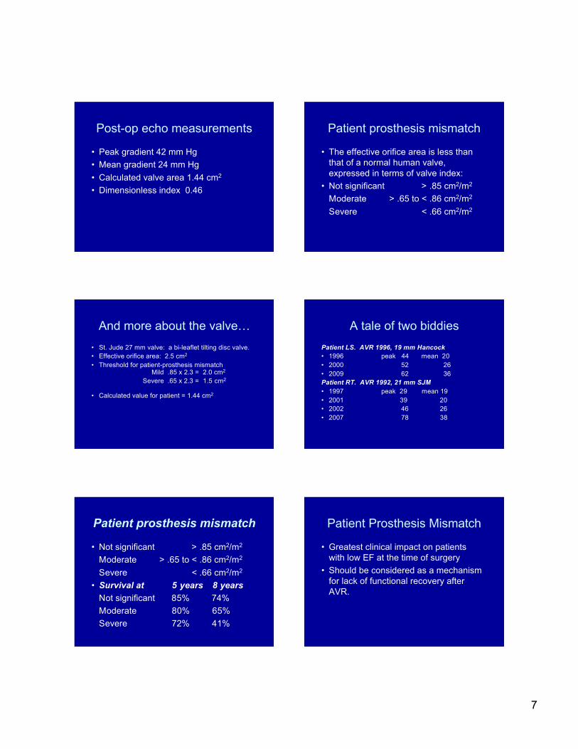
7
Post-op echo measurements
• Peak gradient 42 mm Hg• Mean gradient 24 mm Hg• Calculated valve area 1.44 cm2
• Dimensionless index 0.46
Patient prosthesis mismatch
• The effective orifice area is less thanthat of a normal human valve,expressed in terms of valve index:
• Not significant > .85 cm2/m2
Moderate > .65 to < .86 cm2/m2
Severe < .66 cm2/m2
And more about the valve…• St. Jude 27 mm valve: a bi-leaflet tilting disc valve.• Effective orifice area: 2.5 cm2
• Threshold for patient-prosthesis mismatch Mild .85 x 2.3 = 2.0 cm2
Severe .65 x 2.3 = 1.5 cm2
• Calculated value for patient = 1.44 cm2
A tale of two biddiesPatient LS. AVR 1996, 19 mm Hancock• 1996 peak 44 mean 20• 2000 52 26• 2009 62 36Patient RT. AVR 1992, 21 mm SJM• 1997 peak 29 mean 19• 2001 39 20• 2002 46 26• 2007 78 38
Patient prosthesis mismatch
• Not significant > .85 cm2/m2
Moderate > .65 to < .86 cm2/m2
Severe < .66 cm2/m2
• Survival at 5 years 8 yearsNot significant 85% 74%Moderate 80% 65%Severe 72% 41%
Patient Prosthesis Mismatch
• Greatest clinical impact on patientswith low EF at the time of surgery
• Should be considered as a mechanismfor lack of functional recovery afterAVR.

8
Mitral RegurgitationMoving Beyond The Obsessionof Visual Estimation of Severity
Karthik Ananthasubramaniam, MD FACC FASE FASNCAssociate Professor of Medicine, Wayne State University
Director, Echocardiography and Nuclear CardiologyLaboratory
Heart and Vascular InstituteHenry Ford Hospital, Detroit
[email protected] ©2008 American College of Cardiology Foundation. Restric tions may apply.
Management Strategy for Patients With Chronic Severe Mitral Regurgitation
Is it severe MR or Not?That is the question
Bonow, R. O. et al. J AmColl Cardiol 2008;52:e1-e142
Hemodynamic Determinants of MR Hemodynamic Determinants of MR
√MPG LV-LA pressure gradient :Good news: both change in same direction with altered hemodyanmicsSo the ratio is largely unchanged
Since RVol is related to √MPG( eg : Change in LV-LA MPG of 144 to 100 mm hg
will only change the RV by 20% (√144= 12 to √100 =10 )Nevertheless important to factor this in . Measure BP at time of MR exam
Duration of MR : very important. A common cause of severityoverestimation visually in MVPS given late systolic prediliction of MR
Limitations and Precautions MR Quantification
Timing and duration ofjet is a underappreciatedparameter
Very important to factorin when trying to makesense of numbers whichdon’t make sense
Apical CW signal timingas well a color M-mode inPLAX and PSAX veryuseful for timing MR
Large PISALarge vena contracta? Severe MR
Color Jet of MR : TheObsession
• What is the color ? : It is just spatial distribution ofvelocities of cells
• Since orifice is small there is always a spray effect (finger over thumb hose effects)
• Central jets larger than eccentric jets ( recruitmentphenomenon and Coanda /entrainment effect)
• MR jet are/LA orifice area ; age old method poor Spec• Hypertensive pts with with mild MR may have large jet
area• Acute severe MR patients may have small jet rea• Good rule : central jet area < 4 sq cm and < 10% of
LA area : always mild

9
Eccentric jets Vena Contracta• Smallest highest velocity region of flow jet• Just downstream from reg orifice• Always measure in views perpendicular to leaflet
closure ( ideal PLAX )• LSAX and mitral commissural apical views should
not be used as VC width us elongated and large here• < 0.3 mild and > 0.7 severe• Works for central and eccentric jets• Can be fixed or dynamic depending on pathology• If ERO circular VC works well. But if not VC is
problematic
MR Echo Severity Index• Score of 0-3 based following variables 1. CW doppler MR jet width and penetration 2. Color doppler PISA radius 3. CW characteristics of MR jet ( jet density , envelope , cutoff) 4. TR derived PA pressures 5. PW Pulmonary vein flow pattern 6. LA size by 2DScore < 1.7 =mild MRScore > 1.8 suggest moderate- severe MR. ( overlap territory)Score > 2.2 identified severe MR with sens, spec, PPV of 90, 99 and 79%
Other clues1. E velocity > 1.2 m2. E/A pseuodnormal
Thomas, L,et al. J AmColl Cardiol 1999;
33:2016.
Artifacts
Lynn Cronin, MD FACCWest Michigan Heart

10
• Visualization of objects/structures that do notactually exist– Rib artifact– Reverberation artifact– Mirror artifact
• Degradation of visualization of desiredobjects that do exist– Rib artifact
“Wait, is that a dissection???”
• Reverberation artifact—key points– Reflections of sound wave between two objects
• Most often echo-dense structures perpendicular tothe beam
– Vessel walls– Calcified structures– Prosthetic materials
– The “ghost” object is displayed at a distancethat is a multiple of the distance between theactual object and the transducer.• Motion of the “ghost” object is amplified
“Wait, is that a dissection???”
Notegreateramplitude


![[Cronin] Translation in the Digital Age](https://img.pdfslide.us/doc/110x75/577c78291a28abe0548efcdb/cronin-translation-in-the-digital-age.jpg)