Embed Size (px)
Citation preview

Antitumor Activities of Dammarane Triterpene Saponins from Bacopa monniera
Ling Peng1, Yun Zhou2*, De Yun Kong3 and Wei Dong Zhang4
1Department of Oncology, Jinling Hospital, Medical School of Nanjing University, No. 305 East Zhongshan Road, Nanjing, Jiangsu Province 210002, PR China2Zhejiang Food and Drug Administration, No. 27 Wenbei Lane, Moganshan Road, Hangzhou, Zhejiang Province 310012, PR China3Shanghai Institute of Pharmaceutical Industry, No. 1320 West Beijing Road, Shanghai 200040, PR China4School of Pharmacy, Second Military Medical University, No. 325 Guohe Road, Shanghai 200433, PR China
Bioassay-guided methods were used to test the antitumor activity of methanol extract of the whole plant of Bacopa monniera (L.) Wettst. and four different fractions (petroleum ether, CHCl3, EtOAc, and n-BuOH fractions) of the methanol extract. Among the fi ve crude samples, n-BuOH fraction was noted to have the highest antitumor activity. The dammarane triterpene saponins isolated from n-BuOH fraction, bacopaside É (1) and bacopaside VII (3), had potential antitumor effect. 1 and 3 showed cytotoxicity of all the tested human tumor cell lines MDA-MB-231, SHG-44, HCT-8, A-549 and PC-3M in MTT assay in vitro, and showed 90.52 % and 84.13 % inhibition in mouse implanted with sarcoma S180 in vivo at the concentration of 50 μmol/kg, respectively. The remaining two compounds, bacopaside II (2) and bacopasaponin C (4) were found to be much less potent compared with 1 and 3. 1 and 3 signifi cantly inhibited human breast cancer cell line MDA-MB-231 adhesion, migration and Matrigel invasion in vitro at the concentration of 50 μmol/L. Since no anti-tumor activities about the monomers from Bacopa monniera (L.) Wettst. have been reported, these results indicate that the mechanism of action of 1 and 3 needs further study. Copyright © 2009 John Wiley & Sons, Ltd.
Keywords: Bacopa monniera (L.) Wettst; antitumor activity; dammarane triterpene saponins.
INTRODUCTION
Bacopa monniera (L.) Wettst. has been used in the Ayurvedic system of medicine for centuries. Tradition-ally, it was used as a brain tonic to enhance memory development, learning, and concentration, and to provide relief to patients with anxiety or epileptic dis-orders (Chopra RN, 1958). The plant has also been used in India and Pakistan as a cardiac tonic, a digestive aid, and to improve respiratory function in cases of bron-choconstriction (Nadkarni, 1988). Research on anxiety, epilepsy, bronchitis and asthma, irritable bowel syn-drome, and gastric ulcers also supports the Ayurvedic uses of Bacopa. Standardized extract of B. monniera (Bhattacharya et al., 2000), rich in active constituents referred to as saponins (mainly bacoside A) (Garai et al., 1996), have been developed as a clinical drug for memory and intellect improvement (Bhattacharya et al., 2000). Our study and previous reports have demon-strated that the saponins of B. monniera possess anxio-lytic (Bhattacharya et al., 1998; Zhou et al., 2007) and antioxidant effects (Tripathi et al., 1996; Bhattacharya et al., 2000). Both of these activities have benefi cial effects in treatment of tumor as psychological (Levy et al., 1985; 1987; Andersen et al., 1994) and free radicals factors (Pawar et al., 2001) which have been implicated in the genesis of the tumor. In vitro research previously
demonstrated Bacopa crude extract has cytotoxic activ-ity for sarcoma-180 cells (Elangovan et al., 1995). In order to screen its effective extract fractions and active monomers of antitumor activity, bioassay-guided methods were used on the level of intact animal and molecular pharmacology.
MATERIALS AND METHODS
Plant. Whole plants of Bacopa monniera (L.) Wettst. were collected in Zhangzhou, Fujian Province in July 2002, and authenticated by Prof. Han-Chen Zheng of the Department of Pharmacognosy, School of Phar-macy, Second Military Medical University, Shanghai, China. A voucher specimen (No. 0211-11) was depos-ited in the Herbarium of the School of Pharmacy, Second Military Medical University, Shanghai, China.
Cell lines and animals. The cell lines used for the cyto-toxic activity assay included 5 tumor cell lines originat-ing from human neoplasias, which were human breast cancer cell line MDA-MB-231, human glioma cell line SHG-44, human ileocecal adenocarcinoma cell line HCT-8, human lung adenocarcinoma cell line A-549 and human prostate cancer cell line PC-3M. The cell lines were purchased from Shanghai Medical and Phar-macological Academy (Shanghai, PR China). S180 cells were obtained from Cell Library of Chinese Academy of Sciences and have been maintained in our laboratory for several years as the ascites form by several passages intraperitoneally (i.p.) in balb/c mice. Female balb/c
* Correspondence to: Yun Zhou Food and Drug Administration, No. 27 Wenbei Lane, Moganshan Road, Hangzhou, Zhejiang Province 310012, PR China.E-mail: [email protected]
Received 07 July 2009Revised 16 August 2009
Copyright © 2009 John Wiley & Sons, Ltd. Accepted 01 September 2009
PHYTOTHERAPY RESEARCHPhytother. Res. 24: 864–868 (2010)Published online 3 December 2009 in Wiley InterScience(www.interscience.wiley.com) DOI: 10.1002/ptr.3034
ANTITUMOR ACTIVITIES OF BACOPA MONNIERA 865
Copyright © 2009 John Wiley & Sons, Ltd. Phytother. Res. 24: 864–868 (2010)DOI: 10.1002/ptr
mice, 6–8 weeks old, were purchased from Animal Center of Chinese Academy of Sciences, and housed under specifi c pathogen-free conditions. Animal treat-ment and maintenance were conducted in accordance with the Principle of Laboratory Animal Care. The experiments were carried out under the approval of the Committee of Experimental Animal Administration of the Laboratory.
Bioassay-guided fractionation and isolation. The dried and powdered whole plant of B. monniera (8 kg) was extracted with MeOH (15 L × 3) at room temperature. The MeOH extract (743 g) was partitioned with petro-leum ether (1.5 L × 3), CHCl3 (1.5 L × 3), EtOAc (1.5 L × 3), and n-BuOH (1.5 L × 3), respectively. Since the n-BuOH-soluble fraction exhibited high cytotoxicity against all the tested cell lines, this fraction was investi-gated extensively. The n-BuOH extract (380 g) was sub-mitted to macroporous resin column (10 × 80 cm, 1000 g) chromatography and washed with H2O (5.0 L), 10 % EtOH (10.0 L), 30 % EtOH (5.0 L), 50 % EtOH (10.0 L), 70 % EtOH (10.0 L), and 95 % EtOH (5.0 L) (v/v), respectively. The combined 50% and 70% EtOH eluants afforded a saponin fraction (170 g), which was applied to column chromatography on silica gel (10 × 80 cm, 1000 g) with gradient CHCl3/CH3OH (20 : 1, 3 L; 15 : 1, 2 L; 10 : 1, 2 L; 6 : 1, 2 L; 4 : 1, 2 L; 2 : 1, 2 L; 1 : 1, 2 L) as eluents. All subfractions were repeatedly sub-jected to Sephadex LH-20 (MeOH) and RP silica gel (ODS) column chromatography (H2O/CH3OH, 100 : 0 → 0 : 100) and fi nally purifi ed by semipreparative HPLC (MeOH/H2O, 65 : 35, fl ow rate of 3 mL/min) column chromatography to afford 1 (750 mg, purity 98.8%, mp 259∼261°C, (α)D −35.6°, (MeOH, c 0.2)), 2 (650 mg, purity 98.2%, mp 249∼252°C, (α)D−34.3°, (MeOH, c 0.4)), 3 (490 mg, purity 98.4%, mp 250∼252°C, (α)D −41.5°, (MeOH, c 0.5)), and 4 (510 mg, purity 98.7%, mp 220∼222°C, (α)D −47.5°, (MeOH, c 1.2)).
In vitro cytotoxic activity assay. The cytotoxicity assay was performed according to a methyl thiazole tetrazo-lium (MTT) method as previously described (Carmichael et al., 1987). MTT was purchased from Sigma Chemical Co. (St Louis, MO, USA). Briefl y, the cells were washed twice with phosphate-buffered saline (PBS) and incubated at a density of 5 × 10⁴ cells/mL in fl at-bottomed 96-well microtiter plates in 100 μL of Dulbecco’s modifi ed Eagle’s medium (DMEM) with 10% fetal bovine serum (FBS). Several dilutions of the tested compounds in 100 μL of DMEM with 10% FBS were added to the wells. The fi nal concentration of dimethyl sulfoxide (DMSO) was 0.2% (v/v). After incu-bation for 48 h, 100 μL of medium was removed from each well and 20 μL MTT solution (5 mg/mL PBS) was added. After incubation for 4 h, the optical density was evaluated at 540 and 630 nm. Fluorouracil (5-Fu) was used as positive control. The growth inhibition rate was calculated as percentage of parallel negative con-trols. Each experiment was performed three times.
In vivo tumor growth. Ascites of mice implanted sarcoma S180 were diluted with normal saline in the ratio of 1 : 4. Then 0.2 ml dilution was subcutaneously injected into each mouse. All mice were randomized and divided into 6 groups: blank control, 5-Fu control and four of the testing groups. The blank control group
had 20 mice while the other groups had 10 mice each. The dose of 5-Fu and those of the extract and fractions were 50 μmol/kg. Reagents were infused to stomach once a day for 7 days, from the day after implantation. On the 10th day after implantation, mice were scarifi ed by means of cervical dislocation. Tumors were isolated and weighed to calculate the inhibition ratio.
Cell adhesion, migration and Matrigel invasion assays. The 96-well microtitre plates were coated with 10 μg/ml of fi bronectin, followed by treatment with 1% bovine serum albumin (BSA) for 1 h at 37 °C. Uncoated BSA-blocked wells were used as negative control. To each well, 5 × 10⁴ MDA-MB-231 cells were added in the presence or absence of the tested compounds. After incubation for 2 h at 37 °C, the medium was removed and washed twice with PBS gently. The adherent cells were fi xed in 1 % methanol, stained with 0.5 % crystal violet and lysed with 2 % Triton X-100. The absorbance was measured at OD595 nm. Wells were set up in tripli-cate and all experiments were repeated at least 3 times. The results were calculated as average ± SEM.
Cell migration ability was measured using scratch wound healing assay. Monolayers of MDA-MB-231 cells were cultured to near confl uence (>90%) in 12-well plates in triplicate. Streaks were made on the mono-layer culture with 10-μl pipette tips. Experiment was performed with and without the tested samples. The cell migrations were monitored for 24 h. The migration of cells was determined by the number of cells that crossed into the wound area from their reference point at time zero.
Tumor cell invasion through reconstituted basement membrane (Matrigel) was assayed according to previ-ous method (Bauer et al., 1992). In 24-well Transwell cell culture chambers (Costar), polycarbonate fi lters of 8 μm pore size were precoated with 1 μg of fi bronectin on the lower surface and then 5 μg/10 μl of Matrigel (Sigma, St Louis, MO, USA) was applied to the upper surface of the fi lters. Uncoated wells were used as nega-tive control. The fi lters were dried and washed in phos-phate-buffered saline (PBS). Following rehydration, MDA-MB-231 cells (5 × 10⁵) were suspended in RPMI 1640 containing 0.1% BSA, pretreated with the tested samples for 30 min on ice, added to the upper chamber, and then incubated at 37 °C for 12 h. After 12 h incuba-tion, the cells which invaded to the lower chamber and attached to the lower surface of the fi lter were stained and counted. Four high-power fi elds were counted for each well.
Statistics. The data were analyzed using the unpaired t test with two-tailed P-value or ANOVA (for multiple comparisons) to calculate the statistical signifi cance between control group and treated groups. The results are presented as means ± SEM. Statistically signifi cant differences were defi ned as having a P < 0.05.
RESULTS
Methanol extract was found to exhibit cytotoxicity against all the tested human tumor cell lines at different concentrations. The methanol extract was successively fractionated into petroleum ether, CHCl3, EtOAc and n-BuOH fractions, and the fractions were subjected to
Copyright © 2009 John Wiley & Sons, Ltd. Phytother. Res. 24: 864–868 (2010)DOI: 10.1002/ptr
866 L. PENG ET AL.
bioassay-guided fractionation. The in vitro effect of the crude extract against human tumor cell lines was deter-mined. The extract which showed IC50 values greater than 25 μg/ml for all tumor cells tested were considered to be non-cytotoxic. 5-Fluorouracil, used as positive control, showed IC50 values in the range 0.455∼0.702 μg/ml for the different cell lines, respectively. The IC50 value of the methanol extract to the different tumor cell lines were as follows: 3.31 μg/ml for MDA-MB-231, 4.92 μg/ml for SHG-44, 5.62 μg/ml for HCT-8, 7.82 μg/ml for A-549, and 8.63 μg/ml for PC-3M. The petroleum ether fraction showed little cytotoxic activity even at the highest concentration (>25 μg/ml for all tumor cell lines tested, respectively). CHCl3 fraction was found to be highly sensitive to MDA-MB-231 (9.16 μg/ml), and sensitive to SHG-44 (10.65 μg/ml) and HCT-8(14.45 μg/ml), but not sensitive to A-549 and PC-3M (>25 μg/ml, respectively). EtOAc fraction showed no cytotoxic activity against all the tested cell lines (>25 μg/ml, respectively). The n-BuOH fraction showed high cytotoxicity against all the tested cell lines in vitro
(5.46 μg/ml for MDA-MB-231, 6.13 μg/ml for SHG-44, 5.14 μg/ml for HCT-8, 6.31 μg/ml for A-549, and 6.83 μg/ml for PC-3M, respectively) and 90.18 % inhibition of S180 at the concentration of 50 μmol/kg in vivo, respec-tively. The antitumor activity of n-BuOH fraction was most close to, but not higher than, the activity of metha-nol extract. The cytotoxicity of the methanol extract was concentrated in the n-BuOH fraction. So it is probable that the antitumor constituents should be isolated from the polar fractions in the following research.
The n-BuOH fraction was chromatographed and purifi ed to afford 1–4. Compounds 1–4 were identifi ed as bacopaside I (Chakravarty et al., 2001), bacopaside II (Chakravarty et al., 2001), bacopaside VII (Zhou et al., 2007), and bacopasaponin C (Garai et al., 1996) (Fig. 1) by comparison of their spectral data with those of reported values in the literature.
The compounds were tested for in vitro cytotoxicity against human tumor cell lines. The cytotoxicity (IC50) of the compounds against the tumor cell lines is shown in Table 1. 1 and 3 were the strongest growth inhibitors
1
2
34 5
67
89
10
1112
1314
15
16
17
1819
20
21
2223
28 29
30R1O
O
HO
O
R2
R3
R1 R2 R3
1
O
OO
O
OH
OHHO
OH
OOSO2OH
HOHO OH
24
25
26
27
H
2
O
OO
O
OH
OHHO
OOH
HOHO
OH
HO
OH
H
3
O
O
O
O
OH
OHHO
OOH
HOHO
OH
OH
H24
25
26
27
4
O
O
O
O
OH
OHHO
OOH
HOHO
OH
OH
H
Figure 1. Chemical structures of bacopaside É (1), bacopaside IÉ (2), bacopaside VII (3), and bacopasaponin C (4).

ANTITUMOR ACTIVITIES OF BACOPA MONNIERA 867
Copyright © 2009 John Wiley & Sons, Ltd. Phytother. Res. 24: 864–868 (2010)DOI: 10.1002/ptr
against the tumor cells, 2 and 4 were less potent com-pared with 1 and 3.
The inhibition of the extracts and compounds of S180 is shown in Table 2. 1 and 3 showed 90.52 % and 84.13 % inhibition in mouse implanted with sarcoma S180 in vivo at the dosage of 50 μmol/kg, respectively.
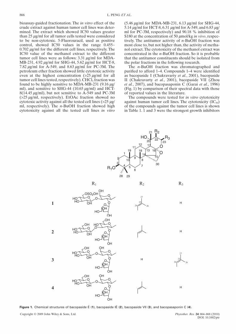
The compounds were then analyzed for their ability to inhibit breast cancer cell adhesion. 1 and 3 signifi -cantly inhibited MDA-MB-231 cell adhesion to fi bro-nectin-coated microtiter plate at the concentration of 50 μmol/L, while 2 and 4 did not have inhibitory effects at the concentration range from 25 to 100 μmol/L (Fig. 2). The effect of the tested samples on tumor cell inva-sion of Matrigel was investigated in a Transwell chamber assay. 1 and 3 signifi cantly suppressed tumor cell inva-sion through Matrigel-coated fi lters at the concentra-tion of 50 μmol/L (Fig. 3). While 2 and 4 showed no signifi cant effects on cell invasion at the concentration range from 25 to 100 μmol/L in this study. Cells exposed to the compounds were analyzed for their rate of migra-tion. In the control group, more cells appeared in the wounded gap, which represented enhanced healing of the wounded area. The cells which crossed the line during 6, 12 and 24 h were taken as an index of wound healing. As shown in Fig. 4, MDA-MB-231 cells that treated with 1 or 3 at the concentration of 50 μmol/L migrated across an artifi cial in vitro wound much less effi ciently than 2 and 4 after 24 h.
Table 1. In vitro cytotoxic activities against human tumor cell lines
Sample
IC50 (μmol/L)
MDA-MB-231 SHG-44 HCT-8 A-549 PC-3M
5-Fu 3.5 3.5 4.7 5.4 4.61 12.3 9.7 11.3 8.9 13.92 32.4 36.9 40.3 44.4 45.43 14.3 15.9 9.8 9.7 10.14 34.9 47.3 58.3 56.4 48.2
BSAPBS 100 50 25 100 50 25 100 50 25 100 50 25
0.0
0.1
0.2
0.3
0.4
0.5
0.6
0.7
0.8
0.9
1.0
1.1
#
##
Abs
orba
nce
(595
nm
)
(μmol/L)(μmol/L) (μmol/L)(μmol/L) 4321
#
Figure 2. Inhibition of cell adhesion in human breast cancer cell line MDA-MB-231 by compound 1 and 3. Statistical analysis of the tested samples at different concentrations (25, 50, 100 μmol/L). A signifi cant decrease of cells that adhered to the microtitre plate coated with fi bronectin was observed of MDA-MB-231 cells treated with 1 and 3 (50 and 100 μmol/L). Uncoated BSA-blocked wells were used as negative control. #: P < 0.05.
BSAPBS 100 50 25 100 50 25 100 50 25 100 50 25
0
20
40
60
80
100
(μmol/L)(μmol/L)(μmol/L)(μmol/L)
#
#
#
432
Cel
ls/F
ield
1
#
Figure 3. Inhibition of cell invasion ability through Matrigel of human breast cancer cell line MDA-MB-231 by compound 1 and 3. Statistical analysis of the tested samples at different concen-trations (25, 50, 100 μmol/L). A signifi cant decrease of cells migrated through Matrigel to the lower chamber of the Tran-swell system was observed of MDA-MB-231 cells treated with 1 and 3 (50 and 100 μmol/L). Uncoated BSA-blocked wells were used as negative control. #: P < 0.05.
0
25
50
75
100
125
150
175
##
#
Cel
ls c
ross
ing
th
e re
fere
nce
po
int/
Fie
ld
6 hrs 12 hrs 24 hrs
#
1 2 3 4 PBS
Figure 4. Inhibition of cell migration of wound healing in human breast cancer cell line MDA-MB-231 by compound 1 and 3. MDA-MB-231 cells were treated with the tested samples (50 μmol/L) at 0 h after creation of a wound by scratch technique and 24 h after subjected to migration. Statistical analysis is provided at 6, 12 and 24 h. A signifi cant decrease in migration of MDA-MB-231 cells treated with 1 and 3 were observed at 12 and 24 h. #: P < 0.05.
DISCUSSION
In the present study, the in vitro and in vivo antitumor activities were studied of the extracts and compounds from B. monniera. In vitro cytotoxic assay was done to

Copyright © 2009 John Wiley & Sons, Ltd. Phytother. Res. 24: 864–868 (2010)DOI: 10.1002/ptr
868 L. PENG ET AL.
screen the extract of B. monniera, and extraction and isolation were performed according to their biological effects. The inhibitory effects of 1 and 3 were shown on human tumor cells of different origin, not specifi c to a single tumor cell line. Among the different cell lines, 1 and 3 showed marked inhibition on human breast cancer cell line MDA-MB-231.
Based on the results of MTT assay in vitro and S180 implanted sarcoma in vivo, the function of 1 and 3 on the cell adhesion, invasion through Matrigel and migration ability of MDA-MB-231 cells in vitro were tested. MDA-MB-231 cell line is a metastatic human breast cancer cell line. Detachment and migration from the primary tumor, and invasion of surrounding blood or lymphatic vessels are critical and complex steps in clinically metastatic diseases.
Scratch wound healing assay was used to determine the effi cacy of the tested samples in inhibiting tumor cell migration. Matrigel invasion assay was used to determine the ability of tumor cells to cross the base-ment membrane. In studies of cell attachment, migra-tion and invasion in vitro, 1 and 3 successfully inhibited these functions, which simulated the in vivo conditions of animals.
Since no antitumor activities about monomers of saponins from B. monniera has been reported, these results indicated that the mechanism of action of 1 and 3 needs further study. Our studies were preliminary evaluation of the extract and compounds, however, these studies provided further clues for the research and development of antitumor activities of extracts and monomers of B. monniera.
Table 2. Inhibition ratio of sarcoma S180 implanted in mouse
SampleDose
(μmol/kg)Animal number
(start/end) Animal weight(g)
(cut of tumor) ± SDTumor weight(g)
± SDInhibition ratio
(%)
Blank – 20/20 28.82 ± 2.36 2.00 ± 0.76 –5-Fu 50 10/10 27.66 ± 1.43 0.18 ± 0.07* 91.121 50 10/10 26.56 ± 4.61 0.19 ± 0.06* 90.522 50 10/10 26.89 ± 4.58 1.68 ± 0.81� 16.273 50 10/10 28.96 ± 2.93 0.32 ± 0.44* 84.134 50 10/10 27.74 ± 3.93 1.29 ± 0.38� 35.51
� P > 0.05; * P < 0.01, compared with the blank control.
Andersen BL, Kiecolt-Glaser JK, and Glaser R. 1994. A biobe-havioral model of cancer stress and disease course. Am Psychol 49(5): 389–404.
Bauer JS, Schreiner CL, Giancotti FG, Ruoslahti E, Juliano RL. 1992. Motility of fi bronectin receptor–defi cient cells on fi bronectin and vitronectin: collaborative interactions among integrins. J Cell Biol 116(2): 477–487.
Bhattacharya SK, Bhattacharya A, Kumar A, Ghosal S. 2000. Antioxidant activity of Bacopa monniera in rat frontal cortex, striatum and hippocampus. Phytother Res 14(3): 174–179.
Bhattacharya SK and Ghosal S. 1998. Anxiolytic activity of a standardized extract of Bacopa Monniera: an experimental study. Phytomedicine 5: 77–82.
Carmichael J, DeGraff WG, Gazdar AF, Minna JD, Mitchell JB. 1987. Evaluation of a tetrazolium-based semiautomated colorimetric assay: assessment of chemosensitivity testing. Cancer Res 47(4): 936–942.
Chakravarty AK, Sarkar T, Masuda K, Shiojima K, Nakane T, Kawahara N. 2001. Bacopaside I and II: two pseudojujubo-genin glycosides from Bacopa monniera. Phytochemistry 58(4): 553–556.
Chopra RN. 1958. Indigenous drugs of India. 2nd ed. UN Dhur and Sons: Calcutta, India; 341.
Elangovan V, Govindasamy S, Ramamoorthy N, Balasubrama-nian K. 1995. In vitro studies on the anticancer activity of Bacopa monniera. Fitoterapia 66: 211–215.
REFERENCES
Garai S, Mahato SB, Ohtani K, Yamasaki K. 1996. Dammarane-type triterpenoid saponins from Bacopa monniera. Phyto-chemistry 42(3): 815–820.
Levy S, Herberman R, Lippman M, d'Angelo T. 1987. Correlation of stress factors with sustained depression of natural killer cell activity and predicted prognosis in patients with breast cancer. J Clin Oncol 5(3): 348–353.
Levy SM, Herberman RB, Maluish AM, Schlien B, Lippman M. 1985. Prognostic risk assessment in primary breast cancer by behavioral and immunological parameters. Health Psychol 4(2): 99–113.
Nadkarni KM. 1988. The Indian Materia Medica. South Asia Books: Columbia, MO; 624–625.
Pawar R, Gopalakrishnan C, Bhutani KK. 2001. Dammarane tri-terpene saponin from Bacopa monniera as the superoxide inhibitor in polymorphonuclear cells. Planta Med 67(8): 752–754.
Tripathi YB, Chaurasia S, Tripathi E, Upadhyay A, Dubey GP. 1996. Bacopa monniera Linn. as an antioxidant: mechanism of action. Indian J Exp Biol 34(6): 523–526.
Zhou Y, Shen YH, Zhang C, Su J, Liu RH, Zhang WD. 2007. Triterpene saponins from Bacopa monnieri and their anti-depressant effects in two mice models. J Nat Prod 70(4): 652–655.