Embed Size (px)
Citation preview
Antiphospholipid Syndrome (APS) and
Anticardiolipin Syndrome (ACL)
Robert I. Fox, M.D., Ph.D.Scripps/XiMED Medical Center
Scripps Memorial Hospital and Research Foundation La Jolla (San Diego), USA
[email protected]://www.robertfoxmd.com
Learning Objectives
At the end of this talk, participants will be able to:
1. Define research diagnostic criteria for APS.2. List 3 laboratory abnormalities of APS3. List treatment interventions for APS during
pregnancy and after pregnancy.4. List treatment interventions for Catastrophic
Cardiolipin Syndrome (CPS).
Anti-Phospholipid Syndrome (APS)
• Also known as “Hughe’s Syndrome,” after Graham Hughes (a Rheumatologist in London)
• Autoimmune hypercoagulable state• Caused by anti-phospholipid antibodies
(particularly IgG anti-B2 glycoprotein-I)• Provokes blood clots in both arteries and veins
APS Pregnancy Related Complications
• Miscarriage• Stillbirth• Fetal growth disruption and pre-term delivery• Placenta integrity deterioration• Pre-eclampsia and eclampsia
Anti-Cardiolipin Antibodies (ACA)
• Cardiolipin is an important component of the inner mitrochondrial membrane (almost 20% of mitochondrial lipids.
• Anti-cardiolipin antibodies (ACA) are antibodies often found in several diseases, including syphilis, SLE, Behcet’s, TBC.
• False positive RPR suggests ACA.• In clinical use, terms APS and ACL are
interchangeable.
Lupus Anticoagulant Studies
• A positive test for LA is a clotting assay (LA test) that demonstrates effects of these antibodies on the phospholipid-dependent factors in the coagulation cascade.
• The most common screening tests employed as the first step of a LA test are the aPTT, and the dilute Russell Viper Venom Time (dRVVT).
• A prolonged screening test alone is not adequate for LA positivity.
• Confirmatory steps with mixing studies (in order to rule out factor deficiencies).
.
Anti-Phospholipid Syndrome (APS)
• Usually refers to anti-phospholipid, anti-cardiolipin, or lupus anti-coagulant.
• Coagulation more common when associated with other coagulopathies (Factor V-L, Factor II mutations), hyper-homocysteine, associated SLE, or metabolic factors such as hypertension or diabetes.
Take Home Lesson-1
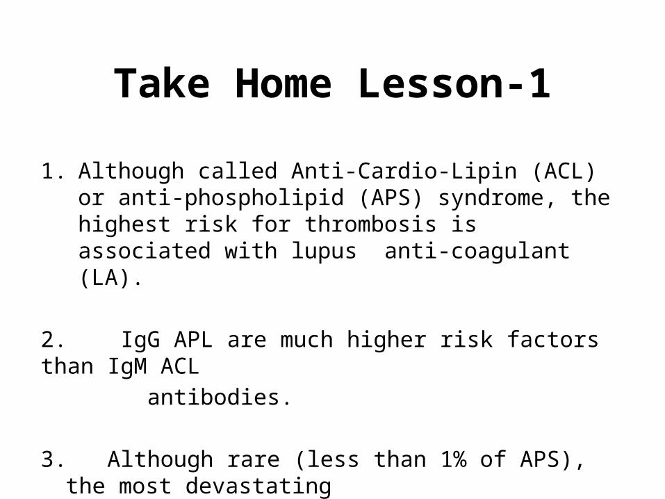
1. Although called Anti-Cardio-Lipin (ACL) or anti-phospholipid (APS) syndrome, the highest risk for thrombosis is associated with lupus anti-coagulant (LA).
2. IgG APL are much higher risk factors than IgM ACL antibodies.
3. Although rare (less than 1% of APS), the most devastating manifestation is catastrophic anti-cardiolipin syndrome
(multi-organ thrombosis).
Take Home Lesson-2
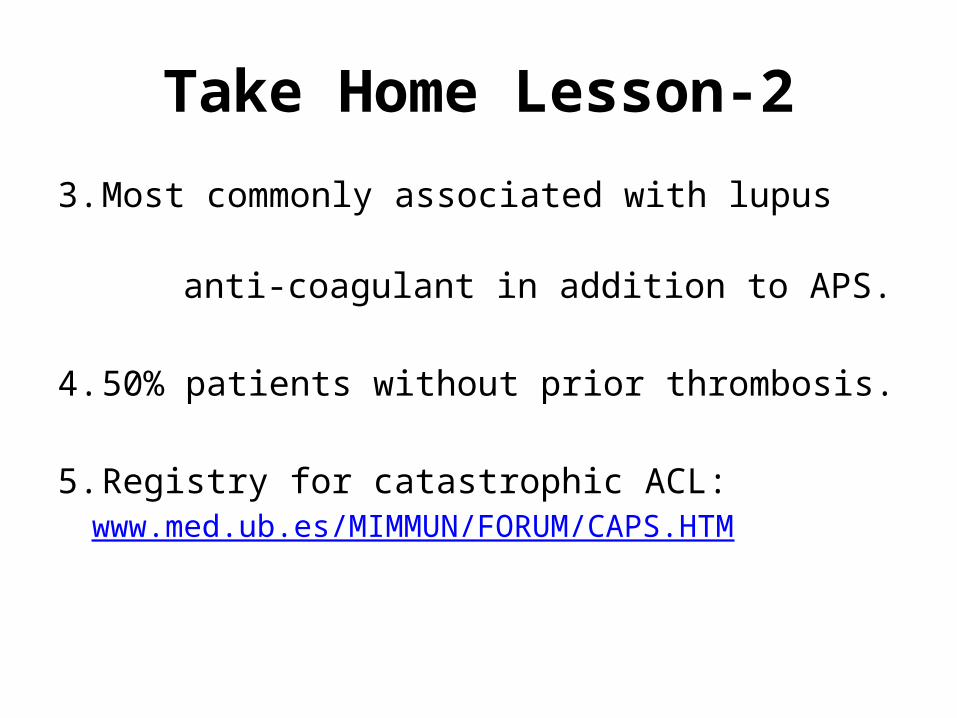
3. Most commonly associated with lupus anti-coagulant in addition to APS.
4. 50% patients without prior thrombosis.
5. Registry for catastrophic ACL:
www.med.ub.es/MIMMUN/FORUM/CAPS.HTM
Take Home Lesson-3
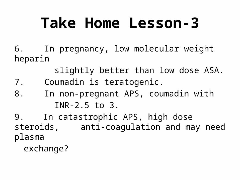
6. In pregnancy, low molecular weight heparin slightly better than low dose ASA.7. Coumadin is teratogenic.8. In non-pregnant APS, coumadin with INR-2.5 to 3.9. In catastrophic APS, high dose steroids,
anti-coagulation and may need plasma exchange?

History
• Antiphospholipid syndrome was described in full in the 1980s, after various previous reports of specific antibodies in people with systemic lupus erythematosus and thrombosis.
• Recognition of primary and secondary APS.• Standardization of antibody assays and
criteria.
Research Criteria
• Originally referred to in 1999 as the Sapporo criteria.
• Subsequently modified at a workshop conducted in Sydney in 2006.
(The next slide addresses your first “learning objective.”)
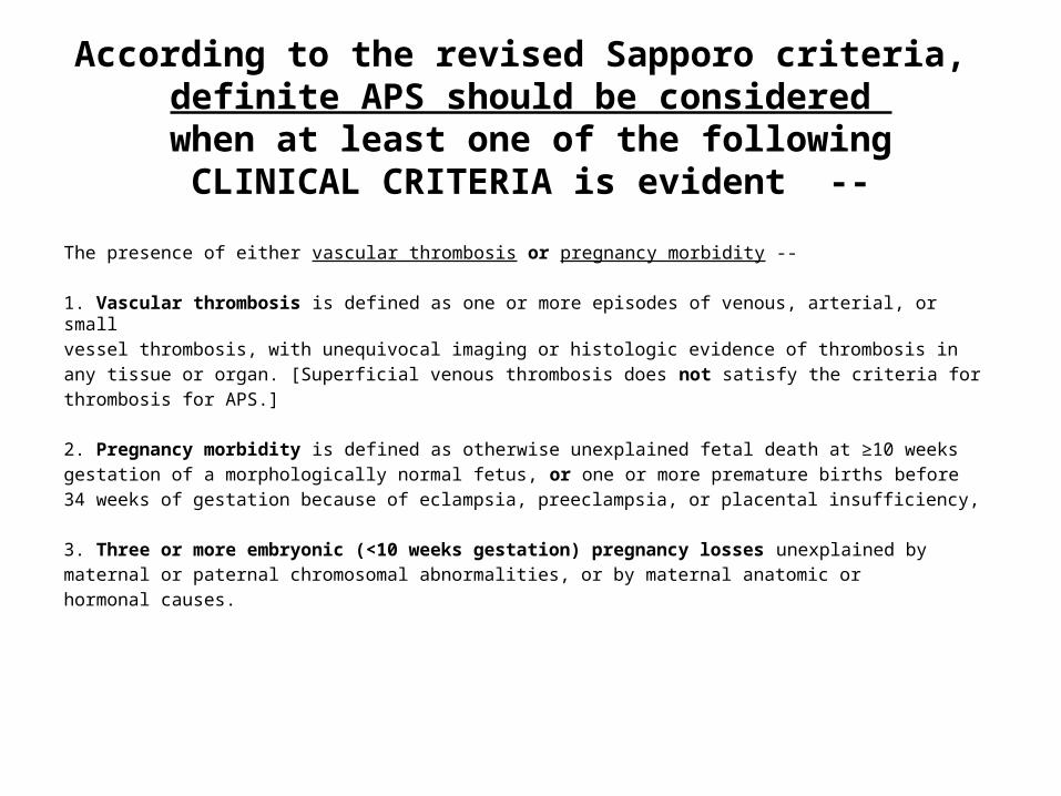
According to the revised Sapporo criteria, definite APS should be considered when at least one of the following
CLINICAL CRITERIA is evident --
The presence of either vascular thrombosis or pregnancy morbidity --
1. Vascular thrombosis is defined as one or more episodes of venous, arterial, or smallvessel thrombosis, with unequivocal imaging or histologic evidence of thrombosis inany tissue or organ. [Superficial venous thrombosis does not satisfy the criteria forthrombosis for APS.]
2. Pregnancy morbidity is defined as otherwise unexplained fetal death at ≥10 weeksgestation of a morphologically normal fetus, or one or more premature births before34 weeks of gestation because of eclampsia, preeclampsia, or placental insufficiency,
3. Three or more embryonic (<10 weeks gestation) pregnancy losses unexplained bymaternal or paternal chromosomal abnormalities, or by maternal anatomic orhormonal causes.
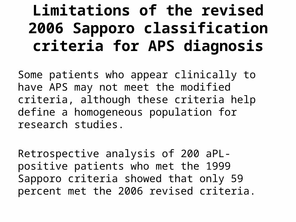
Limitations of the revised 2006 Sapporo classification criteria for APS diagnosis
Some patients who appear clinically to have APS may not meet the modified criteria, although these criteria help define a homogeneous population for research studies.
Retrospective analysis of 200 aPL-positive patients who met the 1999 Sapporo criteria showed that only 59 percent met the 2006 revised criteria.
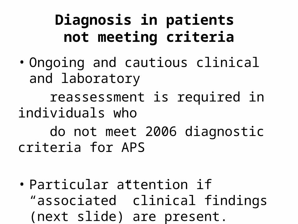
Diagnosis in patients not meeting criteria
• Ongoing and cautious clinical and laboratory reassessment is required in individuals who do not meet 2006 diagnostic criteria for APS
• Particular attention if “associated” clinical findings (next slide) are present.
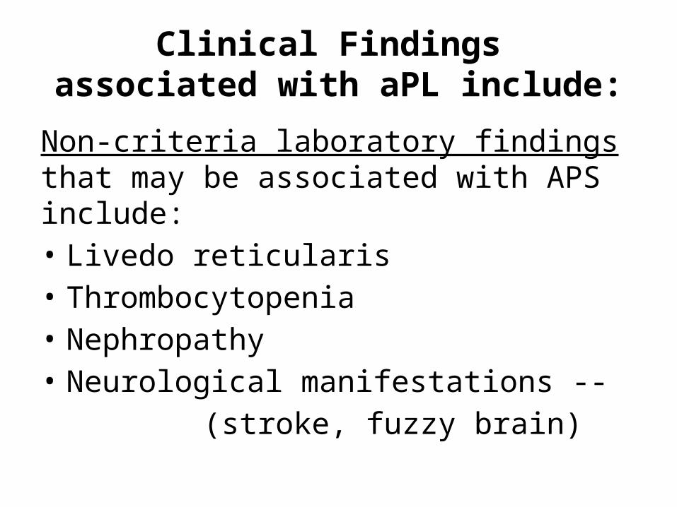
Clinical Findings associated with aPL include:
Non-criteria laboratory findings that may be associated with APS include:• Livedo reticularis• Thrombocytopenia• Nephropathy• Neurological manifestations -- (stroke, fuzzy brain)
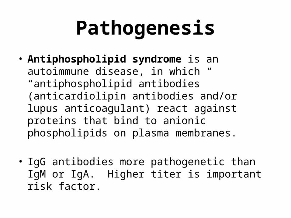
Pathogenesis• Antiphospholipid syndrome is an autoimmune
disease, in which “antiphospholipid antibodies” (anticardiolipin antibodies and/or lupus anticoagulant) react against proteins that bind to anionic phospholipids on plasma membranes.
• IgG antibodies more pathogenetic than IgM or IgA. Higher titer is important risk factor.
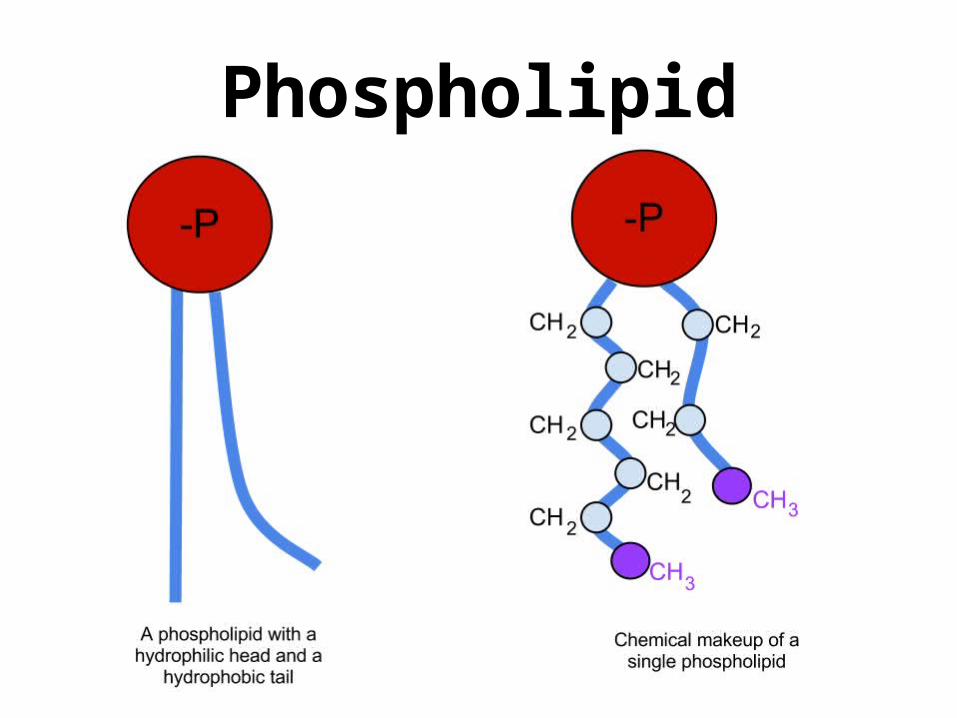
Phospholipid
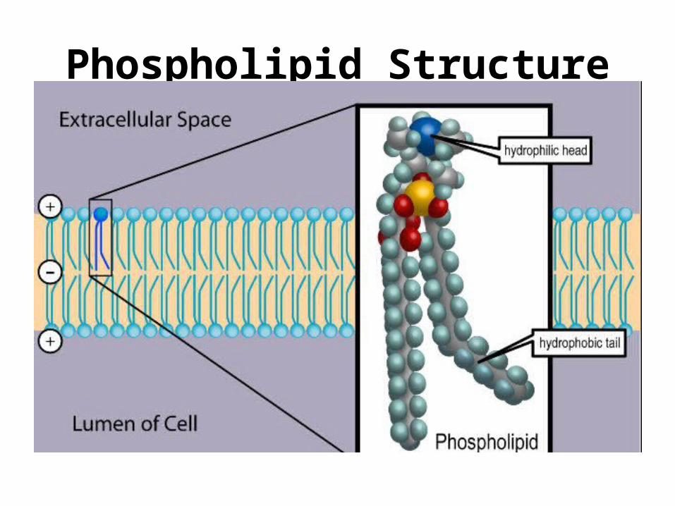
Phospholipid Structure
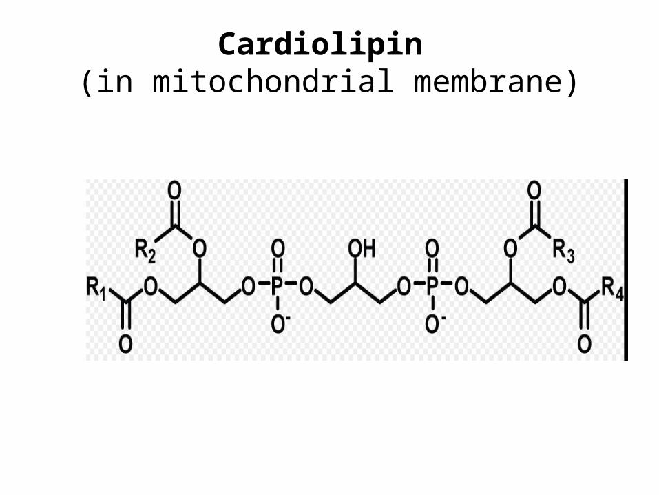
Cardiolipin (in mitochondrial membrane)
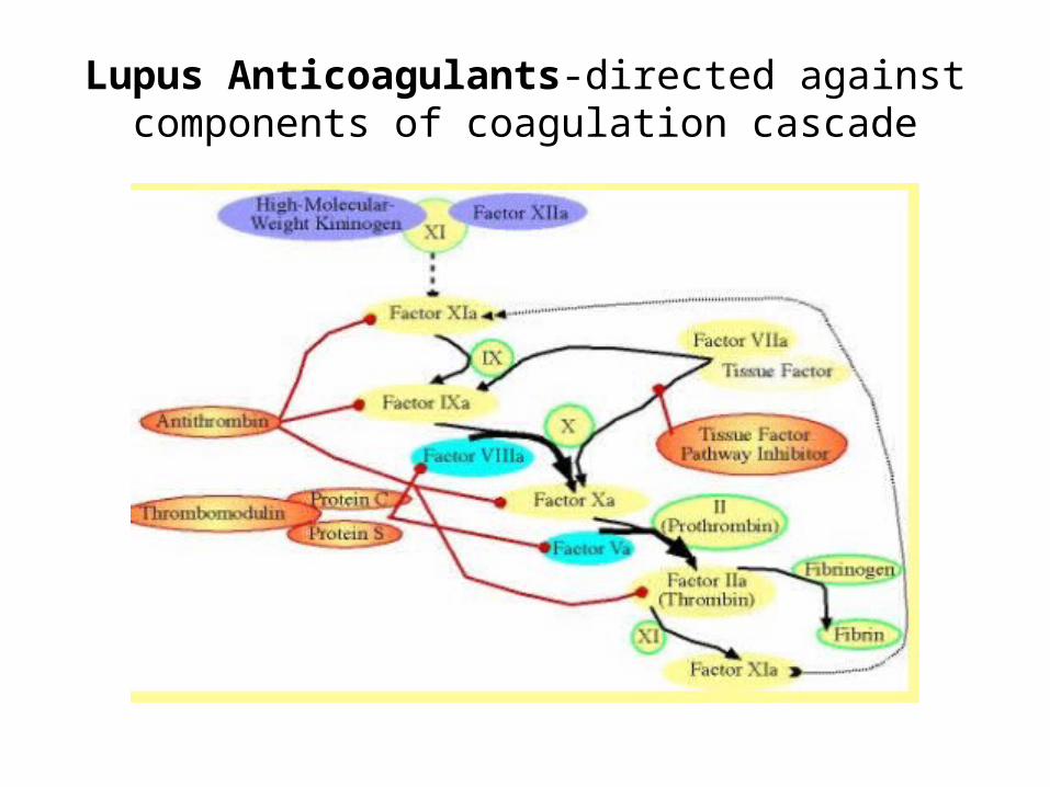
Lupus Anticoagulants-directed against components of coagulation cascade
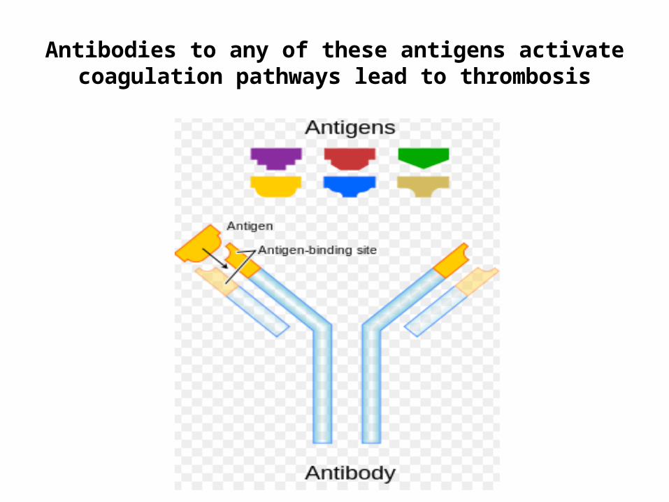
Antibodies to any of these antigens activate coagulation pathways lead to thrombosis
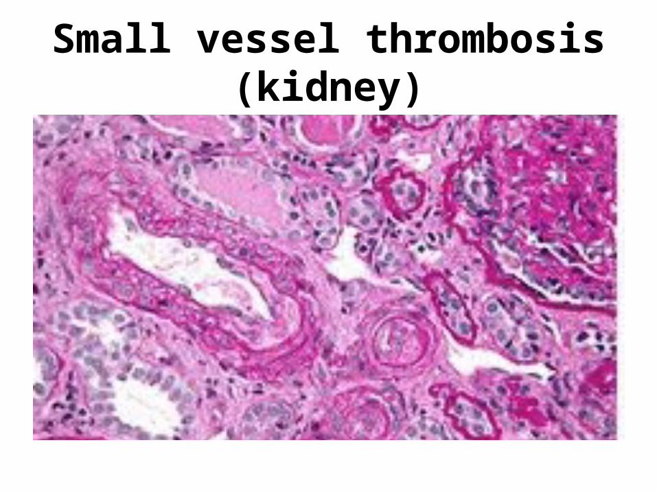
Small vessel thrombosis (kidney)
Clinical Presentations
There are no pathognomonic physical findings of APS; however, look for:• Ischemia livedo reticularis (and particularly
livedo racemosa)• Digital ischemia or gangrene, • Deep venous thrombosis (DVT), • Neurological lesions consistent with a stroke.

Livido Reticularis
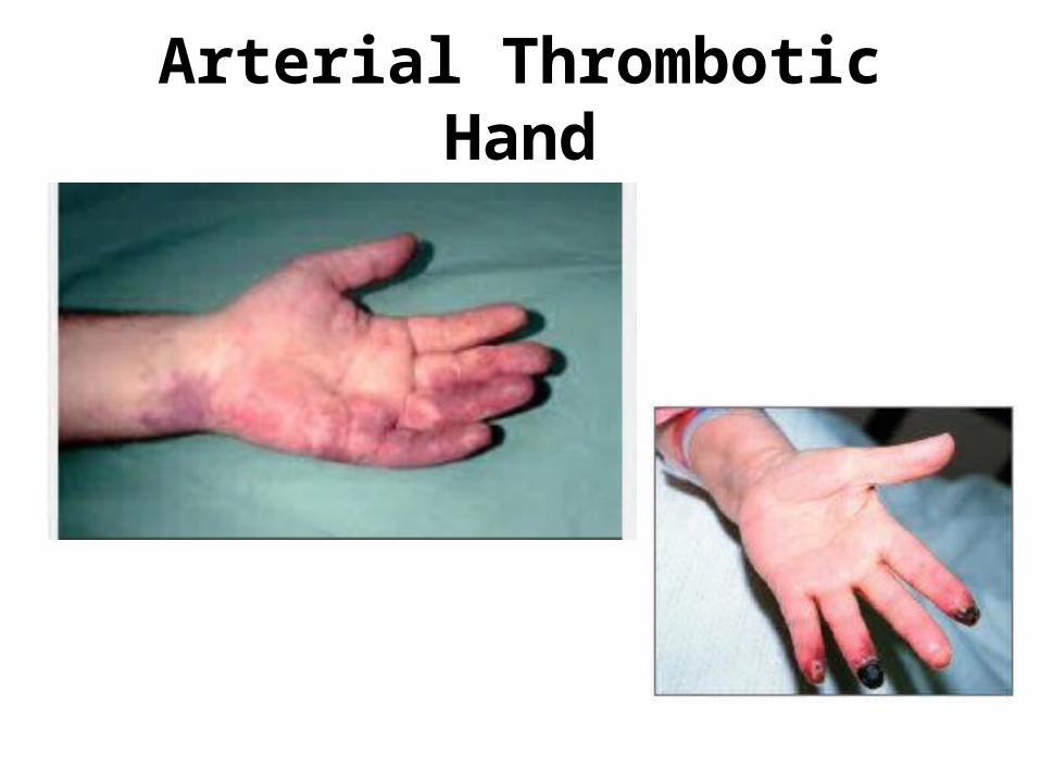
Arterial Thrombotic Hand

Arterial Thrombotic Foot
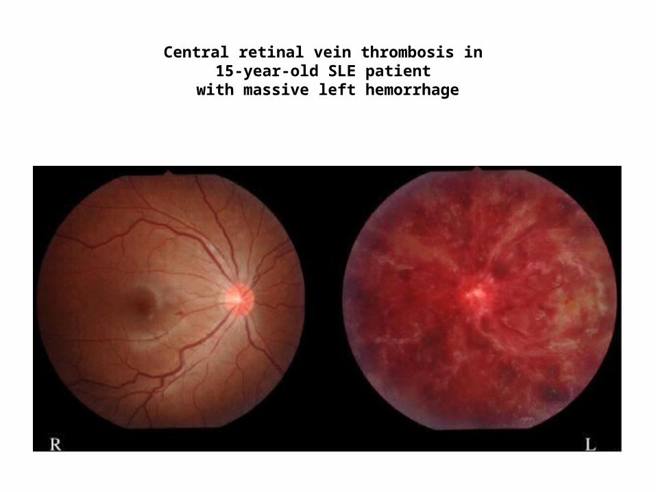
Central retinal vein thrombosis in 15-year-old SLE patient
with massive left hemorrhage
Diagnostic Evaluation-1
History to look for:
– One or more specific adverse outcomes related to pregnancy
– Criteria for SLE or Sjogren’s Syndrome– Detailed obstetric history– Post-operative thromboses
Initial Laboratory Testing
• IgG and IgM anticardiolipin antibodies (aCL) by enzyme-linked immunosorbent assay (ELISA).
• IgG and IgM anti-beta2-glycoprotein (GP)-I antibodies by ELISA.
• Lupus Anticoagulant testing (dilute Russell viper venom time [dRVVT] and activated partial thromboplastin
• Confirmation by by the 1:1 mixing study
Additional Laboratory Testing
Confirmatory aPL testing – In patients with initial positive testing for aPL: • The test should be repeated after at least
12 weeks to confirm persistence of the aCL, anti-beta2-GPI, or LA test.
• Confirmatory testing is required to satisfy the laboratory criteria for APS.
Follow-Up Lab Evaluation-1
• Otherwise unexplained thrombocytopenia or prolongation of a test of blood coagulation
(e.g., activated partial thromboplastin time [aPTT])
If unexplained PT or PTT: Has a 1:1 mixing with normal sera been performed?
Follow Up Lab Testing-2
Alternatively…
Some experts prefer to perform testing for anti-beta2-GPI antibodies only in patients suspected of APS in whom the IgG and IgM aCL and lupus anticoagulant (LA) testing are negative.
Limitations: Transiently elevated levels of IgG or IgM aCL,
can occur in otherwise normal individuals and in the setting of viral or other infections.
• In one study of 522 randomly selected normal blood donors, the prevalence of IgG and IgM aCL on a first test was 6.5 and 9.4 percent, respectively.
• However, repeatedly positive tests, were present in only 22 and 14 percent of those with an initial positive test (i.e., 1.4 and 1.3 percent of the total population), after nine months.
In patients with a strongly suggestive clinical history…
If initial test result are positivebut whose second test is negative:
• Perform a third test after several weeks, and use the third result to help guide decision making.
• Testing should be repeated if the patient has a clinical event.
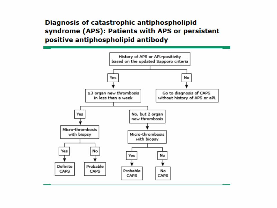
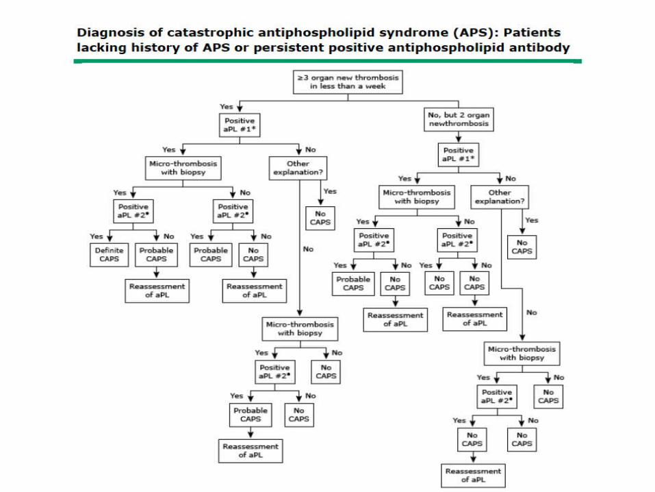
Catastrophic APS
A very small subset (1-2%) of patients with APS has widespread thrombotic disease with multiorgan failure, termed "catastrophic APS---– Three or more new organ thromboses within a
week.– Biopsy confirmation of a microthrombus.– Exclusion of other causes of multiple organ
thromboses or microthromboses* [* algorithm listed in appendix]
Clinical Manifestations of Catastrophic APS-1
[Also known as Asherson’s Syndrome or CAPS]• Peripheral thrombosis may be encountered affecting veins and
arteries. • Intra-abdominal thrombosis may lead to pain.• Cardiovascular, nervous, heart, renal and pulmonary system
complications are common. • The patient may exhibit skin purpura and necrosis. • Cerebral manifestations may lead to encephalopathy and
seizures. • Myocardial infarctions may occur. • Strokes may occur due to the arterial clotting involvement. • Adrenal involvement may lead to Addisonian crisis.

Catastrophic APS-2
• Almost half of the patients who develop Catastrophic APS have not had a prior history of thrombosis.
• Thus, a high level of suspicion and testing for aPL are necessary in these clinical settings.
• Algorithms help to distinguish these conditions.
Treatment of CAPS
Specific therapy includes: Specific therapy includes use of: • intravenous heparin and corticosteroids • possibly plasma exchanges and intravenous
immunoglobulin.• Additional steps may have to be taken to
manage circulatory problems, renal failure, adrenal failure and respiratory distress.
Differential Diagnosis
The differential diagnosis of the APS is broad, and includes venous or arterial thrombotic events or pregnancy morbidity due to other causes:• Abnormal lab tests due to drugs or infection. • Additionally, aPL may coexist with other
conditions including clotting factor deficiency.• A synergistic effect on the development of
clinical events.
Differential of Arterial Thrombosis
• Heparin induced thrombocytopenia• Defective clot lysis due to dysfibrinogenemia or
plasminogen activator deficiency• Homocysteinemia• Myeloproliferative disorders, polycythemia vera (P vera), or
paroxysmal nocturnal Hemoglobinuria• Hyperviscosity due to P-vera, Waldenstrom's
macroglobulinemia, sickle cell disease• Systemic vasculitis, such as those associated with
antineutrophil cytoplasmic antibodies• Paradoxical embolism
Differential Diagnosis
• Venous thrombosis — In patients with venous thrombosis or pulmonary embolic disease
• Factor Deficiencies (V-Leyden, Factor II, Protein C/S)
These conditions can be distinguished from aPL by the lack of initial positive testing for aPL or failure to confirm positive APS results after 12 weeks.
Differential of Thrombotic Angiopathies*
• TTP-deficiency of Adam13• HUS- reaction to shigella toxin or E. Coli• HELLP syndrome (of hemolysis, elevated liver
enzymes, and low platelets with pregnancy)• Sepsis with multiorgan failure and
disseminated intravascular coagulation (DIC) [* listed in appendix]
Treatment APS-1
In pregnancy:
• Low-molecular heparin and low-dose aspirin are used instead of warfarin because of warfarin’s teratogenicity.
• Women with recurrent miscarriage are often advised to take aspirin, and to start low-molecular weight heparin treatment after missing a menstrual cycle.
Treatment of APS-2
• In patients with recurrent DVT, prophylactic treatment with Coumadin is to maintain the patient’s INR between 2.0 and 3.0.
• Duration of Coumadin is usually 6-12 months if single uncomplicated DVT.
• If associated pulmonary emboli, then usually maintain anti-coagulation for life.
Treatment of APS-3
• Anticoagulation appears to prevent miscarriage in pregnant women.
• In refractory cases of pregnancy-related effects (eclampsia, thromocytopenia), high-dose steroids and plasmapheresis may be used.
Rituximab (Anti-CD20) and APS
• Refractory APS or catastrophic ACS patients have received steroids plus rituximab (1000 mg IV) and have normalized bleeding and thrombosis, as well as APS antibody titers*.
• Double-blind studies have not been reported.
*Lupus October, 2011 20(10):1106-1108*Lupus July 2013 22: 865-867
SUMMARY AND RECOMMENDATIONS
Clinical suspicion for Anti-Phospholipid Syndrome (APS) should be raised in the presence of the following:
• Occurrence of one or more otherwise unexplained thrombotic or thromboembolic events;
• One or more specific adverse outcomes related to pregnancy;
• Otherwise unexplained thrombocytopenia or prolongation of a test of blood coagulation (e.g., prothrombin time [PT] or activated partial thromboplastin time
• [aPTT])
Learning Objective-1[Define Research Diagnostic Criteria for APS]
1. Vascular thrombosis is defined as one or more episodes of venous, arterial, or smallvessel thrombosis, with unequivocal imaging or histologic evidence of thrombosis inany tissue or organ. Superficial venous thrombosis does not satisfy the criteria forthrombosis for APS.
2. Pregnancy morbidity is defined as otherwise unexplained fetal death at ≥10 weeksgestation of a morphologically normal fetus, or one or more premature births before34 weeks of gestation because of eclampsia, preeclampsia, or placental insufficiency,
3. Three or more embryonic (<10 weeks gestation) pregnancy losses unexplained bymaternal or paternal chromosomal abnormalities or by maternal anatomic orhormonal causes.
(Learning Objective 2)List 3 laboratory abnormalities of APS
• IgG and IgM anticardiolipin antibodies (aCL) by enzyme-linked immunosorbent assay (ELISA).
• IgG and IgM anti-beta2-glycoprotein (GP)-I antibodies by ELISA.
• Lupus Anticoagulant testing (dilute Russell viper venom time [dRVVT] and activated partial thromboplastin
• Confirmation by by the 1:1 mixing study

(Learning Objective 3)List treatment interventions for APS during pregnancy and after pregnancy.
Low-molecular heparin and low-dose aspirin are used instead of warfarin because of warfarin’s teratogenicity.
Women with recurrent miscarriage are often advised to take aspirin, and to start low-molecular weight heparin treatment after missing a menstrual cycle.
Anticoagulation appears to prevent miscarriage in pregnant women.
In refractory cases of pregnancy-related effects (eclampsia, thromocytopenia), high-dose steroids and plasmapheresis may be used.
(Learning Objective 4)
List treatment interventions for Catastrophic Cardiolipin Syndrome (CPS).
Specific therapy includes use of: • intravenous heparin and corticosteroids • possibly plasma exchanges and intravenous
immunoglobulin.• Additional steps may have to be taken to manage
circulatory problems, renal failure, adrenal failure and respiratory distress.

Sunset over San Diego at Salk Institute ---Thank you for inviting me---
References-1
• Pengo V, Tripodi A, Reber G, et al. Update of the guidelines for lupus anticoagulant detection. Standardisation Committee of the International Society on Thrombosis and Haemostasis. J Thromb Haemost 2009; 7:1737.
• Pierangeli SS, de Groot PG, Dlott J, et al. 'Criteria' aPL tests: report of a task force. Lupus 2011; 20:182.
References-2
• Vila P, Hernández MC, López-Fernández MF, Batlle J. Prevalence, follow-up and clinical significance of the anticardiolipin antibodies in normal subjects. Thromb Haemost 1994;72:209.
• Kaul M, Erkan D, Sammaritano L, Lockshin MD. Assessment of the 2006 revised antiphospholipid syndrome classification criteria. Ann Rheum Dis 2007; 66:927