Embed Size (px)
Citation preview
g, Antioxidant for Fats and Oils from Canary Seed: Sterol and Triterpene Alcohol Esters of Caffeic Acid
T. TAKAGI and T. I IDA, Dept. of Chemistry, Faculty of Fisheries, Hokkaido University, Hakodate, Japan
ABSTRACT Several kinds of seeds used as bird feed were extracted successively with hexane, ether and methanol. In the antioxidant test with ex- tracts, the ether extract from canary seeds showed the highest activ- ity. The antioxidant fraction separated from it by thin layer chro- matography showed excellent activity for lard and sardine oil. The effective components were identified by gas chromatography and gas chromatography-mass spectrometry of the hydrolyzed prod- uets as the esters of caffeic acid with cycloartenol, gramisterol, sitosterol and campesterol with the minor amounts of 24-methyl- enecyr obtusifoliol, brassieasterol and A7-stigmastenol.
INTRODUCTION
Caffeic acid and its esters have been found in a wide variety of plant materials and their excellent ant ioxidant activity is well known (1). However, most of them are water-soluble substances which have low solubility in fats and oils and have a bi t ter taste. These properties have inhibited their use as antioxidants by direct addit ion to edible oils.
In this study we found a lipid-soluble ant ioxidant which consists of sterol and tri terpene alcohol esters of caffeic acid in canary seeds (Pbalaris canariensis, Gramineae). The seeds were products of Australia and possibly contain small amounts of P. tuberosa and P. arundinaccea.
Lipid-soluble caffeic acid esters have been found in oats (2-5) and in Sopbora subprostrata Chun and T. Chen roots (6), Leguminosae (Chinese drug: San Dou Gen). In oats, 24 phenolic ant ioxidant components have been detected by thin layer chromatography (TLC). Eight of them were identified as the mixed esters of caffeic and ferulic acid with fat ty alcohols, a, c0-fatty diols and co-hydroxy fat ty acids with 26 and 28 carbons. From the S. subprostrata, C20-C26 (mainly C22) fat ty alcohol esters of caffeic acid have been isolated. The ant ioxidant from oats is presumed to have lower activity, since it contains ferulic acid esters which are less effective than caffeic acid esters (4). San Dou Gen is a special material for medical use and contains the caffeic acid esters at a lower level. Canary seeds are available in large quantity, since they are a popular bird feed. Another advantage of the ant ioxidant from canary seeds over the aliphatic esters of caffeic acid is higher melting point, which facilitates solvent removal at higher temperatures and refining by recrystallization. Based on subjective testing the ant ioxidant does not have appre- ciable bitterness. Consequently, the canary seed extract has potential as an ant ioxidant for edible oils.
Sterol and tri terpene alcohol esters of caffeic acid were found in the natural products for the first t ime in this study, although the occurrence of the corresponding esters of ferulic acid (methyl ether of caffeic acid at 3-OH) in vegetable oils and related materials has been reported in many papers (7).
MATERIALS AND METHODS
Antioxidant Test
The ant ioxidant test was performed by the oven test using lard and sardine oil. A glass tube (50 ml, 3 cm id) with a
flat bo t tom containing 3 -+ 0.01 g oil and 3 or 1.5 • 0.01 mg extract was placed into an incubator kept at 60 -+ 1 C. The lard, which included no antioxidant , was obtained from a lard refining plant. The sardine oil, obtained from an industrial plant, was used after refining by column chro- matography with silicic acid as packing material and n- hexane as eluent. The periodical change of the peroxide value (POV) was measured using colorimetric iodine method (8).
Effects of ant ioxidants were indicated by the protect ion factor (PF n) described elsewhere (9) and shown by the following equation: PF n = Tn/T n. In this equation, Tn is the t ime for POV of fats to attain n and Tn is the Tn in the control test. Values of PF n are obtained from the curve of the periodical change of POV. At the ends of the induction periods, POV values were measured at intervals of ca. 1 0 4 0 hr.
Thin Layer Chromatography
In all TLC, Silica Gel G plates (20 x 20 cm) were used, and the thicknesses of the layers for analytical and preparative TLC were 0.25 and 0.5 mm, respectively. In the analytical TLC, hexane/ether/acet ic acid (80:20:1, v/v/v) was used in development. All spots were visualized after spraying with 50% sulfuric acid with subsequent charring at 100 C. In preparative TLC, development was carried out using the solvents shown in Figure 1 and the bands were detected under ultraviolet (UV) light after spraying with Rhodamine 6G (0.1% ethanol solution). Each band was scraped and extracted with ether.
Spectroscopy
UV spectra were obtained with a Hitachi 124 spect rophoto- meter (Hitachi Seisakusho Co. Ltd., Tokyo) , infrared (IR) spectra were run in KBr pellets on a Nippon Bunk6K6gyo Co. Ltd., Tokyo) . The 100-MHz pro ton magnetic resonance (PMR) spectra were recorded with Nippon Denshi FX-100 (Nippon Denshi Co. Ltd., Tokyo) in CDC13 with Si(CH3)4 as a reference marker.
Gas Liquid Chromatography (GLC) and Gas Chromato- graphy-Mass Spectrometry (GC-MS)
GLC was done with a Shimadzu GC 6AMPF with a flame ionization de tec tor and an integrator Shimadzu E1A (Shi- madzu Seisakusho Co. Ltd., Kyoto) . GLC of sterols and tri- terpene alcohols were run with a 1.5 m x 3 mm glass column packed with 2% OV-17 on 80/100 mesh Chromo- sorb W at column temperature 270 C. GLC of the tr imeth- ylsi lylated (TMS) product from the phenolic acid, the frac- tion C in Figure 1, was done under the same condit ions except the column temperature (190 C) after conversion to TMS derivatives with the reagent, dry pyr id ine /hexamethyl - disi lazane/tr imethylchlorosilane (9: 3:1, v/v/v) (10). G LC of methyl esters of the fa t ty acid from the hexane extract of canary seeds and sardine oil were run with a 2 m x 3 mm glass column packed with 3% SILAR 10 C on 100/120 mesh Gas Chrom Q at a column temperature of 190 C, with 10% diethyleneglycol succinate polyester on 80/100
326 / JAOCS October 1980
CAFFEIC ACID ESTER ANTIOXIDANTS
mesh Chromosorb W at a co lumn tempera tu re o f 170 C, respectively. In every run, the carrier gas was ni t rogen.
GC-MS was done with a Hitachi M60 ins t rument coupled to a c o m p u t e r Hitachi 002B. The GLC in GC-MS of sterols and t r i terpene alcohols and the TMS p roduc t of C were per- fo rmed on a 2 m x 3 m m glass co lumn packed with 2% OV-17 on 80 /100 mesh Gas Chrom (2. The co lumn temper- ature was 250 C for the sterols and t r i te rpene alcohols and 200 C for the TMS produc t of C. The spectra were taken at an ionizing voltage of 20eV; carrier gas was hel ium.
RESULTS AND DISCUSSION
Antioxidant Activity of the Extracts
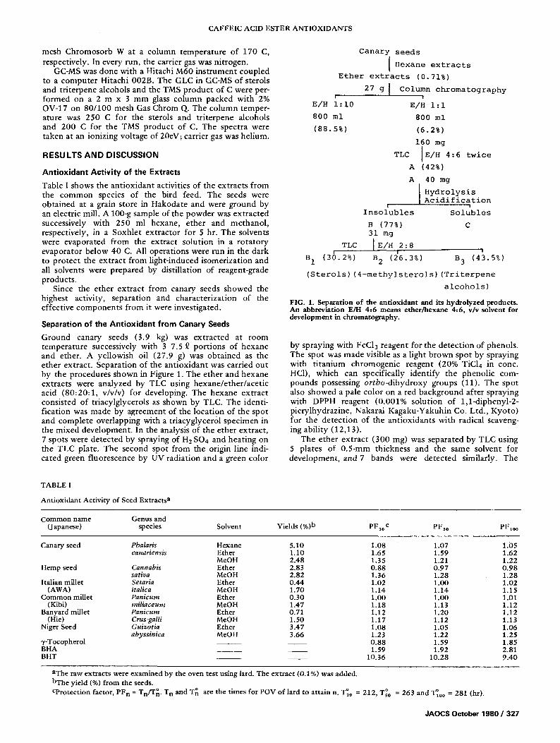
Table I shows the an t iox idan t activities of the ext rac ts f rom the c o m m o n species of the bird feed. The seeds were obta ined at a grain store in Hakodate and were ground by an electric mill. A 100-g sample of the powder was ex t rac ted successively with 250 ml hexane, e ther and methano l , respectively, in a Soxh le t ex t rac tor for 5 hr. The solvents were evapora ted f rom the extract solut ion in a ro ta to ry evapora tor be low 40 C. All operat ions were run in the dark to p ro tec t the ex t rac t f rom l ight- induced isomer iza t ion and all solvents were prepared by disti l lation of reagent-grade products.
Since the ether ex t rac t f rom canary seeds showed the highest activity, separat ion and charac ter iza t ion of the effect ive componen t s f rom it were investigated.
Separation of the Antioxidant from Canary Seeds
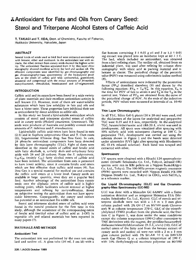
Ground canary seeds (3.9 kg) was ex t rac ted at room tempera ture successively with 3 7.5 ~ por t ions of hexane and ether. A yel lowish oil (27.9 g) was obta ined as the ether extract . Separat ion of the an t iox idan t was carried ou t by the procedures shown in Figure 1. The e ther and hexane extracts were analyzed by TLC using hexane /e the r / ace t i c acid (80 :20 :1 , v/v/v) for developing. The hexane ex t rac t consisted of tr iacylglycerols as shown by TLC. The identi- f icat ion was made by agreement of the locat ion of the spot and comple te overlapping with a triacyglycer01 spec imen in the mixed deve lopment . In the analysis of the ether extract , 7 spots were de tec ted by spraying of H2 SO4 and heat ing on the TLC plate. The second spot f rom the origin line indi- cated green f luorescence by UV radiat ion and a green color
Canary seeds
I H e x a n e e x t r a c t s
Ether extracts (0.71%)
g I Column chromatography 27 I I
E/H i:i0 E/H i:I
800 ml 800 ml
(88.5%) (6.2%)
160 mg |
TLC IE/H 4:6 twice I
A (42%)
A 40 mg
B 1
I Hydrolysis Acidification
I I
Insolubles Solubles
B (77%) 31 mg
TLC I E/H 2 : 8 '1
(36.2%) B 2 (2'6.3%) B 3 (43.5%)
(Sterols) (4-methylsterols) (Triterpene
alcohols)
FIG. 1. Separation of the antioxidant and its hydrolyzed products. An abbreviation E/H 4.-6 means ether/hexane 4..6, v/v solvent for development in chromatography.
by spraying with FeC13 reagent for the de tec t ion of phenols. The spot was made visible as a light b rown spot by spraying with t i t an ium chromogen ic reagent (20% TIC14 in conc. He1), which can specifically ident i fy the phenol ic com- pounds possessing or tbo-d ihydroxy groups (11). The spot also showed a pale color on a red background af ter spraying with DPPH reagent (0.001% solut ion of 1,1-diphenyl-2- picrylhydrazine, Nakarai Kagaku-Yakuhin Co. Ltd. , Kyo to ) for the de tec t ion of the an t ioxidants wi th radical scaveng- ing abi l i ty (12,13).
The ether ex t rac t (300 mg) was separated by TLC using 5 plates of 0.5-ram thickness and the same solvent for deve lopment , and 7 bands were de tec ted similarly. The
TABLE I
Antioxidant Activity of Seed Extracts a
Common name Genus and (Japanese) species Solvent Yields (%)b PF2o c PFs0 PFlo ~
Canary seed Pbalaris Hexane 5.10 1.08 1.07 1.05 canariensis Ether 1.10 1.65 1.59 1.62
MeOH 2.48 1.35 1.21 1.22 Hemp seed Cannabis Ether 2.83 0.88 0.97 0.98
sativa MeOH 2.82 1.36 1.28 1.28 Italian millet Setaria Ether 0.44 1.02 1.00 1.02
(AWA) italica Me OH 1.70 1.14 1.14 1.15 Common millet Panicum Ether 0.30 1.00 1.00 1.01
(Kibi) miliaceum MeOH 1.47 1.18 1.13 1.12 Banyard millet Panicu m Ether 0.71 1.12 1.20 1.12
(Hie) Crus-galli MeOH 1.50 1.17 1.12 1.13 Niger Seed Guizotia Ether 3.47 1.08 1.05 1.06
abyssinica MeOH 3.66 1.23 1.22 1.25 7-Tocopherol 0.88 1.59 1.85 BHA 1.59 1.92 2.81 BHT 10.36 10.28 9.40
aThe raw extracts were examined by the oven test using lard. The extract (0.1%) was added. bThe yield (%) from the seeds.
o o Cprotection factor, PF n = Tn/T g. T n and T~ are the times for POV of lard to attain n. T2o = 212, Tso = 263 and T~,0o = 281 (hr).
JAOCS October 1980 / 327
T. TAKAGI AND T. IIDA
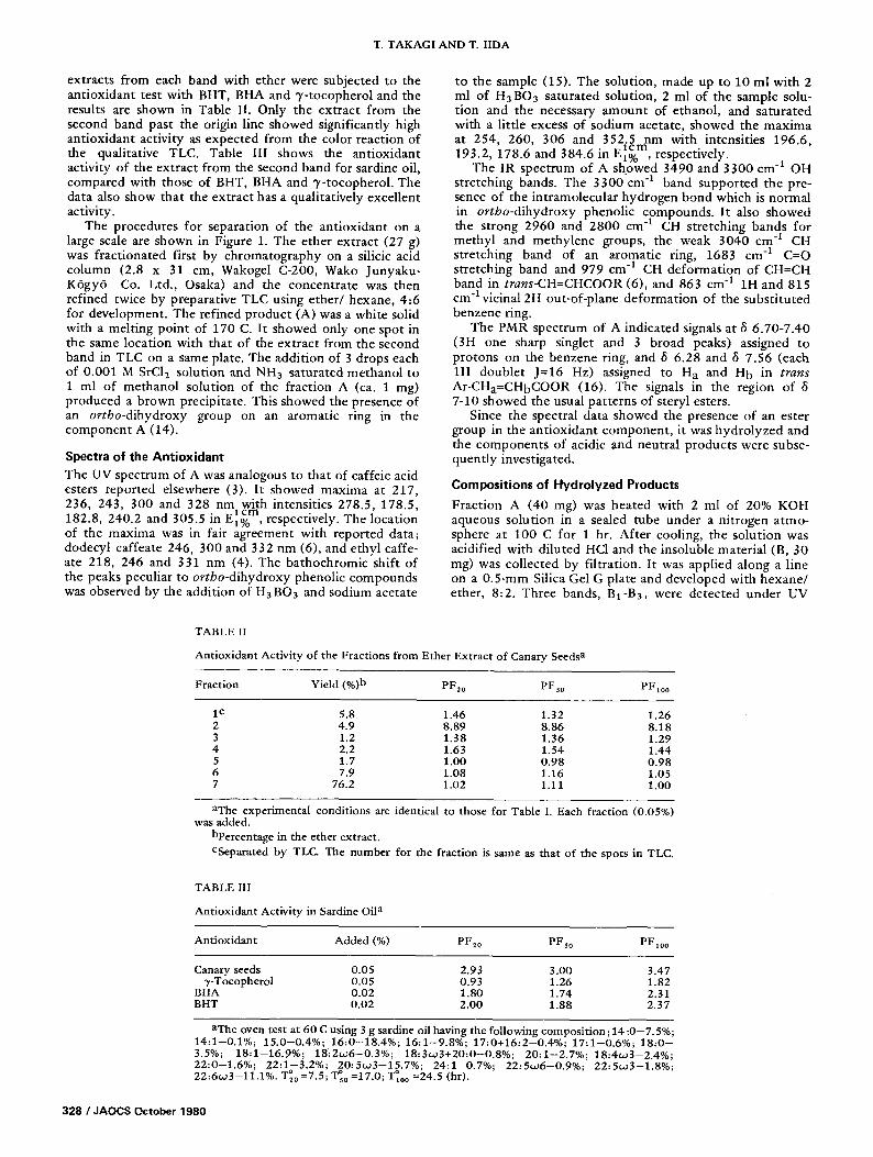
extracts from each band with ether were subjected to the antioxidant test with BHT, BHA and 3'-tocopherol and the results are shown in Table I1. Only the extract from the second band past the origin line showed significantly high antioxidant activity as expected from the color reaction of the qualitative TLC. Table III shows the antioxidant activity of the extract from the second band for sardine oil, compared with those of BHT, BHA and 7-tocopherol. The data also show that the extract has a qualitatively excellent activity.
The procedures for separation of the antioxidant on a large scale are shown in Figure 1. The ether extract (27 g) was fractionated first by chromatography on a silicic acid column (2.8 x 31 cm, Wakogel C-200, Wako Junyaku- K6gy6 Co. Ltd., Osaka) and the concentrate was then refined twice by preparative TLC using e the r /hexane , 4:6 for development. The refined product (A) was a white solid with a melting point of 170 C. It showed only one spot in the same location with that of the extract from the second band in TLC on a same plate. The addition of 3 drops each of 0.001 M SrC12 solution and NH3 saturated methanol to 1 ml of methanol solution of the fraction A (ca. 1 mg) produced a brown precipitate. This showed the presence of an ortbo-dihydroxy group on an aromatic ring in the component A (14).
Spectra of the Antioxidant The UV spectrum of A was analogous to that of caffeic acid esters reported elsewhere (3). It showed maxima at 217, 236, 243, 300 and 328 nm with intensities 278.5, 178.5,
h i c m 182.8, 240.2 and 305.5 in v~1% , respectively. The location of the maxima was in fair agreement with reported data; dodecyl caffeate 246, 300 and 332 nm (6), and ethyl caffe- ate 218, 246 and 331 nm (4). The bathochromic shift of the peaks peculiar to ortbo-dihydroxy phenolic compounds was observed by the addition of H3 BO3 and sodium acetate
to the sample (15). The solution, made up to 10 ml with 2 ml of H3BO 3 saturated solution, 2 ml of the sample solu- tion and the necessary amount of ethanol, and saturated with a little excess of sodium acetate, showed the maxima at 254, 260, 306 and 352.5 nm with intensities 196.6,
. l c m 193.2, 178.6 and 384.6 m E1% , respectwely.
The IR spectrum of A showed 3490 and 3300 cm -1 OH stretching bands. The 3300"cm "1 band supported the pre- sence of the intramolecular hydrogen bond which is normal in ortbo-dihydroxy phenolic compounds. It also showed the strong 2960 and 2800 cm -l CH stretching bands for methyl and methylene groups, the weak 3040 cm -I CH stretching band of an aromatic ring, 1683 cm -1 C=O stretching band and 979 cm -1 CH deformation of CH=CH band in trans-CH=CHCOOR (6), and 863 cm "1 1H and 815 cm "1 vicinal 2H out-of-plane deformation of the substituted benzene ring.
The PMR spectrum of A indicated signals at 8 6.70-7.40 (3H one sharp singlet and 3 broad peaks) assigned to protons on the benzene ring, and 8 6.28 and 8 7.56 (each 1H doublet J=16 Hz) assigned to H a and H b in trans Ar-CHa=CHbCOOR (16). The signals in the region of 8 7-10 showed the usual patterns of steryl esters.
Since the spectral data showed the presence of an ester group in the antioxidant component , it was hydrolyzed and the components of acidic and neutral products were subse- quently investigated.
Compositions of Hydrolyzed Products
Fraction A (40 mg) was heated with 2 ml of 20% KOH aqueous solution in a sealed tube under a nitrogen atmo- sphere at 100 C for 1 hr. After cooling, the solution was acidified with diluted He1 and the insoluble material (B, 30 mg) was collected by filtration. It was applied along a line on a 0.5-ram Silica Gel G plate and developed with hexane/ ether, 8:2. Three bands, BI-B3, were detected under UV
TABLE 1I
Antioxidant Activity of the Fractions from Ether Extract of Canary Seeds a
Fraction Yield (%)b PF20 PFso PFl0 ~
1 c 5.8 1.46 1.32 1.26 2 4.9 8.89 8.86 8.18 3 1.2 1.38 1.36 1.29 4 2.2 1.63 1.54 1.44 5 1.7 1.00 0.98 0.98 6 7.9 1.08 1.16 1.05 7 76.2 1.02 1.11 1.00
aThe experimental conditions are identical to those for Table I. Each fraction (0.05%) was added.
bpercentage in the ether extract. CSeparated by TLC The number for the fraction is same as that of the spots in TLC.
TABLE III
Antioxidant Activity in Sardine Oil a
Antioxidant Added (%) PF20 PFs0 PF1o 0
Canary seeds 0.05 2.93 3.00 3.47 ~,-Tocopherol 0.05 0.93 1.26 1.82
BHA 0.02 1.80 1.74 2.31 BHT 0.02 2.00 1.88 2.37
aThe oven test at 60 C using 3 g sardine oil having the following composition ; 14:0-7.5%; 14:1-0.1%; 15.0-0.4%; 16:0-18.4%; 16:1-9.8%; 17:0+16:2-0.4%; 17:1-0.6%; 18:0- 3.5%; 18:1-16.9%; 18:2to6-0.3%; 18:3to3+20:0-0.8%; 20:1-2.7%; 18:4w3-2.4%; 22:0-1.6%; 22:1-3.2%; 20:5to3-15.7%; 24:1-0.7%; 22:5w6-0.9%; 22:5w3-1.8%;
o o o 22:6to3-11.1%. T20 =7.5; Ts0 =17.0; T100 =24.5 (hr).
328 / JAOCS October 1980
CAFFEIC ACID ESTER ANTIOXIDANTS
light after spraying with Rhodamine 6G solution, and were then extracted with ether. The extracts from each were identified as sterol (B 1 ), 4-methylsterol (B2) and tr i terpene alcohol (B3) fractions, respectively, by comparing their location on a TLC plate with those of reference specimens obtained from the unsaponifiables of wheat germ oil.
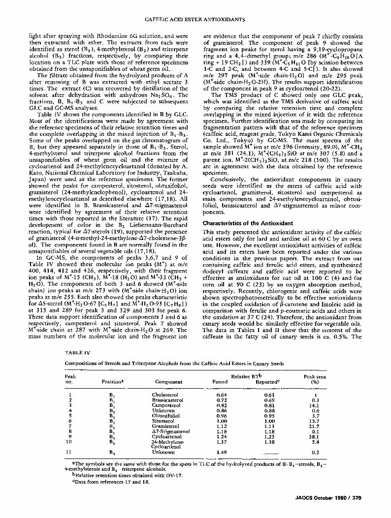
The fil trate obtained from the hydrolyzed products of A after removing of B was extracted with ethyl acetate 3 times. The extract (C) was recovered by distillation of the solvent after dehydrat ion with anhydrous Na2SO4. The fractions, B, B1-B3 and C were subjected to subsequent GLC and GC-MS analyses.
Table IV shows the components identif ied in B by GLC. Most of the identifications were made by agreement with the reference specimens of their relative retent ion times and the complete overlapping in the mixed injection of Ba-B3. Some of the peaks overlapped on the gas chromatogram of B, but they appeared separately in those of B1-B3. Sterol, 4-methylsterol and tri terpene alcohol fractions from the unsaponifiables of wheat germ oil and the mixture of cycloartenol and 24-methylenecycloartanol (donated by A. Kato, National Chemical Laboratory for Industry, Tsukuba, Japan) were used as the reference specimens. The former showed the peaks for campesterol, sitosterol, obtusifoliol, gramisterol (24-methylenelophenol) , cycloartenol and 24- methylenecycloar tanol as described elsewhere (17,18). All were identified in B. Brassicasterol and A7-stigmastenol were identified by agreement of their relative retention times with those reported in the l i terature (17). The rapid development of color in the B2 Liebermann-Burchard reaction, typical for A7-sterols (19), supported the presence of gramisterol (4-a-methyl-24-methylene-A7-cholestene-3/3- ol). The components found in B are normally found in the unsaponifiables of several vegetable oils (17,18).
In GC-MS, the components of peaks 3,6,7 and 9 of Table IV showed their molecular ion peaks (M +) at m/e 400, 414, 412 and 426, respectively, with their fragment ion peaks of M+-15 (CH3), M§ (H20) and M+-33 (CH3 + H20). The components of both 3 and 6 showed (M§ chain) ion peaks at m/e 273 with (M§ chain-H20) ion peaks at m/e 255. Each also showed the peaks characteristic for A5-sterol (M+-H20-67 [CsH7] and M+-H2 O-93 [C7H9] ) at 315 and 289 for peak 3 and 329 and 303 for peak 6. These data support identification of components 3 and 6 as respectively, campesterol and sitosterol. Peak 7 showed M+-side chain at 287 with M+-side chain-H20 at 269. The mass numbers of the molecular ion and the fragment ion
are evidence that the component of peak 7 chiefly consists of gramisterol. The component of peak 9 showed the fragment ion peaks for sterol having a 9,19-cyclopropane ring and a 4 ,4 -d imethy l group; m/e 286 (M§ 0 [A ring + 19 CH2 ] ) and 339 (M+-Cs H n O [by scission between 1-C and 2-C, and between 4-C and 5-C]). It also showed m/e 297 peak (M+-side chain-H20) and m/e 295 peak (M+-side chain-H20-2H). The results support identif icat ion of the component in peak 9 as cycloartenol (20-22).
The TMS product of C showed only one GLC peak, which was identified as the TMS derivative of caffeic acid by comparing the relative retent ion time and complete overlapping in the mixed injection of it with the reference specimen. Fur ther identification was made by comparing its fragmentation pat tern with that of the reference specimen (caffeic acid, reagent grade, Tokyo Kasei Organic Chemicals Co. Ltd., Tokyo) by GC-MS. The mass spectra of the sample showed M + ion at m/e 396 (intensity, 89.0), M+-CH3 at m/e 381 (24.1), M+-(CH3)aSiO at m/e 307 (5.8) and a parent ion, M+-2(CH3)aSiO, at m/e 218 (100). The results are in agreement with the data obtained by the reference specimen.
Conclusively, the ant ioxidant components in canary seeds were identif ied as the esters of caffeic acid with cycloartenol, gramisterol, sitosterol and campesterol as main components and 24-methylenecycloartanol , obtusi- foliol, brassicasterol and A7-stigmasternol as minor com- ponents.
Character ist ics o f the A n t i o x i d a n t
This study presented the ant ioxidant activity of the caffeic acid esters only for lard and sardine oil at 60 C by an oven test. However, the excellent ant ioxidant activities of caffeic acid and its esters have been reported under the various condit ions in the previous papers. The extract from oat containing caffeic and ferulic acid esters, and synthesized dodecyl caffeate and caffeic acid were repor ted to be effective as ant ioxidants for oat oil at 100 C (4) and for corn oil at 90 C (23) by an oxygen absorption method, respectively. Recently, chlorogenic and caffeic acids were shown spectrophotometr ical ly to be effective ant ioxidants in the coupled oxidat ion of/3-carotene and linoleic acid in comparison with ferulic and p-coumaric acids and others in the oxidat ion at 37 C (24). Therefore, the ant ioxidant from canary seeds would be similarly effective for vegetable oils. The data in Tables I and II show that the content of the caffeate in the fat ty oil of canary seeds is ca. 0.5%. The
TABLE IV
Compositions of Sterols and Triterpene Alcohols from the Caffeic Acid Esters in Canary Seeds
Peak Relative RT b Peak area no. Fraction a Component Found Reported c (%)
1 B 1 Cholesterol 0.64 0.61 t 2 B t Brassicasterol 0.72 0.69 O. 3 3 B t Campesterol 0.82 0.81 14.1 4 B 2 Unknown 0.86 0.88 0.6 5 B 2 Obtusifoliol 0.96 0.95 3.7 6 BI Sitosterol 1.00 1.00 15.7 7 B2 Gramisterol 1.12 1.13 21.7 8 Bx A 7-Stigmastenol 1.18 1.18 0.1 9 B a Cycloartenol 1.24 1.23 38.1
10 B a 24-Methylene- 1.37 1.38 5.4 Cycloartenol
11 B a Unknown 1.49 0.3
aThe symbols are the same with those for the spots in TLC of the hydrolyzed products of B: Bx--sterols , 4-methylsterols and B 3 --triterpene alcohols.
bRelative retention times obtained with OV-17. CData from references 17 and 18.
B 2 --
JAOCS October 1980 / 329
T. TAKAGI AND T. IIDA
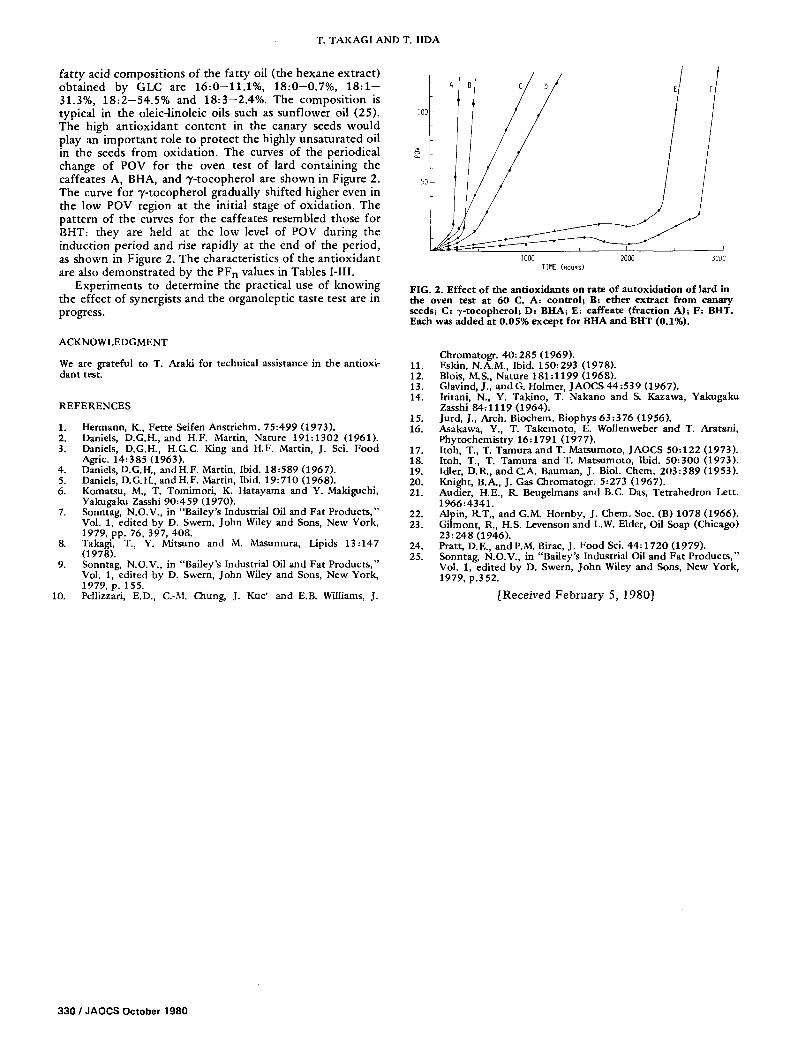
fa t ty acid compos i t ions of the fat ty oil ( the hexane ext rac t ) obta ined by GLC are 1 6 : 0 - 1 1 . 1 % , 1 8 : 0 - 0 . 7 % , 18:1-- 31.3%, 1 8 : 2 - 5 4 . 5 % and 1 8 : 3 - 2 . 4 % . The compos i t ion is typical in the oleic-linoleic oils such as sunf lower oil (25). The high an t iox idan t con ten t in the canary seeds would play an impor t an t role to p ro tec t the highly unsa tura ted oil in the seeds f rom oxidat ion. The curves of the periodical change of POV for the oven test of lard conta in ing the caffeates A, BHA, and 7- tocophero l are shown in Figure 2. The curve for 7 - tocophero l gradually shifted higher even in the low POV region at the initial stage of oxidat ion . The pat tern of the curves for the caffeates resembled those for BHT: they are held at the low level of POV during the induct ion per iod and rise rapidly at the end of the per iod, as shown in Figure 2. The characterist ics of the an t iox idan t are also demons t ra ted by the PF n values in Tables I-III.
Exper iments to de te rmine the practical use of knowing the effect of synergists and the organolept ic taste test are in progress.
ACKNOWLEDGMENT
We are grateful to T. Araki for technical assistance in the antioxi- dant test.
REFERENCES
1. Hermann, K., Fette Seifen Anstrichm. 75:499 (1973). 2. Daniels, D.G.H., and H.F. Martin, Nature 191:1302 (1961). 3. Daniels, D.G.H., H.G.C. King and H.F. Martin, J. Sci. Food
Agric. 14:385 (1963). 4. Daniels, D.G.H., and H.F. Martin, Ibid. 18:589 (1967). 5. Daniels, D.G.H., and H.F. Martin, Ibid. 19:710 (1968). 6. Komatsu, M., T. Tomimori, K. Hatayama and Y. Makiguchi,
Yakugaku Zasshi 90:459 (1970). 7. Sonntag, N.O.V., in "Bailey's Industrial Oil and Fat Products,"
Vol. 1, edited by D. Swern, John Wiley and Sons, New York, 1979, pp. 76, 397, 408.
8. Takagi, T., Y. Mitsuno and M. Masumura, Lipids 13:147 (1978).
9. Sonntag, N.O.V., in "Bailey's Industrial Oil and Fat Products," Vol. 1, edited by D. Swern, John Wiley and Sons, New York, 1979, p. 155.
10. Pellizzari, E.D., C.-M. Chung, J. Kuc' and E.B. Williams, J.
A B C D
I00
> 2
50 !
1000 2000 TIME (HOURS}
5000
FIG. 2. Effect o f the antloxidants on rate of autoxidat lon o f lard in the oven test at 60 C. A: control; B: ether extract from canary seeds; C: -r-tocopherol; D: BHA; E: caffeate (fraction A); F: BHT. Each was added at 0.05% except for BHA and BHT (0.1%).
Chromatogr. 40:285 (1969). 11. Eskin, N.A.M., Ibid. 150:293 (1978). 12. Blois, M.S., Nature 181:1199 (1968). 13. Glavind, J., and G. Holmer, JAOCS 44:539 (1967). 14. Iritani, N., Y. Takino, T. Nakano and S. Kazawa, Yakugaku
Zasshi 84:1119 (1964). 15. Jurd, J., Arch. Biochem. Biophys 63:376 (1956). 16. Asakawa, Y., T. Takemoto, E. Wollenweber and T. Aratani,
Phytochemistry 16:1791 (1977). 17. Itoh, T., T. Tamura and T. Matsumoto, JAOCS 50:122 (1973). 18. Itoh, T., T. Tamura and T. Matsumoto, Ibid. 50:300 (1973). 19. Idler, D.R., and C.A. Banman, J. Biol. Chem. 203:389 (1953). 20. Knight, B.A., J. Gas Chromatogr. 5:273 (1967). 21. Audier, H.E., R. Beugelmans and B.C. Das, Tetrahedron Lett.
1966:4341. 22. Alpin, R.T., and G.M. Hornby, J. Chem. Soc. (B) 1078 (1966). 23. Gilmont, R., H.S. Levenson and L.W. Elder, Oil Soap (Chicago)
23:248 (1946). 24. Pratt, D.E., and P.M. Birac, J. Food Sci. 44:1720 (1979). 25. Sonntag, N.O.V., in "Bailey's Industrial Oil and Fat Products,"
Vol. 1, edited by D. Swern, John Wiley and Sons, New York, 1979, p.352.
[Received Februa ry 5, 1980]
330 / JAOCS October 1980