Embed Size (px)
Citation preview
Food Sci. Biotechnol. 23(4): 1341-1348 (2014)
DOI 10.1007/s10068-014-0184-3
Antioxidant, Antimutagenic, and In vivo Buccal Mucosa Cancer
Preventive Effects of Fructus Malvae
Guijie Li, Qiang Wang, Yu Qian, and Xin Zhao
Received June 16, 2013; revised October 8, 2013; accepted October 9, 2013; published online August 31, 2014
© KoSFoST and Springer 2014
Abstract Fructus Malvae is functional food known for
antioxidant, anti-mutagenic, and in vivo anticancer effects.
Fructus Malvae extracts demonstrated high antioxidant
activities in DPPH and hydroxyl radical scavenging activity
assays. In an Ames mutagenicity test, Fructus Malvae
exhibited antimutagenicty in association with MNNG (N-
methyl-N'-nitro-N-nitrosoguanidine) in Salmonella Typhimurium
TA100 cells. U14 squamous carcinoma cells were injected
into the buccal mucosa of KM (Kunming) mice. The
wound at the injection site was smeared with a Fructus
Malvae solution, which was also administered to mice by
gavage. Tumor volumes were reduced and tissue section
analysis of buccal mucosa cancer and cervical lymph node
tissues showed anti-cancer effects in Fructus Malvae
treated groups. Fructus Malvae possesses good antioxidant
and antimutagenic activities and exerts a preventive effect
against buccal mucosa cancer in vivo.
Keywords: antimutagenicity, Fructus Malvae, antioxidant,
anticancer, mice
Introduction
Fructus Malvae is a Mongolian medical herb functional
food from the recurring, unisexual plant Malva verticillata
L., a winter dry ripe fruit. The thin, short stalks of the fruit
are 1.4-2.5 mm in diameter, the surface is yellow-white or
yellowish brown, and uplift of the fine veins and the seed
kidney is tan or dark brown (1,2). Fructus Malvae is used
for making health foods and products, such as Fructus
Malvae dietic tea and Fructus Malvae skin care power, and
is also used for cooking in a stew. In ancient times, Fructus
Malvae was used as a Chinese traditional medicine to treat
edema, thirst, and urinary infections. In recent studies,
Fructus Malvae polysaccharides have displayed a reticulo-
endothelial system activity and a strong antioxidant effect
related to removal of oxygen free radicals (3,4).
Buccal mucosa cancer is the most common cancer of the
oral cavity (5). The U14 mouse tumor is a squamous cell
carcinoma that is ectopically induced by treating the
uterine cervix with 20-methylcholanthrene (6). U14 cell
transplantation into mice caused buccal mucosa cancer (7).
In the present study, the antioxidant and antimutagenic
activities of Fructus Malvae were investigated. The cancer
preventive effect of Fructus Malvae was also evaluated
using a mouse model of buccal mucosa cancer. Fructus
Malvae was shown to have antioxidant, antimutagenic, and
anticancer effects. As a functional medicine, Fructus
Malvae demonstrated oral health benefits in mice (3).
Materials and Methods
Fructus Malvae extract preparation Medicinal Fructus
Malvae was purchased from Neimenggu Mongolian
Medicine Corporation (Tongliao, China) in the Inner
Mongolia Autonomous Region of China. Fructus Malvae
samples were freeze-dried and powdered to prepare boiled
water extracts. A 10× volume of boiling water was added
to powdered samples, followed by extraction twice by
shaking. The water extract was evaporated using a rotary
evaporator (N-1100; Eyela, Tokyo, Japan).
DPPH free radical assay The DPPH radical scavenging
activity was determined according to the method of Blois
(8). An amount of 4 mL of different concentrations of
Guijie Li, Qiang Wang, Yu Qian, Xin Zhao (�)Department of Biological and Chemical Engineering, ChongqingUniversity of Education, Chongqing 400067, ChinaTel, Fax: +86-23-62658256E-mail: [email protected]
RESEARCH ARTICLE
1342 Li et al.
sample solutions was added to 1.0 mL of a DPPH methanol
solution (1.5/104 M). After storage at room temperature for
30 min, the absorbance of the solution was determined at
520 nm using a spectrophotometer (iMark; Bio-Rad,
Hercules, CA, USA), and the remaining DPPH was
measured. Results are expressed as the mean values of
triplicates.
Hydroxyl radical assay Hydroxyl radical scavenging
activities were determined as described by Banerijee et al.
(9). The reaction system (1.4 mL) contained extracts,
deoxyribose (6 mM, 0.2 mL), 0.2 mL of a sodium
phosphate buffer solution (20 mM, pH 7.4), 0.2 mL of iron
chloride, anhydrous (FeCl3) (400 µM), 0.2 mL of FeSO4,
EDTA (400 µM), 0.2 mL of H2O2 (3 mM), 0.2 mL of
ascorbic acid (400 µM), and 0.2 mL of the extracts. After
incubation in a 37oC water bath for 60 min, the reaction
was stopped by adding 1 mL of trichloroacetic acid and
1 mL of 2-thiobarbituric acid to the 1.4 mL reaction
system. The solution was then boiled for 20-25 min at 90oC.
The absorbance was measured at 532 nm. All analyses were
performed in triplicate and mean values are reported.
Antimutagenic analysis The Salmonella Typhimurium
strain TA100, a histidine-requiring mutant bacterium, was
maintained as described by Maron and Ames (10). In brief,
0.5 mL of phosphate buffer containing the direct mutagen
MNNG (N-methyl-N'-nitro-N-nitrosoguanidine) was
distributed in sterilized, capped tubes, then 0.1 mL of a test
bacterial suspension from an overnight culture (1-2×10⁹
cells/mL) and 0.1 mL of a test sample compound (50 µL of
the mutagen and/or 50 µL of a test sample) was added.
After gentle vortexing (Vortex-Genie 2 Digital; Scientific
Industries Inc., Springfield, MA, USA) and preincubation
at 37oC for 30 min, 2 mL of top agar supplemented with L-
histidine and D-biotin kept at 45oC was added to each tube
and vortexed for 3 s. The entire resulting mixture was
overlaid on a minimal agar plate, followed by incubation at
37oC for 48 h, then revertant bacterial colonies on each
plate were counted.
Animals Seven-week-old female KM (Kunming) mice
were purchased from the Experimental Animal Center of
Chongqing Medical University (Chongqing, China). Mice
were maintained in a temperature-controlled (temperature
23±1oC, relative humidity 50±5%) facility with a 12-h
light/dark cycle and unlimited access to a standard mouse
chow diet and water.
Cell preparation U14 squamous carcinoma cells obtained
from the Chinese Academy of Medical Sciences (Beijing,
China) were used. Cells were cultured in RPMI-1640
medium (Gibco Co., Birmingham, MI, USA) supplemented
with 10% fetal bovine serum (FBS) and 1% penicillin-
streptomycin (Gibco-BRL, Grand Island, NY, USA) at
37oC in a humidified atmosphere with 5% CO2 (incubator
model 311 S/N29035; Forma, Waltham, MA, USA). The
medium was changed 2 or 3 times a week. In vitro cultured
U14 cells (5×10⁶/mouse) were injected into the abdominal
cavity of 7-week-old female KM mice. After 1 week,
carcinoma ascites were collected and diluted in sterile
saline to a concentration of 1×10⁷/mL.
Induction of buccal mucosa cancer To investigate the
preventive effects of Fructus Malvae against buccal
mucosa cancer that is induced by injection of U14 cells,
mice were divided into 2 treatment groups and 2 control
groups with 10 mice in each group. The experimental
design included 2 treatment groups, called A and B, and 2
control groups, one for group A and one for group B.
Fructus Malvae solutions were administered to group A at
500 mg/kg and to group B at 1,000 mg/kg via gavage. The
corresponding control groups for A and B did not receive
Fructus Malvae solution administration. The control and
Fructus Malvae treatment group mice were then inoculated
in the buccal mucosa with 0.05 mL of suspended cancer
cells (1×10⁷/mL). The buccal mucosa tissue injection
wound site of mice in the treatment groups was also
smeared with Fructus Malvae solutions (group A, 200 mg/
mL; group B, 400 mg/mL) every 12 h for 14 days. Mice
were sacrificed 14 days after the beginning of treatment
and tumor volumes and lymph node metastasis rates were
determined, as previously described (11). All experimental
procedures followed protocols approved by the Animal
Ethics Committee of Chongqing Medical University
(Chongqing, China).
Histological grading of buccal mucosa cancer Buccal
mucosa tissues were removed and embedded in paraffin
for histological analysis using hematoxylin and eosin
(H&E) staining. Buccal mucosa cancer was graded as i)
well-differentiated carcinoma; cells resemble adjacent
benign squamous epithelium, ii) moderately differentiated
carcinoma; cells form large anastomosing areas in which
keratin pearls are formed but are not numerous, and the
main component consists of cells with pronounced
cytonuclear atypia, and iii) poorly differentiated carcinoma;
cells have lost the majority of their squamous epithelial
characteristics and architecture (12).
Reverse transcription-polymerase chain reaction (RT-
PCR) analysis Total RNA from buccal mucosa tissues
of mice was isolated using Trizol reagent (Invitrogen,
Carlsbad, CA, USA) according to the manufacturer’s
recommendations. RNA was digested using RNase-free
DNase (Roche, Basel, Switzerland) for 15 min at 37oC and
Anticancer preventive effects of Fructus Malvae 1343
purified using an RNeasy kit (Qiagen, Hilden, Germany)
according to the manufacturer’s protocol. cDNA was
synthesized using 2 µg of total RNA via incubation at 37oC
for l h with avian myeloblastosis virus reverse transcriptase
(GE Healthcare, Little Chalfont, UK) with random hexa-
nucleotides, according to the manufacturer’s instructions.
Sequences of primers used to specifically amplify the
genes of interest are shown in Table 1. Amplification was
performed in a thermal cycler (Eppendorf, Hamburg,
Germany). PCR products were separated in 1.0% agarose
gel and visualized using ethidium bromide staining (13).
RT-PCR results were quantified, and mean values were
subjected to statistical analysis.
Protein extraction and western blot analysis Total cell
lysates were obtained using an extraction buffer as previously
described (14). Protein concentrations were determined
using a protein assay kit (Bio-Rad, Hercules, CA, USA).
For Western blot analysis, cell lysates were separated via
12% SDS-PAGE, transferred to a polyvinylidene fluoride
membrane (GE Healthcare), blocked using 5% skim milk,
and incubated with primary antibodies (1:1,000 dilution).
Antibodies against Bax, Bcl-2, caspase-3, caspase-9, NF-
κB, IκB-α, iNOS, and COX-2 were obtained from Santa
Cruz Biotechnology, Inc. (Santa Cruz, CA, USA). After
incubation with horseradish peroxidase-conjugated secondary
antibody at room temperature, immunoreactive proteins
were detected using a chemiluminescent enhanced chemilu-
minescence assay kit (GE Healthcare) according to the
manufacturer’s instructions. Bands in blots were visualized
using a LAS3000 luminescent image analyzer (Fujifilm
Life Science, Tokyo, Japan).
Statistical analysis Data are presented as mean±standard
deviation. Differences between mean values for individual
groups were assessed using a one-way analysis of variance
(ANOVA) with Duncan’s multiple range test. Statistical
significance was defined as p<0.05. The SAS version 9.1
statistical software package (SAS Institute Inc., Cary, NC,
USA) was used for analysis.
Results and Discussion
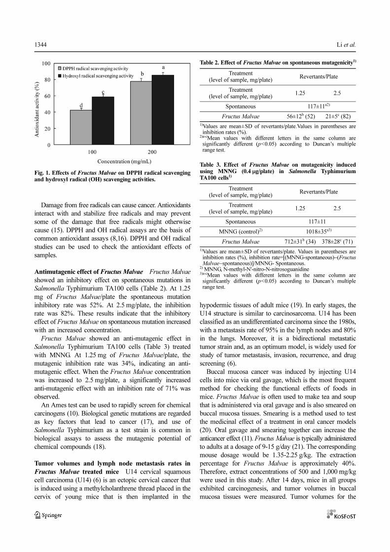
DPPH radical and OH radical scavenging activities of
Fructus Malvae The radical scavenging effects of Fructus
Malvae on DPPH radicals was tested (Fig. 1). Fructus
Malvae showed scavenging activities of 42.2% and 77.9%
at concentrations of 100 and 200 mg/mL, respectively,
indicating that the radical scavenging activity of Fructus
Malvae increased at higher concentrations.
The effects of Fructus Malvae on the hydroxyl scavenging
activity were determined based on deoxyribose damage
induced using a Fe3+/ascorbate/EDTA/H2O2 system, and
measured using the TBA method. Deoxyribose degrades
into fragments that react with TBA upon heating at a low
pH to form a pink color. The inhibitory effects of Fructus
Malvae on deoxyribose damage are shown in Fig. 1.
Inhibition with the 200 mg/mL extract was 85.4%, higher
than inhibition with the 100 mg/mL (58.8%) extract.
Table 1. Sequences of RT-PCR primers used in this study
Gene name Sequence
BaxForward: 5'-AAG CTG AGC GAG TGT CTC CGG CG-3'
Reverse: 5'-CAG ATG CCG GTT CAG GTA CTC AGT C-3'
Bcl-2Forward: 5'-CTC GTC GCT ACC GTC GTG ACT TGG-3'
Reverse: 5'-CAG ATG CCG GTT CAG GTA CTC AGT C-3'
Caspase-3Forward: 5'-CAA ACT TTT TCA GAG GGG ATC G-3'
Reverse: 5'-GCA TAC TGT TTC AGC ATG GCA-3'
Caspase-9Forward: 5'-GGC CCT TCC TCG CTT CAT CTC-3'
Reverse: 5'-GGT CCT TGG GCC TTC CTG GTA T-3'
NF-κBForward: 5'-CAC TTA TGG ACA ACT ATG AGG TCT CTG G-3'
Reverse: 5'-CTG TCT TGT GGA CAA CGC AGT GGA ATT TTA GG-3'
IκB-αForward: 5'-GCT GAA GAA GGA GCG GCT ACT-3'
Reverse: 5'-TCG TAC TCC TCG TCT TTC ATG GA-3'
iNOSForward: 5'-AGA GAG ATC GGG TTC ACA-3'
Reverse: 5'-CAC AGA ACT GAG GGT ACA-3'
COX-2Forward: 5'-TTA AAA TGA GAT TGT CCG AA-3'
Reverse: 5'-AGA TCA CCT CTG CCT GAG TA-3'
GAPDHForward: 5'-CGG AGT CAA CGG ATT TGG TC-3'
Reverse: 5'-AGC CTT CTC CAT GGT CGT GA-3'
1344 Li et al.
Damage from free radicals can cause cancer. Antioxidants
interact with and stabilize free radicals and may prevent
some of the damage that free radicals might otherwise
cause (15). DPPH and OH radical assays are the basis of
common antioxidant assays (8,16). DPPH and OH radical
studies can be used to check the antioxidant effects of
samples.
Antimutagenic effect of Fructus Malvae Fructus Malvae
showed an inhibitory effect on spontaneous mutations in
Salmonella Typhimurium TA100 cells (Table 2). At 1.25
mg of Fructus Malvae/plate the spontaneous mutation
inhibitory rate was 52%. At 2.5 mg/plate, the inhibition
rate was 82%. These results indicate that the inhibitory
effect of Fructus Malvae on spontaneous mutation increased
with an increased concentration.
Fructus Malvae showed an anti-mutagenic effect in
Salmonella Typhimurium TA100 cells (Table 3) treated
with MNNG. At 1.25 mg of Fructus Malvae/plate, the
mutagenic inhibition rate was 34%, indicating an anti-
mutagenic effect. When the Fructus Malvae concentration
was increased to 2.5 mg/plate, a significantly increased
anti-mutagenic effect with an inhibition rate of 71% was
observed.
An Ames test can be used to rapidly screen for chemical
carcinogens (10). Biological genetic mutations are regarded
as key factors that lead to cancer (17), and use of
Salmonella Typhimurium as a test strain is common in
biological assays to assess the mutagenic potential of
chemical compounds (18).
Tumor volumes and lymph node metastasis rates in
Fructus Malvae treated mice U14 cervical squamous
cell carcinoma (U14) (6) is an ectopic cervical cancer that
is induced using a methylcholanthrene thread placed in the
cervix of young mice that is then implanted in the
hypodermic tissues of adult mice (19). In early stages, the
U14 structure is similar to carcinosarcoma. U14 has been
classified as an undifferentiated carcinoma since the 1980s,
with a metastasis rate of 95% in the lymph nodes and 80%
in the lungs. Moreover, it is a bidirectional metastatic
tumor strain and, as an optimum model, is widely used for
study of tumor metastasis, invasion, recurrence, and drug
screening (6).
Buccal mucosa cancer was induced by injecting U14
cells into mice via oral gavage, which is the most frequent
method for checking the functional effects of foods in
mice. Fructus Malvae is often used to make tea and soup
that is administered via oral gavage and is also smeared on
buccal mucosa tissues. Smearing is a method used to test
the medicinal effect of a treatment in oral cancer models
(20). Oral gavage and smearing together can increase the
anticancer effect (11). Fructus Malvae is typically administered
to adults at a dosage of 9-15 g/day (21). The corresponding
mouse dosage would be 1.35-2.25 g/kg. The extraction
percentage for Fructus Malvae is approximately 40%.
Therefore, extract concentrations of 500 and 1,000 mg/kg
were used in this study. After 14 days, mice in all groups
exhibited carcinogenesis, and tumor volumes in buccal
mucosa tissues were measured. Tumor volumes for the
Fig. 1. Effects of Fructus Malvae on DPPH radical scavengingand hydroxyl radical (OH) scavenging activities.
Table 2. Effect of Fructus Malvae on spontaneous mutagenicity1)
Treatment(level of sample, mg/plate)
Revertants/Plate
Treatment(level of sample, mg/plate)
1.25 2.5
Spontaneous 117±11a2)
Fructus Malvae 56±12b (52) 21±5c (82)
1)Values are mean±SD of revertants/plate.Values in parentheses areinhibition rates (%).
2)a-cMean values with different letters in the same column aresignificantly different (p<0.05) according to Duncan’s multiplerange test.
Table 3. Effect of Fructus Malvae on mutagenicity inducedusing MNNG (0.4 µg/plate) in Salmonella TyphimuriumTA100 cells1)
Treatment(level of sample, mg/plate)
Revertants/Plate
Treatment(level of sample, mg/plate)
1.25 2.5
Spontaneous 117±110
MNNG (control)2) 1018±35a3)
Fructus Malvae 712±31b (34) 378±28c (71)
1)Values are mean±SD of revertants/plate. Values in parentheses areinhibition rates (%), inhibition rate=[(MNNG-spontaneous)−(FructusMalvae−spontaneous)]/MNNG- Spontaneous.
2) MNNG, N-methyl-N'-nitro-N-nitrosoguanidine3)a-cMean values with different letters in the same column aresignificantly different (p<0.05) according to Duncan’s multiplerange test.
Anticancer preventive effects of Fructus Malvae 1345
Fructus Malvae control groups A and group B were 8.7
and 4.4 mm3, respectively (Table 4). A total of 6 mice
demonstrated lymph node metastasis in control groups, 3
in Fructus Malvae control group A, and 1 in Fructus
Malvae control group B. Consequently, the lymph node
metastasis rates were 60, 30, and 10%, respectively, for
control, A and B groups. These results demonstrate that
Fructus Malvae is effective in impeding carcinogenesis,
proliferation, and metastasis.
With growth of tumor tissue, open and expanding peri-
cancer lymphatic vessel growth increases. Vessel walls
were thinner, and gaps or nicks appeared in the thin vessel
walls. This phenomenon is usually observed in areas where
tumor cells are concentrated and is favorable in helping
tumor cells enter the lymphatic lumen (22).
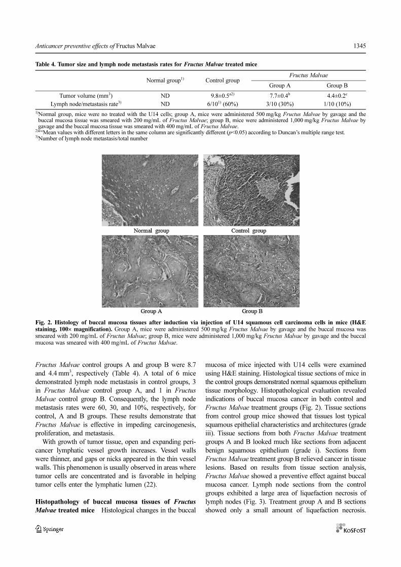
Histopathology of buccal mucosa tissues of Fructus
Malvae treated mice Histological changes in the buccal
mucosa of mice injected with U14 cells were examined
using H&E staining. Histological tissue sections of mice in
the control groups demonstrated normal squamous epithelium
tissue morphology. Histopathological evaluation revealed
indications of buccal mucosa cancer in both control and
Fructus Malvae treatment groups (Fig. 2). Tissue sections
from control group mice showed that tissues lost typical
squamous epithelial characteristics and architectures (grade
iii). Tissue sections from both Fructus Malvae treatment
groups A and B looked much like sections from adjacent
benign squamous epithelium (grade i). Sections from
Fructus Malvae treatment group B relieved cancer in tissue
lesions. Based on results from tissue section analysis,
Fructus Malvae showed a preventive effect against buccal
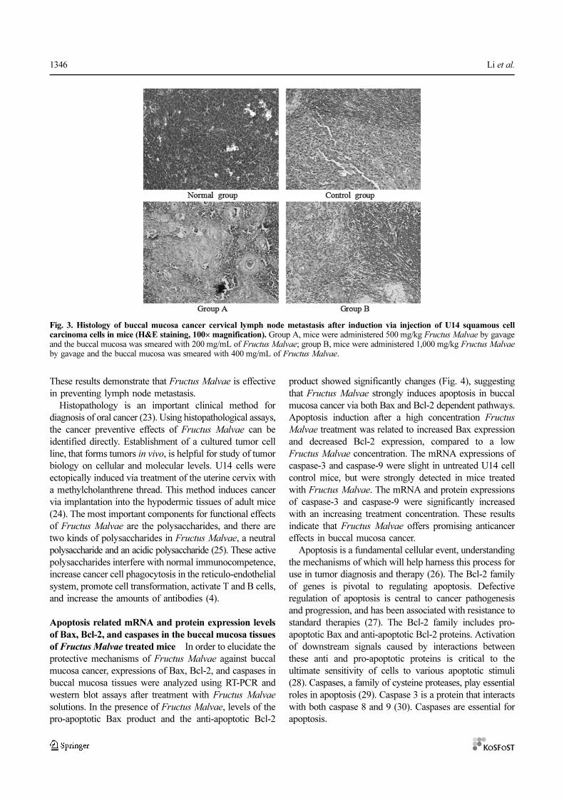
mucosa cancer. Lymph node sections from the control
groups exhibited a large area of liquefaction necrosis of
lymph nodes (Fig. 3). Treatment group A and B sections
showed only a small amount of liquefaction necrosis.
Fig. 2. Histology of buccal mucosa tissues after induction via injection of U14 squamous cell carcinoma cells in mice (H&Estaining, 100× magnification). Group A, mice were administered 500 mg/kg Fructus Malvae by gavage and the buccal mucosa wassmeared with 200 mg/mL of Fructus Malvae; group B, mice were administered 1,000 mg/kg Fructus Malvae by gavage and the buccalmucosa was smeared with 400 mg/mL of Fructus Malvae.
Table 4. Tumor size and lymph node metastasis rates for Fructus Malvae treated mice
Normal group1) Control groupFructus Malvae
Group A Group B
Tumor volume (mm3) ND 9.8±0.5a2) 7.7±0.4b 4.4±0.2c
Lymph node/metastasis rate3) ND 6/101) (60%) 3/10 (30%) 1/10 (10%)
1)Normal group, mice were no treated with the U14 cells; group A, mice were administered 500 mg/kg Fructus Malvae by gavage and thebuccal mucosa tissue was smeared with 200 mg/mL of Fructus Malvae; group B, mice were administered 1,000 mg/kg Fructus Malvae bygavage and the buccal mucosa tissue was smeared with 400 mg/mL of Fructus Malvae.
2)a-cMean values with different letters in the same column are significantly different (p<0.05) according to Duncan’s multiple range test.3)Number of lymph node metastasis/total number
1346 Li et al.
These results demonstrate that Fructus Malvae is effective
in preventing lymph node metastasis.
Histopathology is an important clinical method for
diagnosis of oral cancer (23). Using histopathological assays,
the cancer preventive effects of Fructus Malvae can be
identified directly. Establishment of a cultured tumor cell
line, that forms tumors in vivo, is helpful for study of tumor
biology on cellular and molecular levels. U14 cells were
ectopically induced via treatment of the uterine cervix with
a methylcholanthrene thread. This method induces cancer
via implantation into the hypodermic tissues of adult mice
(24). The most important components for functional effects
of Fructus Malvae are the polysaccharides, and there are
two kinds of polysaccharides in Fructus Malvae, a neutral
polysaccharide and an acidic polysaccharide (25). These active
polysaccharides interfere with normal immunocompetence,
increase cancer cell phagocytosis in the reticulo-endothelial
system, promote cell transformation, activate T and B cells,
and increase the amounts of antibodies (4).
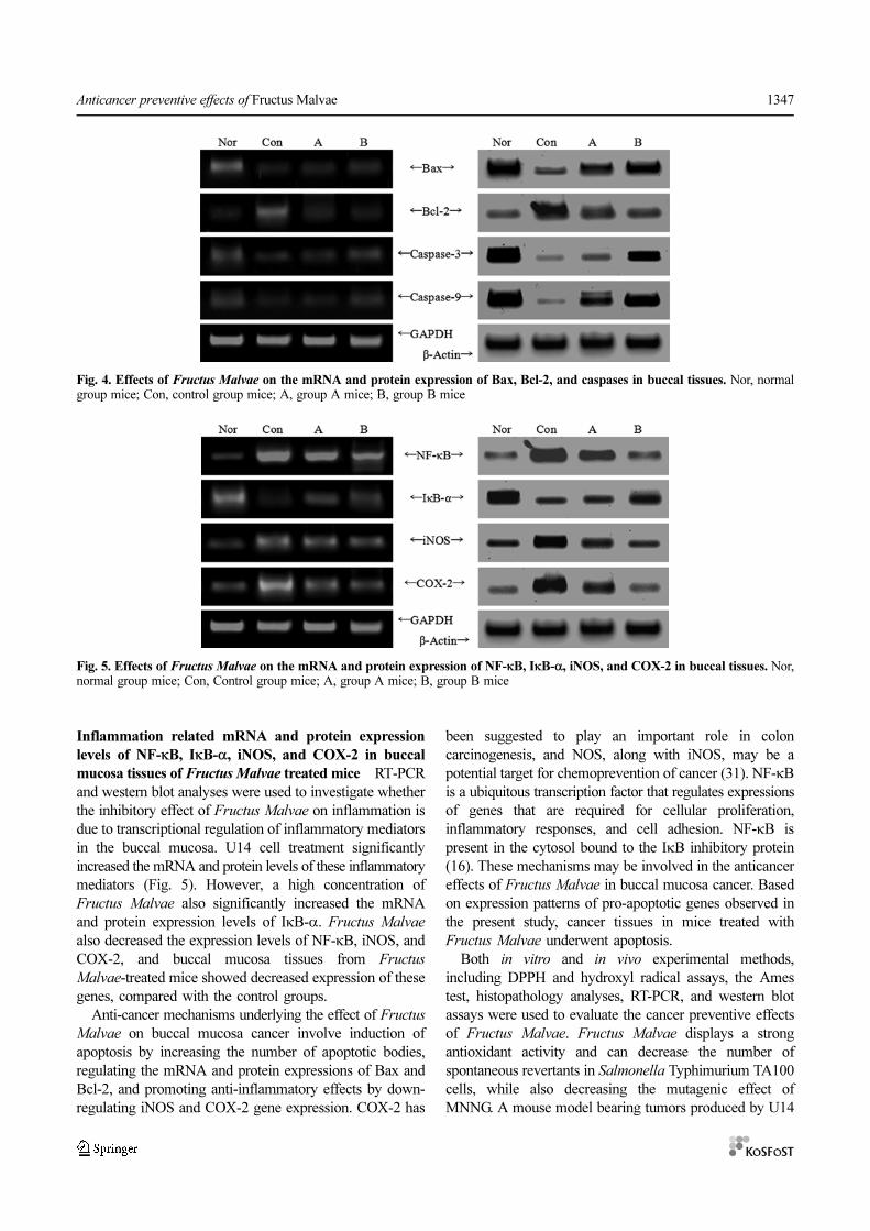
Apoptosis related mRNA and protein expression levels
of Bax, Bcl-2, and caspases in the buccal mucosa tissues
of Fructus Malvae treated mice In order to elucidate the
protective mechanisms of Fructus Malvae against buccal
mucosa cancer, expressions of Bax, Bcl-2, and caspases in
buccal mucosa tissues were analyzed using RT-PCR and
western blot assays after treatment with Fructus Malvae
solutions. In the presence of Fructus Malvae, levels of the
pro-apoptotic Bax product and the anti-apoptotic Bcl-2
product showed significantly changes (Fig. 4), suggesting
that Fructus Malvae strongly induces apoptosis in buccal
mucosa cancer via both Bax and Bcl-2 dependent pathways.
Apoptosis induction after a high concentration Fructus
Malvae treatment was related to increased Bax expression
and decreased Bcl-2 expression, compared to a low
Fructus Malvae concentration. The mRNA expressions of
caspase-3 and caspase-9 were slight in untreated U14 cell
control mice, but were strongly detected in mice treated
with Fructus Malvae. The mRNA and protein expressions
of caspase-3 and caspase-9 were significantly increased
with an increasing treatment concentration. These results
indicate that Fructus Malvae offers promising anticancer
effects in buccal mucosa cancer.
Apoptosis is a fundamental cellular event, understanding
the mechanisms of which will help harness this process for
use in tumor diagnosis and therapy (26). The Bcl-2 family
of genes is pivotal to regulating apoptosis. Defective
regulation of apoptosis is central to cancer pathogenesis
and progression, and has been associated with resistance to
standard therapies (27). The Bcl-2 family includes pro-
apoptotic Bax and anti-apoptotic Bcl-2 proteins. Activation
of downstream signals caused by interactions between
these anti and pro-apoptotic proteins is critical to the
ultimate sensitivity of cells to various apoptotic stimuli
(28). Caspases, a family of cysteine proteases, play essential
roles in apoptosis (29). Caspase 3 is a protein that interacts
with both caspase 8 and 9 (30). Caspases are essential for
apoptosis.
Fig. 3. Histology of buccal mucosa cancer cervical lymph node metastasis after induction via injection of U14 squamous cellcarcinoma cells in mice (H&E staining, 100× magnification). Group A, mice were administered 500 mg/kg Fructus Malvae by gavageand the buccal mucosa was smeared with 200 mg/mL of Fructus Malvae; group B, mice were administered 1,000 mg/kg Fructus Malvae
by gavage and the buccal mucosa was smeared with 400 mg/mL of Fructus Malvae.
Anticancer preventive effects of Fructus Malvae 1347
Inflammation related mRNA and protein expression
levels of NF-κB, IκB-α, iNOS, and COX-2 in buccal
mucosa tissues of Fructus Malvae treated mice RT-PCR
and western blot analyses were used to investigate whether
the inhibitory effect of Fructus Malvae on inflammation is
due to transcriptional regulation of inflammatory mediators
in the buccal mucosa. U14 cell treatment significantly
increased the mRNA and protein levels of these inflammatory
mediators (Fig. 5). However, a high concentration of
Fructus Malvae also significantly increased the mRNA
and protein expression levels of IκB-α. Fructus Malvae
also decreased the expression levels of NF-κB, iNOS, and
COX-2, and buccal mucosa tissues from Fructus
Malvae-treated mice showed decreased expression of these
genes, compared with the control groups.
Anti-cancer mechanisms underlying the effect of Fructus
Malvae on buccal mucosa cancer involve induction of
apoptosis by increasing the number of apoptotic bodies,
regulating the mRNA and protein expressions of Bax and
Bcl-2, and promoting anti-inflammatory effects by down-
regulating iNOS and COX-2 gene expression. COX-2 has
been suggested to play an important role in colon
carcinogenesis, and NOS, along with iNOS, may be a
potential target for chemoprevention of cancer (31). NF-κB
is a ubiquitous transcription factor that regulates expressions
of genes that are required for cellular proliferation,
inflammatory responses, and cell adhesion. NF-κB is
present in the cytosol bound to the IκB inhibitory protein
(16). These mechanisms may be involved in the anticancer
effects of Fructus Malvae in buccal mucosa cancer. Based
on expression patterns of pro-apoptotic genes observed in
the present study, cancer tissues in mice treated with
Fructus Malvae underwent apoptosis.
Both in vitro and in vivo experimental methods,
including DPPH and hydroxyl radical assays, the Ames
test, histopathology analyses, RT-PCR, and western blot
assays were used to evaluate the cancer preventive effects
of Fructus Malvae. Fructus Malvae displays a strong
antioxidant activity and can decrease the number of
spontaneous revertants in Salmonella Typhimurium TA100
cells, while also decreasing the mutagenic effect of
MNNG. A mouse model bearing tumors produced by U14
Fig. 4. Effects of Fructus Malvae on the mRNA and protein expression of Bax, Bcl-2, and caspases in buccal tissues. Nor, normalgroup mice; Con, control group mice; A, group A mice; B, group B mice
Fig. 5. Effects of Fructus Malvae on the mRNA and protein expression of NF-κB, IκB-α, iNOS, and COX-2 in buccal tissues. Nor,normal group mice; Con, Control group mice; A, group A mice; B, group B mice
1348 Li et al.
squamous cell carcinoma cells was used to study the in
vivo effects of Fructus Malvae. A strong anticancer activity
against buccal mucosa cancer was observed. Overall,
Fructus Malvae showed in vitro anti-mutagenic effects and
in vivo anticancer activities. In conclusion, increased
Fructus Malvae concentrations should be used to increase
the oral cancer preventive effect.
Acknowledgments The Chongqing Innovative Research
Team in University (KJTD201325), China provided support
for this research.
Disclosure The authors declare no conflict of interest.
References
1. Dong Y, Ma Q, Na SS, Li X, Li SM. Quantitative determination ofcaffeic acid in Dongkuiguo (Fructus Malvae, Mongolian medicatedherb) by HPLC. J. Beijing Univ. Trad. Chinese Med. 33: 117-119(2010)
2. Gan RY, Kuang L, Xu XR, Zhang Y, Xia EQ, Song FL, Li BH.Screening of natural antioxidants from traditional Chinese medicinalplants associated with treatment of rheumatic disease. Molecules 15:5988-5997 (2010)
3. Wu LGRL, Zhao J, Ba HS. Antioxidant effect of Fructus Malvaepolysaccharides. Nat. Prod. Res. Dev. 24: 536-538 (2012)
4. Nose M, Terawaki K, Ogihara Y. The role of a crude polysaccharidefraction in the macrophage activation by “Shosaikoto”. Phytomedicine4: 23-26 (1997)
5. Kolanjiappan K, Ramachandran CR, Manoharan S. Biochemicalchanges in tumor tissues of oral cancer patients. Clin. Biochem. 36:61-65 (2003)
6. Gu B, Feng HL, Dong JH, Zhang H, Bian XC, Liu YQ. TheEstablishment and characterization of a continuous cell line ofmouse cervical carcinoma. Chinese J. Clin. Oncol. 5: 44-48 (2008)
7. Pang L, Qiu LH, Gao Z, Li P, Xu P, Luo DP. Experimental study oncontrast-enhanced ultrasound imaging of metastatic lymph modes ofcheek carcinoma. J. Ultrasound Clin. Med. 13: 581-583 (2011)
8. Kang HS, Chung HY, Jung JH, Kang SS, Choi JS. Antioxidanteffect of Salvia miltiorrhiza. Arch. Pharm. Res. 20: 496-500 (1997)
9. Banerijee A, Dasgupta N, Bratati D. In vitro study of antioxidantactivity of Syzygium cumini fruit. Food Chem. 90: 727-733 (2005)
10. Maron DM, Ames BN. Revised methods for the Salmonellamutagenicity test. Mutat. Res. 113: 173-215 (1983)
11. Zhao X, Deng XX, Park KY, Qiu LH, Pang L. Purple bamboo salthas anticancer activity in TCA8113 cells in vitro and preventiveeffects on buccal mucosa cancer in mice in vivo. Exp. Ther. Med. 5:549-554 (2013)
12. Schrader M, Laberke HG. Differential diagnosis of verrucouscarcinoma in the oral cavity and larynx. J. Laryngol. Otol. 102: 700-703 (1998)
13. Zhao X, Kim SY, Park KY. Bamboo salt has in vitro anti-canceractivity in HCT-116 cells and exerts anti-metastatic effects in vivo. J.Med. Food 16: 9-19 (2013)
14. Zhao X. Hawk tea (Litsea coreana Levl. var. lanuginose) attenuatesCCl4-induced hepatic damage in Sprague-Dawley rats. Exp. Ther.Med. 5: 555-560 (2013)
15. Valko M, Rhodes CJ, Moncol J, Izakovic M, Mazur M. Freeradicals, metals and antioxidants in oxidative stress-induced cancer.Chem-Biol. Interact. 160: 1-40 (2006)
16. Baeuerle PA. IkappaB-NF-kappaB structures: At the interface ofinflammation control. Cell 95: 729-731 (1998)
17. Hwang KM, Jung KO, Song CH, Park KY. Increased antimutagenicand anticlastogenic effects of doenjang (Korean fermented soybeanpaste) prepared with bamboo salt. J. Med. Food 11: 717-722 (2008)
18. Mortelmans K, Zeiger E. The Ames Salmonella/microsomemutagenicity assay. Mutat. Res. 455: 29-60 (1999)
19. Wang LF, Wu YX, Zhang YP, Tang W. Antitumor effects ofpolyethylene glycol-modified recombinant human interleukin-2 onmouse uterine cervical carcinoma in vivo. Chinese J. Cancer Res. 9:28-31 (1997)
20. Li N, Chen XX, Han C, Chen JS, Yang ZS. Chemopreventive effectof tea and curcumin on DMBA-induced oral carcinogenesis inhamsters. J. Hyg. Res. 31: 354-357 (2002)
21. Wang HW, Wang SW, Dong Y, Li SR. The experimental study ontraditional Mongolianmateria medica of Fructus Malvae. J. InnerMongolia Med. Coll. 34: 69-72 (2012)
22. Xuan M, Weng YM, Wang CM, Li XQ. Pathologic changes oflymphatic capillaries after inoculation of U14 cells in rat tongueperineoplastic area. West China J. Stomatol. 18: 5-8 (2000)
23. Sankaranarayanan R, Ramadas K, Thomas G, Muwonge R, Thara S,Mathew B, Rajan B. Effect of screening on oral cancer mortality inKerala, India: A cluster-randomised controlled trial. Lancet 365:1927-1933 (2005)
24. Zhao X, Pang L, Qian Y, Wang Q, Li Y, Wu M, Ouyang Z, Gao Z,Qiu L. An animal model of buccal mucosa cancer and cervicallymph node metastasis induced by U14 squamous cell carcinomacells. Exp. Ther. Med. 5: 1083-1088 (2013)
25. Li MH, Fang YS, Chen JC, Yang XQ, Peng L, Ding ZT. GC-MSanalysis of volatile constituents from the seeds of euryale feroxsalisb and Malva verticillata L. Yunnan Chem. Technol. 34: 47-49(2007)
26. Milanezi F, Leitao D, Ricardo S, Augusto I, Schmitt F. Evaluationof HER2 in breast cancer: Reality and expectations. Expert Opin.Med. Diagn. 3: 607-620 (2009)
27. Chao DT, Korsmeyer SJ. Bcl-2 family: Regulators of cell death.Annu. Rev. Immunol. 16: 395-419 (1998)
28. Oltvai ZN, Milliman CL, Korsmeyer SJ. Bcl-2 Heterodimerizes invivo with a conserved homolog, Bax, that accelerates programedcell death. Cell 74: 609-619 (1993)
29. Thornberry NA. The caspase family of cysteine proteases. Brit.Med. Bull. 53: 478-490 (1997)
30. Alnemri ES, Livingston DJ, Nicholson DW, Salvesen G, ThornberryNA, Wong WW, Yuan J. Human ICE/CED-3 proteasenomenclature. Cell 87: 171 (1996)
31. Delić R, Štefanović M. Optimal laboratory panel for predictingpreeclampsia. J. Matern-Fetal. Neo. Med. 23: 96-102 (2010)