Embed Size (px)
Citation preview
Antimicrobial Susceptibility Testing
Even before the discovery of true antibiotics, scientists have been working on ways to determine the action of noxious agents on bacteria. Since then, both the methods and the agents in use have progressed a long way. Determination of the antimicrobial susceptibility of signifi cant bacterial pathogens is now one of the principle functions of clinical microbiology laboratories.
The primary objective of susceptibility testing in the clinical setting is to predict the outcome of treatment of an infected individual with the chosen antibiotics. Results must be derived in a timely manner with a high degree of accuracy and reproducibility. Clinical laboratories can choose to determine the susceptibility of bacteria by a number of methods including disc diffusion, breakpoint testing, broth micro-dilution or antibiotic gradients, most of which can be performed manually or with automation.
The disc diffusion method is one of the most widely adopted susceptibility testing methods worldwide, offering a simple technique with fl exibility of choice for the compounds tested, at a relatively low cost. The advantage of this method is that the results obtained are simple, qualitative, interpretative results (sensitive, intermediate or resistant) that are easily understood by clinicians.
Breakpoint testing is also a popular technique with large laboratories for multiple sample testing, e.g. for testing urinary isolates. The benefi t of this method is that it is easily automated and can be very cost effective. However, subtle changes in susceptibility will not be detected by this technique and the end points are not always clear-cut making it diffi cult to read or interpret the result.
Advances in Antimicrobial Susceptibility Testing with Oxoid Minimum Inhibitory Concentration Evaluators (M.I.C.E.TM)
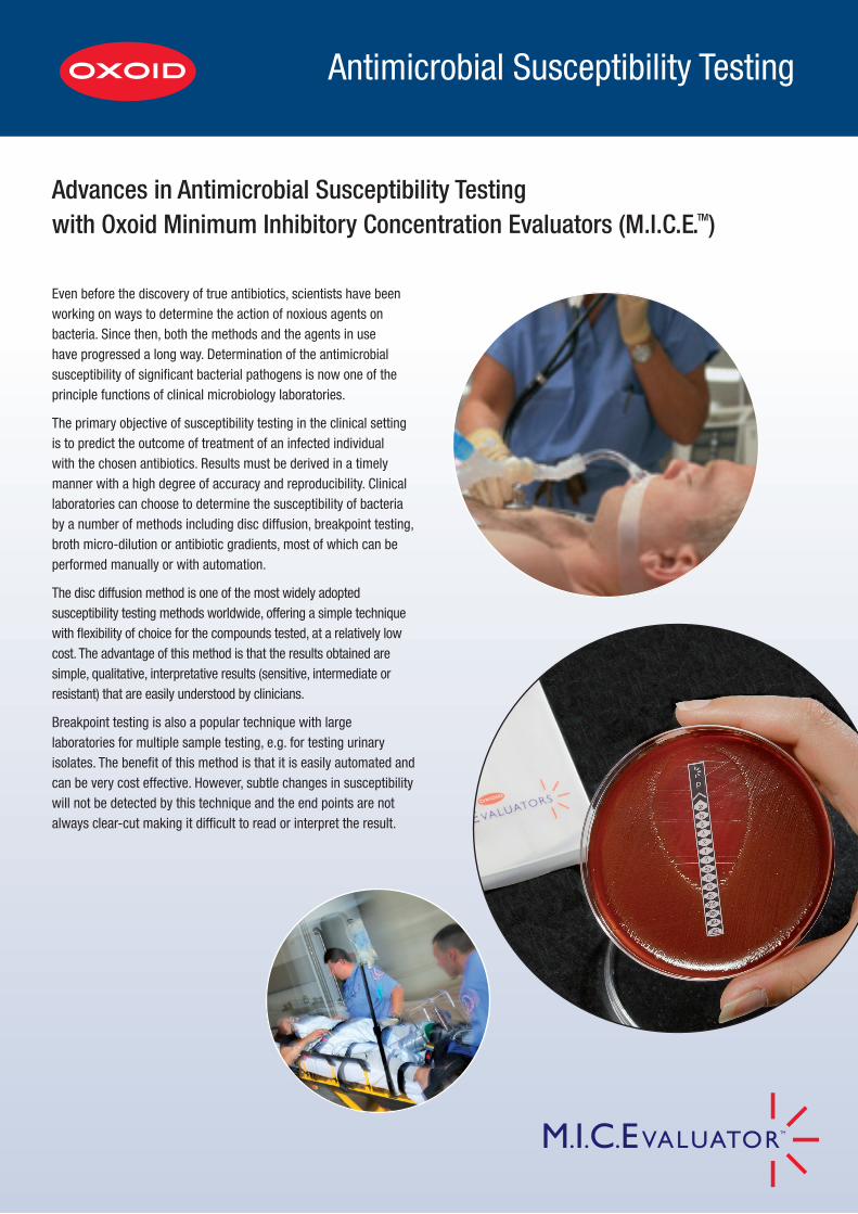
1 Inoculate the appropriate agar plate by swabbing the test organism in at least three different directions, taking care to ensure no gaps are left in the deposited inoculum. 2 Open the sachet by peeling apart at the corners indicated by
the blue arrows.
4 Apply the strip by placing the end with the lowest concentration onto the plate fi rst.
A Step by Step Guide to M.I.C.E. Strips
5 Carefully roll the strip onto the agar surface to ensure good contact along the entire length of the strip. Alternatively, carefully place the lower end of the M.I.C.E. strip onto the agar and gently drop the strip. Using sterile forceps gently smooth the strip onto the agar. TAKE CARE NOT TO MOVE THE POSITION OF THE STRIP.
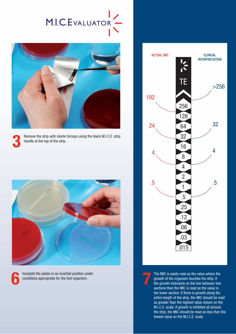
3 Remove the strip with sterile forceps using the black M.I.C.E. strip handle at the top of the strip.
6 Incubate the plates in an inverted position under conditions appropriate for the test organism. 7 The MIC is easily read as the value where the
growth of the organism touches the strip. If the growth intersects on the line between two sections then the MIC is read as the value in the lower section. If there is growth along the entire length of the strip, the MIC should be read as greater than the highest value shown on the M.I.C.E. scale. If growth is inhibited all around the strip, the MIC should be read as less than the lowest value on the M.I.C.E. scale.
TE>256
32
4
.5
192
24
4
.5
256
128
64
32
16
8
4
2
1
.5
.25
.12
.06
.03
.015
CLINICALINTERPRETATION
ACTUAL MIC
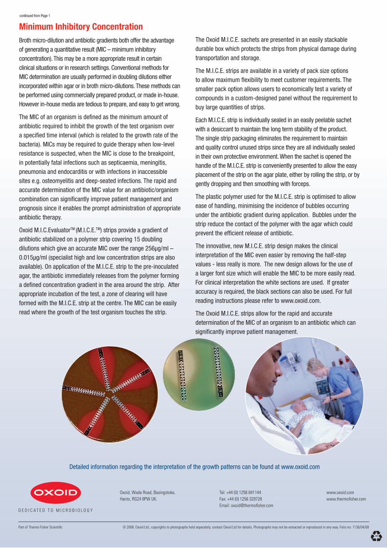
Detailed information regarding the interpretation of the growth patterns can be found at www.oxoid.com
Minimum Inhibitory ConcentrationBroth micro-dilution and antibiotic gradients both offer the advantage of generating a quantitative result (MIC – minimum inhibitory concentration). This may be a more appropriate result in certain clinical situations or in research settings. Conventional methods for MIC determination are usually performed in doubling dilutions either incorporated within agar or in broth micro-dilutions. These methods can be performed using commercially prepared product, or made in-house. However in-house media are tedious to prepare, and easy to get wrong.
The MIC of an organism is defi ned as the minimum amount of antibiotic required to inhibit the growth of the test organism over a specifi ed time interval (which is related to the growth rate of the bacteria). MICs may be required to guide therapy when low-level resistance is suspected, when the MIC is close to the breakpoint, in potentially fatal infections such as septicaemia, meningitis, pneumonia and endocarditis or with infections in inaccessible sites e.g. osteomyelitis and deep-seated infections. The rapid and accurate determination of the MIC value for an antibiotic/organism combination can signifi cantly improve patient management and prognosis since it enables the prompt administration of appropriate antibiotic therapy.
Oxoid M.I.C.EvaluatorTM (M.I.C.E.TM) strips provide a gradient of antibiotic stabilized on a polymer strip covering 15 doubling dilutions which give an accurate MIC over the range 256µg/ml – 0.015µg/ml (specialist high and low concentration strips are also available). On application of the M.I.C.E. strip to the pre-inoculated agar, the antibiotic immediately releases from the polymer forming a defi ned concentration gradient in the area around the strip. After appropriate incubation of the test, a zone of clearing will have formed with the M.I.C.E. strip at the centre. The MIC can be easily read where the growth of the test organism touches the strip.
continued from Page 1
The Oxoid M.I.C.E. sachets are presented in an easily stackable durable box which protects the strips from physical damage during transportation and storage.
The M.I.C.E. strips are available in a variety of pack size options to allow maximum fl exibility to meet customer requirements. The smaller pack option allows users to economically test a variety of compounds in a custom-designed panel without the requirement to buy large quantities of strips.
Each M.I.C.E. strip is individually sealed in an easily peelable sachet with a desiccant to maintain the long term stability of the product. The single strip packaging eliminates the requirement to maintain and quality control unused strips since they are all individually sealed in their own protective environment. When the sachet is opened the handle of the M.I.C.E. strip is conveniently presented to allow the easy placement of the strip on the agar plate, either by rolling the strip, or by gently dropping and then smoothing with forceps.
The plastic polymer used for the M.I.C.E. strip is optimised to allow ease of handling, minimising the incidence of bubbles occurring under the antibiotic gradient during application. Bubbles under the strip reduce the contact of the polymer with the agar which could prevent the effi cient release of antibiotic.
The innovative, new M.I.C.E. strip design makes the clinical interpretation of the MIC even easier by removing the half-step values - less really is more. The new design allows for the use of a larger font size which will enable the MIC to be more easily read. For clinical interpretation the white sections are used. If greater accuracy is required, the black sections can also be used. For full reading instructions please refer to www.oxoid.com.
The Oxoid M.I.C.E. strips allow for the rapid and accurate determination of the MIC of an organism to an antibiotic which can signifi cantly improve patient management.
Oxoid, Wade Road, Basingstoke, Hants, RG24 8PW UK.
www.oxoid.comwww.thermofi sher.com
Tel: +44 (0) 1256 841144Fax: +44 (0) 1256 329728Email: oxoid@thermofi sher.com
D E D I C A T E D T O M I C R O B I O L O G Y
Part of Thermo Fisher Scientifi c © 2008, Oxoid Ltd.; copyrights to photographs held separately; contact Oxoid Ltd for details. Photographs may not be extracted or reproduced in any way. Folio no: 1139/04/08