Embed Size (px)
Citation preview
Volume 2, Issue 1 June 2017
1
CLSI Subcommittee on Antimicrobial Susceptibility Testing
CLSI AST News Update
The CLSI Outreach Working Group (ORWG) is providing this Newsletter to highlight some recent issues related to antimicrobial susceptibility testing (AST) and reporting. We are listing links to some new educational materials and reminding you where you can find information about the CLSI AST Subcommittee proceedings.
Members:Janet A. Hindler (Co-Chairholder), UCLA Health System, USAAudrey N. Schuetz (Co-Chairholder), Mayo Clinic, Rochester, USAApril Abbott, Deaconess Health System, USAStella Antonara, Nationwide Children’s Hospital, USAApril Bobenchik, Lifespan Academic Medical Center, USAMariana Castanheira, JMI Laboratories, USAAngella Charnot-Katsikas, University of Chicago, USAMarcelo Galas, National Institute of Infectious Disease, ArgentinaRomney Humphries, UCLA Health System, USAVioleta Rekasius, Loyola University Medical Center, USANicole Scangarella-Oman, GlaxoSmithKline, USALars Westblade, Weill Cornell Medical College, USA
What does the CLSI AST Subcommittee do?The first edition of this newsletter described details about the organization and operation of the CLSI AST Subcommittee.
• Access that newsletter here.
• To learn more about upcoming or past meetings, click here.
• CLSI posts meeting minutes and summaries for public access here.
Interested in becoming a CLSI volunteer? Learn more here.Please remember that CLSI AST Subcommittee welcomes suggestions from you about any aspect of CLSI documents, educational materials, or this Newsletter.
Volume 2, Issue 1 June 2017
The One Health Initiative ............................... 4
What is the Antibiotic Resistance Laboratory Network (ARLN)? .............................................. 5
A New Phenotypic Test for Detection of Carbapenemases – The Modified Carbapenem Inactivation Method (mCIM) ............................................... 7
Carbapenems and Enterobacteriaceae: What is the difference between CRE vs Carbapenemase-Producing (CP)-CRE vs non-CP-CRE? .............................................................. 9
Newer Developments in Antifungal Susceptibility Testing - Species-Specific Breakpoints for Candida spp. .......................10
The 21st Century Cures Act and the Future of AST ...................................................13
Hot Topic – OXA Carbapenemases ..............14
In Memoriam – Dr. Paul Schreckenberger ..............................17
Inside This Issue:
Volume 2, Issue 1 June 2017
2
CLSI AST Subcommittee PartnershipsRepresentatives with expertise in antimicrobials from the following organizations attend and participate in CLSI AST Subcommittee meetings and aid in dissemination of information regarding CLSI decisions and AST issues.
American Society for Microbiology (ASM)
Association of Public Health Laboratories (APHL)
ASTM International
College of American Pathologists (CAP)
European Committee on Antimicrobial Susceptibility Testing (EUCAST)
Infectious Diseases Society of America (IDSA)
Pediatric Infectious Diseases Society (PIDS)
Society for Healthcare Epidemiology of America (SHEA)
Susceptibility Testing Manufacturers Association (STMA)
Updated CLSI AST Documents – What’s new? M100S 27th ed. Major changes in “Performance Standards for Antimicrobial Susceptibility Testing; Twenty-Seventh Informational Supplement” include:
• CLSI now uses only the term “breakpoint”; discontinued use of “interpretive criteria”
• Clarified testing/reporting for:– Colistin/polymyxin with P. aeruginosa and Acinetobacter baumannii
– Atypical (poor growing) S. aureus
– Coagulase-negative staphylococci and oxacillin• Expanded definition/discussion of ECVs and added ECVs for:
– Colistin – Enterobacteriaceae– Azithromycin – Neisseria gonorrhoeae
• Added mCIM for detecting carbapenemases in Enterobacteriaceae• Quality control/Quality assurance
– Modified disk diffusion QC ranges: Cefepime - P. aeruginosa ATCC 27853 Meropenem - E. coli ATCC 25922 – Modified MIC QC ranges: Meropenem - P. aeruginosa ATCC 27853 Tedizolid - S. aureus ATCC 29213– Added QC ranges for several new drugs that are in development (not yet available for human use)– Expanded MIC Troubleshooting Guide
• Added suggestions for confirmation of resistant antimicrobial results for B. fragilis
• Deleted tetracycline as intrinsic “R” for M. morganii
More information on availability of M00 including the Read Only web version can be found here.
M100Performance Standards for Antimicrobial Susceptibility Testing
This document includes updated tables for the Clinical and
Laboratory Standards Institute antimicrobial susceptibility testing
standards M02-A12, M07-A10, and M11-A8.
An informational supplement for global application developed through the Clinical and Laboratory Standards Institute
consensus process.
27th Edition
Archive of Retired Breakpoints An archive of breakpoints removed from M100 since 2010 together with the rationale for their removal is now available here.
Volume 2, Issue 1 June 2017
3
WebinarsFor information on webinars please go here. Recently archived CLSI webinars can be accessed on demand. Learn more about program availability here.
On-Demand Webinars:• Practical Recommendations for Antifungal Susceptibility Testing and Reporting in Clinical Laboratories: New Drugs, New
Breakpoints, New Guidelines• Facts and Fiction about Colistin from Clinical and Public Health Perspectives• Verification of Commercial Microbial Identification and Antimicrobial Susceptibility Testing Systems• Navigating CLSI Document M100: Antimicrobial Susceptibility Testing Made Easy• CLSI 2017 Antimicrobial Susceptibility Testing Update
Free WebinarsWebinars are available free of charge six to twelve months after the scheduled event for CLSI members. Please contact CLSI for more information about accessing these on-demand webinars.
Upcoming Webinars (registration details will be posted soon!)
October 17, 20173rd Annual CAP-CLSI Webinar – Digging Deeper into AST Challenges
Angella Charnot-Katsikas, MDUniversity of Chicago, Chicago, Illinois
Romney M. Humphries, PhD D(ABMM)UCLA Health, Los Angeles, California
October 19, 20175th Annual CLSI-SIDP Webinar – Merging Microbiology and Stewardship: CLSI updates on antimicrobial susceptibility testing and advances in detecting and managing Clostridium difficile
Jennifer Dien-Bard, PhD D(ABMM)Children’s Hospital, Los Angeles, California
Dhara Surati, PharmD, BCPSUniversity of Houston, Houston, Texas
ASM/CLSI 2017 AST Webinar Series ASM and CLSI have been coordinating a webinar series entitled
“A Comprehensive Course in Antimicrobial Susceptibility Testing” which is geared towards bench level technologists. This program is being presented in three parts with a series of 5 or 6 talks in each part. The first two parts have been completed and presentations are available on demand for a fee. Click on the links below to learn more:
Part I. Fundamentals of Susceptibility Testing, Reporting, and Test Validation
Part II. Mechanisms of Resistance, Antimicrobial Stewardship, and Infection Prevention
Part III. Special Antimicrobial Susceptibility Tests - scheduled to be presented Fall 2017
Check It Out! Educational Workshops held at CLSI meetingsTo coincide with the January and June CLSI Committee Weeks, the ORWG coordinates a “live” Educational Workshop, typically held on the Saturday evening prior to the start of the AST Subcommittee Working Group meetings.
The next workshop to be held Saturday June 24, 2017, in Philadelphia is entitled “New and Successful Approaches to Antimicrobial Stewardship: The Role of the Microbiology Laboratory.” Clinical microbiology laboratories play a critical role in implementing, influencing, and executing successful antimicrobial stewardship programs with the ultimate goal of improving patient care. Some of the topics to be highlighted during this workshop include: antimicrobial stewardship in the hospital, long-term care and outpatient settings, the role of rapid diagnostics and interpretive reporting, and the role of antibiograms in antimicrobial stewardship.
The January 2017 Workshop was focused on “One Health – One Medicine”- Linking Human, Animal and Environmental Health (see article in this Newsletter on page 4).
PowerPoint presentations from past workshops can be found here.
Future CLSI AST Meetings!
June 25-27, 2017 Philadelphia, Pennsylvania, USA
January 28-30, 2018Dallas, Texas, USA
June 3-5, 2018San Diego, California, USA
Volume 2, Issue 1 June 2017
4
The One Health Initiative The One Health Initiative is a movement that was launched in 2006 and is “a worldwide strategy for expanding interdisciplinary collaborations and communications in all aspects of health care for humans, animals, and the environment.” CLSI has been involved in the One Health Initiative in a practical way for 25 years through its antimicrobial susceptibility testing (AST) subcommittees that establish testing standards for the human and veterinary laboratory testing sectors. Bacteria isolated from animals may become zoonotic and cause disease in people, either via foodborne transmission (eg, Salmonella or Campylobacter), direct contact (eg, Staphylococcus pseudintermedius), or possibly through environmental contamination. Likewise, many common human pathogens are anthroponotic (i.e., carried by humans but then transferred to animals; an example is Cryptosporidium) and may produce colonization and disease in domestic and companion animals. In recognition of the 25th anniversary of the Veterinary Antimicrobial Susceptibility Testing (VAST) Subcommittee, an educational workshop featuring four speakers was held during the January 2017 CLSI Committee Week. Each speaker discussed the shared interests between the VAST and Human AST Subcommittees and between human and animal laboratory medicine testing.
Dr. Jeff Watts, Zoetis Animal Health, outlined key recommendations from the WHO (World Health Organization), OIE (World Organization for Animal Health) and FAO (Food and Agricultural Organization) which address responsible antibiotic use guidelines and national antimicrobial resistance monitoring (AMR) programs with harmonized AST methodology. Responsible use guidelines require the identification and AST of animal pathogens as for human pathogens. The VAST methods are described in VET01-A4 (Performance Standards for Antimicrobial Disk and Dilution Susceptibility Tests for Bacteria Isolated From Animals; Approved Standard—Fourth Edition) and breakpoints are listed in VET01S-Ed3. These documents encompass both companion and food animal species, the pathogens for many major diseases, and antibiotics used to treat them. Dr. Watts discussed the value that national AMR programs may find in the VET05-R document (Generation, Presentation, and Application of Antimicrobial Susceptibility Test Data for Bacteria of Animal Origin; A Report), which emphasizes the use of CLSI methods to ensure comparability of cumulative AST data. A recent blog on the One Health Commission website amplified the key role of CLSI AST methodology in comparing AST data across institutions and can be accessed here.
Dr. Mark Papich, Professor of Veterinary Pharmacology at North Carolina State University, reviewed how the unique pharmacokinetics in animals require careful consideration to properly establish appropriate clinical breakpoints, which may not be the same as those set for humans. He noted all current clinical breakpoints for veterinary medicine can be found in VET01S-Ed3 and VET03/VET04-S2 (only applicable to aquatic animals). Access to species-specific clinical breakpoints is a key component to the foundations of “Responsible Use” by veterinarians, since reliance on human clinical breakpoints could result in inappropriate use, emergence of resistance and/or therapeutic failure.
Dr. Tom Fritsche, Marshfield Clinic, Marshfield, Wisconsin, related his One Health – One Lab experience in creating a single lab entity for performing human and animal microbiology testing by integrating mass spectrometry and broth microdilution AST for all specimens. Cross-training of technical staff, partnership with in-house veterinary pathologists, and a common laboratory information system were key factors in successfully streamlining laboratory operations and improving client satisfaction. Benefits realized from this One Health lab approach has included recognition of new pathogens common to both humans and animals, and comparative antibiogram analyses for detecting emerging resistance in both populations.
The final speaker, Dr. Ron Miller, FDA Center for Veterinary Medicine, shared a glimpse of the future comparing Whole Genome Sequencing (WGS) data with phenotypic MIC data for key food-borne bacteria. An exceptionally high correlation was determined between the genotypic and phenotypic data. The new concept of a Genotypic Cutoff Value (GCV) was explored as a counterpart to validate and confirm more traditional MIC distribution analyses used to establish epidemiological cutoff values (ECVs).1
The presentations collectively provided insight into the overlap between the human and animal laboratory sectors and how awareness and cooperation can benefit overall health. Slides from this educational workshop can be found here.
Reference
1 Tyson GH, et al. Establishing genotypic cutoff values to measure antimicrobial resistance in Salmonella. Antimicrob Agents Chemother. 2017;61(3): pii: e02140-16. PMID 27993845.
Volume 2, Issue 1 June 2017
5
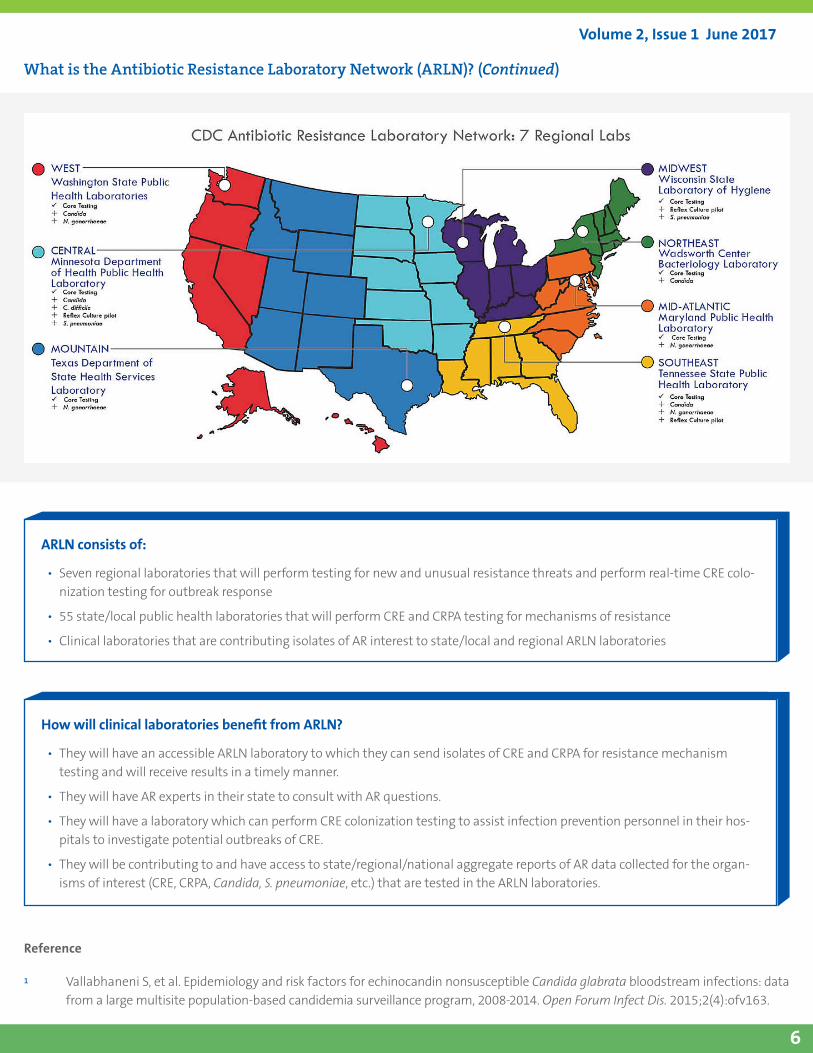
What is The Antibiotic Resistance Laboratory Network (ARLN)?In August 2016, the Centers for Disease Control and Prevention (CDC) launched a significant new effort that will dramatically impact our understanding of the landscape of antimicrobial resistance (AR) in the US. This new venture will provide an avenue to combat persistent and emerging resistance in targeted microorganisms identified as critical AR threats. The new Antibiotic Resistance Laboratory Network, or the ARLN, is the structure for great things to come.
The CDC has responded to the March 2015 National Strategy for Combating Antibiotic-Resistant Bacteria, a National Action Plan put forth by the White House that called for “…creation of a regional public health laboratory network.” CDC’s insightful planning and rapid implementation of a simple yet robust network of laboratories is ambitious and necessary. To allow for faster identification and response to outbreaks, this network provides public health laboratories in all 50 states and 5 cities/territories with funding and a framework to fortify connections with clinical laboratory partners and to start building testing capacity for carbapenem resistant Enterobacteriaceae (CRE) and carbapenem resistant Pseudomonas aeruginosa (CRPA) isolates from their jurisdictions; in most cases, this capacity never existed. This effort includes performing susceptibility testing as well as characterizing resistance mechanisms, such as KPC, NDM, and OXA-48-like genes in Enterobacteriaceae and KPC, NDM, and VIM in P. aeruginosa.
Adding to this solid base of 55 state/local laboratories, are the seven ARLN regional laboratories. The seven regional laboratories listed on the map accept referral isolates with new or unusual resistance from state laboratories within their region. They have the capacity to perform additional characterization and recognize emerging or novel resistance mechanisms such as mcr-1 and detect other, difficult-to-treat microorganisms such as Acinetobacter. The goal of the regional labs is to support rapid detection of existing and emerging resistance more effectively, by closing the gap between hospital-based laboratory testing and the need for pertinent information to guide infection control and prevention across the spectrum of care. Thus, a responsibility of the regional labs is to perform outbreak detection in the form of rapid rectal swab testing for carbapenemase-producing bacteria. Hospital and other healthcare facilities’ infection control personnel now have a resource for laboratory testing that is required to investigate possible outbreaks of carbapenemase-producing (CP) CRE and CRPA. This testing is rapid, taking no
longer than 2 days, but mostly same-day, so that infection prevention and control measures can be implemented to stop transmission of CP-CRE or CP-CRPA.
Select regional laboratories perform additional resistance testing of Neisseria gonorrhoeae, Candida species, and Streptococcus pneumoniae. Beginning in August 2017, all regional laboratories will characterize certain Candida species such as C. glabrata that show unusual resistance patterns (eg, resistance to an echinocandin) and boost efforts to detect and prevent the emergence of Candida auris.1 Also in August 2017, two laboratories will be funded to perform whole genome sequencing to track and detect emergence of drug-resistant Mycobacterium tuberculosis.
The ARLN regional laboratories are also tasked with providing consistent and substantial communication, coordination and consultation for the healthcare system within their regions about laboratory testing and AR. They also help CDC to track all pertinent organisms detected, with the promise of impactful pathogen-specific solutions. An important product of this work is the FDA-CDC AR Isolate Bank. This repository will continue to grow through the submission of isolates from the ARLN regional laboratories, so that researchers, diagnostic device developers, antimicrobial drug manufacturers, and clinical laboratories will have well-characterized pathogens to support their work of combatting AR. In addition, as the ARLN matures, regional laboratories will provide AR training to state/local ARLN and clinical laboratories.
Finally, it is necessary that the ARLN regional laboratories be nimble, not only to build capacity and expertise for CRE and CRPA, but to be able to turn on a dime to respond to the next
“nightmare bacteria” lurking in our health care hallways. Will that be another mechanism of resistance such as mcr-1, or an emerging resistant yeast like Candida auris? Only time will tell, but with the capacity and expertise that is being brought to the fore, there will be more actionable data, quicker detection of emerging threats leading to faster implementation of control measures, and a growing knowledge of AR, ultimately ensuring a healthier public. More information on ARLN can be found here.
Volume 2, Issue 1 June 2017
6
ARLN consists of:
• Seven regional laboratories that will perform testing for new and unusual resistance threats and perform real-time CRE colo-nization testing for outbreak response
• 55 state/local public health laboratories that will perform CRE and CRPA testing for mechanisms of resistance
• Clinical laboratories that are contributing isolates of AR interest to state/local and regional ARLN laboratories
How will clinical laboratories benefit from ARLN?
• They will have an accessible ARLN laboratory to which they can send isolates of CRE and CRPA for resistance mechanism testing and will receive results in a timely manner.
• They will have AR experts in their state to consult with AR questions.
• They will have a laboratory which can perform CRE colonization testing to assist infection prevention personnel in their hos-pitals to investigate potential outbreaks of CRE.
• They will be contributing to and have access to state/regional/national aggregate reports of AR data collected for the organ-isms of interest (CRE, CRPA, Candida, S. pneumoniae, etc.) that are tested in the ARLN laboratories.
Reference
1 Vallabhaneni S, et al. Epidemiology and risk factors for echinocandin nonsusceptible Candida glabrata bloodstream infections: data from a large multisite population-based candidemia surveillance program, 2008-2014. Open Forum Infect Dis. 2015;2(4):ofv163.
What is the Antibiotic Resistance Laboratory Network (ARLN)? (Continued)
Volume 2, Issue 1 June 2017
7
A New Phenotypic Test for Detection of Carbapenemases – The Modified Carbapenem Inactivation Method (mCIM) Several phenotypic methods for detecting carbapenemase production are described in CLSI M100S 27th Ed, Table 3D. The newest of these is the modified carbapenem inactivation method or mCIM.1
When might mCIM be used?
If there is a need to know if a carbapenem resistant Enterobacteriaceae (CRE) isolate is a carbapenemase producer, the mCIM is an option. As a reminder, it is not necessary to perform a test for carbapenemase when reporting patient results using the current CLSI carbapenem breakpoints. However, because genes encoding for carbapenemase production are generally located on highly transmissible plasmids, CRE that are resistant due to this mechanism versus alternative mechanisms (eg, other β-lactamases and porin changes) are often considered a greater threat for transmission between patients. Consequently, a laboratory may be asked to determine if a CRE isolate is a carbapenemase producer for infection control or epidemiological purposes.
How does mCIM work?
When a meropenem disk is placed in a suspension of carbapenemase-producing bacteria and incubated for a few hours, the carbapenem in the disk will be hydrolyzed by the carbapenemase. When the disk is then transferred to a plate that has just been inoculated with a meropenem-susceptible E. coli and the plate is incubated overnight, there will be no zone or a very small zone of inhibition around the meropenem disk. In contrast, if the test bacterium is not a carbapenemase producer, the meropenem disk will retain its activity and be available to inhibit growth of E. coli as evidenced by a zone of inhibition around the disk.
How is mCIM actually performed?
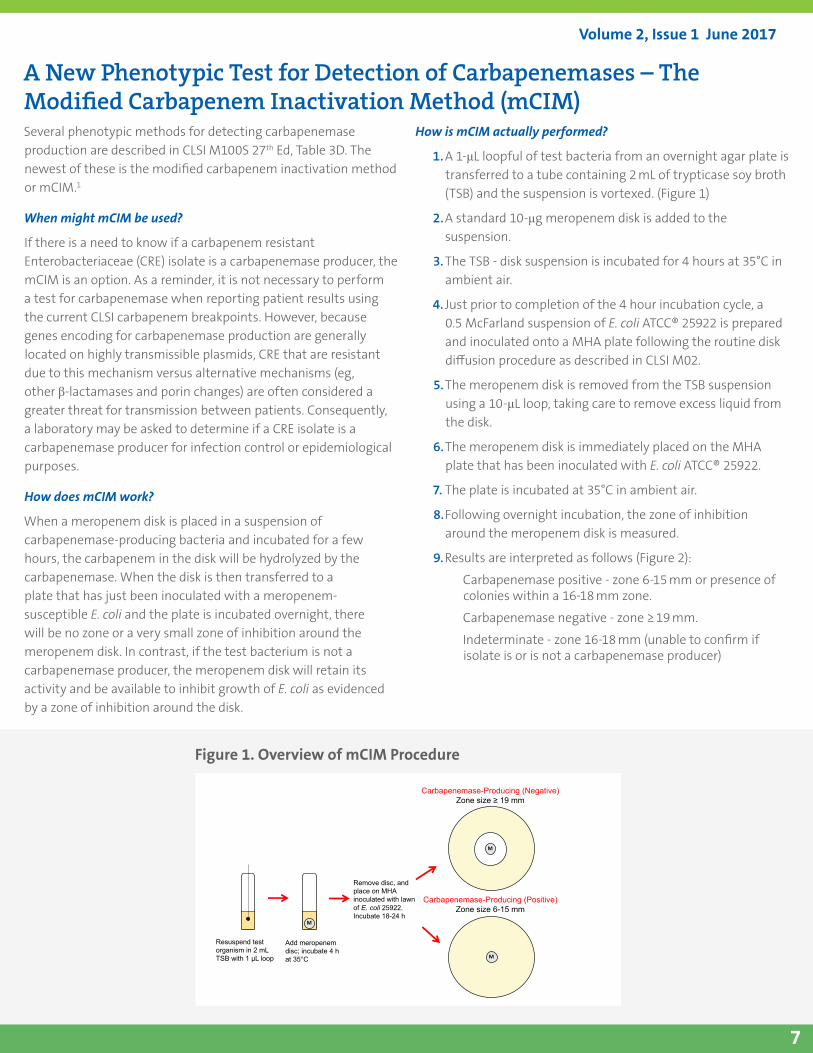
1. A 1-μL loopful of test bacteria from an overnight agar plate is transferred to a tube containing 2 mL of trypticase soy broth (TSB) and the suspension is vortexed. (Figure 1)
2. A standard 10-μg meropenem disk is added to the suspension.
3. The TSB - disk suspension is incubated for 4 hours at 35°C in ambient air.
4. Just prior to completion of the 4 hour incubation cycle, a 0.5 McFarland suspension of E. coli ATCC® 25922 is prepared and inoculated onto a MHA plate following the routine disk diffusion procedure as described in CLSI M02.
5. The meropenem disk is removed from the TSB suspension using a 10-μL loop, taking care to remove excess liquid from the disk.
6. The meropenem disk is immediately placed on the MHA plate that has been inoculated with E. coli ATCC® 25922.
7. The plate is incubated at 35°C in ambient air.
8. Following overnight incubation, the zone of inhibition around the meropenem disk is measured.
9. Results are interpreted as follows (Figure 2):
Carbapenemase positive - zone 6-15 mm or presence of colonies within a 16-18 mm zone.
Carbapenemase negative - zone ≥ 19 mm.
Indeterminate - zone 16-18 mm (unable to confirm if isolate is or is not a carbapenemase producer)
Figure 1. Overview of mCIM Procedure
M
Resuspend test organism in 2 mL TSB with 1 μL loop
Add meropenem disc; incubate 4 h at 35°C
Remove disc, and place on MHA inoculated with lawn of E. coli 25922.Incubate 18-24 h
Carbapenemase-Producing (Negative)Zone size ≥ 19 mm
Carbapenemase-Producing (Positive)Zone size 6-15 mm
Overview of mCIM Procedure
M
M
Volume 2, Issue 1 June 2017
8
A New Phenotypic Test for Detection of Carbapenemases (Continued)
What else should we know about mCIM?
The mCIM test is simple to perform with minimal hands-on time (< 5 min per isolate when testing multiple isolates) and uses laboratory supplies that are readily available. In fact, there are several advantages of mCIM over other phenotypic tests previously described for carbapenemase production (see Table). The mCIM has > 99% sensitivity and > 99% specificity for detection of carbapenemase producing Enterobacteriaceae. For additional details on performing mCIM (including QC recommendations), please see M100S 27th Ed.1 For results on the performance of mCIM as demonstrated in a controlled multicenter study, please see a recent publication.2
Does a verification need to be performed prior to implementation of mCIM?
Yes, a limited verification of at least 30 isolates (15 carbapenemase positive, 15 carbapenemase negative) needs to be performed to measure accuracy and precision of the assay in your laboratory. Characterized isolates (known carbapenemase producers/non-producers) from your own institution should be used, if possible. For laboratories lacking these isolates or those desiring a greater variety of carbapenem resistance mechanisms than available in house, a source for well-characterized isolates is included in the “Gram-negative Carbapenemase Detection Panel” from the FDA-CDC Antimicrobial Resistance Isolate Bank.
What about use of mCIM for Pseudomonas aeruginosa and Acinetobacter spp.?
The mCIM test has been shown to be reliable for detection of carbapenemase production in P. aeruginosa and a slight modification of the above-mentioned procedure will be included
for P. aeruginosa in M100S in 2018. Unfortunately, the mCIM as described here is not reliable for detecting carbapenemase production in Acinetobacter baumannii complex.
mCIM Key Points
• mCIM is a new phenotypic method for detection of carbapenemase producing Enterobacteriaceae and described in detail in CLSI M100S 27th ed.
• mCIM uses readily available laboratory reagents.
• Results from mCIM are available within 24 hours following testing of isolated colonies suspicious for carbapenemase production.
• mCIM is not reliable for testing Acinetobacter spp.
References
1 CLSI. Performance Standards for Antimicrobial Susceptibility Testing. 27th ed. CLSI document M100S. Wayne, PA: Clinical and Laboratory Standards Institute; 2017.
2 Pierce VM, et al. The modified carbapenem inactivation method (mCIM) for phenotypic detection of carbapene-mase production among Enterobacteriaceae. J Clin Microbiol. 2017;Epub Apr 5.
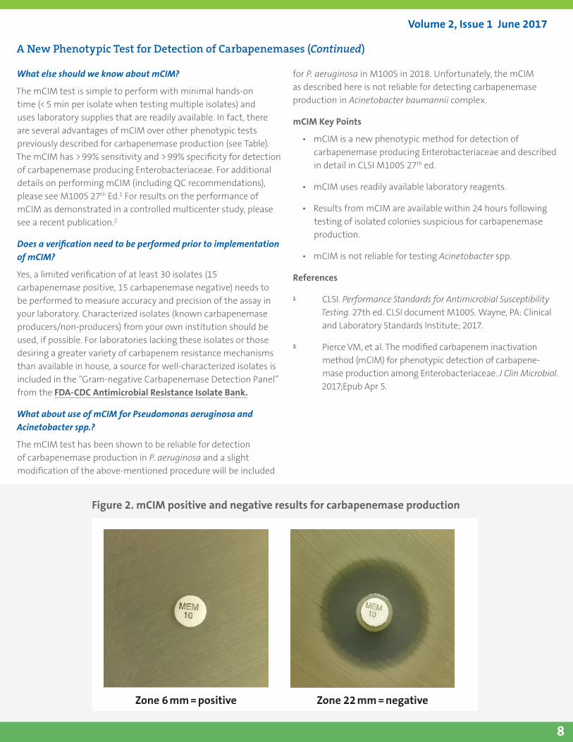
Figure 2. mCIM positive and negative results for carbapenemase production
Zone 6 mm = positive Zone 22 mm = negative
Volume 2, Issue 1 June 2017
9
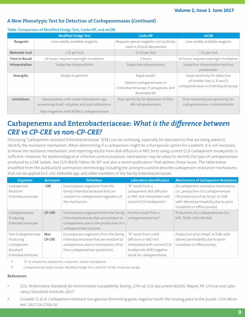
Table. Comparison of Modified Hodge Test, Carba NP, and mCIMModified Hodge Test Carba NP mCIM
Reagents Uses readily available reagents Requires special reagents, not routinely used in clinical laboratories
Uses readily available reagents
Materials Cost < $1 per test $2-10 per test < $1 per test
Time to Result 24 hours, requires overnight incubation 2 hours 24 hours, requires overnight incubation
Interpretation Subjective interpretation Subjective interpretation Subjective interpretation but less problematic
Strengths Simple to perform Rapid results
Detects carbapenemases in Enterobacteriaceae, P. aeruginosa, and
Acinetobacter
Good sensitivity for detection of Ambler class A, B and D
carbapenemases in Enterobacteriaceae
Limitations False positives with some Enterobacter spp. possessing AmpC enzymes and porin alterations
False negatives with NDM-1 carbapenemases
Poor sensitivity for detection of OXA-48 carbapenemases
Poor sensitivity and specificity for carbapenemases in Acinetobacter
Discussing “carbapenem resistant Enterobacteriaceae” (CRE) can be confusing, especially for laboratories that are being asked to identify the resistance mechanism. When determining if a carbapenem might be a therapeutic option for a patient, it is not necessary to know the resistance mechanism, and reporting results from disk diffusion or MIC tests using current CLSI carbapenem breakpoints is sufficient. However, for epidemiological or infection control purposes, laboratories may be asked to identify the type of carbapenemase produced by a CRE isolate. See CLSI M100 Tables 3A-3D1 and also a recent publication2 that address these issues. The table below (modified from the publication) summarizes terminology including the acronyms used to describe carbapenem resistance mechanisms that can be applied to E. coli, Klebsiella spp. and other members of the family Enterobacteriaceae.
a “R” to ertapenem, doripenem, imipenem, and/or meropenemb Carbapenemase tests include: Modified Hodge Test, carba NP, mCIM, molecular assays.
Carbapenems and Enterobacteriaceae: What is the difference between CRE vs CP-CRE vs non-CP-CRE?
Organism Acronyms Definition Laboratory Identification Mechanisms of Carbapenem Resistance
Carbapenem Resistant Enterobacteriaceae
CRE Encompasses organisms from the family Enterobacteriaceae that are resistant to carbapenems regardless of the mechanism.
“R” result from a carbapenem disk diffusion or MIC test interpreted with current CLSI breakpoints.a
All carbapenem resistance mechanisms (i.e., production of a carbapenemase OR production of an AmpC or ESBL with altered permeability due to porin mutations or efflux pumps)
Carbapenemase Producing Enterobacteriaceae
CP-CRE Encompasses organisms from the family Enterobacteriaceae that are resistant to carbapenems due to the production of a carbapenemase enzyme.
Positive result from a carbapenemase test.b
Production of a carbapenemase (i.e., KPC, NDM, OXA-48-like)
Non Carbapenemase Producing Carbapenem Resistant Enterobacteriaceae
Non CP-CRE
Encompasses organisms from the family Enterobacteriaceae that are resistant to carbapenems due to mechanisms other than carbapenemase production.
“R” result from a disk diffusion or MIC test interpreted with current CLSI breakpoints AND negative result for carbapenemase.
Production of an AmpC or ESBL with altered permeability due to porin mutations or efflux pumps.
References
1 CLSI. Performance Standards for Antimicrobial Susceptibility Testing. 27th ed. CLSI document M100S. Wayne, PA: Clinical and Labo-ratory Standards Institute; 2017.
2 Gniadek TJ, et al. Carbapenem-resistant non-glucose-fermenting gram-negative bacilli: the missing piece to the puzzle. J Clin Micro-biol. 2017;54:1700-10.
A New Phenotypic Test for Detection of Carbapenemases (Continued)
Volume 2, Issue 1 June 2017
10
Newer Developments in Antifungal Susceptibility Testing - Species-Specific Breakpoints for Candida spp.In November 2016, the ORWG presented the webinar “Practical Recommendations for Antifungal Susceptibility Testing and Reporting in Clinical Laboratories: New Breakpoints, New Drugs, New Guidelines.” Below are some highlights from the program that can be accessed through the CLSI webinar archives here, under Microbiology webinars.
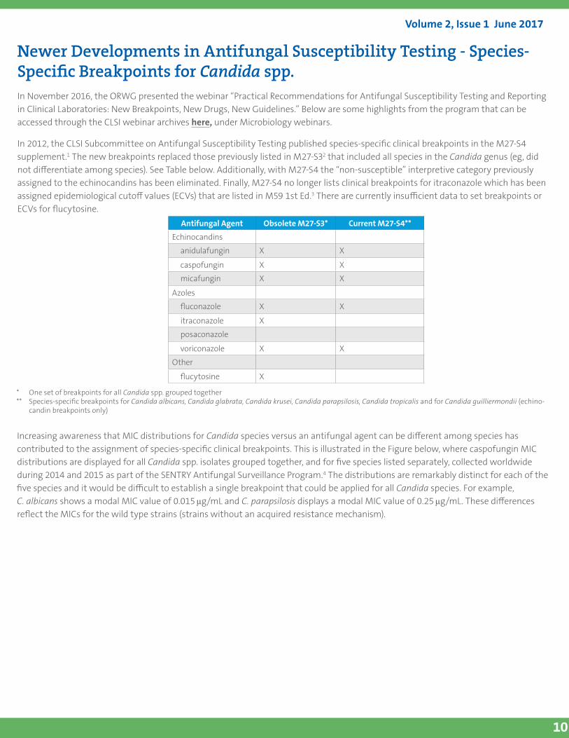
In 2012, the CLSI Subcommittee on Antifungal Susceptibility Testing published species-specific clinical breakpoints in the M27-S4 supplement.1 The new breakpoints replaced those previously listed in M27-S32 that included all species in the Candida genus (eg, did not differentiate among species). See Table below. Additionally, with M27-S4 the “non-susceptible” interpretive category previously assigned to the echinocandins has been eliminated. Finally, M27-S4 no longer lists clinical breakpoints for itraconazole which has been assigned epidemiological cutoff values (ECVs) that are listed in M59 1st Ed.3 There are currently insufficient data to set breakpoints or ECVs for flucytosine.
Antifungal Agent Obsolete M27-S3* Current M27-S4**
Echinocandins
anidulafungin X X
caspofungin X X
micafungin X X
Azoles
fluconazole X X
itraconazole X
posaconazole
voriconazole X X
Other
flucytosine X
* One set of breakpoints for all Candida spp. grouped together** Species-specific breakpoints for Candida albicans, Candida glabrata, Candida krusei, Candida parapsilosis, Candida tropicalis and for Candida guilliermondii (echino-
candin breakpoints only)
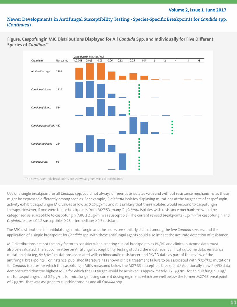
Increasing awareness that MIC distributions for Candida species versus an antifungal agent can be different among species has contributed to the assignment of species-specific clinical breakpoints. This is illustrated in the Figure below, where caspofungin MIC distributions are displayed for all Candida spp. isolates grouped together, and for five species listed separately, collected worldwide during 2014 and 2015 as part of the SENTRY Antifungal Surveillance Program.4 The distributions are remarkably distinct for each of the five species and it would be difficult to establish a single breakpoint that could be applied for all Candida species. For example, C. albicans shows a modal MIC value of 0.015 µg/mL and C. parapsilosis displays a modal MIC value of 0.25 µg/mL. These differences reflect the MICs for the wild type strains (strains without an acquired resistance mechanism).
Volume 2, Issue 1 June 2017
11
Newer Developments in Antifungal Susceptibility Testing - Species-Specific Breakpoints for Candida spp. (Continued)
Figure. Caspofungin MIC Distributions Displayed for All Candida Spp. and Individually for Five Different Species of Candida.*
CaspofunginMIC(µg/mL)Organism No.tested ≤0.0080.0150.030.060.120.250.51248>8
AllCandida spp. 2765
Candidaalbicans 1310
Candidaglabrata 514
Candidaparapsilosis 417
Candidatropicalis 264
Candidakrusei 93
Use of a single breakpoint for all Candida spp. could not always differentiate isolates with and without resistance mechanisms as these might be expressed differently among species. For example, C. glabrata isolates displaying mutations at the target site of caspofungin activity exhibit caspofungin MIC values as low as 0.25 µg/mL and it is unlikely that these isolates would respond to caspofungin therapy. However, if one were to use breakpoints from M27-S3, many C. glabrata isolates with resistance mechanisms would be categorized as susceptible to caspofungin (MIC ≤ 2 µg/ml was susceptible). The current revised breakpoints (µg/ml) for caspofungin and C. glabrata are: ≤ 0.12 susceptible; 0.25 intermediate; ≥ 0.5 resistant.
The MIC distributions for anidulafungin, micafungin and the azoles are similarly distinct among the five Candida species, and the application of a single breakpoint for Candida spp. with these antifungal agents could also impact the accurate detection of resistance.
MIC distributions are not the only factor to consider when creating clinical breakpoints as PK/PD and clinical outcome data must also be evaluated. The Subcommittee on Antifungal Susceptibility Testing studied the most recent clinical outcome data, resistance mutation data (eg, fks1/fks2 mutations associated with echinocandin resistance), and PK/PD data as part of the review of the antifungal breakpoints. For instance, published literature has shown clinical treatment failure to be associated with fks1/fks2 mutations for Candida isolates for which the caspofungin MICs measured below the M27-S3 susceptible breakpoint.5 Additionally, new PK/PD data demonstrated that the highest MICs for which the PD target would be achieved is approximately 0.25 µg/mL for anidulafungin, 1 µg/mL for caspofungin, and 0.5 µg/mL for micafungin using current dosing regimens, which are well below the former M27-S3 breakpoint of 2 µg/mL that was assigned to all echinocandins and all Candida spp.
* The new susceptible breakpoints are shown as green vertical dotted lines.
Volume 2, Issue 1 June 2017
12
Despite the rationale behind the new breakpoints, according to supplemental questions from the 2016 College of American Pathologists (CAP) Fungal proficiency testing survey, approximately one-third of subscribing laboratories still used the obsolete M27-S3 guidelines. While a minority (25/107; 23%) of these laboratories stated using the old M27-S3 guidelines in conjunction with the current M27-S4 guidelines, the majority of these laboratories (82/107, 77%) reported using solely the old guidelines. It is important for clinical laboratories to discontinue using M27-S3.
References
1 CLSI. Reference Method for Broth Dilution Antifungal Susceptibility Testing of Yeasts; Fourth Informational Supplement. CLSI document M27-S4. Wayne, PA: Clinical and Laboratory Standards Institute; 2012.
2 CLSI. Reference Method for Broth Dilution Antifungal Susceptibility Testing of Yeasts; Third Informational Supplement. CLSI document M27-S3. Wayne, PA: Clinical and Laboratory Standards Institute; 2008.
3 CLSI. Epidemiological Cutoff Values for Antifungal Susceptibility Tests; 1st edition. CLSI Supplement M59. Wayne, PA: Clinical and Laboratory Standards Institute; 2008.
4 Castanheira M, et al. Monitoring antifungal resistance in a global collection of invasive yeasts and moulds: application of the recently published CLSI epidemiological cutoff values and whole genome sequencing analysis for detection of resistance mechanisms. Manuscript submitted.
5 Pfaller MA, et al. Clinical breakpoints for the echinocandins and Candida revisited: integration of molecular, clinical, and microbiological data to arrive at species-specific interpretive criteria. Drug Resist Update. 2011;14(3):164-176.
Newer Developments in Antifungal Susceptibility Testing - Species-Specific Breakpoints for Candida spp. (Continued)
Volume 2, Issue 1 June 2017
13
Many of these challenges are in part related to the fact that in the United States, two organizations set breakpoints: FDA and CLSI. FDA breakpoints are listed in the drug prescribing information (package insert or drug label), which can be found at www.dailymed.nlm.nih.gov, whereas CLSI breakpoints are listed in CLSI’s M100S document.
Per US law, FDA can only approve an AST system using FDA breakpoints, and only for those organisms listed in the drug labeling to have activity both in vitro and in clinical infections. This latter piece is an important consideration, as FDA may have assigned breakpoints for the Enterobacteriaceae, but if no or few infections caused by Enterobacter aerogenes were encountered during the clinical trial (eg, as is the case for ceftazidime-avibactam), an AST system cannot be cleared by FDA for this particular species. These restrictions have led to the challenges listed in the table and much frustration for diagnostic manufacturers who cannot market tests that are analytically precise, have a CLSI (or even FDA) breakpoint and are critical to patient care.
The Good News!
The 21st Century Cures Act, which was passed in November 2016, and was signed into law by President Obama on December 13th, 2016, has the potential to provide solutions to these challenges (Sec. 3044). In particular, Sec 3044 of the act includes updates to Sec. 511 of the Federal Food, Drug and Cosmetic Act (21 USC 360a), to allow FDA to recognize breakpoints established by breakpoint-setting organizations, like CLSI, providing they uphold certain standards to mitigate potential conflicts of interest and maintain transparency in decision making processes. Furthermore, the intent is for all breakpoints to be listed on a website established by FDA, and breakpoints will be removed from drug prescribing information (eg, package insert or drug label). Diagnostics manufacturers can then submit data to FDA to have their AST system cleared using breakpoints listed on the new FDA website.
Importantly, this allows a streamlined process for FDA to recognize breakpoints established by CLSI (or other breakpoint-setting organizations), as this website must be updated at minimum every 6 months. Furthermore, it will provide more transparency regarding which breakpoints must be used by diagnostic manufacturers for FDA clearance. Manufacturers will be able to request FDA clearance for drugs that currently lack FDA breakpoints (eg, colistin). Finally, because the breakpoints will no longer be associated with the ‘indications for use’ listed in the drug package insert, many are hopeful that ASTs for drug/bug combinations without an FDA indication for use could be cleared by FDA.
The Timeline:
The intended timeline for these changes is rapid: FDA must establish the breakpoint website by November 2017. The drug manufacturers will have until November 2018 to delete breakpoints from their drug prescribing information. While promising, there remain some unanswered questions, including which breakpoint setting organization(s) will be recognized by FDA, and how quickly diagnostic manufacturers will be able to update their systems. If the delay between FDA updating Enterobacteriaceae carbapenem and cephem breakpoints and the diagnostic manufacturers updating these on their AST systems is any indication, it may be several years before these changes will have a positive impact on the clinical laboratory. On the flip side, many manufacturers of AST systems have a global presence and may be able to rapidly assemble and/or supplement the data that was submitted to non-US agencies for clearance in other countries, for submission to FDA.
The 21st Century Cures Act and the Future of ASTCurrent Challenges:
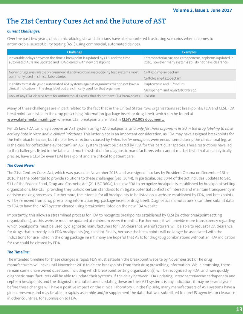
Over the past few years, clinical microbiologists and clinicians have all encountered frustrating scenarios when it comes to antimicrobial susceptibility testing (AST) using commercial, automated devices.
Challenge Examples
Inexorable delays between the time a breakpoint is updated by CLSI and the time automated ASTs are updated and FDA-cleared with new breakpoint
Enterobacteriaceae and carbapenems, cephems (updated in 2010, however many systems still do not have clearance)
Newer drugs unavailable on commercial antimicrobial susceptibility test systems most commonly used in clinical laboratories
Ceftazidime-avibactam
Ceftolozane-tazobactam
Inability to test drugs on automated AST systems against organisms that do not have a clinical indication in the drug label but are clinically used for that organism
Daptomycin and E. faecium
Meropenem and Acinetobacter spp.
Lack of any FDA-cleared tests for antimicrobial agents that do not have FDA breakpoints Colistin
Volume 2, Issue 1 June 2017
14
The 21st Century Cures Act and the Future of AST (Continued)
…to find current FDA Breakpoints and clinical indications listed in “Prescribing Information” (aka “Drug Label” or “Package Insert”), proceed as follows:
1. Go to: https://dailymed.nlm.nih.gov/dailymed/.
2. Enter a drug name and you will be directed towards one or more links to the Prescribing Information for the drug. If your search doesn’t yield results, try using the drug’s trade name. There is a separate link for each manufacturer of the drug. This means many links for generic drugs (ceftriaxone has 4 pages of links!). Select any of these to access the drug’s Prescribing Information. Eg, daptomycin.
3. Once inside the “Prescribing Information” document, search for “Clinical Pharmacology” section and “Microbiology” Subsection. Scroll through this subsection to find breakpoints and QC ranges.
4. Under “Microbiology,” check out the “Activity” subsection where it states something like “Drug X has been shown to be active against most isolates of the following microorganisms both in vitro and in clinical infections. The drug manufacturer has clinical data supporting positive outcomes for these species only.” For daptomycin and enterococci, you will see Enterococcus faecalis (VSE only) listed. This is sometimes called the “A” list.
5. And, then check out a second list of organisms, introduced with something like “at least 90% of the following bacteria exhibit an in vitro MIC less than or equal to the susceptible breakpoint for Drug X against isolates of similar genus or organism group. However, the efficacy of Drug X in treating clinical infections due to these bacteria has not been established in adequate and well-controlled clinical trials.” For daptomycin and enterococci, you will see E. faecalis (VRE) and Enterococcus faecium listed. This is sometimes called the “B” list.
Commercial manufacturers can currently seek FDA approval for only organisms in the “A” list for their AST!
Notes:
1. For most antimicrobial agents, there are multiple manufacturers of the drug and there are separate “Prescribing Information” documents for each product.
2. Some manufacturers of these antimicrobial agents have updated their breakpoints in the “Prescribing Information” and others have not. FDA keeps track of those which have updated breakpoints here.
3. Many patients are prescribed drugs for organisms that are not on list A or list B (“off label”). Some estimate up to 40% of critically ill patients are prescribed antibiotics off-label, and clinicians rely on AST results to help guide the decision to use these antibiotics.
Class D (OXA) Carbapenemases: the Achilles Heel of Carbapenemase Detection TestsCarbapenems play an essential role in healthcare. Due to their broad activity and relative stability against β-lactamases, these agents are often reserved for treatment of multi-drug resistant organisms in critically ill patients. A primary mechanism of resistance to carbapenems in gram-negative bacteria is production of carbapenemases—enzymes that confer resistance by hydrolyzing the essential four-membered β-lactam ring of the carbapenem molecules, rendering them inactive. Carbapenemases belong to one of three groups based on their molecular structure: Ambler class A, B, and D.1,2 Class A and D enzymes have serine residues at their active sites, while class B enzymes require zinc ions for activity. Historically, class D enzymes were thought to be mainly restricted to members of the genus Acinetobacter; however, it is now apparent that these enzymes are a significant cause of carbapenem resistance in other genera, including members of the family Enterobacteriaceae.
Hot Topic
Volume 2, Issue 1 June 2017
15
Class D (OXA) Carbapenemases: the Achilles Heel of Carbapenemase Detection Tests (Continued)
The first class D enzymes described were shown to confer resistance to penicillin and oxacillin (hence the prefix OXA). Subsequently in the 1980s, OXA enzymes that hydrolyze carbapenems were reported in Acinetobacter species, especially Acinetobacter baumannii. In the early 2000s, a novel enzyme, termed OXA-48, was isolated from a carbapenem resistant Klebsiella pneumoniae strain obtained from a patient in Istanbul, Turkey.3 OXA-48 and its variants (collectively often termed OXA-48-like β-lactamases) found in Enterobacteriaceae are listed in Table 1. These β-lactamases hydrolyze narrow-spectrum β-lactams (aminopenicillins and ureidopenicillins) and weakly hydrolyze carbapenems, but spare extended-spectrum cephalosporins (eg, ceftriaxone, ceftazidime, cefepime) and are not inhibited by clavulanic acid or the metal ion chelator EDTA. However, in the presence of permeability defects or other β-lactamases, OXA-48-like β-lactamases can impart high levels of carbapenem resistance.4
OXA-48 like β-lactamases are now widespread in Enterobacteriaceae, including K. pneumoniae and E. coli. They are endemic in many European and North African countries. Sporadic outbreaks/occurrences of clustered cases have been documented in the US,1 but given their weak activity against carbapenems, which sometimes results in MICs or zone size values in the susceptible range, their prevalence in the US is probably underestimated.
It can be difficult to identify bacteria harboring OXA-48-like β-lactamases in isolates of Enterobacteriaceae when tested with antimicrobial susceptibility testing systems routinely used in clinical laboratories. Current CLSI-recommended carbapenemase detection tests including the Modified Hodge test (MHT), the Carba NP and the modified carbapenem inactivation method (mCIM), that are based on the hydrolysis of a carbapenem can be falsely negative for isolates that produce OXA-48-like β-lactamases (Table 2).6-11 In contrast, nucleic acid-based assays targeting OXA-48-like genes are generally reliable.12 However, some commercial or laboratory-developed molecular assays may not detect all OXA-48-like β-lactamase genes due to sequence variations between them.13 Thus, the sensitivity of nucleic acid-based assays could be a shortcoming in the setting of changing epidemiology or emergence of novel enzymes. Finally, with the use of “direct from positive blood culture” assays, laboratories may be faced with the detection of an OXA-48-like β-lactamase gene in an isolate belonging to the family Enterobacteriaceae without phenotypic resistance to the carbapenems. In these situations, CLSI recommends that clinical laboratories utilizing current Enterobacteriaceae carbapenem breakpoints should interpret the agents as they test (i.e., if the MIC value corresponds to a susceptible interpretation, report as such).
Plasmid-mediated Ambler class D OXA-48-like β-lactamases have arisen as a significant cause of transmissible carbapenem resistance in members of the family Enterobacteriaceae and can be difficult to detect using routine antimicrobial susceptibility tests and phenotypic tests for carbapenemases. If an isolate demonstrates a susceptibility profile of resistance to narrow-spectrum β-lactams (eg, ampicillin, cefazolin) and shows intermediate or resistant results for carbapenems (using current CLSI breakpoints) but is susceptible to extended-spectrum cephalosporins (eg, ceftriaxone, ceftazidime, cefepime), performance of a molecular or phenotypic assay for carbapenemase should be considered. However, the limitations of phenotypic assays in detecting OXA-48 like β-lactamases must be taken into consideration. Optimally, performance of a molecular assay for OXA-48-like β-lactamases could be performed, particularly if there is any evidence of a potential outbreak. Subsequently, if an isolate is confirmed as a carbapenemase producer using a phenotypic assay or positive for an OXA-48-like gene using a molecular assay, the laboratory should follow their protocol for notification of the Infection Prevention team to help avert transmission of significant resistance genes.
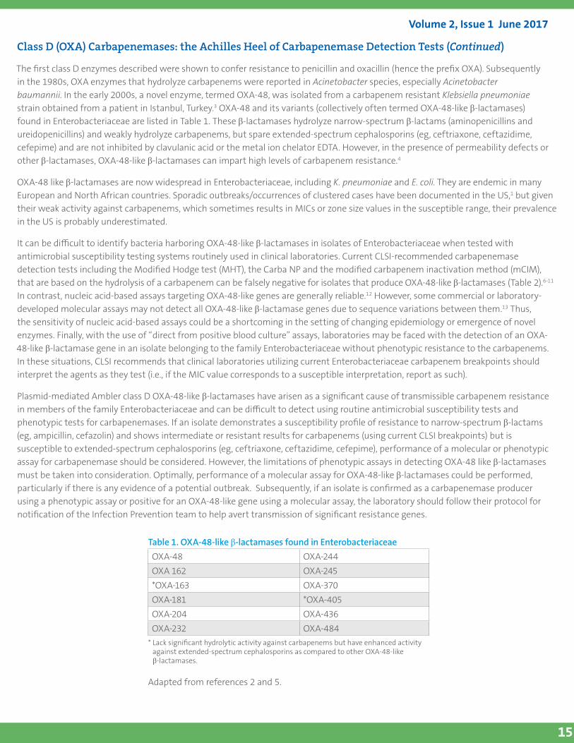
Table 1. OXA-48-like β-lactamases found in EnterobacteriaceaeOXA-48 OXA-244OXA 162 OXA-245*OXA-163 OXA-370OXA-181 *OXA-405OXA-204 OXA-436OXA-232 OXA-484
* Lack significant hydrolytic activity against carbapenems but have enhanced activity against extended-spectrum cephalosporins as compared to other OXA-48-like β-lactamases.
Adapted from references 2 and 5.
Volume 2, Issue 1 June 2017
16
Class D (OXA) Carbapenemases: the Achilles Heel of Carbapenemase Detection Tests (Continued)
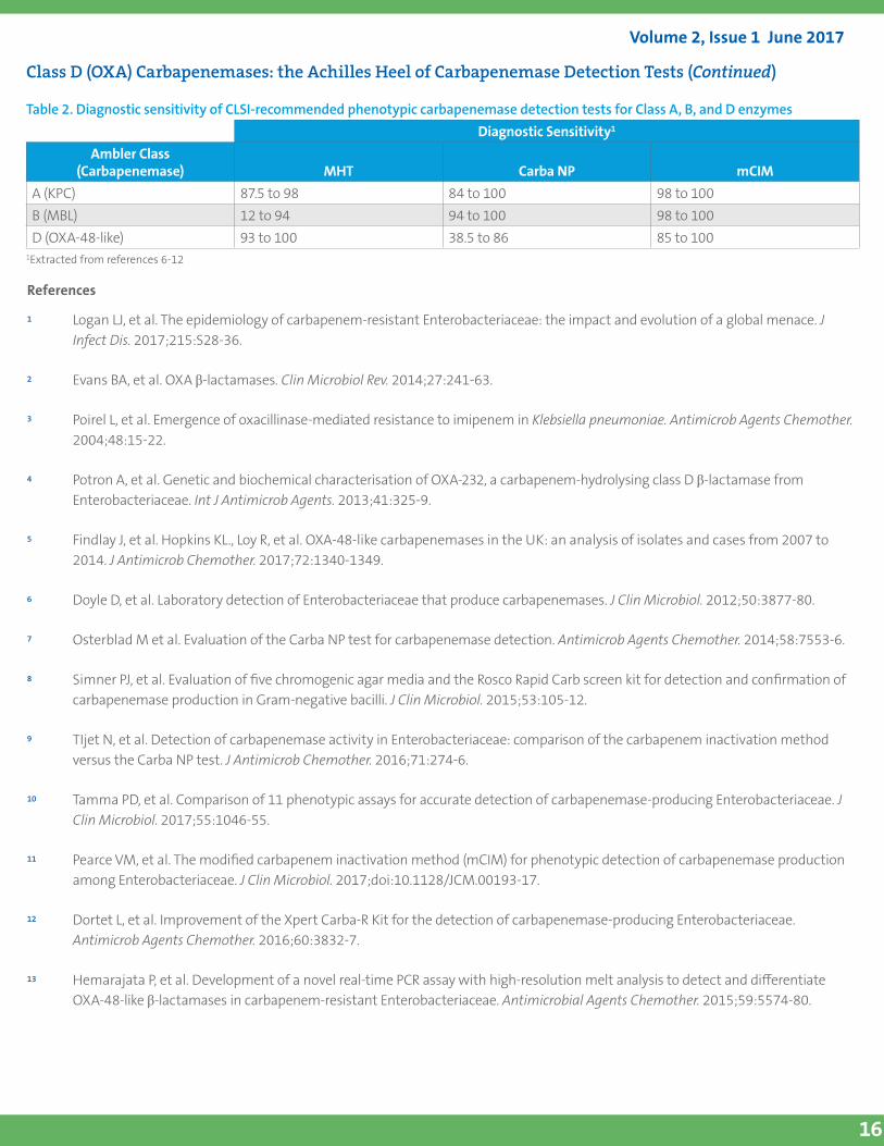
Table 2. Diagnostic sensitivity of CLSI-recommended phenotypic carbapenemase detection tests for Class A, B, and D enzymesDiagnostic Sensitivity1
Ambler Class(Carbapenemase) MHT Carba NP mCIM
A (KPC) 87.5 to 98 84 to 100 98 to 100B (MBL) 12 to 94 94 to 100 98 to 100D (OXA-48-like) 93 to 100 38.5 to 86 85 to 100
1Extracted from references 6-12
References
1 Logan LJ, et al. The epidemiology of carbapenem-resistant Enterobacteriaceae: the impact and evolution of a global menace. J Infect Dis. 2017;215:S28-36.
2 Evans BA, et al. OXA β-lactamases. Clin Microbiol Rev. 2014;27:241-63.
3 Poirel L, et al. Emergence of oxacillinase-mediated resistance to imipenem in Klebsiella pneumoniae. Antimicrob Agents Chemother. 2004;48:15-22.
4 Potron A, et al. Genetic and biochemical characterisation of OXA-232, a carbapenem-hydrolysing class D β-lactamase from Enterobacteriaceae. Int J Antimicrob Agents. 2013;41:325-9.
5 Findlay J, et al. Hopkins KL., Loy R, et al. OXA-48-like carbapenemases in the UK: an analysis of isolates and cases from 2007 to 2014. J Antimicrob Chemother. 2017;72:1340-1349.
6 Doyle D, et al. Laboratory detection of Enterobacteriaceae that produce carbapenemases. J Clin Microbiol. 2012;50:3877-80.
7 Osterblad M et al. Evaluation of the Carba NP test for carbapenemase detection. Antimicrob Agents Chemother. 2014;58:7553-6.
8 Simner PJ, et al. Evaluation of five chromogenic agar media and the Rosco Rapid Carb screen kit for detection and confirmation of carbapenemase production in Gram-negative bacilli. J Clin Microbiol. 2015;53:105-12.
9 TIjet N, et al. Detection of carbapenemase activity in Enterobacteriaceae: comparison of the carbapenem inactivation method versus the Carba NP test. J Antimicrob Chemother. 2016;71:274-6.
10 Tamma PD, et al. Comparison of 11 phenotypic assays for accurate detection of carbapenemase-producing Enterobacteriaceae. J Clin Microbiol. 2017;55:1046-55.
11 Pearce VM, et al. The modified carbapenem inactivation method (mCIM) for phenotypic detection of carbapenemase production among Enterobacteriaceae. J Clin Microbiol. 2017;doi:10.1128/JCM.00193-17.
12 Dortet L, et al. Improvement of the Xpert Carba-R Kit for the detection of carbapenemase-producing Enterobacteriaceae. Antimicrob Agents Chemother. 2016;60:3832-7.
13 Hemarajata P, et al. Development of a novel real-time PCR assay with high-resolution melt analysis to detect and differentiate OXA-48-like β-lactamases in carbapenem-resistant Enterobacteriaceae. Antimicrobial Agents Chemother. 2015;59:5574-80.
Volume 2, Issue 1 June 2017
17
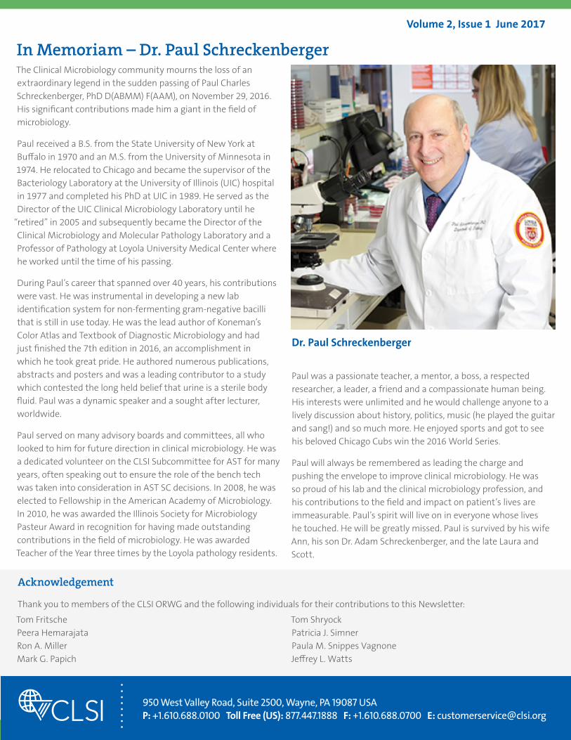
In Memoriam – Dr. Paul SchreckenbergerThe Clinical Microbiology community mourns the loss of an extraordinary legend in the sudden passing of Paul Charles Schreckenberger, PhD D(ABMM) F(AAM), on November 29, 2016. His significant contributions made him a giant in the field of microbiology.
Paul received a B.S. from the State University of New York at Buffalo in 1970 and an M.S. from the University of Minnesota in 1974. He relocated to Chicago and became the supervisor of the Bacteriology Laboratory at the University of Illinois (UIC) hospital in 1977 and completed his PhD at UIC in 1989. He served as the Director of the UIC Clinical Microbiology Laboratory until he
“retired” in 2005 and subsequently became the Director of the Clinical Microbiology and Molecular Pathology Laboratory and a Professor of Pathology at Loyola University Medical Center where he worked until the time of his passing.
During Paul’s career that spanned over 40 years, his contributions were vast. He was instrumental in developing a new lab identification system for non-fermenting gram-negative bacilli that is still in use today. He was the lead author of Koneman’s Color Atlas and Textbook of Diagnostic Microbiology and had just finished the 7th edition in 2016, an accomplishment in which he took great pride. He authored numerous publications, abstracts and posters and was a leading contributor to a study which contested the long held belief that urine is a sterile body fluid. Paul was a dynamic speaker and a sought after lecturer, worldwide.
Paul served on many advisory boards and committees, all who looked to him for future direction in clinical microbiology. He was a dedicated volunteer on the CLSI Subcommittee for AST for many years, often speaking out to ensure the role of the bench tech was taken into consideration in AST SC decisions. In 2008, he was elected to Fellowship in the American Academy of Microbiology. In 2010, he was awarded the Illinois Society for Microbiology Pasteur Award in recognition for having made outstanding contributions in the field of microbiology. He was awarded Teacher of the Year three times by the Loyola pathology residents.
Dr. Paul Schreckenberger
Paul was a passionate teacher, a mentor, a boss, a respected researcher, a leader, a friend and a compassionate human being. His interests were unlimited and he would challenge anyone to a lively discussion about history, politics, music (he played the guitar and sang!) and so much more. He enjoyed sports and got to see his beloved Chicago Cubs win the 2016 World Series.
Paul will always be remembered as leading the charge and pushing the envelope to improve clinical microbiology. He was so proud of his lab and the clinical microbiology profession, and his contributions to the field and impact on patient’s lives are immeasurable. Paul’s spirit will live on in everyone whose lives he touched. He will be greatly missed. Paul is survived by his wife Ann, his son Dr. Adam Schreckenberger, and the late Laura and Scott.
Tom FritschePeera HemarajataRon A. Miller Mark G. Papich
Tom ShryockPatricia J. Simner Paula M. Snippes VagnoneJeffrey L. Watts
Acknowledgement
Thank you to members of the CLSI ORWG and the following individuals for their contributions to this Newsletter:
4
950 West Valley Road, Suite 2500, Wayne, PA 19087 USA P: +1.610.688.0100 Toll Free (US): 877.447.1888 F: +1.610.688.0700 E: [email protected]