Embed Size (px)
Citation preview
Proc. Natl. Acad. Sci. USAVol. 93, pp. 2149-2154, March 1996Biochemistry
Anticoagulant repertoire of the hookworm Ancylostoma caninum(Ascaris-like inhibitors/blood coagulation/serine proteases/factors Xa and Vlla)
PATRICK STANSSENSt, PETER W. BERGUMt, YANNICK GANSEMANSt, LAURENT JESPERSt, YVES LAROCHEt,STEPHEN HUANGt, STEVEN MAKIt, JORIS MESSENSt, MARC LAUWEREYSt, MICHAEL CAPPELLO§,PETER J. HOTEZ§, IGNACE LASTERSt, AND GEORGE P. VLASUKt¶1Corvas International Inc., San Diego, CA 92121; §Departments of Pediatrics and Epidemiology and Public Health, Yale University School of Medicine, NewHaven, CT 06520; and tCorvas International N.V., Ghent, Belgium
Communicated by Laszlo Lorand, Northwestern University Medical School, Chicago, IL, December 7, 1995 (received for review November 9, 1995)
ABSTRACT Hookworms are hematophagous nematodesthat infect a wide range of mammalian hosts, includinghumans. There has been speculation for nearly a century as tothe identity of the anticoagulant substance(s) used by theseorganisms to subvert host hemostasis. Using molecular clon-ing, we describe a family of potent small protein (75-84 aminoacids) anticoagulants from the hookworm Ancylostoma cani-num termed AcAP (A. caninum anticoagulant protein). Tworecombinant AcAP members (AcAP5 and AcAP6) directlyinhibited the catalytic activity of blood coagulation factor Xa(fXa), while a third form (AcAPc2) predominantly inhibitedthe catalytic activity of a complex composed of blood coagu-lation factor Vlla and tissue factor (fVIIa/TF). The inhibitionof fVIIa/TF was by a unique mechanism that required theinitial formation of a binary complex of the inhibitor with fXaat a site on the enzyme that is distinct from the catalytic center(exo-site). The sequence ofAcAPc2 as well as the utilization ofan exo-site on fXa distinguishes this inhibitor from themammalian anticoagulant TFPI (tissue factor pathway inhib-itor), which is functionally equivalent with respect to fXa-dependent inhibition of fVIIa/TF. The relative sequencepositions of the reactive site residues determined for AcAP5with the homologous regions in AcAP6 and AcAPc2 as well asthe pattern of 10 cysteine residues present in each of theinhibitors suggest that the AcAPs are distantly related to thefamily of small protein serine protease inhibitors found in thenonhematophagous nematode Ascaris lumbricoides var. suum.
Hookworm infection is a leading cause of iron deficiencyanemia in developing countries, affecting approximately onebillion people worldwide. Hookworms cause anemia in theirhost by extracting their blood meal from lacerated capillariesin the mucosa of the small intestine over an extended periodof time (1). Like other hematophagous invertebrates such asticks (2, 3) and leeches (4, 5), hookworms have evolved highlyeffective anticoagulant strategies to facilitate the acquisition oftheir required blood meal. Although the anticoagulant prop-erties of adult hookworm extracts have been the subject ofscientific investigation for nearly a century (6-8), only recentlyhas an attempt been made to characterize the molecularentities responsible for this function.We have previously described the identification (9) and
purification (10) of a potent inhibitor of blood coagulationfactor Xa (fXa) (termed AcAP for Ancylostoma caninumanticoagulant peptide) that represents a major anticoagulantactivity present in soluble protein extracts of adult A. caninumhookworms. Determination of the first 30 amino acids ofpurified AcAP revealed a unique partial sequence with het-erogeneity at two distinct positions, suggesting that more thanone protein may be responsible for its anticoagulant activity
The publication costs of this article were defrayed in part by page chargepayment. This article must therefore be hereby marked "advertisement" inaccordance with 18 U.S.C. §1734 solely to indicate this fact.
(10). We sought to determine the extent of molecular heter-ogeneity in the original AcAP preparation purified from A.caninum. Here, we report the cloning, recombinant expres-sion, and characterization of three homologous small proteinsthat specifically inhibit different key blood coagulation serineproteases.11 From these studies, it appears that the hookwormA. caninum has evolved an effective strategy for manipulationof the mammalian blood coagulation pathway that is mecha-nistically and structurally distinct from those strategies utilizedby other hematophagous parasites or the mammalian host.
MATERIALS AND METHODScDNA Isolation and Sequencing. An oligo(dT)-primed
cDNA library was constructed in the Agtll Sfi-Not vector(Promega) and screened by described procedures (11). ThemRNA used for cDNA synthesis was isolated from adult A.caninum hookworms (Antibody Systems, Bedford, TX). Re-combinant phage (_ 106) were probed by standard procedures(11) with the radiolabeled oligonucleotide 5'-AAR GCI TAYCCI GAR TGY GGI GAR AAY GAR TGG-3' (where R =A or G and Y = T or C). The cDNA inserts of five candidateclones were subcloned on pGEM phagemids (Promega) andboth strands were sequenced by using the Autoread kit and theA.L.F. sequencer (Pharmacia). The sequence data revealedtwo different types of cDNAs corresponding to AcAP5 andAcAP6. Restriction analysis of additional positive cDNAclones demonstrated that they all belong to one or the othertype and that the two cDNAs are approximately equallyrepresented in the library.
Production of Recombinant (r)AcAPs. The correspondingproteins encoded by the AcAP cDNAs were produced in theyeast Pichia pastoris using the vector pYAM7SP8 as described(12). In each case, a His' transformant of strain GS115 (13)that secreted a high level of inhibitory activity (see below)upon induction and exhibited a wild-type, methanol utilizationphenotype, was selected. All three recombinant proteins werepurified from the yeast culture medium as follows. The pH ofthe culture supernatant was adjusted to pH 3.0 and theconductivity was brought to <10 mS/cm. The diluted super-natant was applied to a Poros2OHS (Perseptive Biosystems,Framingham, MA) column preequilibrated with cation buffer(50 mM sodium citrate, pH 3). The column was washed withthe same buffer and subsequently eluted with cation buffercontaining 1 M NaCl. Material with inhibitory activity was
Abbreviations: AcAP, Ancylostoma caninum anticoagulant peptide; f,human blood coagulation factor; PT, prothrombin time; rAcAP,recombinant AcAP; TF, human tissue factor; PLV, phospholipidvesicle.ITo whom reprint requests should be addressed at: Corvas Interna-tional Inc., 3030 Science Park Road, San Diego, CA 92121.'The sequences reported in this paper have been deposited in theGenBank data base [accession nos. U30795 (AcAP5), U30794(AcAP6), and U30793 (AcAPc2)].
2149

2150 Biochemistry: Stanssens et al.
pooled and fractionated on Superdex 30 PG equilibrated with10 mM sodium phosphate, pH 7.4/150 mM NaCl. The pooledinhibitory activity was fractionated by reverse-phase HPLCusing a C18 column (model 218TP54; Vydac, Hesperia, CA)developed with a linear gradient of 10-35% CH3CN in 0.1%trifluoroacetic acid. Fractions containing inhibitory activitywere pooled and lyophilized. Each rAcAP was judged >95%pure using analytical reverse-phase HPLC, electrospray massspectrometry, and quantitative amino acid analysis and wasshown to be correctly processed by N-terminal sequenceanalysis (data not shown). Each protein was quantified byamino acid analysis.Prothrombin Time Clotting Assay. The anticoagulant prop-
erties of the rAcAP proteins were measured ex vivo, using theprothrombin time (PT) clotting assay. Dilutions of each rA-cAP were made in freshly thawed normal human plasma(George King Biomedical, Overland Park, KS) and assayedusing the Coag-A-Mate RA4 automated coagulometer (Gen-eral Diagnostics, Organon Technika) and Simplastin Excel asthe initiating reagent according to the manufacturer's instruc-tions (Organon Technika).
Serine Protease Specificity. The selectivity profile of therAcAP anticoagulants was examined against 11 serine pro-teases: human-derived proteins, fXa, a-thrombin, plasmin,fXIa, fXIIa, recombinant tissue plasminogen activator, acti-vated protein C, urokinase, plasma kallikrein, and the bovine-derived proteins trypsin and chymotrypsin. The sources for theenzymes and the conditions for the chromogenic assays wereas described (10), except that the final concentrations ofenzyme and inhibitor were 0.75 and 75 nM, respectively. Theresults are expressed as percentage inhibition compared to thecontrol (uninhibited) substrate hydrolysis rate.
fXa and Prothrombinase Inhibition Assays. The assays forinhibition of free fXa amidolytic activity and of thrombingeneration by the preformed prothrombinase complex wereperformed as described (10). The velocity of substrate hydro-lysis in the presence (Vi) and absence (VO) of inhibitor wasmeasured. Values for the apparent equilibrium dissociationconstant for the El complex (Ki*) were determined from theratios of Vl/Vo as described (10) using the algorithm derivedfor analysis of tight-binding inhibition (14).fVIIa/TF Inhibition Assays. The enzymatic activity of a
complex of recombinant fVIIa (Novo-Nordisk, Copenhagan)and recombinant full-length tissue factor (TF; Corvas Inter-national) was determined in the presence and absence of eitherfXa or the catalytically inactive derivative EGR-fXa (i.e., fXatreated with the irreversible active-site inhibitor Glu-Gly-Arg-chloromethylketone; Hematologic Technologies, Essex Junc-tion, VT). EGR-fXa was determined to have <0.05% of theamidolytic activity of untreated fXa. TF was incorporated intophospholipid vesicles of uniform size (PLV; 120 nm averagediameter) as described (15). A complex of TF/PLV and fVIIawas formed for 10 min in 10 mM Hepes, pH 7.5/150 mM NaCl(HBS) containing 0.1% bovine serum albumin and 3 mMCaCl2 (HBSA) prior to the addition of inhibitor and/orsubstrate.Activation of iX. The fIX zymogen (Hematologic Technol-
ogies) was depleted of residual fVIl and fX by sequentialmonoclonal antibody affinity chromatography prior to reduc-tive tritiation (16). The resulting 3H-flX preparation had aspecific activity of 2 x 10⁸ dpm/mg and retained 97% of theoriginal clotting activity measured in fIX-deficient plasma(George King Biomedical). To measure 3H-fIX activation,preformed fVIIa/TF, inhibitor and fXa, or EGR-fXa werepreincubated for 60 min prior to initiating the reaction by theaddition of the 3H-fIX substrate. The final concentration ofreactants common to all the assays in a total vol of 420 Al ofHBSA was as follows: fVIIa, 50 pM; TF, 2.7 nM; PLV, 6.4 jIM;fXa or EGR-fXa, 0-500 pM; rAcAP inhibitors, 0-1 nM; and3H-fIX, 200 nM (-5 times Kmn). The rate of 3H-fIX activation
was determined by linear regression analysis of the acid-soluble 3H counts (i.e., activation peptide) released over timeunder steady-state conditions, where <5% of the radiolabeledsubstrate was consumed. The reaction was stopped at specifictime points (8, 16, 24, 32, and 40 min) by diluting 80 p,l of thereaction mixture with an equal vol of HBS containing 50 mMEDTA and 0.5% BSA followed by addition of an equal vol of6% (wt/vol) trichloroacetic acid at 0°C. After centrifugation,the radioactivity contained in the resulting supernatant wasquantitated. The background (<1.0% of uninhibited controlvelocity) was subtracted. Kj* values were determined from theratios of the inhibited/uninhibited initial velocities (Vl/Vo) asdescribed above.Amidolytic activity. Preformed fVIIa/TF was incubated with
inhibitor and EGR-fXa for 30 min at ambient temperatureprior to addition of the peptidyl chromogenic substrate S-2288(D-Ile-Pro-Arg-p-nitroaniline; Kabi Pharmacia Hepar, Frank-lin, OH). The final concentration of reactants in a total vol of200 ,ul of HBSA was as follows: fVIIa, 750 pM; TF, 3.0 nM;PLV, 6.4 ,uM; EGR-FXa, 2.5 nM; rAcAP, 0-25 nM; andS-2288, 3.0 mM (-3 times Km). Initial velocities were mea-sured as a linear increase in the absorbance at 405 nm over 10min, using a Thermomax kinetic microplate reader (MolecularDevices), under steady-state conditions, where <5% of thesubstrate was consumed.
Cleavage of rAcAP5 by fXa. Purified rAcAP5 was incubatedwith fXa at a final molar ratio of 22:1 for 8 hr at 37°C in 0.1M citrate buffer (pH 5.0). Following disulfide reduction withdithiothreitol and alkylation with iodoacetamide, the sampleswere evaluated by SDS/PAGE using a 16% Tricine gel(NOVEX, San Diego) and visualized by Coomassie bluestaining. Identification of the fXa cleavage site in rAcAP5 wasdetermined by N-terminal sequence analysis following purifi-cation of nonreduced material by reverse-phase HPLC on aC18 column (Vydac). Two sequences were detected, the firststarting at the authentic N terminus and the second beginningwith Gly-41.
RESULTS AND DISCUSSIONCloning of AcAP-Related cDNA Sequences. AcAP-related
cDNA clones were initially identified by hybridization, using anoligonucleotide probe specific for the predominant N-terminalsequence determined for purified AcAP (10). Sequence anal-ysis of two representative isolates identified with this proberevealed unique open reading frames encoding highly homol-ogous polypeptides, designated AcAP5 and AcAP6 (Fig. 1).The translated sequences were in agreement with the sequencefor the first 30 amino acids determined for purified AcAP andconfirmed that the heterogeneity observed at positions 14 and18 was attributable to the presence of two distinct geneproducts (Fig. 1). Initial attempts to express the AcAP5/6cDNAs utilized transient expression in COS cells and resultedin the secretion of proteins that carry an Arg-Thr-Val-ArgN-terminal extension when compared to A. caninum-derivedAcAP (data not shown). This result suggests that the AcAP5/6primary translation products contain a 19-residue secretionsignal followed by a 4-residue prosequence, which is apparentlynot processed by COS cells (Fig. 1). The mature sequences forAcAP5 and AcAP6 contain 77 and 75 amino acids, respec-tively, and their calculated molecular sizes are similar to thatdetermined for purified AcAP (10). The sequences of bothmature proteins include 10 cysteine residues and predict anacidic pl.
Using a filimentous phage display, we previously selected anA. caninum cDNA based on the binding of the encodedproduct (fusedtt the C terminus of coat protein VI) to fXa(see ref. 19). Based on a comparison with AcAPS/6, the cDNAtranslation product is predicted to contain the completemature sequence of a third AcAP-like protein with a partial
Proc. Natl. Acad. Sci. USA 93 (1996)
Proc. Natl. Acad. Sci. USA 93 (1996) 2151
AcAP5AcAP6AcAPc2
ATI
ACAP5ACAP6AcAPc2
ATI
- 1MKMLYAIAIMFLLVSLCSA RTVR KAYPEC-GENEWLDDCGTQKPCEAKC----NEEPPMKMLYAIAIMFLLVSLCST RTVR KAYPEC-GENEWLDVCGTKKPCEAKC----SEE--............ .LVSYCSG KATMQC-GENEKYDSCGSKE-CDKKCKYDGVEE-E
EAEKCTKPNEQWTKCG ---GCEGTCAQK-
EEEDPI--CRSRGCLLPPACVCKD--GFYRDTVIGDCVREEEC--DQHEIIHV 77EEEDPI--CRSFSCPGPAACVCED--GFYRDTVIGDCVKEEEC--DQHEIIHV 75DDEEPNVPCLVRVCHQD--CVCEE--GFYRNK-DDKCVSAEDCELDNMDFIYPGTRN 84
-----IVP-CTRECK-PPRCECIASAGFVRDA-QGNCIKFEDCPK 62
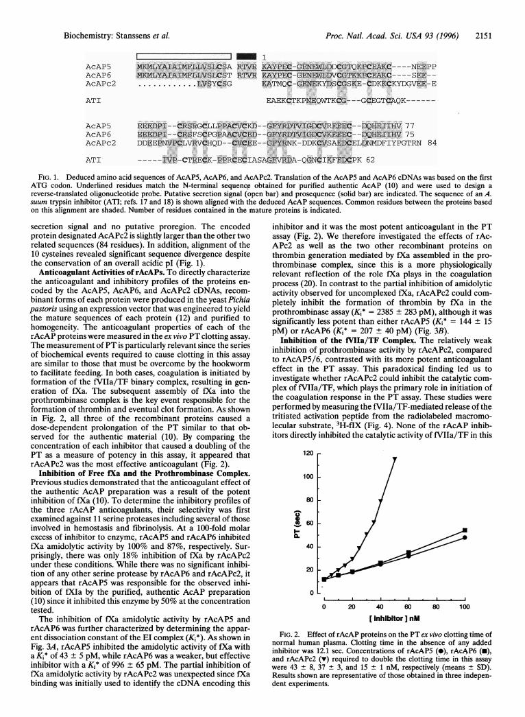
FIG. 1. Deduced amino acid sequences of AcAP5, AcAP6, and AcAPc2. Translation of the AcAP5 and AcAP6 cDNAs was based on the firstATG codon. Underlined residues match the N-terminal sequence obtained for purified authentic AcAP (10) and were used to design areverse-translated oligonucleotide probe. Putative secretion signal (open bar) and prosequence (solid bar) are indicated. The sequence of an A.suum trypsin inhibitor (ATI; refs. 17 and 18) is shown aligned with the deduced AcAP sequences. Common residues between the proteins basedon this alignment are shaded. Number of residues contained in the mature proteins is indicated.
secretion signal and no putative proregion. The encodedprotein designated AcAPc2 is slightly larger than the other tworelated sequences (84 residues). In addition, alignment of the10 cysteines revealed significant sequence divergence despitethe conservation of an overall acidic pl (Fig. 1).
Anticoagulant Activities of rAcAPs. To directly characterizethe anticoagulant and inhibitory profiles of the proteins en-coded by the AcAP5, AcAP6, and AcAPc2 cDNAs, recom-binant forms of each protein were produced in the yeast Pichiapastoris using an expression vector that was engineered to yieldthe mature sequences of each protein (12) and purified tohomogeneity. The anticoagulant properties of each of therAcAP proteins were measured in the ex vivo PT clotting assay.The measurement of PT is particularly relevant since the seriesof biochemical events required to cause clotting in this assayare similar to those that must be overcome by the hookwormto facilitate feeding. In both cases, coagulation is initiated byformation of the fVIIa/TF binary complex, resulting in gen-eration of fXa. The subsequent assembly of fXa into theprothrombinase complex is the key event responsible for theformation of thrombin and eventual clot formation. As shownin Fig. 2, all three of the recombinant proteins caused adose-dependent prolongation of the PT similar to that ob-served for the authentic material (10). By comparing theconcentration of each inhibitor that caused a doubling of thePT as a measure of potency in this assay, it appeared thatrAcAPc2 was the most effective anticoagulant (Fig. 2).
Inhibition of Free fXa and the Prothrombinase Complex.Previous studies demonstrated that the anticoagulant effect ofthe authentic AcAP preparation was a result of the potentinhibition of fXa (10). To determine the inhibitory profiles ofthe three rAcAP anticoagulants, their selectivity was firstexamined against 11 serine proteases including several of thoseinvolved in hemostasis and fibrinolysis. At a 100-fold molarexcess of inhibitor to enzyme, rAcAP5 and rAcAP6 inhibitedfXa amidolytic activity by 100% and 87%, respectively. Sur-prisingly, there was only 18% inhibition of fXa by rAcAPc2under these conditions. While there was no significant inhibi-tion of any other serine protease by rAcAP6 and rAcAPc2, itappears that rAcAP5 was responsible for the observed inhi-bition of fXIa by the purified, authentic AcAP preparation(10) since it inhibited this enzyme by 50% at the concentrationtested.The inhibition of fXa amidolytic activity by rAcAP5 and
rAcAP6 was further characterized by determining the appar-ent dissociation constant of the El complex (Ki*). As shown inFig. 3A, rAcAP5 inhibited the amidolytic activity of fXa witha Kj* of 43 + 5 pM, while rAcAP6 was a weaker, but effectiveinhibitor with a Kj* of 996 ± 65 pM. The partial inhibition offXa amidolytic activity by rAcAPc2 was unexpected since fXabinding was initially used to identify the cDNA encoding this
inhibitor and it was the most potent anticoagulant in the PTassay (Fig. 2). We therefore investigated the effects of rAc-APc2 as well as the two other recombinant proteins onthrombin generation mediated by fXa assembled in the pro-thrombinase complex, since this is a more physiologicallyrelevant reflection of the role fXa plays in the coagulationprocess (20). In contrast to the partial inhibition of amidolyticactivity observed for uncomplexed fXa, rAcAPc2 could com-pletely inhibit the formation of thrombin by fXa in theprothrombinase assay (Ki* = 2385 + 283 pM), although it wassignificantly less potent than either rAcAP5 (Ki* = 144 + 15pM) or rAcAP6 (Ki* = 207 + 40 pM) (Fig. 3B).
Inhibition of the fVIIa/TF Complex. The relatively weakinhibition of prothrombinase activity by rAcAPc2, comparedto rAcAP5/6, contrasted with its more potent anticoagulanteffect in the PT assay. This paradoxical finding led us toinvestigate whether rAcAPc2 could inhibit the catalytic com-plex of fVIIa/TF, which plays the primary role in initiation ofthe coagulation response in the PT assay. These studies wereperformed by measuring the fVIIa/TF-mediated release of thetritiated activation peptide from the radiolabeled macromo-lecular substrate, 3H-flX (Fig. 4). None of the rAcAP inhib-itors directly inhibited the catalytic activity of fVIIa/TF in this
120 r
100 -
80 -
a)0@1
I~-0.
60 -
40 k
20 -
0
0 20 40 60
[ Inhibitor ] nM80 100
FIG. 2. Effect of rAcAP proteins on the PT ex vivo clotting time ofnormal human plasma. Clotting time in the absence of any addedinhibitor was 12.1 sec. Concentrations of rAcAP5 (0), rAcAP6 (m),and rAcAPc2 (v) required to double the clotting time in this assaywere 43 ± 8, 37 + 3, and 15 + 1 nM, respectively (means + SD).Results shown are representative of those obtained in three indepen-dent experiments.
Biochemistry: Stanssens et al.
2152 Biochemistry: Stanssens et alt
A 1.0
0.8
02-1
0.6
0.4
0.2
0.0
B 1.0
0.8
0el5-
0.6
0.4
A 100FC0
4)
SCe-
0
x4
CI
c..0
C._
80 1
60 k
40 1
20 1
05 10 15 20 25
0 5 10 1 5 20 25
B 1.0
I e0 100 200 300 400 500
[ fXa or EGR-fXa ] pM
0.8 V
02>
0.6 1
0.4 k0.2
0.0
0 5 10 15 20 25
[Inhibitor] nM
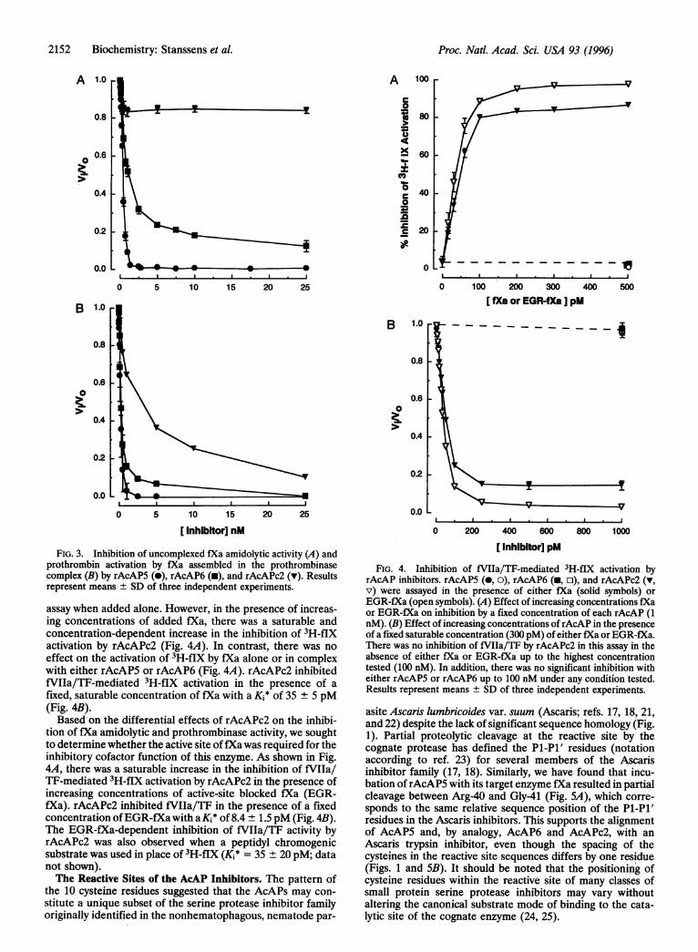
FIG. 3. Inhibition of uncomplexed fXa amidolytic activity (A) andprothrombin activation by fXa assembled in the prothrombinasecomplex (B) by rAcAP5 (0), rAcAP6 (-), and rAcAPc2 (v). Resultsrepresent means ± SD of three independent experiments.
assay when added alone. However, in the presence of increas-ing concentrations of added fXa, there was a saturable andconcentration-dependent increase in the inhibition of 3H-fIXactivation by rAcAPc2 (Fig. 4A). In contrast, there was noeffect on the activation of 3H-flX by fXa alone or in complexwith either rAcAP5 or rAcAP6 (Fig. 4A). rAcAPc2 inhibitedfVIIa/TF-mediated 3H-flX activation in the presence of afixed, saturable concentration of fXa with a Kj* of 35 ± 5 pM(Fig. 4B).Based on the differential effects of rAcAPc2 on the inhibi-
tion of fXa amidolytic and prothrombinase activity, we soughtto determine whether the active site offXa was required for theinhibitory cofactor function of this enzyme. As shown in Fig.4A, there was a saturable increase in the inhibition of fVIIa/T1-mediated 3H-flX activation by rAcAPc2 in the presence ofincreasing concentrations of active-site blocked fXa (EGR-fXa). rAcAPc2 inhibited fVIIa/TF in the presence of a fixedconcentration ofEGR-fXa with aKi* of 8.4 ± 1.5 pM (Fig. 4B).The EGR-fXa-dependent inhibition of fVIIa/TF activity byrAcAPc2 was also observed when a peptidyl chromogenicsubstrate was used in place of 3H-flX (Ki* = 35 + 20 pM; datanot shown).The Reactive Sites of the AcAP Inhibitors. The pattern of
the 10 cysteine residues suggested that the AcAPs may con-stitute a unique subset of the serine protease inhibitor familyoriginally identified in the nonhematophagous, nematode par-
0.2 L
o.o L
0 200 400 600 800 1000
[Inhibitor] pM
FIG. 4. Inhibition of fVIIa/TF-mediated 3H-fIX activation byrAcAP inhibitors. rAcAPS (0, o), rAcAP6 (-, o), and rAcAPc2 (v,v) were assayed in the presence of either fXa (solid symbols) orEGR-fXa (open symbols). (A) Effect of increasing concentrations fXaor EGR-fXa on inhibition by a fixed concentration of each rAcAP (1nM). (B) Effect of increasing concentrations ofrAcAP in the presenceof a fixed saturable concentration (300 pM) of either fXa or EGR-fXa.There was no inhibition of fVIIa/TF by rAcAPc2 in this assay in theabsence of either fXa or EGR-fXa up to the highest concentrationtested (100 nM). In addition, there was no significant inhibition witheither rAcAP5 or rAcAP6 up to 100 nM under any condition'tested.Results represent means ± SD of three independent experiments.
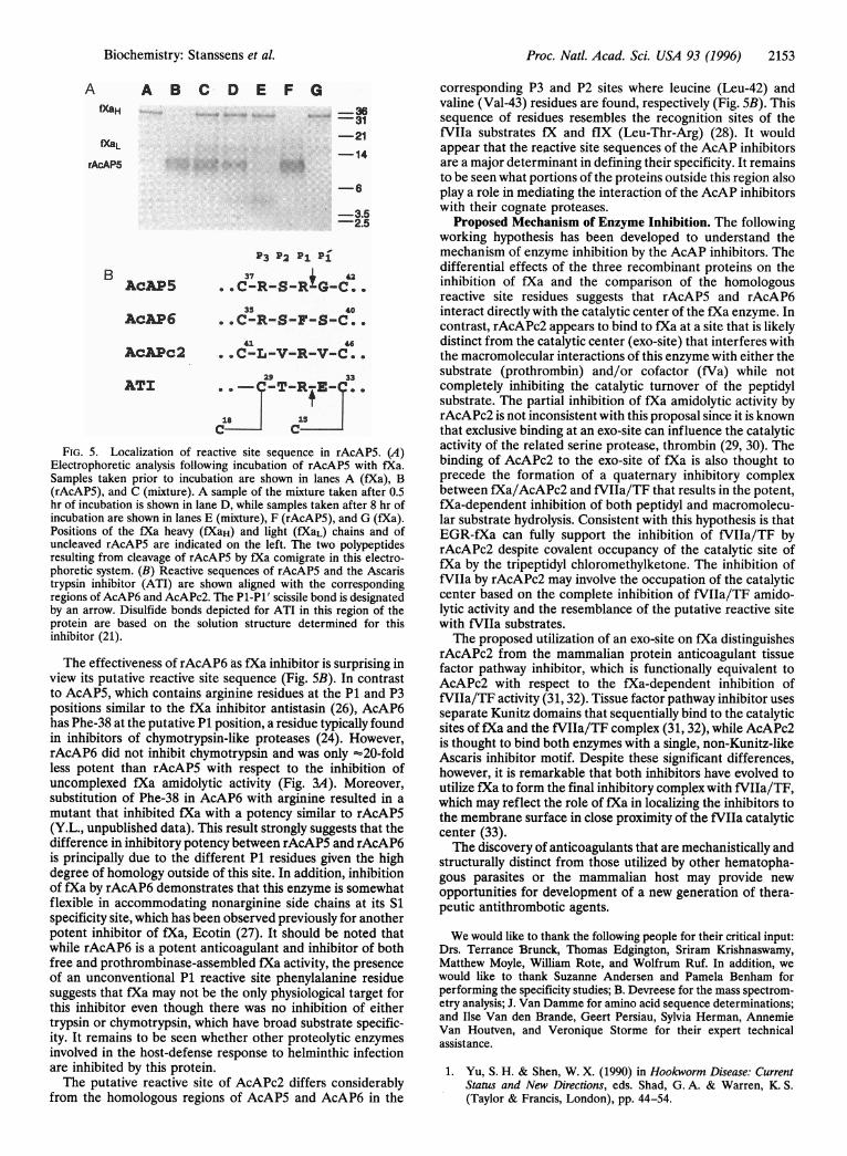
asite Ascaris lumbricoides var. suum (Ascaris; refs. 17, 18, 21,and 22) despite the lack of significant sequence homology (Fig.1). Partial proteolytic cleavage at the reactive site by thecognate protease has defined the Pi-Pl' residues (notationaccording to ref. 23) for several members of the Ascarisinhibitor family (17, 18). Similarly, we have found that incu-bation ofrAcAP5 with its target enzyme fXa resulted in partialcleavage between Arg-40 and Gly-41 (Fig. 5A), which corre-sponds to the same relative sequence position of the P1-Pl'residues in the Ascaris inhibitors. This supports the alignmentof AcAP5 and, by analogy, AcAP6 and AcAPc2, with anAscaris trypsin inhibitor, even though the spacing of thecysteines in the reactive site sequences differs by one residue(Figs. 1 and SB). It should be noted that the positioning ofcysteine residues within the reactive site of many classes ofsmall protein serine protease inhibitors may vary withoutaltering the canonical substrate mode of binding to the cata-lytic site of the cognate enzyme (24, 25).
Proc. Natl. Acad. Sci. USA 93 (1996)
Proc. Natl. Acad. Sci. USA 93 (1996) 2153
A A B C D E F GfXaH
fXaL
rAcAP5
BACAP5
AcAP6
AcAPc2
ATI
-36
31
-21
-14
-6
-3.52.5
P3 P2 PI Pi37 + 42
. .C-R-S-R-G-C..35 40
. .C-R-S-F-S-C..43. 46
. .C-L-V-R-V-C..
29 33.._ -T-RjE- ..18 15
C C
FIG. 5. Localization of reactive site sequence in rAcAP5. (A)Electrophoretic analysis following incubation of rAcAP5 with fXa.Samples taken prior to incubation are shown in lanes A (fXa), B(rAcAP5), and C (mixture). A sample of the mixture taken after 0.5hr of incubation is shown in lane D, while samples taken after 8 hr ofincubation are shown in lanes E (mixture), F (rAcAP5), and G (fXa).Positions of the fXa heavy (fXaH) and light (fXaL) chains and ofuncleaved rAcAP5 are indicated on the left. The two polypeptidesresulting from cleavage of rAcAP5 by fXa comigrate in this electro-phoretic system. (B) Reactive sequences of rAcAP5 and the Ascaristrypsin inhibitor (ATI) are shown aligned with the correspondingregions of AcAP6 and AcAPc2. The P1-P1' scissile bond is designatedby an arrow. Disulfide bonds depicted for ATI in this region of theprotein are based on the solution structure determined for thisinhibitor (21).
The effectiveness of rAcAP6 as fXa inhibitor is surprising inview its putative reactive site sequence (Fig. 5B). In contrastto AcAP5, which contains arginine residues at the P1 and P3positions similar to the fXa inhibitor antistasin (26), AcAP6has Phe-38 at the putative P1 position, a residue typically foundin inhibitors of chymotrypsin-like proteases (24). However,rAcAP6 did not inhibit chymotrypsin and was only "20-foldless potent than rAcAP5 with respect to the inhibition ofuncomplexed fXa amidolytic activity (Fig. 3A). Moreover,substitution of Phe-38 in AcAP6 with arginine resulted in a
mutant that inhibited fXa with a potency similar to rAcAP5(Y.L., unpublished data). This result strongly suggests that thedifference in inhibitory potency between rAcAP5 and rAcAP6is principally due to the different P1 residues given the highdegree of homology outside of this site. In addition, inhibitionof fXa by rAcAP6 demonstrates that this enzyme is somewhatflexible in accommodating nonarginine side chains at its Sispecificity site, which has been observed previously for anotherpotent inhibitor of fXa, Ecotin (27). It should be noted thatwhile rAcAP6 is a potent anticoagulant and inhibitor of bothfree and prothrombinase-assembled fXa activity, the presenceof an unconventional P1 reactive site phenylalanine residuesuggests that fXa may not be the only physiological target forthis inhibitor even though there was no inhibition of eithertrypsin or chymotrypsin, which have broad substrate specific-ity. It remains to be seen whether other proteolytic enzymesinvolved in the host-defense response to helminthic infectionare inhibited by this protein.The putative reactive site of AcAPc2 differs considerably
from the homologous regions of AcAP5 and AcAP6 in the
corresponding P3 and P2 sites where leucine (Leu-42) andvaline (Val-43) residues are found, respectively (Fig. SB). Thissequence of residues resembles the recognition sites of thefVIIa substrates fX and fIX (Leu-Thr-Arg) (28). It wouldappear that the reactive site sequences of the AcAP inhibitorsare a major determinant in defining their specificity. It remainsto be seen what portions of the proteins outside this region alsoplay a role in mediating the interaction of the AcAP inhibitorswith their cognate proteases.
Proposed Mechanism of Enzyme Inhibition. The followingworking hypothesis has been developed to understand themechanism of enzyme inhibition by the AcAP inhibitors. Thedifferential effects of the three recombinant proteins on theinhibition of fXa and the comparison of the homologousreactive site residues suggests that rAcAP5 and rAcAP6interact directly with the catalytic center of the fXa enzyme. Incontrast, rAcAPc2 appears to bind to fXa at a site that is likelydistinct from the catalytic center (exo-site) that interferes withthe macromolecular interactions of this enzyme with either thesubstrate (prothrombin) and/or cofactor (fVa) while notcompletely inhibiting the catalytic turnover of the peptidylsubstrate. The partial inhibition of fXa amidolytic activity byrAcAPc2 is not inconsistent with this proposal since it is knownthat exclusive binding at an exo-site can influence the catalyticactivity of the related serine protease, thrombin (29, 30). Thebinding of AcAPc2 to the exo-site of fXa is also thought toprecede the formation of a quaternary inhibitory complexbetween fXa/AcAPc2 and fVIIa/TF that results in the potent,fXa-dependent inhibition of both peptidyl and macromolecu-lar substrate hydrolysis. Consistent with this hypothesis is thatEGR-fXa can fully support the inhibition of fVIIa/TF byrAcAPc2 despite covalent occupancy of the catalytic site offXa by the tripeptidyl chloromethylketone. The inhibition offVIIa by rAcAPc2 may involve the occupation of the catalyticcenter based on the complete inhibition of fVIIa/TF amido-lytic activity and the resemblance of the putative reactive sitewith fVIla substrates.The proposed utilization of an exo-site on fXa distinguishes
rAcAPc2 from the mammalian protein anticoagulant tissuefactor pathway inhibitor, which is functionally equivalent toAcAPc2 with respect to the fXa-dependent inhibition offVIIa/TF activity (31, 32). Tissue factor pathway inhibitor usesseparate Kunitz domains that sequentially bind to the catalyticsites of fXa and the fVIIa/TF complex (31, 32), while AcAPc2is thought to bind both enzymes with a single, non-Kunitz-likeAscaris inhibitor motif. Despite these significant differences,however, it is remarkable that both inhibitors have evolved toutilize fXa to form the final inhibitory complex with fVIIa/TF,which may reflect the role of fXa in localizing the inhibitors tothe membrane surface in close proximity of the fVIIa catalyticcenter (33).The discovery of anticoagulants that are mechanistically and
structurally distinct from those utilized by other hematopha-gous parasites or the mammalian host may provide newopportunities for development of a new generation of thera-peutic antithrombotic agents.
We would like to thank the following people for their critical input:Drs. Terrance Brunck, Thomas Edgington, Sriram Krishnaswamy,Matthew Moyle, William Rote, and Wolfrum Ruf. In addition, wewould like to thank Suzanne Andersen and Pamela Benham forperforming the specificity studies; B. Devreese for the mass spectrom-etry analysis; J. Van Damme for amino acid sequence determinations;and Ilse Van den Brande, Geert Persiau, Sylvia Herman, AnnemieVan Houtven, and Veronique Storme for their expert technicalassistance.
1. Yu, S. H. & Shen, W. X. (1990) in Hookworm Disease: CurrentStatus and New Directions, eds. Shad, G. A. & Warren, K. S.(Taylor & Francis, London), pp. 44-54.
Biochemistry: Stanssens et al.

2154 Biochemistry: Stanssens et al.
2. Waxman, L., Smith, D. E., Arcuri, K. E. & Vlasuk, G. P. (1990)Science 248, 593-596.
3. Vlasuk, G. P. (1993) Thromb. Haemostasis 70, 212-216.4. Markwardt, F. (1957) Hoppe-Seylers Z. Physiol. Chem. 308,
147-156.5. Nutt, E., Gasic, T., Rodkey, J., Gasic, G. J., Jacobs, J. W.,
Friedman, P. A. & Simpson, E. (1988) J. Biol. Chem. 263,10162-10167.
6. Loeb, L. & Fleisher, M. S. (1910) J. Infect. Dis. 7, 625-631.7. Eiff, J. A. (1966) J. Parasitol. 52, 833-843.8. Spellman, G. G. & Nossel, H. L. (1971) Am. J. Physiol. 220,
922-927.9. Cappello, M., McPhedran, L. P. & Hotez, P. J. (1993) J. Infect.
Dis. 167, 1474-1477.10. Cappello, M., Vlasuk, G. P., Bergum, P. W., Huang, S. & Hotez,
P. J. (1995) Proc. Natl. Acad. Sci. USA 92, 6152-6156.11. Sambrook, J., Fritsch, E. F. & Maniatis, T. (1989) Molecular
Cloning: A Laboratory Manual (Cold Spring Harbor Lab. Press,Plainview, NY).
12. Laroche, Y., Storme, V., De Meutter, J., Messens, J. & Lauw-ereys, M. (1994) Bio/Technology 12, 1119-1124.
13. Cregg, J. M., Barringer, K. J., Hessler, A. Y. & Madden, K. R.(1985) Mol. Cell. Biol. 5, 3378-3385.
14. Morrison, J. F. (1969) Biochim. Biophys. Acta 185, 269-286.15. Ruf, W., Miles, D. J., Rehemtulla, A. & Edgington, T. S. (1993)
Methods Enzymol. 222, 209-224.16. Usharani, P., Cramer, B. J. W., Casper, C. K. & Bajaj, S. P.
(1985) J. Clin. Invest. 75, 76-83.17. Peanasky, R. J., Martzen, M. R., Homandberg, G. A., Cash,
J. M., Babin, D. R. & Litweiler, B. (1987) in Paradigms forEradicating Helminthic Parasites, ed. Macinnis, A. J. (Liss, NewYork), pp. 349-366.
18. Goodman, R. B., Martzen, M. R. & Peanasky, R. J. (1983) ActaBiochim. Pol. 30, 233-244.
19. Jespers, L. S., Messens, J. H., De Keyser, A., Eeckhout, D., VanDen Brande, I., Gansemans, Y. G., Lauwereys, M. J., Vlasuk,G. P. & Stanssens, P. E. (1995) BiolTechnology 13, 378-382.
20. Mann, K. G., Krishnaswamy, S. & Lawson, J. H. (1992) Semin.Hematol. 29, 213-226.
21. Grasberger, B. L., Clore, G. M. & Gronenborn, A. M. (1994)Structure 2, 669-678.
22. Huang, K., Strynadka, N. C. J., Bernard, V. D., Peanasky, R. J. &James, M. N. G. (1994) Structure 2, 679-689.
23. Schechter, I. & Berger, A. (1967) Biochem. Biophys. Res. Com-mun. 27, 157-162.
24. Laskowski Jr., M. & Kato, I. (1980) Annu. Rev. Biochem. 49,593-626.
25. Bode, W. & Huber, R. (1991) Biomed. Biochim. Acta 50,437-446.26. Dunwiddie, C. T., Thornberry, N., Bull, H., Sardana, M., Fried-
man, P., Jacobs, J. & Simpson, E. (1989) J. Biol. Chem. 264,16694-16699.
27. Seymour, J. L., Linquist, R. N., Dennis, M. S., Moffat, B., Yan-sura, D., Reilly, D., Wessinger, M. E. & Lazarus, R. A. (1994)Biochemistry 33, 3949-3958.
28. Butenas, S., Ribarik, N. & Mann, K. G. (1993) Biochemistry 32,6531-6538.
29. Naski, M. C., Fenton, J. W., Maraganore, J. M., Olson, S. T. &Shafer, J. A. (1990) J. Biol. Chem. 265, 13484-13489.
30. Liu, L., Vu, T.-K., Esmon, C. T. & Coughlin, S. R. (1991)J. Biol.Chem. 266, 16977-16980.
31. Broze, G. J., Girard, T. J. & Novotny, W. F. (1990) Biochemistry29, 7539-7546.
32. Huang, Z.-F., Wun, T.-C. & Broze, G. J. (1993) J. Biol. Chem.268, 26950-26955.
33. Broze, G. J., Warren, L. A., Novotny, W. F., Higuchi, D. A.,Girard, J. J. & Miletich, J. P. (1988) Blood 71, 335-343.
Proc. Natl. Acad. Sci. USA 93 (1996)