Embed Size (px)
Citation preview
VOL 31 (2) 2020: 131–143 | RESEARCH ARTICLE
131
Indonesian Journal of Pharmacy
Indonesian J Pharm 31(2), 2020, 131-143 | DOI: 10.14499/indonesianjpharm31iss2pp131 indonesianjpharm.farmasi.ugm.ac.id Copyright © 2020 THE AUTHOR(S). This article is distributed under a Creative Commons Attribution-ShareAlike 4.0 International (CC BY-SA 4.0)
Anti-stress activity of some plants extracts of the North Caucasus flora
Dmitry I Pozdnyakov*1, Similla L Adzhiahmetova2, Nadezhda M Chervonnaya2, Andey V MamLeev1, Eduard T Oganesyan2. 1. Department pharmacology with course of clinical pharmacology Pyatigorsk Medical Pharmaceutical
Institute - a branch of VolgGMU of the Ministry of Health of Russia; working postal address: Pyatigorsk, Kalinin Ave., 11, 357532,
2. Department of organic chemistry of the Pyatigorsk Medical Pharmaceutical Institute - a branch of VolgGMU of the Ministry of Health of Russia; working postal address: Pyatigorsk, Kalinin Ave., 11, 357532.
Info Article ABSTRACT Submitted: 28-03-2020 Revised: 20-06-2020 Accepted: 23-06-2020 *Corresponding author Pozdnyakov Dmitry Email: pozdniackow.dmitry @yandex.ru
To date, stress is a common medical and socially significant disease that requires rational pharmacotherapeutic correction.The anti-stress properties of ethnolic and aqueous extracts obtained from leaves of Ribes nigrum L., inflorescences of Gaillardia pulchella Foug., stems of Lysimachia punctata L were studied in this work. Acute stress was modeled by immobilization of rats for 2h. The test-extracts were administered per os prophylactically in a dose of 1/20 of LD50 (2000mg/kg). The following parameters were evaluated: organs mass coefficient (adrenal glands, thymus, spleen), the number of stomach erosion; the biochemical changes in the blood serum (adrenaline, cortisol, total protein and glucose concentration); the mitochondrial function parameters in brain and myocardium (evaluation of mitochondrial pore transitional permeability opening and mitochondrial membrane potential). 70% ethanol extract from Gaillardia pulchella Foug. inflorescences has the highest anti-stress activity, the course application of which contributed to the normalization of the weight index of organs, a decrease in glucose concentration by 64.5% (p <0.05), and cortisol by 73.7% (p <0.05) and adrenaline - 78.9% (p <0.05) in the blood, an increase in total protein level by 62.5% (p <0.05) and stabilization of mitochondrial function. The study showed a high anti-stress activity of 70 % ethanol extract from Gaillardia pulchella Foug. inflorescences. Keywords: stress; plant extracts; mitochondria.
INTRODUCTION
In the course of life, a modern person is constantly exposed to unfavorable factors that could provoke a stress disorders (Yeap et al., 2015). Stress is a normal physiological reaction of the body aimed at mobilizing available resources and limiting the impact on the body of negative factors (Doreddula et al., 2014). However, stress that goes beyond the physiological norm causes a violation of neuroendocrine, behavioral, emotional reactions and homeostasis changes. Chronic stress is the cause of the development of much psychosomatic pathology, for example, depression, anxiety, coronary heart disease, dementia (Joshi et al., 2012). The main pathogenic
mechanisms of stress are activation of the hypothalamic-pituitary-adrenal and sympatho-adrenal systems, which is accompanied by an increase in the concentration of corticosteroids and adrenaline in the blood (Gold, 2005). It is known that neurohumoral regulation of organs and systems is important in body adapting to the action of a stressor. The uncontrolled releasing of catecholamines and corticosteroids into the bloodstream has a damaging effect due to the depletion of energy cells reserves. Moreover, the alternative action, in addition to the classical «Selye triad» (thymo-lymphatic apparatus inflation, adrenal hypertrophy, ulcerative lesions of the stomach), is usually directed to organs with
Anti-stress activity of some plants
132 Volume 31 Issue 2 (2020)
high metabolic activity, primarily the brain and myocardium (Tan &Yip, 2018). The brain is the central organ responsible for the adaptation of the body to stress, involving the autonomic and endocrine systems in anti-stress reactions (McEwen, 2006). However, under conditions of excessive stress on the body, glucocorticoids overproduction causes an imbalance in neurotransmitter cerebral systems with a predominance of the activating mediators influence (glutamate and aspartate). As a result, there is deterioration in the neuroplasticity processes, an increase in glial reactions, a decrease in the density of synapses and a violation of the transduction of the synaptic signal (McEwen and Chattarji, 2007) was noted. Therefore, the controlling function of the brain is lost, and stress goes into the phase of maladaptation. The negative effect of the stressor on the myocardium is expressed in the depletion of the heart muscle under the influence of high concentrations of catecholamines. Sensitization of adrenoceptors causes a disruption in the functioning of energy-producing systems in the myocardium, which negatively affects on the general level of blood flow. Moreover, the consequences of insufficient blood supply to organs and tissues can be quite variable (Bergh et al., 2015).
Individuals constantly exposed to stress have cardiac arrhythmias, increased blood pressure, worsening microcirculation, a drop in overall physical activity, obesity, and depression (Montgomery et al., 2000).
The high medical and social role of stress disorders necessitates a targeted search of medicines that can increase the adaptive capacity of the organism. Medicinal plants have long been an inexhaustible source of biologically active compounds (Upadhyay et al., 2016). Almost every region has its own traditions of herbal medicine, associated, as a rule, with the use of medicinal plants widespread in this area. Numerous experimental and clinical data indicate the high efficiency and safety of the phytotherapeutic approach in the treatment and prevention of many diseases, including the central nervous system, cardiovascular and neuroendocrine systems disorders ( Othman et al., 2013).
In previous studies the antihypoxic and anti-ischemic properties of some extracts obtained from the leaves of Ribes nigrum L., inflorescences of Gaillardia pulchella Foug., stems of Lysimachia punctata L. were noted. It was found that the use of these extracts reduced the level of ischemic
damage to the brain and also reduced tissue hypoxia due to restoration of mitochondrial functions (Pozdnyakov et al. ,2019). Also, extracts obtained from black currant are characterized by the presence of endotheliotropic action aimed at increasing the activity of endothelial nitric oxide synthase (eNOS), and also exhibit the properties of acetylcholinesterase inhibitors and have anti-inflammatory activity, realized by suppressing the production of pro-inflammatory cytokines:tumor necrosis factor-α, IL-1β and CINC-1, which in turn can be an essential component of the stress-protective action of these extracts (Staszowska-Karkut and Materska, 2020). Extracts obtained from Gaillardia pulchella Foug are characterized by the presence of large amounts of apigenin and also have anti-inflammatory and antioxidant effects, which can favorably affect the course of both acute and chronic stress disorders (Moharram et al, 2017). The antioxidant properties of extracts obtained from Lysimachia punctata L are also widely known, but existing research is usually focused on in vitro evaluation of the antioxidant properties of extracts from Lysimachia punctata L. (Toth et al, 2014; Toth et al, 2018)
As it is known, normalization of mitochondrial functions may be one of the promising approaches of stress-protective therapy. Under stress conditions, mitochondrial damage induces lipid peroxidation, decreased ATP synthesis, and initiates programmed cell death (Hsu et al., 2018). In this regard, extracts from the leaves of R. nigrum, inflorescences of G. pulchella, stems of L. punctata L. were used in this study as the test-objects.
The aim of this study is to evaluate anti-stress activity of Ribes nigrum L., inflorescences of Gaillardia pulchella Foug., stems of Lysimachia punctata L. at course administration in experimental conditions. The originality of this study is expressed in the fact that the anti-stress properties of ethanol and water extracts from the leaves of R. nigrum, inflorescences of G. pulchella, stems of L. punctata were studied. The effect of the test-extracts on the processes of opening of the mitochondrial pore of transition permeability and the value of mitochondrial membrane potential were also evaluated. The choice of defined parameters was based primarily on the significant role of hormonal, biochemical and functional abnormalities that occur during the course of the stress response. It was found that the primary compensatory reaction in response to the action of
Dmitry I Pozdnyakov
Volume 31 Issue 2 (2020) 133
stressors is hyperproduction of catecholamines (adrenaline) and corticosteroids, which with the continued influence of the corresponding stress factor on the organism is negative and manifests itself as disorders of protein and carbohydrate metabolism, functional disorders of organs and systems of organs, primarily the hypothalamic-pituitary-adrenal axis, gastrointestinal tract and and thymolymphatic apparatus. (Möstl Palme, 2002). In addition, adrenaline is known as a strong disconnecting agent of oxidation and phosphorylation reactions, which adversely affects the function of mitochondria, especially the brain and myocardium. (Mishra et al., 2019) In this regard, the studied parameters in this research were: the concentration of adrenaline, cortisol, glucose and total protein in the blood serum, the mass coefficient of the thymus, spleen, adrenal glands, the number of erosions on the gastric mucosa, as well as some parameters that allow us to assess the change in mitochondrial function: the value of the mitochondrial membrane potential and the latent opening time of the mitochondrial transition permeability pore.
MATERIAL AND METHODS Test objects
The test-objects in this study were ethanol and aqueous extracts of R. nigrum leaves, inflorescences of G. pulchella, and stems of L. punctata. The plant materials were sampled from the Stavropol region territory (Russia; 44.095783, 43.025125). The identification of plant species was carried out by specialists of the Department of Pharmacognosy with a course in the technology of herbal remedies (herbarium samples №PG1171; №PG1189; №PG1210). Ethanol in various concentrations (95%, 70%, 40%), as well as purified water, were used for extraction. Additionally, leaf samples of plants were extracted. A total of 16 extracts were obtained.
Extraction: 1g (precise weight) of the crushed raw material was placed in a 100mL round-bottom flask with a thin grind, 30mL of ethyl alcohol in different concentration (or water) was added, connect with the reflux condenser and heated in a boiling water bath for 1h. After cooling, the resulting extract was filtered through a filter paper into a 100mL volumetric flask. Extraction was repeated twice under the conditions described above. The extract was filtered through the same filter into the same volumetric flask. After cooling, the volume was adjusted with ethyl alcohol (or water) to the mark and mix.
Determination of the total content of antioxidants
The total content of antioxidants was performed on a «Tsvet-01-AA» liquid chromate-graph. The mass concentration of antioxidants was measured amperometrically using a calibration graph of the output signal versus the concentration of quercetin and / or gallic acid. The method is based on the detection of free radicals resulting from electro chemical oxidation according to the following scheme (Dementieva et al., 2014). flavonoid-О-Н → flavonoid О + е–-(oxidation at maximum potential) flavonoid-О-Н → flavonoid-О • + Н •(free radical capture)
Laboratory animals The experiment was performed on 150male
Wistar rats weighing 220-240g. The animals were obtained from the vivarium of the Pyatigorsk Medical and Pharmaceutical Institute and were kept under controlled conditions: ambient temperature 20±2ᵒC, relative humidity 60±5%, with a natural change in the daily cycle (12h a day, 12h a night). At the time of the experiment, laboratory rats were kept in macrolon cages (5 animals in cage) and received full-nutrition balanced by proteins, fats, carbohydrates, vitamins and trace elements feed, as well as tap water. Animals were not restricted in their intake of food and water (ad libithum). Rats were removed from the experiment by cervical dislocation under chloral hydrate anesthesia (350mg/kg, intra-peritoneally). The keeping and handling of animals was in accordance with international ethical standards of experimental practice (Directive 2010/63 / EU of the European Parliament and of the council on the protection of animals used for scientific purposes, September 22, 2010). The research concept was approved by the local ethics committee (protocol No. 25 of 09/29/2019).
Study design. The toxicity of the test-extracts was
previously evaluated in an acute experiment with determination of LD50. Subsequently, at the stage of evaluating the anti-stress action, the following experimental groups were formed (n=10, each group):sham-operated animals (SO, placed in an immobilization machine and then immediately removed); group of animals of negative control (NC) - treated by purified water at the rate of 1mL/ 100g of animal weight; a group of rats treated by the reference drug – tincture of ginseng in a dose of 1mL/kg.
Anti-stress activity of some plants
134 Volume 31 Issue 2 (2020)
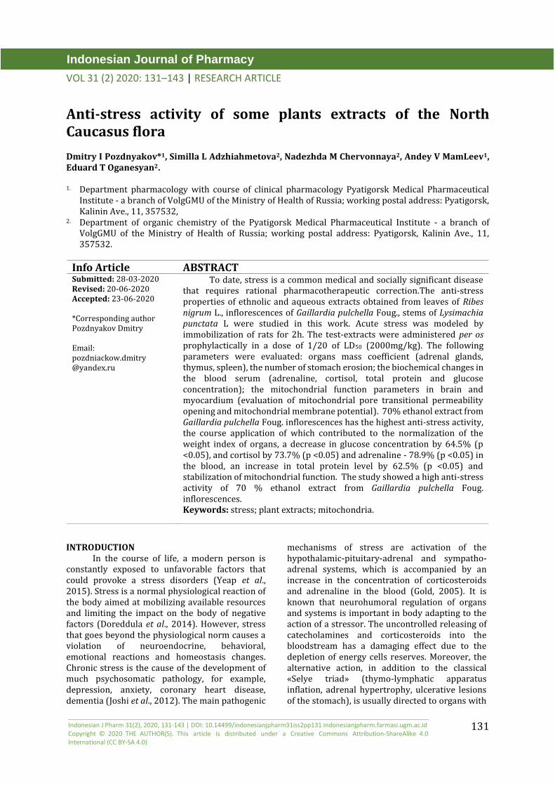
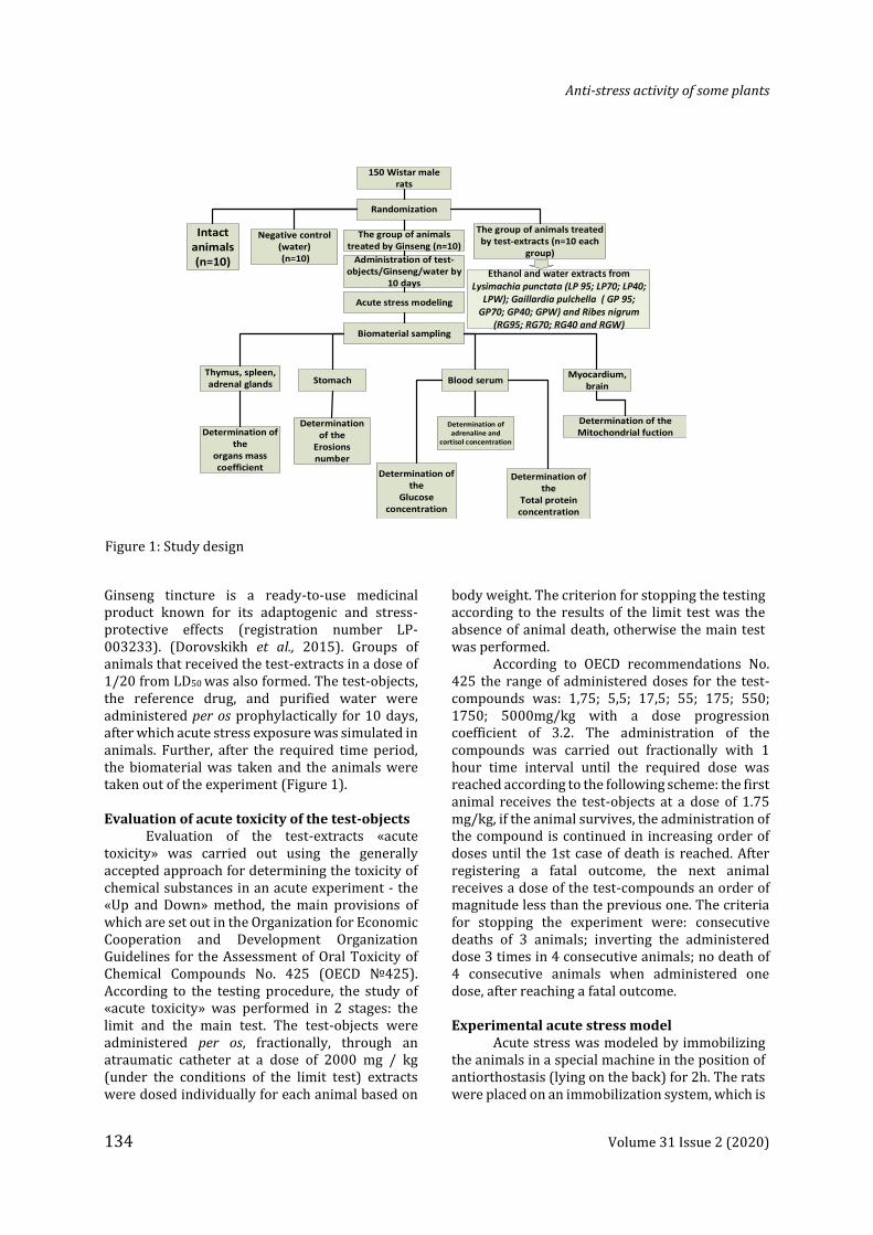
Ginseng tincture is a ready-to-use medicinal product known for its adaptogenic and stress-protective effects (registration number LP-003233). (Dorovskikh et al., 2015). Groups of animals that received the test-extracts in a dose of 1/20 from LD50 was also formed. The test-objects, the reference drug, and purified water were administered per os prophylactically for 10 days, after which acute stress exposure was simulated in animals. Further, after the required time period, the biomaterial was taken and the animals were taken out of the experiment (Figure 1).
Evaluation of acute toxicity of the test-objects
Evaluation of the test-extracts «acute toxicity» was carried out using the generally accepted approach for determining the toxicity of chemical substances in an acute experiment - the «Up and Down» method, the main provisions of which are set out in the Organization for Economic Cooperation and Development Organization Guidelines for the Assessment of Oral Toxicity of Chemical Compounds No. 425 (OECD №425). According to the testing procedure, the study of «acute toxicity» was performed in 2 stages: the limit and the main test. The test-objects were administered per os, fractionally, through an atraumatic catheter at a dose of 2000 mg / kg (under the conditions of the limit test) extracts were dosed individually for each animal based on
body weight. The criterion for stopping the testing according to the results of the limit test was the absence of animal death, otherwise the main test was performed.
According to OECD recommendations No. 425 the range of administered doses for the test-compounds was: 1,75; 5,5; 17,5; 55; 175; 550; 1750; 5000mg/kg with a dose progression coefficient of 3.2. The administration of the compounds was carried out fractionally with 1 hour time interval until the required dose was reached according to the following scheme: the first animal receives the test-objects at a dose of 1.75 mg/kg, if the animal survives, the administration of the compound is continued in increasing order of doses until the 1st case of death is reached. After registering a fatal outcome, the next animal receives a dose of the test-compounds an order of magnitude less than the previous one. The criteria for stopping the experiment were: consecutive deaths of 3 animals; inverting the administered dose 3 times in 4 consecutive animals; no death of 4 consecutive animals when administered one dose, after reaching a fatal outcome.
Experimental acute stress model
Acute stress was modeled by immobilizing the animals in a special machine in the position of antiorthostasis (lying on the back) for 2h. The rats were placed on an immobilization system, which is
150 Wistar male rats
Randomization
Intact animals (n=10)
Negative control (water) (n=10)
The group of animals treated by test-extracts (n=10 each
group)
The group of animals treated by Ginseng (n=10)
Administration of test-objects/Ginseng/water by
10 days
Acute stress modeling
Biomaterial sampling
Determination of the
organs mass coefficient
Determination of the
Erosions number
Determination of the
Glucose concentration
Determination of the
Total protein concentration
Determination of the Mitochondrial fuction
Determination of adrenaline and
cortisol concentration
Thymus, spleen, adrenal glands Stomach Blood serum
Myocardium, brain
Ethanol and water extracts from Lysimachia punctata (LP 95; LP70; LP40;
LPW); Gaillardia pulchella ( GP 95; GP70; GP40; GPW) and Ribes nigrum
(RG95; RG70; RG40 and RGW)
Figure 1: Study design

Dmitry I Pozdnyakov
Volume 31 Issue 2 (2020) 135
a table with 4 branches for fixing the limbs and a soft substrate under the body and head of the animal. The upper and lower extremities of rats were fixed with adhesive tape (Yeap et al., 2015). Biomaterial sampling
After 2h of stress exposure, the animals were weighed and recorded body weight in grams to the nearest 1g. Then the rats were taken out of the study by cervical dislocation, the abdominal and thoracic cavities were opened, and then the following organs were removed: thymus, spleen, stomach, left and right adrenal glands and myocardium. To extract the brain, the cranium was opened. After that, the mass coefficient of the thymus, spleen, left and right adrenal glands were determined. The stomach was opened according to slight curvature, the contents were removed, washed with purified water and the number of erosive-ulcerative lesions was estimated. Heart and brain homogenized in a mechanical Potter homogenizer in a selection medium in the ratio 1:5 (1mmol EDTA, 215mmol mannitol, 75mmol sucrose, 0.% BSA solution, 20mmol HEPES, with a pH of 7.2). The mitochondrial fraction was obtained by differential centrifugation, for which the obtained biogenic homogenate was centrifuged in the mode of 1.400g → 3min. at 40ᵒC, after which the supernatant was transferred to 2 mL tubes. Next, the resulting supernatant was centrifuged at 13,000g → 10min and the supernatant (culture contains native mitochondria) was removed for determining the parameters of mitochondrial function. Blood sampling in animals was carried out portion wise from the abdominal aorta into a vacuum tube with citrate filling. The first part was centrifuged in 3000RPM - 10min .mode to obtaining serum and determining the content of glucose and total protein, the second part was centrifuged at 1000g for 20min and removed for ELISA analysis. Control of the mitochondrial fraction obtaning was performed by measuring respiratory control (data are not reflected in the work) (Sullivan et al., 2007). Determination of the organs mass coefficient
Thymus, spleen, left and right adrenal glands were separated from adipose tissue, accompanying connective tissue and weighed on a Scout Pro electronic balance (Ohaus, USA) with an accuracy of 1 mg. The mass of organs was fixed. The mass coefficient was calculated by the following formula [1].
К (mass coefficient)= Organs weight
………[1] Animal weight
Determination of glucose concentration in blood serum
Serum glucose was determined by glucose oxidase method with spectrophotometric detection. This method is based on the oxidation of glucose by glucose oxidase with the formation of hydrogen peroxide, which is with the participation of peroxidase contributes to the oxidative azo coupling of phenol and 4-aminoantipyrine reaction with colored complex formation (Boyer, 1975). Moreover, the color intensity is proportional to the concentration of glucose in the sample. The incubation medium contained: EDTA 2 mmol/L; phenol - 11mmol/L; 4-aminoantipyrine - 0.016mmol/L; glucose oxidase – 30.000 U/L; mutarotase - 200 U/L; peroxidase - 1800 U / L; BSA - 0.05g/L; test sample - 0.01mL. The calibrator solution contains 10mmol/L glucose. The absorbance of the resulting mixture was recorded at a wavelength of λ = 500nm. The serum glucose concentration was calculated by the formula [2].
C= Ax
……………………………………….…[2] Ao
Where :Ax - is the absorbance of the test sample; Ao - is the absorbance of the calibrator solution; 10 - glucose concentration in a calibrator solution.
Determination of the total protein concentration in blood serum
The total protein content in blood serum was evaluated by the biuret method with spectrophotometric detection. The principle of the method is based on the formation of a red-colored complex of copper ions with peptide bonds of proteins, the concentration of which is directly proportional to the total protein content in blood serum (Parvin et al., 1965). The incubation medium contained: copper sulfate - 120mmol/Ll; potassium iodide -300 mmol/L; sodium potassium tartrate - 20mmol/L; sodium hydroxide - 3mol/L; 100μL of the analyzed sample. The calibration solution contained 60g/L of BSA. The absorbance of the resulting mixture was recorded at λ=540nm. The protein concentration in the test sample was calculated by the formula [3].
C= Ax
X 60 ………………………….…[3] Ao
Where: Ax - is the absorbance of the test sample; Ao - is the absorbance of the calibrator solution; 60 –total protein concentration in a calibrator solution
Anti-stress activity of some plants
136 Volume 31 Issue 2 (2020)
Study of mitochondrial pore transitional permeability opening (mPTP)
The effect of the test-extracts and the reference drug on the opening of the mitochondrial pore was evaluated by spectrophotometric method. The incubation medium contained: 0.5mL of the analyzed supernatant, 200mM KCl, 0.5mL of a 1μm solution of cyclosporin A. The resulting mixture was adjusted to 2mL with HEPES buffer solution with a pH of 7.4. The optical density of the mixture was recorded at λ=540nm, then the resulting solution was incubated for 25min at room temperature with constant stirring. At the same time, the latent time of opening of the mitochondrial pore in seconds was evaluated (by changing the optical density of the incubation medium) (Zhyliuk et al., 2015).
Study of mitochondrial membrane potential
Mitochondrial membrane potential was evaluated by spectrophotometric method. The incubation medium contained: 0.5mL of the analyzed supernatant, 0.5mL of a 9μm solution of safranin O. The resulting mixture was adjusted to 2mL with HEPES buffer solution with a pH of 7.4. The optical density of the mixture was recorded at λ=51nm and λ=525nm. The proton moving force (transmembrane electrochemical gradient, ∆Ψ) was determined by the difference of the optical density: ∆Ψ=A515-A525. (Zhyliuk et al.,2015).
ELISA study In this study, the concentration of
adrenaline and cortisol was determined by ELISA in blood serum. The experiment was performed using species-specific reagent kits manufactured by Cloud clone (USA). The analysis, preparation of reagents, standard images and the test-material corresponded to the manufacturer's instructions attached to each kit. The results were recorded using an Infinite F50 microplate reader (Tecan, Austria).
Statistical analysis
Statistical processing of the obtained data was carried out in the software package of statistical analysis STATISTICA 6.0 for Windows (StatSoft, USA). The results were expressed as M (mean) ± SEM. Data was checked for normal distribution (Shapiro-Wilk test). Comparison of the means groups was performed by the method of one-way analysis of variance (ANOVA) with post-processing of Newman-Keuls (normal distribution) or Kruskall-Wallis (abnormal distribution).
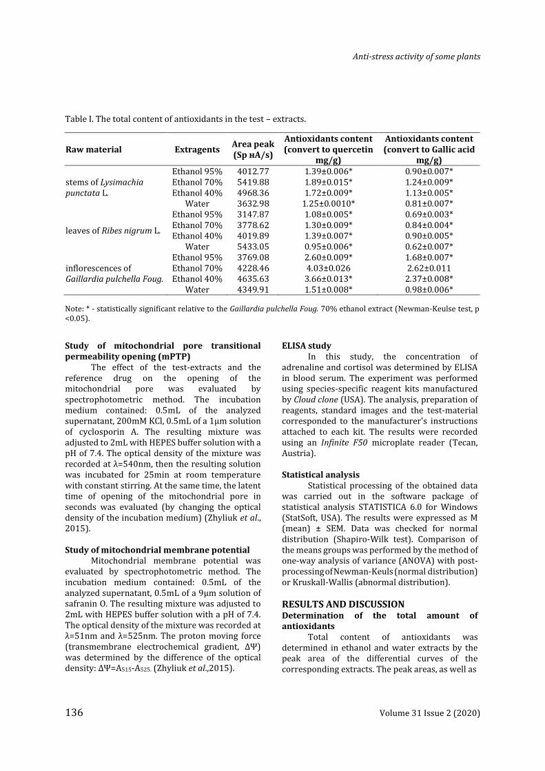
RESULTS AND DISCUSSION Determination of the total amount of antioxidants
Total content of antioxidants was determined in ethanol and water extracts by the peak area of the differential curves of the corresponding extracts. The peak areas, as well as
Table I. The total content of antioxidants in the test – extracts.
Raw material Extragents Area peak (Sp нА/s)
Antioxidants content (convert to quercetin
mg/g)
Antioxidants content (convert to Gallic acid
mg/g)
stems of Lysimachia punctata L.
Ethanol 95% Ethanol 70% Ethanol 40%
Water
4012.77 5419.88 4968.36 3632.98
1.39±0.006* 1.89±0.015* 1.72±0.009*
1.25±0.0010*
0.90±0.007* 1.24±0.009* 1.13±0.005* 0.81±0.007*
leaves of Ribes nigrum L.
Ethanol 95% Ethanol 70% Ethanol 40%
Water
3147.87 3778.62 4019.89 5433.05
1.08±0.005* 1.30±0.009* 1.39±0.007* 0.95±0.006*
0.69±0.003* 0.84±0.004* 0.90±0.005* 0.62±0.007*
inflorescences of Gaillardia pulchella Foug.
Ethanol 95% Ethanol 70% Ethanol 40%
Water
3769.08 4228.46 4635.63 4349.91
2.60±0.009* 4.03±0.026 3.66±0.013* 1.51±0.008*
1.68±0.007* 2.62±0.011 2.37±0.008* 0.98±0.006*
Note: * - statistically significant relative to the Gaillardia pulchella Foug. 70% ethanol extract (Newman-Keulse test, p <0.05).
Dmitry I Pozdnyakov
Volume 31 Issue 2 (2020) 137
the concentration of antioxidants in terms of quercetin and gallic acid (Table I). Based on the experimental data presented (Table I), assumes that the maximum content of the total antioxidants was detected in the inflorescences of G. pulchella extracts.
Evaluation of «acute toxicity» of the test-extracts
When assessing the «acute toxicity» of the test-extracts, it was found that in the limit test conditions with the administration of the test-objects at a dose of 2000mg/kg (per os), no animal deaths were noted. At the same time, in animals treated by the test-extracts, there were no significant deviations in behavior, sensory perception, and motor function in relation to the control group of mice. Since no deaths were observed during the limiting test, the main test are not carried out, and the value LD50 was taken to be 2000mg/kg. Thus, for further studies, the dose of the test-extracts was 100mg/kg (per os).
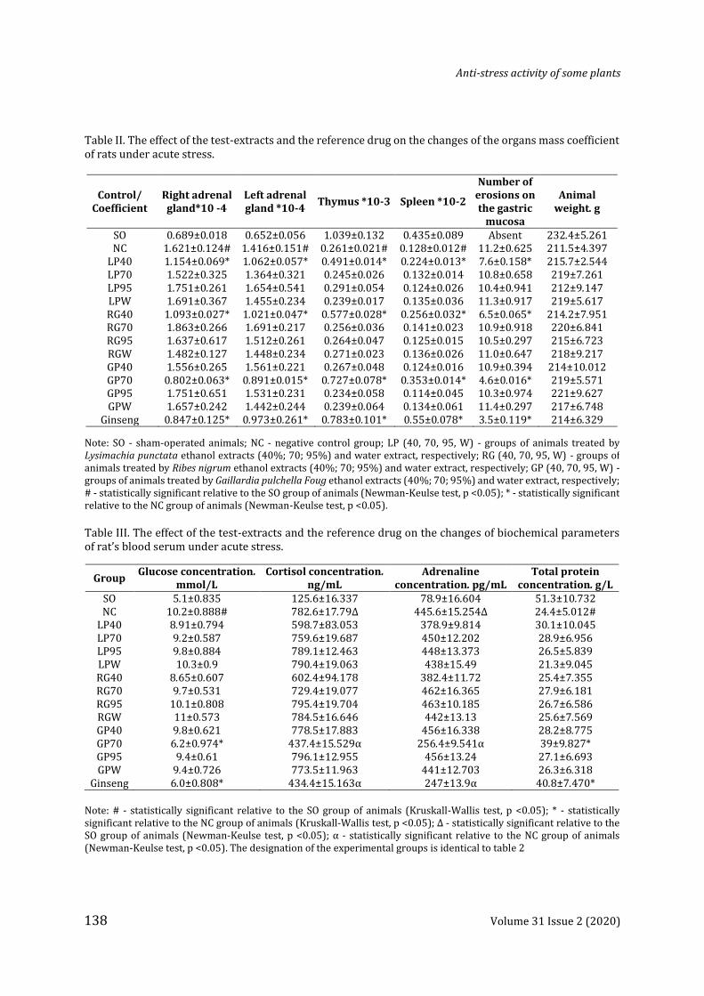
Evaluation of the test-extracts effect on the change in the mass coefficient of rat organs under acute stress
The results of this block of work are presented (Table II). When studying the effect of the test-extracts on the change in the mass coefficient of stressed rats organs, it was found that the most significant changes in the studied parameters were obtained with the administration of ethanol extracts of L. punctata (40%), G. pulchella (70%), R. nigrum (40%). So when using 40% ethanol extract of L. punctata (LP40) compared with the NC group of rats, a decrease in the mass coefficient of the left and right adrenal glands was found, as well as an increase in the weight indexes of the thymus and spleen. Moreover, in animals treated by LP40 extract, the amount of erosion on the gastric mucosa was 47.3% (p<0.05) lower than that of a group of rats lacking pharmacological support (Table II). Against the background of 40% ethanolic extract of R. nigrum (RG40) administration to animals, the decrease in the involution of the thymus and spleen (the mass coefficient in this group of rats was 2.2 times (p<0.05) and 2 times (p<0.05), respectively, higher than that of the NC group) was noted. In addition, in animals that were treated by RG40, there was a decrease in the weight index of the left and right adrenal glands, as well as the number of erosive defects in the stomach. When using the test 70% ethanol extract of G. pulchella
(GP70) in comparison with the NC group of animals, a decrease in the mass coefficient of the adrenal glands, as well as an increase in the weight indexes of the spleen and thymus was noted. Moreover, in rats treated with GP70 extract, the number of ulcerative damage on the gastric mucosa decreased in relation to the group of animals lacking pharmacological support. The administration of the remaining test-extracts did not significantly affect the change in the estimated parameters. Against the background of the use of ginseng tincture in rats with respect to the NC group of animals, a decrease in the mass coefficient of the adrenal glands and also an increase in the weight index of the spleen and thymus respectively, with a decrease in the number of erosions on the gastric mucosa was observed. Evaluation of the effect of test-extracts on the change of the biochemical parameters in blood plasma of rats under acute stress conditions.
In animals of the NC group, in conditions of acute stress, an increase in the concentration of glucose, cortisol and adrenaline in the blood serum was 2 times (p <0.05); 6.2 times (p <0.05) and 5.6 times (p <0.05), respectively was noted, while the protein content in rats lacking pharmacological support, was 2.1 times ( p <0.05) less than that in SO animals (Table III).
The concentration of glucose and adrenaline was statistically significant in relation to the NC group of animals changed with the administration of the GP70 test - extract (there was a decrease in these indicators by 64.5% (p <0.05) and 73.7% (p <0.05), respectively). Against the background of the remaining studied objects administration, the content of glucose and adrenaline in the blood serum of rats was not statistically significant relative to the NC group of animals (Table III). At the same time, the use of ginseng tincture contributed to a decrease in the concentration of glucose and adrenaline in the blood serum of rats in relation to the NC group of animals by 70% (p<0.05) and 80.3% (p<0.05), respectively. Against the background of the test-objects LP 40, RG 40 and GP 70 administration, a decrease in the concentration of cortisol in blood serum in rats by 30.7 (p<0.05); 29.9% (p<0.05) and 78.9% (p<0.05), respectively, compared with the NC group of animals was noted. Moreover, when using ginseng tincture, the serum cortisol content decreased by 80.1% (p<0.05) relative to the index of the group of rats lacking pharmacological support.
Anti-stress activity of some plants
138 Volume 31 Issue 2 (2020)
Table II. The effect of the test-extracts and the reference drug on the changes of the organs mass coefficient of rats under acute stress.
Control/ Coefficient
Right adrenal gland*10 -4
Left adrenal gland *10-4
Thymus *10-3 Spleen *10-2
Number of erosions on the gastric
mucosa
Animal weight. g
SO 0.689±0.018 0.652±0.056 1.039±0.132 0.435±0.089 Absent 232.4±5.261 NC 1.621±0.124# 1.416±0.151# 0.261±0.021# 0.128±0.012# 11.2±0.625 211.5±4.397
LP40 1.154±0.069* 1.062±0.057* 0.491±0.014* 0.224±0.013* 7.6±0.158* 215.7±2.544 LP70 1.522±0.325 1.364±0.321 0.245±0.026 0.132±0.014 10.8±0.658 219±7.261 LP95 1.751±0.261 1.654±0.541 0.291±0.054 0.124±0.026 10.4±0.941 212±9.147 LPW 1.691±0.367 1.455±0.234 0.239±0.017 0.135±0.036 11.3±0.917 219±5.617 RG40 1.093±0.027* 1.021±0.047* 0.577±0.028* 0.256±0.032* 6.5±0.065* 214.2±7.951 RG70 1.863±0.266 1.691±0.217 0.256±0.036 0.141±0.023 10.9±0.918 220±6.841 RG95 1.637±0.617 1.512±0.261 0.264±0.047 0.125±0.015 10.5±0.297 215±6.723 RGW 1.482±0.127 1.448±0.234 0.271±0.023 0.136±0.026 11.0±0.647 218±9.217 GP40 1.556±0.265 1.561±0.221 0.267±0.048 0.124±0.016 10.9±0.394 214±10.012 GP70 0.802±0.063* 0.891±0.015* 0.727±0.078* 0.353±0.014* 4.6±0.016* 219±5.571 GP95 1.751±0.651 1.531±0.231 0.234±0.058 0.114±0.045 10.3±0.974 221±9.627 GPW 1.657±0.242 1.442±0.244 0.239±0.064 0.134±0.061 11.4±0.297 217±6.748
Ginseng 0.847±0.125* 0.973±0.261* 0.783±0.101* 0.55±0.078* 3.5±0.119* 214±6.329
Note: SO - sham-operated animals; NC - negative control group; LP (40, 70, 95, W) - groups of animals treated by Lysimachia punctata ethanol extracts (40%; 70; 95%) and water extract, respectively; RG (40, 70, 95, W) - groups of animals treated by Ribes nigrum ethanol extracts (40%; 70; 95%) and water extract, respectively; GP (40, 70, 95, W) - groups of animals treated by Gaillardia pulchella Foug ethanol extracts (40%; 70; 95%) and water extract, respectively; # - statistically significant relative to the SO group of animals (Newman-Keulse test, p <0.05); * - statistically significant relative to the NC group of animals (Newman-Keulse test, p <0.05).
Table III. The effect of the test-extracts and the reference drug on the changes of biochemical parameters of rat’s blood serum under acute stress.
Group Glucose concentration.
mmol/L Cortisol concentration.
ng/mL Adrenaline
concentration. pg/mL Total protein
concentration. g/L SO 5.1±0.835 125.6±16.337 78.9±16.604 51.3±10.732 NC 10.2±0.888# 782.6±17.79Δ 445.6±15.254Δ 24.4±5.012#
LP40 8.91±0.794 598.7±83.053 378.9±9.814 30.1±10.045 LP70 9.2±0.587 759.6±19.687 450±12.202 28.9±6.956 LP95 9.8±0.884 789.1±12.463 448±13.373 26.5±5.839 LPW 10.3±0.9 790.4±19.063 438±15.49 21.3±9.045 RG40 8.65±0.607 602.4±94.178 382.4±11.72 25.4±7.355 RG70 9.7±0.531 729.4±19.077 462±16.365 27.9±6.181 RG95 10.1±0.808 795.4±19.704 463±10.185 26.7±6.586 RGW 11±0.573 784.5±16.646 442±13.13 25.6±7.569 GP40 9.8±0.621 778.5±17.883 456±16.338 28.2±8.775 GP70 6.2±0.974* 437.4±15.529α 256.4±9.541α 39±9.827* GP95 9.4±0.61 796.1±12.955 456±13.24 27.1±6.693 GPW 9.4±0.726 773.5±11.963 441±12.703 26.3±6.318
Ginseng 6.0±0.808* 434.4±15.163α 247±13.9α 40.8±7.470*
Note: # - statistically significant relative to the SO group of animals (Kruskall-Wallis test, p <0.05); * - statistically significant relative to the NC group of animals (Kruskall-Wallis test, p <0.05); Δ - statistically significant relative to the SO group of animals (Newman-Keulse test, p <0.05); α - statistically significant relative to the NC group of animals (Newman-Keulse test, p <0.05). The designation of the experimental groups is identical to table 2
Dmitry I Pozdnyakov
Volume 31 Issue 2 (2020) 139
With the administration of the remaining studied objects, statistically significant changes in the concentration of cortisol relative to the NC group of animals were not established. The use of the GP70 test extract also contributed to the elimination of hypoproteinemia, which was expressed in an increase in the concentration of total protein in the blood of rats treated by GP70 extract, compared with the NC group of animals by 62.5% (p<0.05). The remaining studied extracts did not significantly affect the change in the total protein in the blood of rats (Table III). Against the background of the administration of ginseng tincture to animals, the total protein content in the blood in rats increased by 65.4% (p<0.05) relative to the NC group of animals.
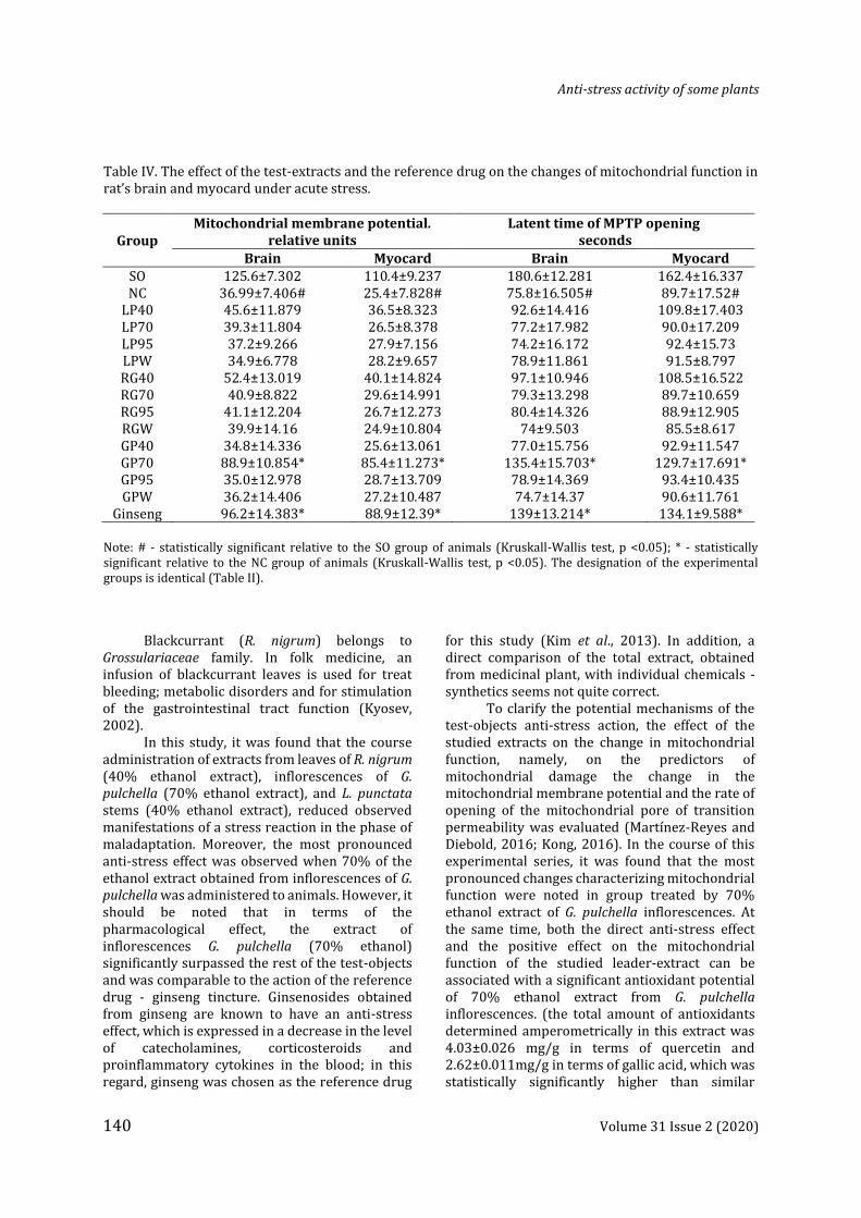
Evaluation of the studied extracts effect on the change in the mitochondrial function of the rat brain and myocardium under acute stress
In animals lacking pharmacological support compared with intact rats, a decrease in mitochondrial membrane potential in the brain and myocardium by 3.4 times (p<0.05) and 4.3 times (p<0.05), respectively was observed (Table IV). Against the background of the test objects use RG40; LP40 and GP70 showed an increase (relative to the NC group of animals) of the mitochondrial membrane potential in the rat brain by 23.5% (p<0.05); 42% (p<0.05) and 2.4times (p<0.05), respectively. At the same time, the mitochondrial membrane potential increased by 43.7% (p<0.05); 57.9% (p<0.05) and 3.4 times (p<0.05) compared with the same indicator of the NC group of animals in the myocardium when using the test extracts RG40; LP40 and GP70. At the same time, against the background of the administration of ginseng tincture to rats, an increase in the mitochondrial membrane potential in the brain and myocardium by 2.6 times (p<0.05) and 3.5 times (p<0,05) in comparison with the group of animals lacking pharmacological support, respectively was observed (Table IV).
The opening time of mPTP in the myocardium and brain in the NC group of rats decreased compared with SO animals by 81% (p<0.05) and 2.4times (p<0.05), respectively. With the use of the test LP40 extract, the latent opening time of mPTP in the brain and myocardium increased relative to the NC group by 22% (p<0.05) and 48% (p<0.05), respectively. The time period for the mPTP formation in the myocardium and brain of rats receiving the studied RG40 extract increased in relation to animals lacking
pharmacological support by 20.9% (p<0.05) and 49.7% (p<0.05), respectively . When using LP40 extract, an increase in the latent time of mPTP formation in the brain and myocardium of animals was observed relative to the NC group by 78.6% (p<0.05) and 44.6% (p<0.05), respectively, at the same time with the administration of ginseng tincture to animals, these indicators increased by 83.2% (p<0.05) and 50.1% (p<0.05), respectively (Table IV).
However, the pace of modern life, the constant effect on the body of adverse exogenous and endogenous factors (noise, vibration, bacterial toxins, viruses, radiation, microwaves), as well as intense social the situation can be stress-provocative factors (Patel et al., 2011; Oladipo and Ibukun, 2016.). Stress beyond physiological adaptation is sharply negative and adversely affects activities of organs and systems, primarily those in which there is a high metabolic activity - the brain and myocardium, as well as the thymo-lymphatic apparatus (Oladipo and Ibukun, 2016). Currently, stress-protective therapy is usually limited to the use of tranquilizers. However, representatives of this pharmacotherapeutic group have an extensive spectrum of adverse reactions, usually affecting on the activity of the central nervous system. Excessive sedation, decreased intensity of motor and mental reactions, impaired neurotransmitter metabolism in the central nervous system, respiratory system disorders are the most characteristic drug-induced adverse reactions that occur in response to the tranquilizers administration (Bighelli et al., 2016). At the same time an affordable and safe alternative to an anxiolytic drug maybe the administration of herbal origin medicines. It has been established that medicinal plant-based products have pronounced stress-protective properties that are not inferior to those of benzodiazepines (Kumar et al., 2008). In this regard, this study focused on studying the antistress activity of ethanol and water extracts obtained from plants growing in the North Caucasus - R. nigrum, G. pulchella, L. punctata. G. pulchella belongs to the Asteraceae family. The sesquiterpenes of the lipophilic fraction of Gailardia pulchella inflorescences were studied (Moharram et al., 2017).
L. punctata is a perennial herb belonging to the Primulaceae family. Infusions and decoctions from this plant are used in folk medicine for gastrointestinal disorders, skin inflammations and lung diseases (Abramova et al., 2018).
Anti-stress activity of some plants
140 Volume 31 Issue 2 (2020)
Blackcurrant (R. nigrum) belongs to Grossulariaceae family. In folk medicine, an infusion of blackcurrant leaves is used for treat bleeding; metabolic disorders and for stimulation of the gastrointestinal tract function (Kyosev, 2002).
In this study, it was found that the course administration of extracts from leaves of R. nigrum (40% ethanol extract), inflorescences of G. pulchella (70% ethanol extract), and L. punctata stems (40% ethanol extract), reduced observed manifestations of a stress reaction in the phase of maladaptation. Moreover, the most pronounced anti-stress effect was observed when 70% of the ethanol extract obtained from inflorescences of G. pulchella was administered to animals. However, it should be noted that in terms of the pharmacological effect, the extract of inflorescences G. pulchella (70% ethanol) significantly surpassed the rest of the test-objects and was comparable to the action of the reference drug - ginseng tincture. Ginsenosides obtained from ginseng are known to have an anti-stress effect, which is expressed in a decrease in the level of catecholamines, corticosteroids and proinflammatory cytokines in the blood; in this regard, ginseng was chosen as the reference drug
for this study (Kim et al., 2013). In addition, a direct comparison of the total extract, obtained from medicinal plant, with individual chemicals - synthetics seems not quite correct.
To clarify the potential mechanisms of the test-objects anti-stress action, the effect of the studied extracts on the change in mitochondrial function, namely, on the predictors of mitochondrial damage the change in the mitochondrial membrane potential and the rate of opening of the mitochondrial pore of transition permeability was evaluated (Martínez-Reyes and Diebold, 2016; Kong, 2016). In the course of this experimental series, it was found that the most pronounced changes characterizing mitochondrial function were noted in group treated by 70% ethanol extract of G. pulchella inflorescences. At the same time, both the direct anti-stress effect and the positive effect on the mitochondrial function of the studied leader-extract can be associated with a significant antioxidant potential of 70% ethanol extract from G. pulchella inflorescences. (the total amount of antioxidants determined amperometrically in this extract was 4.03±0.026 mg/g in terms of quercetin and 2.62±0.011mg/g in terms of gallic acid, which was statistically significantly higher than similar
Table IV. The effect of the test-extracts and the reference drug on the changes of mitochondrial function in rat’s brain and myocard under acute stress.
Group Mitochondrial membrane potential.
relative units Latent time of MPTP opening
seconds
Brain Myocard Brain Myocard SO 125.6±7.302 110.4±9.237 180.6±12.281 162.4±16.337 NC 36.99±7.406# 25.4±7.828# 75.8±16.505# 89.7±17.52#
LP40 45.6±11.879 36.5±8.323 92.6±14.416 109.8±17.403 LP70 39.3±11.804 26.5±8.378 77.2±17.982 90.0±17.209 LP95 37.2±9.266 27.9±7.156 74.2±16.172 92.4±15.73 LPW 34.9±6.778 28.2±9.657 78.9±11.861 91.5±8.797 RG40 52.4±13.019 40.1±14.824 97.1±10.946 108.5±16.522 RG70 40.9±8.822 29.6±14.991 79.3±13.298 89.7±10.659 RG95 41.1±12.204 26.7±12.273 80.4±14.326 88.9±12.905 RGW 39.9±14.16 24.9±10.804 74±9.503 85.5±8.617 GP40 34.8±14.336 25.6±13.061 77.0±15.756 92.9±11.547 GP70 88.9±10.854* 85.4±11.273* 135.4±15.703* 129.7±17.691* GP95 35.0±12.978 28.7±13.709 78.9±14.369 93.4±10.435 GPW 36.2±14.406 27.2±10.487 74.7±14.37 90.6±11.761
Ginseng 96.2±14.383* 88.9±12.39* 139±13.214* 134.1±9.588* Note: # - statistically significant relative to the SO group of animals (Kruskall-Wallis test, p <0.05); * - statistically significant relative to the NC group of animals (Kruskall-Wallis test, p <0.05). The designation of the experimental groups is identical (Table II).
Dmitry I Pozdnyakov
Volume 31 Issue 2 (2020) 141
indicators in the other studied extracts). However, it should be noted that the content of antioxidants in terms of quercetin and gallic acid in other ethanol extracts was comparable, which may be explained by the nature of the available biologically active substances (for example, gallic acid), which can be equally extracted from plant raw materials by ethanol of different concentrations (Sut et al, 2019). The influence of antioxidants on the course of stress disorder and mitochondrial dysfunction has been the subject of a significant number of experimental studies that note their high therapeutic efficacy (Ranchordas K et al., 2017). Work Peng et.al. 2019 demonstrated the high antioxidant and anti-stress potential of herbal medicine derived from San-Huang-Xie-Xin.
Agents with antioxidant activity are also promising correctors of mitochondrial dysfunction. Moreover, both chemically modified and native molecules, as well as total extracts, can be pharmacologically active (Apostolova and Victor, 2015). It has been established that, for example, flavonoids (resveratrol) and phenolic acid derivatives (caffeic acid) can have a positive effect on the change in mitochondrial function. Moreover, the action of these biologically active compounds can be mediated both by a direct effect on mitochondria (a change in OXPHOS, membrane potential), and indirectly (through a change in the activity of regulatory systems - AMPK, PPAR) (Kang et al., 2018).
CONCLUSION The study showed that in a series of ethanol
(40%, 70%, 95%) and aqueous extracts obtained from leaves of R. nigrum, inflorescences of G. pulchella, and stems of L. punctata, 70% ethanol extract from inflorescences G. pulchella has the most pronounced anti-stress activity. The course administration of this extract at a dose of 100 mg / kg (orally) contributed to the reduction of typical post-stress effects (characteristic biochemical changes in the form of an increase in the concentration of adrenaline, cortisol and glucose, as well as a decrease in the level of total protein in the blood, the presence of gastric mucosa ulceration, hypertrophy of adrenal glands, involution of the thymus). Also, the administration of 70% ethanol extract, obtained from Gaillardia pulchella Foug. inflorescences helped to restore mitochondrial function, as evidenced by an increase in the mitochondrial membrane potential and a decrease in the latent opening time of the mitochondrial transition permeability pore.
ACKNOWLEDGMENTS The author's team is grateful to the
Pyatigorsk medical and pharmaceutical Institute. The study had no sponsorship.
REFERENCES Abramova ER., Tekeyeva DI., Adzhiakhmetova SL,
2018. The study of polysaccharides of some representatives of the families Primulaceae and Asteraceae. Belikov meetings:104–107. (In Russ.)
Apostolova N., Victor VM., 2015. Molecular strategies for targeting antioxidants to mitochondria: therapeutic implications. Antioxid Redox Signal. 22(8):686–729. doi:10.1089/ars.2014.5952.
Ben OM., Han J., El Omri A., Ksouri R., Neffati M., Isoda H., 2013. Antistress Effects of the Ethanolic Extract from Cymbopogon schoenanthus Wild in Tun Growing isia. Evid Based Complement Alternat Med. 2013:737401. doi:10.1155/2013/737401
Bergh C., Udumyan R., Fall K., Almroth H., Montgomery S., 2015. Stress resilience and physical fitness in adolescence and risk of coronary heart disease in middle age. Heart. 101(8):623–629. doi:10.1136/heartjnl-2014-306703
Bighelli I., Trespidi C., Castellazzi M., 2016. Antidepressants and benzodiazepines for panic disorder in adults. Cochrane Database Syst Rev. 9(9): doi:10.1002/14651858.CD011567.pub2
Boyer PD. 1975.The Enzymes. Academic Press ⅩⅡB: 421
Dementieva TM., Frolova OO., Evseeva OS., Selivanova SL., 2014 Research of antioxidant activity of decoctions of bark and shoots of Babylonian willow and its hybrid with white willow. Healthcare Of The Far East 1:83-87.
Doreddula SK., Bonam SR., Gaddam DP., Desu BS., 2014. Phytochemical analysis, antioxidant, antistress, and nootropic activities of aqueous and methanolic seed extracts of ladies finger (Abelmoschus esculentus L.) in mice. ScientificWorldJournal 2014:519848. doi:10.1155/2014/519848
Dorovskikh VA., Simonova NV., Tonkonogova MS., 2015. Comparative evaluation of phytoadaptogens under oxidative stress .Bull. physical and Res. Pat. 55:95-100.
Gold SM., 2005.The role of stress-response systems for the pathogenesis and progression of MS. Trends Immunol 26(12):644–652.
Anti-stress activity of some plants
142 Volume 31 Issue 2 (2020)
Hsu F., Spannl S., Ferguson C., Hyman AA., 2018. Rab5 and Alsin regulate stress-activated cytoprotective signaling on mitochondria. Elife 7:e32282. doi:10.7554/eLife.32282
Joshi T., Sah SP., Singh A. 2012, Antistress activity of ethanolic extract of Asparagus racemosus willd roots in mice. Indian. J. Experimental Biology. 50(6):419–424.
Kang HW., Lee SG., Otieno D., Ha K., 2018. Flavonoids, Potential Bioactive Compounds, and Non-Shivering Thermogenesis. Nutrients. 10(9):1168. doi:10.3390/nu10091168
Kim EH., Kim IH., Ha JA., Choi KT., Pyo S., Rhee DK., 2013. Antistress effect of red ginseng in brain cells is mediated by TACE repression via PADI4. J Ginseng Res. 37(3):315–323. doi:10.5142/jgr.2013.37.315
Kulkarni MP., Juvekar AR., 2008. Attenuation of Acute and Chronic Restraint Stress-induced Perturbations in Experimental Animals by Nelumbo nucifera Gaertn. Indian J Pharm Sci. 70(3):327–332. doi:10.4103/0250-474X.42982
Kumar S., Madaan R., Sharma A., 2008. Pharmacological evaluation of Bioactive Principle of Turnera aphrodisiaca. Indian J Pharm Sci. 70(6):740–744. doi:10.4103/0250-474X.49095
Kyosev PA., 2002. A complete reference of medicinal plants. EKSMO – Press :992.
Martínez-Reyes I., Diebold LP., Kong H., 2016. TCA Cycle and Mitochondrial Membrane Potential Are Necessary for Diverse Biological Functions. Mol Cell. 61(2):199–209. doi:10.1016/j.molcel.2015.12.002
McEwen BS., 2006. Protective and damaging effects of stress mediators: central role of the brain. Dial. in Clin. Neurosci.Stress. 8:367–381.
McEwen BS., Chattarji S., 2007. Handbook of Neurochemistry and Molecular Neurobiology. Springer-Verlag; 2007. pp. 572–593.
Mishra S., Chattopadhyay A., Naaz S., Ghosh AK., Das AR., Bandyopadhyay D., 2019. Oleic acid ameliorates adrenaline induced dysfunction of rat heart mitochondria by binding with adrenaline: An isothermal titration calorimetry study. Life Sci. 218:96-111. doi:10.1016/j.lfs.2018.12.035
Moharram FA., El Dib RAEM., Marzouk MS., El-Shenawy SM., 2017. New Apigenin
Glycoside, Polyphenolic Constituents, Anti-inflammatory and Hepatoprotective Activities of Gaillardia grandiflora and Gaillardia pulchella Aerial Parts. Pharmacogn Mag. 13(Suppl 2):S244–S249. doi:10.4103/pm.pm_344_16
Moharram FA., El Dib RAEM., Marzouk MS., El-Shenawy SM., Ibrahim HA., 2017. New Apigenin Glycoside, Polyphenolic Constituents, Anti-inflammatory and Hepatoprotective Activities of Gaillardia grandiflora and Gaillardia pulchella Aerial Parts. Pharmacogn Mag. 13(Suppl 2):S244-S249. doi:10.4103/pm.pm_344_16
Montgomery SM., Berney LR., Blane D., 2000. Prepubertal stature and blood pressure in early old age. Arch Dis Child. 82(5):358–363. doi:10.1136/adc.82.5.358
Möstl E., Palme R. 2002. Hormones as indicators of stress. Domest Anim Endocrinol.; 23 (1-2) :67-74. doi:10.1016/s0739-7240(02)00146-7
Oladipo GO., Ibukun EO. 2016, BioActivities of Coturnix japonica (quail) egg yolk and albumen against physiological stress. Food Sci Nutr. 5(2):334–343. doi:10.1002/fsn3.397
Parvin R., Pande SV., Venkitasubramanian TA., 1965. On the colorimetric biuret method of protein determination. Analytical Biochemistry. 12 (2):219-229
Patel NB., Galani VJ., Patel BG., 2011. Antistress activity of Argyreia speciosa roots in experimental animals. J Ayurveda Integr Med. 2(3):129–136. doi:10.4103/0975-9476.85551
Peng W., Du H., Liu G., 2019. Antistress Effects of San-Huang-Xie-Xin Decoction on Restraint-Stressed Mice Revealed by 1H NMR-Based Metabolomics and Biochemistry Analysis. Oxid Med Cell Longev. 2019:5897675. doi:10.1155/2019/5897675
Pozdnyakov DI., Pozdnyakova AE, Adzhiahmetova SL, Chervonnaya NM, Zolotych DS, Lyakhova NS, Miroshnichenko KA, 2019. Antihypoxic and anti-ischemic properties of the North Caucasus flora plant extracts. Bol. latinoam. Caribe plantas med. Aromát. 18:504-517
Ranchordas MK., Rogerson D., Soltani H., Costello JT., 2017. Antioxidants for preventing and reducing muscle soreness after exercise. Cochrane Database Syst Rev. 12(12) doi:10.1002/14651858.CD009789.pub2
Dmitry I Pozdnyakov
Volume 31 Issue 2 (2020) 143
Staszowska-Karkut M., Materska M., 2020. Phenolic Composition, Mineral Content, and Beneficial Bioactivities of Leaf Extracts from Black Currant (Ribes nigrum L.), Raspberry (Rubus idaeus), and Aronia (Aronia melanocarpa). Nutrients.12:463. doi:10.3390/nu12020463
Sullivan PG., Krishnamurthy S., Patel SP., Pandya JD., 2007. Temporal characterization of mitochondrial bioenergetics after spinal cord injury. Journal of neurotrauma. 24:991–999
Sut S., Dall'Acqua S., Zengin G., 2019. Influence of different extraction techniques on the chemical profile and biological properties of Anthemis cotula L.: Multifunctional aspects for potential pharmaceutical applications. J Pharm Biomed Anal.173:75-85. doi:10.1016/j.jpba.2019.05.028
Tan SY., Yip A., 2018. Hans Selye (1907-1982): Founder of the stress theory. Singapore Med J 59(4):170–171. doi:10.11622/smedj. 2018043
Toth A., Riethmuller E., Vegh K., Alberti A., Beni S., Kery A., 2018. Contribution of individual
flavonoids in Lysimachia species to the antioxidant capacity based on HPLC-DPPH assay. Nat Prod Res. 32(17):2058-2061. doi:10.1080/14786419.2017.1359176
Toth A., Toth G., Kery A. 2014. Polyphenol composition and antioxidant capacity of three Lysimachia species. Nat Prod Commun. 9(10):1473-1478.
Upadhyay G., Khoshla S., Kosuru R., Singh S. 2016. Anxiolytic, antidepressant, and antistress activities of the aqueous extract of Cinnamomum tamala Nees and Eberm in rats. Indian J Pharmacol. 48(5):555–561. doi:10.4103/0253-7613.190752
Yeap SK., Beh BK., Ali NM., 2015. Antistress and antioxidant effects of virgin coconut oil in vivo. Exp Ther Med 9(1):39–42. doi:10.3892/etm.2014.2045
Zhyliuk VI., Mamchur VV., Pavlov S. 2015, Role of functional state of neuronal mitochondria of cerebral cortex in mechanisms of nootropic activity of neuroprotectors in rats with alloxan hyperglycemia. Eksp. i klin. farm. 78: 10-4. (In Russ.)


![Anti-inflammatory effects of Nelumbo leaf extracts and … · 2017-07-28 · 266 Anti-inflammatory effects of Nelumbo leaf extracts and thereby exerts antioxidant effects [20]. For](https://img.pdfslide.us/doc/110x75/5ea515630be6904b9618283f/anti-inflammatory-effects-of-nelumbo-leaf-extracts-and-2017-07-28-266-anti-inflammatory.jpg)