Embed Size (px)
Citation preview
RESEARCH ARTICLE Open Access
Anti-influenza virus activity of extracts fromthe stems of Jatropha multifida Linn.collected in MyanmarMasaki Shoji1*, So-Yeun Woo2, Aki Masuda1, Nwet Nwet Win2,3, Hla Ngwe3, Etsuhisa Takahashi4, Hiroshi Kido4,Hiroyuki Morita2, Takuya Ito2* and Takashi Kuzuhara1*
Abstract
Background: To contribute to the development of novel anti-influenza drugs, we investigated the anti-influenzaactivity of crude extracts from 118 medicinal plants collected in Myanmar. We discovered that extract from thestems of Jatropha multifida Linn. showed anti-influenza activity. J. multifida has been used in traditional medicinefor the treatment of various diseases, and the stem has been reported to possess antimicrobial, antimalarial, andantitumor activities. However, the anti-influenza activity of this extract has not yet been investigated.
Methods: We prepared water (H2O), ethyl acetate (EtOAc), n-hexane (Hex), and chloroform (CHCl3) extracts fromthe stems of J. multifida collected in Myanmar, and examined the survival of Madin-Darby canine kidney (MDCK)cells infected with the influenza A (H1N1) virus, and the inhibitory effects of these crude extracts on influenza Aviral infection and growth in MDCK cells.
Results: The H2O extracts from the stems of J. multifida promoted the survival of MDCK cells infected with theinfluenza A H1N1 virus. The EtOAc and CHCl3 extracts resulted in similar, but weaker, effects. The H2O, EtOAc, andCHCl3 extracts from the stems of J. multifida inhibited influenza A virus H1N1 infection; the H2O extract possessedthe strongest inhibitory effect on influenza infection in MDCK cells. The EtOAc, Hex, and CHCl3 extracts all inhibitedthe growth of influenza A H1N1 virus, and the CHCl3 extract demonstrated the strongest activity in MDCK cells.
Conclusion: The H2O or CHCl3 extracts from the stems of J. multifida collected in Myanmar demonstrated thestrongest inhibition of influenza A H1N1 viral infection or growth in MDCK cells, respectively. These results indicatedthat the stems of J. multifida could be regarded as an anti-influenza herbal medicine as well as a potential crudedrug source for the development of anti-influenza compounds.
Keywords: Anti-influenza, Anti-virus, Jatropha multifida, Stem, Herbal medicine
BackgroundIn 1918, the Spanish influenza A (H1N1) virus pandemiccaused 50 million deaths worldwide [1, 2]. In 2009, influ-enza A virus originating in swine (H1N1) caused a pan-demic, and the avian H5N1 and H7N9 influenza A virusesin China are highly pathogenic to humans [1–4]. Cur-rently, the application of three antiviral medicines known
as neuraminidase (NA) inhibitors, oral oseltamivir, zana-mivir, and peramivir, is recommended for the treatment ofinfluenza. However, oseltamivir resistance has been de-tected in some of the 2009-derived H1N1 viruses and theseasonal H1N1 viruses between 2007 and 2009, but littlein H3N2 viruses [5]. In the future, zanamivir- andperamivir-resistant strains, similar to oseltamivir-resistantstrain, will emerge. Therefore, the development of novelanti-influenza drugs to prevent and control future influ-enza epidemics and pandemics is desired.Traditional medicinal plants have been recognized as a
rich source of candidate compounds for the developmentof pharmaceuticals [6, 7]. A large number of natural
* Correspondence: [email protected]; [email protected];[email protected] of Biochemistry, Faculty of Pharmaceutical Sciences, TokushimaBunri University, 180 Yamashiro-cho, Tokushima 770-8514, Japan2Institute of Natural Medicine, University of Toyama, 2630, Sugitani, Toyama930-0194, JapanFull list of author information is available at the end of the article
© The Author(s). 2017 Open Access This article is distributed under the terms of the Creative Commons Attribution 4.0International License (http://creativecommons.org/licenses/by/4.0/), which permits unrestricted use, distribution, andreproduction in any medium, provided you give appropriate credit to the original author(s) and the source, provide a link tothe Creative Commons license, and indicate if changes were made. The Creative Commons Public Domain Dedication waiver(http://creativecommons.org/publicdomain/zero/1.0/) applies to the data made available in this article, unless otherwise stated.
Shoji et al. BMC Complementary and Alternative Medicine (2017) 17:96 DOI 10.1186/s12906-017-1612-8
products and extracts from medicinal plants have been re-ported to possess anti-influenza virus activity [8–10].Therefore, many studies have focused on traditional medi-cinal plants as an important source of candidate com-pounds for the discovery of novel anti-influenza drugs.The abundance of medicinal plants in Myanmar has en-
abled the population to use traditional medicines to main-tain their own health and treat various diseases. Thus, todiscover sources for novel anti-influenza drugs, wescreened extracts from 118 medicinal plants collected inMyanmar to analyze the cell viability of influenza A H1N1virus (A/PR/8/34)-infected MDCK cells using naphtholblue black staining. We identified six medicinal plants thatpromoted the survival of influenza A virus-infected cellsselected by the criteria described at the Methods section.Of these six plants, the activity of extract from the stemsof Jatropha multifida Linn (J. multifida) was strongly pro-nounced. J. multifida, a member of the family Euphorbia-ceae, is a tree of 2–3 m in height, and widely distributedin sub-tropical and tropical areas throughout Asia andAfrica [11]. Popularly known as “Say-ma-khan”, it is com-monly used as a folk medicine in Myanmar and has beenused as a purgative, and against fever, indigestion, colic,wounds, and skin infection [11]. The seed oil, latex, andleaves are effective purgatives and abortifacients, havebeen used as wound dressings, and for the treatment ofneurodermatitis, eczema, and itches [11]. The roots andstems have antimicrobial, antimalarial, antitumor, antil-eishmanial, and antiulcer activities [11, 12]. In addition,previous phytochemical studies on J. multifida reportedthe presence of cyclic peptides, diterpenoids, and phenoliccompounds [11]. However, pharmacological and phyto-chemical investigations of J. multifida stems originatingfrom Myanmar have not yet been conducted, whichattracted us to investigate whether extracts from the stemsof J. multifida, obtained using various solvents, possessedanti-influenza virus activity.
MethodsPlant materialThe stems of J. multifida were purchased from SandhiBrothers Trading Co. Ltd (Yangon, Myanmar) inNovember 2015. A voucher specimen (TMPW 28729)was deposited at the Museum of Materia Medica, Ana-lytical Research Center for Ethnomedicines, Institute ofNatural Medicine, University of Toyama, Japan.
Plant extractionWe performed the plant extraction as described previ-ously [13, 14]. In brief, dried stems of J. multifida werechopped into small pieces (3.0 kg), which were macer-ated four times with 70% aqueous EtOH (7 L) in anultrasonic bath for 90 min at 25 °C. After filtration ofthe suspension, the resulting solution was evaporated
under reduced pressure to yield a crude extract (180 g).The crude extract was suspended in water and parti-tioned into n-hexane (Hex), chloroform (CHCl3), andethyl acetate (EtOAc) fractions, to yield Hex-soluble(12.2 g), CHCl3-soluble (11.0 g), and EtOAc-soluble por-tions (11.0 g), respectively. Finally, the residual aqueouslayer was evaporated under reduced pressure to yield awater (H2O)-soluble portion (140.8 g). The extracts werestored at a concentration of 10 mg/mL in dimethyl sulf-oxide (DMSO).
CellsMadin-Darby canine kidney (MDCK) cells were culturedin high-glucose Dulbecco’s modified Eagle’s medium(DMEM; Wako, Osaka, Japan), supplemented with 10%fetal bovine serum (FBS; Life Technologies, CA, USA),50 units/mL penicillin and 50 μg/mL streptomycin (P/S;Life Technologies), and 4 mM L-glutamine, at 37 °C inthe presence of 5% CO2 [15].
Viral strainThis study used the Puerto Rico 8/34 (A/PR/8/34;H1N1) strain of the influenza A virus. Viral titers weredetermined by immunostaining influenza A viral nucleo-protein (NP) as previously described [16].
Analysis of cell viability of influenza A virus-infectedMDCK cells using naphthol blue black stainingMDCK cells were seeded in a 96-well plate (1 × 10⁴
cells/well). H2O, EtOAc, Hex, or CHCl3 extracts fromthe stems of J. multifida (0.8-25 μg/mL in DMSO) weremixed with influenza A virus in 10% FBS-supplementedgrowth medium at a multiplicity of infection (MOI) of10, and then incubated for 30 min at 37 °C in the pres-ence of 5% CO2. DMSO (0.008-0.5%) and (+)-(S)-baku-chiol (0.8-25 μM in DMSO) were used as negative andpositive controls, respectively, for the inhibition of influ-enza A viral infection [15]. The mixture was added tothe cells, which were then incubated for 4 days at 37 °Cin the presence of 5% CO2. After incubation, the cellswere stained using naphthol blue black as previously de-scribed [15, 17]. The viable cells in each well werestained blue, while dead cells remained unstained. Theselection criteria for the six plants are more than 50%cell survival at 96 h after the viral infection with theconcentration of 50 μg/mL.
Thiazolyl blue tetrazolium bromide (MTT) assayMDCK cells were seeded in each well of a 96-well plate(1 × 10⁴ cells/well). H2O, EtOAc, Hex, or CHCl3 extracts(12.5-100 μg/mL) were prepared in DMSO (12.5 μg/mL,0.125%; 25 μg/mL, 0.25%; 50 μg/mL, 0.5%; 100 μg/mL,1%) and mixed with infection medium (DMEM supple-mented with 1% bovine serum albumin [BSA; Wako,
Shoji et al. BMC Complementary and Alternative Medicine (2017) 17:96 Page 2 of 7

Osaka, Japan], P/S, and 4 mM L-glutamine). The mixturewas added to the cells, which were then incubated for24, 72, or 96 h at 37 °C in the presence of 5% CO2. Afterincubation, cell viability was determined using the MTTcell proliferation assay as previously described [15].
Immunofluorescence staining of influenza A virus-infected cellsMDCK cells were seeded in a 96-well plate (1 × 10⁴ cells/well). H2O, EtOAc, Hex, or CHCl3 extracts from thestems of J. multifida (3.1-25 μg/mL) or (+)-(S)-bakuchiol(3.1-25 μM) were mixed with influenza A virus at a MOIof 0.1 in the infection medium and incubated for 30 minat 37 °C in the presence of 5% CO2. DMSO (0.031-0.25%)was used as the negative control. Each mixture was addedto the cells and incubated for 24 h at 37 °C in the presenceof 5% CO2. The cells were fixed with 4% paraformalde-hyde in PBS for 30 min at 4 °C and then permeabilized bythe addition of 0.3% Triton X-100 for 20 min at 25 °C. Amouse antibody for the detection of the NP of A/PR/8/34(FluA-NP 4 F1; SouthernBiotech, AL, USA) was used asthe primary antibody. Alexa Fluor488-conjugated goatanti-mouse IgG (H + L) antibody (Life Technologies, CA,USA) was used as the secondary antibody. Cell nucleiwere then stained using diamidino-2-phenylindole (DAPI;Life Technologies). The wells were photographed using afluorescence microscope (BIOREVO BZ-9000, Keyence,Osaka, Japan), and the percentage of influenza A NP-positive cells per DAPI-positive cells were calculatedbased on measurements recorded with BZ-H1C software(Keyence).
Influenza A viral growth assayTo explore whether the extracts from the stems of J.multifida affected viral growth in pre-infected cells,MDCK cells were seeded in a 24-well plate (1 × 10⁵
cells/well). The cells were infected with A/PR/8/34(MOI; 0.001) in infection medium for 1 h at 37 °C in thepresence of 5% CO2. The infected cells were washedprior to the addition of H2O, EtOAc, Hex, or CHCl3 J.multifida extracts (12.5 or 25 μg/mL in 0.5% DMSO) tothe cells in infection medium supplemented with 3 μg/mL L-tosylamido-2-phenyl ethyl chloromethyl ketone(TPCK)-treated trypsin (Sigma-Aldrich). DMSO (0.5%)and ribavirin (50 μM in 0.5% DMSO) were the negativeand positive controls, respectively, for the inhibition oninfluenza A viral growth [18]. The cells were then incu-bated for 24, 48, or 72 h at 37 °C in the presence of 5%CO2. Cell culture media were collected from each wellat predetermined time points. Viral titers (plaque form-ing units per mL [PFU/mL]) were determined as previ-ously described [15].
Statistical analysisAll results were expressed as the mean ± the standarderror of the mean (SEM). Differences between morethan two groups were analyzed for statistical significanceby using one-way analysis of variance (ANOVA). Valuesof p < 0.05 were considered statistically significant.
ResultsExtracts from the stems of J. multifida increased thesurvival of influenza A viral-infected MDCK cellsTo evaluate the anti-influenza viral activity of extractsfrom the stems of J. multifida, we first examined the sur-vival of influenza A virus-infected MDCK cells aftertreatment with the H2O, EtOAc, Hex, or CHCl3 extractsfrom the stems of J. multifida. As shown in Fig. 1, cellsexposed to DMSO and infected with A/PR/8/34 werenot stained. However, cells treated with 3.1-25 μM(+)-(S)-bakuchiol or 3.1-25 μg/mL H2O extract and in-fected with A/PR/8/34 were stained blue. Cells exposedto 25 μg/mL EtOAc or 12.5-25 μg/mL CHCl3 extractand infected with A/PR/8/34 were also weakly stainedblue (Fig. 1).To evaluate cytotoxicity, we determined the viability of
MDCK cells after incubation for 24, 72, or 96 h in infec-tion medium containing BSA using the MTT assay(Fig. 2). The viability of MDCK cells treated with H2O,EtOAc, Hex, or CHCl3 extract from the stems of J. mul-tifida was unaffected after 24 h, compared with cells ex-posed to DMSO only (Fig. 2a). After 72 or 96 h ofincubation, the viability of MDCK cells treated with100 μg/mL H2O or 12.5-100 μg/mL CHCl3 extracts sig-nificantly reduced (Fig. 2b), whereas the viability of cellsexposed to ≤ 50 μg/mL H2O, ≤ 100 μg/mL EtOAc and
Fig. 1 Effect of extracts from the stems of J. multifida on the viabilityof MDCK cells infected with influenza A H1N1 virus. H2O, EtOAc, Hex,and CHCl3 extracts from the stems of J. multifida (0.8-25 μg/mL inDMSO) were mixed with or without (virus-) influenza A H1N1 virus (A/PR/8/34) (MOI; 10), and added to MDCK cells. DMSO (0.008-0.5%) and(+)-(S)-bakuchiol (bakuchiol; 0.8-25 μM in DMSO) were used asnegative and positive controls, respectively, for the inhibition ofinfluenza A viral infection. After incubation for 4 days, cell viability wasdetermined by naphthol blue black staining. Data are representative ofthree independent experiments, and the results were found tobe reproducible
Shoji et al. BMC Complementary and Alternative Medicine (2017) 17:96 Page 3 of 7
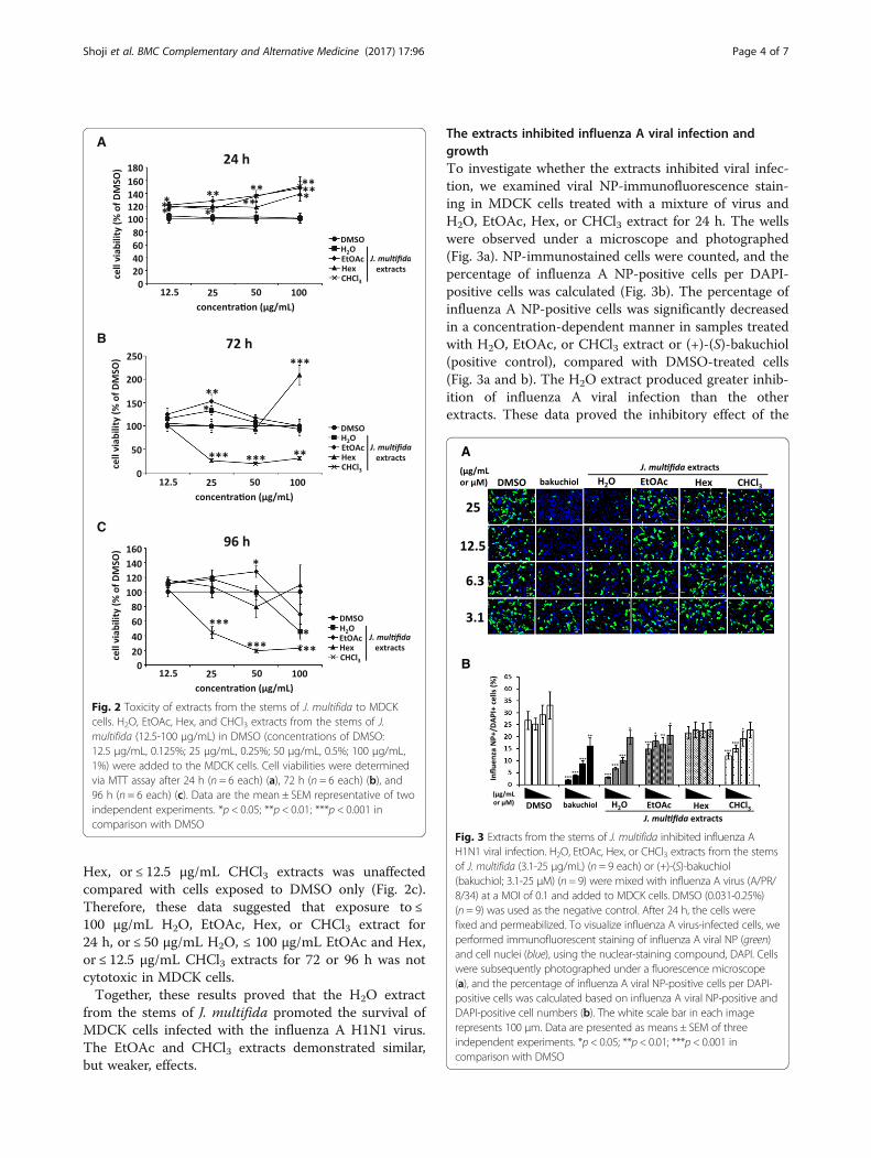
Hex, or ≤ 12.5 μg/mL CHCl3 extracts was unaffectedcompared with cells exposed to DMSO only (Fig. 2c).Therefore, these data suggested that exposure to ≤100 μg/mL H2O, EtOAc, Hex, or CHCl3 extract for24 h, or ≤ 50 μg/mL H2O, ≤ 100 μg/mL EtOAc and Hex,or ≤ 12.5 μg/mL CHCl3 extracts for 72 or 96 h was notcytotoxic in MDCK cells.Together, these results proved that the H2O extract
from the stems of J. multifida promoted the survival ofMDCK cells infected with the influenza A H1N1 virus.The EtOAc and CHCl3 extracts demonstrated similar,but weaker, effects.
The extracts inhibited influenza A viral infection andgrowthTo investigate whether the extracts inhibited viral infec-tion, we examined viral NP-immunofluorescence stain-ing in MDCK cells treated with a mixture of virus andH2O, EtOAc, Hex, or CHCl3 extract for 24 h. The wellswere observed under a microscope and photographed(Fig. 3a). NP-immunostained cells were counted, and thepercentage of influenza A NP-positive cells per DAPI-positive cells was calculated (Fig. 3b). The percentage ofinfluenza A NP-positive cells was significantly decreasedin a concentration-dependent manner in samples treatedwith H2O, EtOAc, or CHCl3 extract or (+)-(S)-bakuchiol(positive control), compared with DMSO-treated cells(Fig. 3a and b). The H2O extract produced greater inhib-ition of influenza A viral infection than the otherextracts. These data proved the inhibitory effect of the
Fig. 2 Toxicity of extracts from the stems of J. multifida to MDCKcells. H2O, EtOAc, Hex, and CHCl3 extracts from the stems of J.multifida (12.5-100 μg/mL) in DMSO (concentrations of DMSO:12.5 μg/mL, 0.125%; 25 μg/mL, 0.25%; 50 μg/mL, 0.5%; 100 μg/mL,1%) were added to the MDCK cells. Cell viabilities were determinedvia MTT assay after 24 h (n = 6 each) (a), 72 h (n = 6 each) (b), and96 h (n = 6 each) (c). Data are the mean ± SEM representative of twoindependent experiments. *p < 0.05; **p < 0.01; ***p < 0.001 incomparison with DMSO
Fig. 3 Extracts from the stems of J. multifida inhibited influenza AH1N1 viral infection. H2O, EtOAc, Hex, or CHCl3 extracts from the stemsof J. multifida (3.1-25 μg/mL) (n = 9 each) or (+)-(S)-bakuchiol(bakuchiol; 3.1-25 μM) (n = 9) were mixed with influenza A virus (A/PR/8/34) at a MOI of 0.1 and added to MDCK cells. DMSO (0.031-0.25%)(n = 9) was used as the negative control. After 24 h, the cells werefixed and permeabilized. To visualize influenza A virus-infected cells, weperformed immunofluorescent staining of influenza A viral NP (green)and cell nuclei (blue), using the nuclear-staining compound, DAPI. Cellswere subsequently photographed under a fluorescence microscope(a), and the percentage of influenza A viral NP-positive cells per DAPI-positive cells was calculated based on influenza A viral NP-positive andDAPI-positive cell numbers (b). The white scale bar in each imagerepresents 100 μm. Data are presented as means ± SEM of threeindependent experiments. *p < 0.05; **p < 0.01; ***p < 0.001 incomparison with DMSO
Shoji et al. BMC Complementary and Alternative Medicine (2017) 17:96 Page 4 of 7
H2O, EtOAc, and CHCl3 extracts from the stems of J.multifida on influenza A virus H1N1 infection.Next, we investigated the inhibition of viral growth by
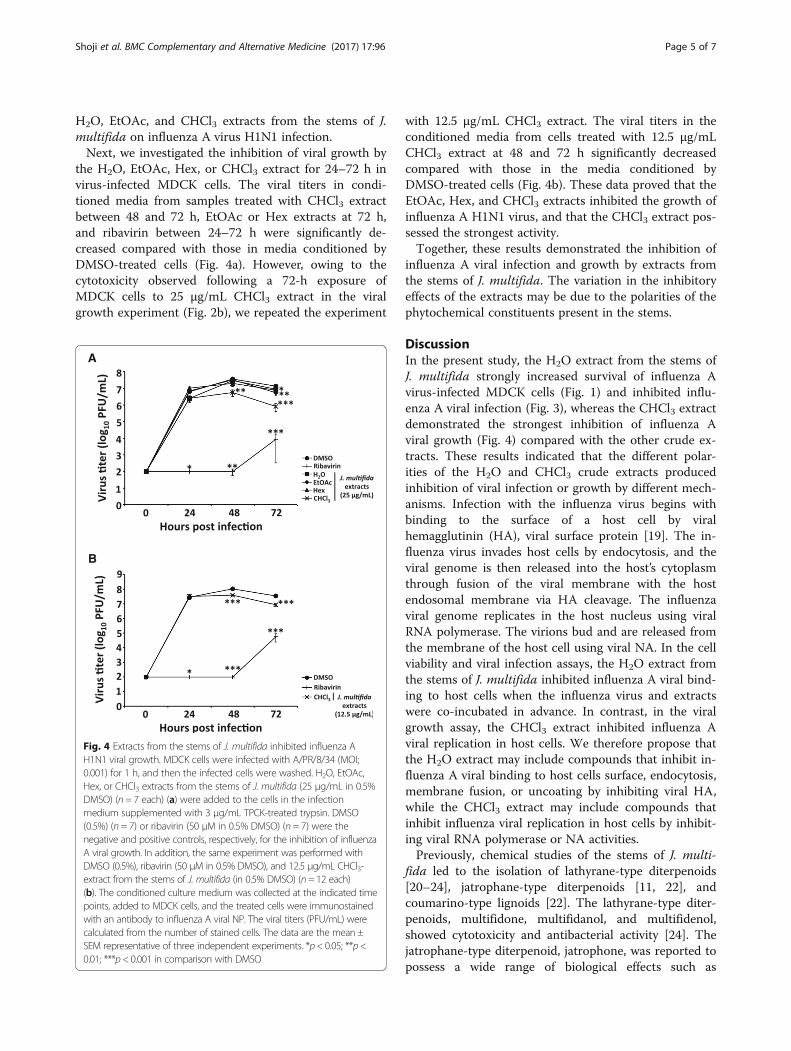
the H2O, EtOAc, Hex, or CHCl3 extract for 24–72 h invirus-infected MDCK cells. The viral titers in condi-tioned media from samples treated with CHCl3 extractbetween 48 and 72 h, EtOAc or Hex extracts at 72 h,and ribavirin between 24–72 h were significantly de-creased compared with those in media conditioned byDMSO-treated cells (Fig. 4a). However, owing to thecytotoxicity observed following a 72-h exposure ofMDCK cells to 25 μg/mL CHCl3 extract in the viralgrowth experiment (Fig. 2b), we repeated the experiment
with 12.5 μg/mL CHCl3 extract. The viral titers in theconditioned media from cells treated with 12.5 μg/mLCHCl3 extract at 48 and 72 h significantly decreasedcompared with those in the media conditioned byDMSO-treated cells (Fig. 4b). These data proved that theEtOAc, Hex, and CHCl3 extracts inhibited the growth ofinfluenza A H1N1 virus, and that the CHCl3 extract pos-sessed the strongest activity.Together, these results demonstrated the inhibition of
influenza A viral infection and growth by extracts fromthe stems of J. multifida. The variation in the inhibitoryeffects of the extracts may be due to the polarities of thephytochemical constituents present in the stems.
DiscussionIn the present study, the H2O extract from the stems ofJ. multifida strongly increased survival of influenza Avirus-infected MDCK cells (Fig. 1) and inhibited influ-enza A viral infection (Fig. 3), whereas the CHCl3 extractdemonstrated the strongest inhibition of influenza Aviral growth (Fig. 4) compared with the other crude ex-tracts. These results indicated that the different polar-ities of the H2O and CHCl3 crude extracts producedinhibition of viral infection or growth by different mech-anisms. Infection with the influenza virus begins withbinding to the surface of a host cell by viralhemagglutinin (HA), viral surface protein [19]. The in-fluenza virus invades host cells by endocytosis, and theviral genome is then released into the host’s cytoplasmthrough fusion of the viral membrane with the hostendosomal membrane via HA cleavage. The influenzaviral genome replicates in the host nucleus using viralRNA polymerase. The virions bud and are released fromthe membrane of the host cell using viral NA. In the cellviability and viral infection assays, the H2O extract fromthe stems of J. multifida inhibited influenza A viral bind-ing to host cells when the influenza virus and extractswere co-incubated in advance. In contrast, in the viralgrowth assay, the CHCl3 extract inhibited influenza Aviral replication in host cells. We therefore propose thatthe H2O extract may include compounds that inhibit in-fluenza A viral binding to host cells surface, endocytosis,membrane fusion, or uncoating by inhibiting viral HA,while the CHCl3 extract may include compounds thatinhibit influenza viral replication in host cells by inhibit-ing viral RNA polymerase or NA activities.Previously, chemical studies of the stems of J. multi-
fida led to the isolation of lathyrane-type diterpenoids[20–24], jatrophane-type diterpenoids [11, 22], andcoumarino-type lignoids [22]. The lathyrane-type diter-penoids, multifidone, multifidanol, and multifidenol,showed cytotoxicity and antibacterial activity [24]. Thejatrophane-type diterpenoid, jatrophone, was reported topossess a wide range of biological effects such as
Fig. 4 Extracts from the stems of J. multifida inhibited influenza AH1N1 viral growth. MDCK cells were infected with A/PR/8/34 (MOI;0.001) for 1 h, and then the infected cells were washed. H2O, EtOAc,Hex, or CHCl3 extracts from the stems of J. multifida (25 μg/mL in 0.5%DMSO) (n = 7 each) (a) were added to the cells in the infectionmedium supplemented with 3 μg/mL TPCK-treated trypsin. DMSO(0.5%) (n = 7) or ribavirin (50 μM in 0.5% DMSO) (n = 7) were thenegative and positive controls, respectively, for the inhibition of influenzaA viral growth. In addition, the same experiment was performed withDMSO (0.5%), ribavirin (50 μM in 0.5% DMSO), and 12.5 μg/mL CHCl3-extract from the stems of J. multifida (in 0.5% DMSO) (n= 12 each)(b). The conditioned culture medium was collected at the indicated timepoints, added to MDCK cells, and the treated cells were immunostainedwith an antibody to influenza A viral NP. The viral titers (PFU/mL) werecalculated from the number of stained cells. The data are the mean ±SEM representative of three independent experiments. *p< 0.05; **p<0.01; ***p< 0.001 in comparison with DMSO
Shoji et al. BMC Complementary and Alternative Medicine (2017) 17:96 Page 5 of 7
cytotoxicity and antitumor activity [25, 26]. However,the anti-influenza activity of the phytochemical constitu-ents of J. multifida has not been yet investigated. Thelathyrane-type diterpenoids from Euphorbia micractinashowed the anti-HIV activity [27]. Dang et al. recentlyreported that the abietane-type tricyclic phenolic diter-penoids, (+)-podcarpic acid and (+)-totarol, inhibited in-fluenza A H1N1 viral infection (A/PR/8/34) [28].Therefore, the diterpenoids contained in J. multifidamay confer the anti-viral effects such as anti-influenzaand anti-HIV activities. It is expected that anti-influenzacompounds will be isolated from the active crudeextracts in our ongoing work.
ConclusionsThe findings of the present study demonstrated that themost polar extract, the H2O extract, from the stems of J.multifida increased survival of MDCK cells infected withthe influenza A H1N1 virus and showed the strongestinhibition of influenza A H1N1 viral infection in MDCKcells. Of the EtOAc, Hex, and CHCl3 extracts, theCHCl3 extract showed the strongest inhibition of influ-enza A H1N1 viral growth in MDCK cells. These resultsindicated that the stems of J. multifida could be used asa herbal medicine for the treatment of influenza andmay be a source of candidate compounds for novel anti-influenza drug development.
AbbreviationsANOVA: One-way analysis of variance; CHCl3: Chloroform; DAPI: Diamidino-2-phenylindole; DMSO: Dimethyl sulfoxide; EtOAc: Ethyl acetate; H2O: Water;Hex: n-hexane; MDCK: Madin-Darby canine kidney; MTT: Thiazolyl bluetetrazolium bromide; NA: Neuraminidase; NP: Nucleoprotein; P/S: Penicillinand streptomycin; PBS: Phosphate-buffered saline; PR: Puerto Rico; TPCK: L-tosylamido-2-phenyl ethyl chloromethyl ketone
AcknowledgementsNot applicable.
FundingThis research was performed as part of the Cooperative Research Projectwith the Institute of Natural Medicine, University of Toyama in 2016 (M.S., T.I.,and T.K.). This work was also supported in part by a Grant-in-Aid for ScientificResearch from the Ministry of Education, Culture, Sports, Science and Tech-nology, Japan (H.M. and T.I.)
Availability of data and materialsThe original data are available from the authors. The availability of allmaterials and reagents is detailed in this manuscript.
Authors’ contributionsThis study was designed by MS, TI, HM, and TK. MS performed all theexperiments and drafted the manuscript. SYW and HM performed the plantextraction. AM performed the experiments with influenza virus. NNW and HNperformed the plant harvesting and extraction. ET and HK performed thegrowth and purification of influenza virus. TI and TK critically reviewed themanuscript. All authors reviewed the manuscript, and read and approved thefinal version.
Competing interestsThe authors declare that they have no conflicts of interest concerning this work.
Consent for publicationAll authors agree to publish the manuscript in its present form.
Ethic approval and consent participateNot applicable.
Author details1Laboratory of Biochemistry, Faculty of Pharmaceutical Sciences, TokushimaBunri University, 180 Yamashiro-cho, Tokushima 770-8514, Japan. 2Institute ofNatural Medicine, University of Toyama, 2630, Sugitani, Toyama 930-0194,Japan. 3Department of Chemistry, University of Yangon, Yangon 11041,Myanmar. 4Division of Pathology and Metabolome Research for InfectiousDisease and Host Defense, Institute for Enzyme Research, University ofTokushima, 3-18-15, Kuramoto-cho, Tokushima 770-8503, Japan.
Received: 29 September 2016 Accepted: 28 January 2017
References1. Horimoto T, Kawaoka Y. Influenza: lessons from past pandemics, warnings
from current incidents. Nat Rev Microbiol. 2005;3(8):591–600.2. Itoh Y, Shinya K, Kiso M, Watanabe T, Sakoda Y, Hatta M, Muramoto Y, Tamura
D, Sakai-Tagawa Y, Noda T, et al. In vitro and in vivo characterization of newswine-origin H1N1 influenza viruses. Nature. 2009;460(7258):1021–5.
3. Neumann G, Noda T, Kawaoka Y. Emergence and pandemic potential ofswine-origin H1N1 influenza virus. Nature. 2009;459(7249):931–9.
4. Dortmans JC, Dekkers J, Wickramasinghe IN, Verheije MH, Rottier PJ, vanKuppeveld FJ, de Vries E, de Haan CA. Adaptation of novel H7N9 influenzaA virus to human receptors. Sci Rep. 2013;3:3058.
5. Fiore AE, Fry A, Shay D, Gubareva L, Bresee JS, Uyeki TM. Centers for DiseaseC, Prevention: Antiviral agents for the treatment and chemoprophylaxis ofinfluenza — recommendations of the Advisory Committee onImmunization Practices (ACIP). MMWR Recomm Rep. 2011;60(1):1–24.
6. Molinari G. Natural products in drug discovery: present status andperspectives. Adv Exp Med Biol. 2009;655:13–27.
7. Harvey AL, Edrada-Ebel R, Quinn RJ. The re-emergence of natural productsfor drug discovery in the genomics era. Nat Rev Drug Discov. 2015;14(2):111–29.
8. Gao L, Sun Y, Si J, Liu J, Sun G, Sun X, Cao L. Cryptoporus volvatus extractinhibits influenza virus replication in vitro and in vivo. PLoS One. 2014;9(12):e113604.
9. Yang CH, Tan DH, Hsu WL, Jong TT, Wen CL, Hsu SL, Chang PC. Anti-influenza virus activity of the ethanolic extract from Peperomia sui. JEthnopharmacol. 2014;155(1):320–5.
10. Rajasekaran D, Palombo EA, Chia Yeo T. Lim Siok Ley D, Lee Tu C, MalherbeF, Grollo L: Identification of traditional medicinal plant extracts with novelanti-influenza activity. PLoS One. 2013;8(11):e79293.
11. Sabandar CW, Ahmat N, Jaafar FM, Sahidin I. Medicinal property, phytochemistryand pharmacology of several Jatropha species (Euphorbiaceae): a review.Phytochemistry. 2013;85:7–29.
12. Falodun A, Imieje V, Erharuyi O, Joy A, Langer P, Jacob M, Khan S, AbaldryM, Hamann M. Isolation of antileishmanial, antimalarial and antimicrobialmetabolites from Jatropha multifida. Asian Pac J Trop Biomed. 2014;4(5):374–8.
13. Win NN, Ito T, Aimaiti S, Imagawa H, Ngwe H, Abe I, Morita H.Kaempulchraols A-H, Diterpenoids from the Rhizomes of Kaempferiapulchra Collected in Myanmar. J Nat Prod. 2015;78(5):1113–8.
14. Ito T, Nisa K, Kodama T, Tanaka M, Okamoto Y. Ismail, Morita H: Two newcyclopentenones and a new furanone from Baeckea frutescens and theircytotoxicities. Fitoterapia. 2016;112:132–5.
15. Shoji M, Arakaki Y, Esumi T, Kohnomi S, Yamamoto C, Suzuki Y, Takahashi E,Konishi S, Kido H, Kuzuhara T. Bakuchiol Is a Phenolic Isoprenoid with NovelEnantiomer-selective Anti-influenza A Virus Activity Involving Nrf2 Activation. JBiol Chem. 2015;290(46):28001–17.
16. Takahashi E, Kataoka K, Indalao IL, Konoha K, Fujii K, Chida J, Mizuno D,Fujihashi K, Kido H. Oral clarithromycin enhances airway immunoglobulin A(IgA) immunity through induction of IgA class switching recombination andB-cell-activating factor of the tumor necrosis factor family molecule onmucosal dendritic cells in mice infected with influenza A virus. J Virol. 2012;86(20):10924–34.
Shoji et al. BMC Complementary and Alternative Medicine (2017) 17:96 Page 6 of 7
17. Shoji M, Takahashi E, Hatakeyama D, Iwai Y, Morita Y, Shirayama R, Echigo N,Kido H, Nakamura S, Mashino T, et al. Anti-influenza activity of c60 fullerenederivatives. PLoS One. 2013;8(6):e66337.
18. Shigeta S, Mori S, Watanabe J, Soeda S, Takahashi K, Yamase T. Synergistic anti-influenza virus A (H1N1) activities of PM-523 (polyoxometalate) and ribavirin invitro and in vivo. Antimicrob Agents Chemother. 1997;41(7):1423–7.
19. von Itzstein M. The war against influenza: discovery and development ofsialidase inhibitors. Nat Rev Drug Discov. 2007;6(12):967–74.
20. Das B, Laxminarayana K, Krishnaiah M, Srinivas Y, Raju TV. Multidione, a novelditerpenoid from Jatropha multifida. Tetrahedron Lett. 2009;50(34):4885–7.
21. Das B, Ravikanth B, Reddy KR, Thirupathi P, Raju TV, Sridhar B. Diterpenoidsfrom Jatropha multifida. Phytochemistry. 2008;69(14):2639–41.
22. Das B, Reddy KR, Ravikanth B, Raju TV, Sridhar B, Khan PU, Rao JV. Multifidone: anovel cytotoxic lathyrane-type diterpene having an unusual six-membered Aring from Jatropha multifida. Bioorg Med Chem Lett. 2009;19(1):77–9.
23. Das B, Satya Kumar A, Narayan Kumar J, Venugopal Raju T. A new macrocyclicditerpenoid from Jatropha multifida. Nat Prod Res. 2010;24(16):1510–3.
24. Kanth BS, Kumar AS, Shinde DB, Babu KH, Raju TV, Kumar CG, Sujitha P, DasB. New bioactive macrocyclic diterpenoids from Jatropha multifida. BioorgMed Chem Lett. 2011;21(22):6808–10.
25. Goel G, Makkar HP, Francis G, Becker K. Phorbol esters: structure, biologicalactivity, and toxicity in animals. Int J Toxicol. 2007;26(4):279–88.
26. Theoduloz C, Rodriguez JA, Pertino M, Schmeda-Hirschmann G.Antiproliferative activity of the diterpenes jatrophone and jatropholone andtheir derivatives. Planta Med. 2009;75(14):1520–2.
27. Tian Y, Xu W, Zhu C, Lin S, Li Y, Xiong L, Wang S, Wang L, Yang Y, Guo Y, etal. Lathyrane diterpenoids from the roots of Euphorbia micractina and theirbiological activities. J Nat Prod. 2011;74(5):1221–9.
28. Dang Z, Jung K, Zhu L, Xie H, Lee KH, Chen CH, Huang L. Phenolic diterpenoidderivatives as anti-influenza a virus agents. ACS Med Chem Lett. 2015;6(3):355–8.
• We accept pre-submission inquiries
• Our selector tool helps you to find the most relevant journal
• We provide round the clock customer support
• Convenient online submission
• Thorough peer review
• Inclusion in PubMed and all major indexing services
• Maximum visibility for your research
Submit your manuscript atwww.biomedcentral.com/submit
Submit your next manuscript to BioMed Central and we will help you at every step:
Shoji et al. BMC Complementary and Alternative Medicine (2017) 17:96 Page 7 of 7


![Pelagia Research Library Comparison of different extracts leaf of … · 2016-09-21 · and vegetable crops by using their buds, inflorescences, leaves, roots, seeds and stems [8]](https://img.pdfslide.us/doc/110x75/5ea4bb809a712d5997091a8c/pelagia-research-library-comparison-of-different-extracts-leaf-of-2016-09-21-and.jpg)