Embed Size (px)
Citation preview

© 2016 Vageesh Revadigar et al. Thisis an open access article distributed under the terms of the Creative Commons Attribution License -NonCommercial-ShareAlikeUnported License (http://creativecommons.org/licenses/by-nc-sa/3.0/).
Journal of Applied Pharmaceutical Science Vol. 7 (05), pp. 103-110, May, 2017
Available online at http://www.japsonline.com
DOI: 10.7324/JAPS.2017.70518
ISSN 2231-3354
Anti-oxidative and cytotoxic attributes of phenolic rich ethanol
extract of Musa balbisiana Colla inflorescence
Vageesh Revadigar
1, Majed Ahmed Al-Mansoub
1, Muhammad Asif
2, Mohammad Razak Hamdan
3,
Amin Malik Shah Abdul Majid2, Mohd Zaini Asmawi
1, Vikneswaran Murugaiyah
1*
1Discipline of Pharmacology, School of Pharmaceutical Sciences, Universiti Sains Malaysia, 11800Penang, Malaysia.
2EMAN Testing and Research Laboratory, School of Pharmaceutical Sciences, Universiti Sains Malaysia, 11800, Penang, Malaysia.
3Centre for Drug Research, Universiti Sains Malaysia, 11800, Penang, Malaysia.
ARTICLE INFO
ABSTRACT
Article history:
Received on: 09/04/2016
Accepted on: 16/09/2016
Available online: 30/05/2017
Inflorescence of Musa species is one of the most commonly consumed vegetables in Southeast Asian region. In
the present study, chemical composition and antioxidant potential of the ethanolic extract of inflorescence of
Musa balbisiana Colla (MbCi) were evaluated. In addition, the extract was also subjected to cytotoxicity testing
on a panel of human cancer cell lines. The ethanolic extract of inflorescence of Musa balbisiana Colla was
evaluated for antioxidant activity using 1,1-diphenyl-2-picryl-hydrazyl assay (DPPH), 2,2'-azino-bis (3-
ethylbenzothiazoline-6-sulphonic acid) (ABTS) and ferric reducing antioxidant power (FRAP) assays. The
extract was chemically characterized for total phenolic (TPC) and total flavonoid (TFC) contents and by gas
chromatography–mass spectrometry (GC-MS) analysis. Cytotoxicity was evaluated by MTT cell viability assay.
The extract showed moderate antioxidant activity in all the antioxidant assays. Chemically, the extract was
found to possess high total phenolic (92.01 ± 0.40 μg gallic acid equivalent/mg extract) but low flavonoid (4.852
± 0.04 μg quercetin equiv./mg extract) contents. In cell viability assay, MbCi extract showed selective
cytotoxicity towards HT-29 cell line. Morphological observations show that MbCi has apoptosis inducing
nature. GC-MS analysis has revealed the presence of 22 compounds, mainly belonging to steroids, fatty acids
and long chain aliphatic compounds, which in part may be responsible for observed antioxidant and cytotoxic
activities of ethanol extract. Our study revealed that MbCi has chemotherapeutic potential activity that warrants
further investigation.
Key words:
Musa balbisiana Colla,
antioxidant, cytotoxicity,
morphological changes,
apoptosis.
INTRODUCTION
Plants from genus Musa contribute as the fourth most
important food in the world today. Musa species are natives to
Indo-Malaysian, Asian and Australian tropics (Nelson et al.,
2006). Inflorescence of Musa species (banana) is much
appreciated vegetable in Malaysia. The inner part of
inflorescence as well as lower soft inner part of pseudostem is
widely used for cooking (Vimala et al., 2003). Decoction
of half ripe fruit of Musa balbisiana Colla (MbC) is used in the
* Corresponding Author
Discipline of Pharmacology, School of Pharmaceutical Sciences,
Universiti Sains Malaysia, 11800 Penang, Malaysia.
Email: vicky @ usm.my
treatment of dysentery. In India, exudates from rhizomatous stem
of MbC are used for the treatment of pinworm infection (Kalita
and Deb, 2004), while the tablets prepared from the fresh and dried
seed paste of MbC are used as contraceptives (Das et al., 2014).
Decoction from the pith of Musa species is used in the treatment of
congestive heart failure and hypertension by Temuan tribe of
Peninsular Malaysia (Azliza et al., 2012). Musa species are studied
extensively for their pharmacological activities. Kumar et al.
(2012), reported that flavonoid leucocyanidin significantly increase
the thickness of the mucous membrane layer of the stomach and
improved gastric ulcer in comparison with antacid. Aqueous
extract from the roots of MbC produced significant size reduction
in albumin induced hind paw edema in Wistar rats in a dose
dependent manner (Ibegbu et al., 2012).
104 Revadigar et al. / Journal of Applied Pharmaceutical Science 7 (05); 2017: 103-110
Fresh dried pulp of the fruit was reported to possess in
vitro as well in vivo antioxidant activity in a dose dependent
manner (Mudoi et al., 2011). Antioxidant activity of eight
Malaysian bananas were reported by Sulaiman et al. (2011), who
found that the chloroform extract of dried pulp possessed good
antioxidant activity. The authors also reported weak correlation
between the antioxidant activity and total phenolic content of the
samples. Vilela et al. (2014) identified GC-MS of dichloromethane
extracts of several Musa species and reported the presence of
lipophilic phytochemicals. Hitherto, there is no data available on
the antioxidant activity and cytotoxicity of inflorescence of Musa
balbisiana Colla (MbCi). Therefore, the present study was
designed to investigate the phytochemical composition,
antioxidant capacity and cytotoxicity of MbCi.
MATERIALS AND METHODS
Plant materials
Inflorescence of MbC was collected from Balik Pulau
hilly area, Penang, Malaysia. A voucher specimen (11559) was
deposited in Herbarium Unit at the School of Biological Sciences,
Universiti Sains Malaysia.
Preparation of MbCi extract
The fresh inflorescences were cut into smaller pieces,
crushed and triturated in mortar and pestle by addition of small
amount of ethanol and made into paste. This mass was further
extracted by maceration for 6 days with absolute ethanol. The
extracts were filtered using whatman filter paper and concentrated
using rotavapor (Heidolph Instruments; Schwabach, Germany).
Then, the extracts were stored in sealed vial at 4 °C until
biological testing. All determinations were done in triplicate and
absorbencies were measured using microplate reader (TECAN
Infinite Pro® M200, Switzerland).
Chemical characterization
Total phenolic content
Total phenolic content was determined using Folin–
Ciocalteu reagent following the method described by Kumaran and
Joel Karunakaran (2007), using gallic acid as a reference standard.
The assay was carried out by mixing Folin–Ciocalteu reagent,
sodium carbonate, standard/extract sample and distilled water in a
test tube in a ratio of 5:15:1:79 to the final volume of 1000 µL.
The tubes were incubated for 2 hour at room temperature and an
aliquot (200 µL) of each mixture was transferred into 96-well
microplate. The amount of test sample was substituted by distilled
water in blank. Absorbencies were taken at 765 nm. The results
are expressed as µg gallic acid equivalent/mg dry extract.
Total flavonoid content
Total flavonoid content was determined by the aluminum
chloride method as described by Orhan et al. (2011) using
quercetin as a reference standard. For the assay, the standard or
extract solutions (100 µL) were mixed with of 10% (w/v)
aluminum chloride (20 µL), 1 mol/L sodium acetate (20 µL)
methanol (300 µL) and distilled water (560 µL). After incubation
at room temperature for 30 min, the absorbance of the reaction
mixture was measured at 415 nm. The results are expressed as µg
quercetin equivalent/mg dry extract.
Gas Chromatography Mass Spectrometry (GC-MS) analysis
The chemical composition of MbCi was determined
using Agilent GC-MS system consisting of a gas chromatograph
(Agilent 6890) coupled to a mass spectrophotometer (Agilent
5973; inert mass selective detector). Separation was achieved on a
HP-5 MS column of 30 m length, 0.25 mm diameter consist of
film thickness 0.25 μm. The injector was set at 70 C for 2 minutes
and steadily increased 20 C up to 285 C. Helium was used as the
carrier gas with the flow rate of 20 mL per minute. An amount of 2
μL of sample was injected. Transfer line was maintained at 250
C. The mass spectrophotometer was operated at 1717.6 eV. The
total run time was 47.75 minutes. The identification of compounds
was done by using NIST 02 library.
Antioxidant assays
DPPH scavenging assay
Free radical scavenging activity was determined using
2,2-Diphenyl-2-Picrylhydrazyl (DPPH) as described by Al-
Mansoub et al. (2014). A 100 µL of the extract sample (0.78 –
200) μg/mL dissolved in DMSO were mixed with 100 µL of
DPPH (200 µmol/L) dissolved in methanol, and the reaction
mixture was incubated at room temperature for 30 min. Ascorbic
acid was used as a reference standard. The absorbance was
measured at 517 nm. The results are expressed as IC50.
ABTS radical scavenging activity assay
ABTS radical scavenging activity was measured by
themodified ABTS cation decolorization assay as described by
Al-Mansoub et al. (2014) as described by Re et al. (1999). ABTS
radical cation (ABTS•+
) solution was prepared by mixing of 14
mM ABTS and 4.9 mM potassium persulfate (K2S2O8) dissolved
in deionized water in equal volumes. This solution was allowed to
react in the dark place at room temperature for 16-20 h before use.
Then, 1 mL of stock ABTS•+
solution was then diluted with 40 mL
of deionized water to yield an absorbance equals to 0.70 ± 0.02 at
734 nm. In Brief, to 180 µL of ABTS radical solution, 20µl of
sample extract (3.13 – 400) μg/mL were added. Ascorbic acid was
used as a reference standard. The absorbance of ABTS•+
sample
extract/standard was taken at 734 nm. The results are expressed as
IC50.
Ferric reducing antioxidant power (FRAP) assay
The FRAP assay was carried out by method of Benzie
and Strain (1996) as developed by Griffin and Bhagooli (2004).
FRAP working solution was prepared by mixing (300 mmol/L)
acetate buffer, pH 3.6 (10 mmol/L) TPTZ in (40 mmol/L) HCl and
(20 mmol/L) FeCl3 in a ratio of 10:1:1. An amount of 150 μL of
Revadigar et al. / Journal of Applied Pharmaceutical Science 7 (05); 2017: 103-110 105
working solution was added to 50 µL standard/extracts. Blank was
done in the same way using methanol instead of test solutions. The
reaction mixture was incubated for 8 min then readings were taken
at 600 nm. Ferrous sulfate (FeSO4.7H2O) was used as reference
standard and the results are expressed as nmol Fe+2
equivalent/mg
dry extract.
In vitro anticancer assays
Cell viability assay
Cytotoxicity of MbCi was tested on EA.hy926 (human
normal endothelial cells) and four cancer cell lines namely, MCF-7
(human breast cancer ATCC® HTB-22), HeLa (human cervical
carcinoma ATCC®
CCL-2), HT-29 (human colorectal
adenocarcinoma ATCC® HTB-38) and HCT 116 (human
colorectal carcinoma ATCC® CCL-247) using MTT assay [3-(4,5-
dimethylthiazol-2-yl)2,5-diphenyl tetrazolium bromide] following
the protocol described by Asif et al. (2016). MCF7 and HeLa cells
were cultured in Dulbecco’s Modified Eagle medium (DMEM)
(Gibco® Invitrogen) whereas HT-29 and HCT-116 were cultured
in Roswell Park Memorial Institute medium (RPMI) (Gibco®
Invitrogen) supplemented with fetal bovine serum (10%) and
penicillin-streptomycin (1%) (Gibco® Invitrogen). Cells were
maintained at 37 C in a water saturated atmosphere containing 5%
CO2. Counting of cells was done using a Neubauer hemocytometer
under light microscope by using trypan blue (Gibco® Invitrogen)
staining method. Cells of 5,000 to 10,000 densities per well seeded
in a 96 well plate and treated with different concentrations (100-
3.125 µg/mL) of ethanolic extract dissolved in DMSO (0.5% in
final concentration). 5-fluorouracil (5-FU) was used as the
reference standard and 0.5% DMSO was used as the negative
control. The treated cells were incubated for 48 hours. The
absorbance was read using microplate reader (TECAN infinite
Pro® M200, Switzerland) at 570 nm using 620 as reference
wavelength. The results were presented as percent viability.
Morphological analysis
Changes in the morphology of cells treatred with MbCi
were studied following well established method of Ebrahim et al.
(2014) with some modifications. In brief, overnight seeded cells
(5×10⁵) were incubated with IC50 vlaues of MbCi for 48 hours in a
6-well tissue culture plate. At the end of treatment period, the
medium was discarded and cells were washed once with PBS. The
changes in the morphology of treated cells were observed using
inverted phase contrast microscope at 10× magnification.
Statistical analysis
Data are presented as mean ± standard error of
mean (SEM). The minimum inhibitory concentration (IC50) was
calculated from the linear regression equations of dose response
curve for each experiment. All statistics analyses were carried out
using SPSS software (20.0 version).
RESULTS
Chemical characterization of ethanol extract
The extract was chemically characterized by colorimetric
and GC-MS methods.
Total phenolic and flavonoid contents
MbCi was revealed to have higher total phenolic content
(92.02 ± 0.40 μg gallic acid equiv./mg extract) and low total
flavonoid content (4.85 ± 0.05 μg quercetin equiv./mg extract)
(Table 1).
GC-MS analysis
The GC-MS analysis has shown the presence of 22
compounds in the extract; among these 16 compounds were found
to be with more than 90% of similarity with NIST 02 library.
These identified compounds belong to the class of steroids, long
chain unsaturated and saturated hydrocarbons, esters and fatty
acids.
On the other hand, compounds with less similarity index
while found to be in major proportion could be predicted as the
polyphenolics by considering the higher TPC value of the extract.
The extract was enriched with major sterols stigmasterol, beta-
sitosterol, and campesterol, in descending order of abundance.
However, the quantitative analysis of the tested extract revealed
that the major dominant peak corresponds to Z-12-Pentacosene
(8.23%), followed by stigmasterol (7.33%), 10-Heneicosene
(5.73%), and beta-sitosterol (5.23%) (Table 2, Figure 1).
Antioxidant Assays
Finding of the present study shows that MbCi has
moderate antioxidant activity in DPPH, ABTS free radical
scavenging, while ferric reducing antioxidant power of the MbCi
in FRAP assay demonstrated good activity.
The antioxidant results of MbCi in these respective
assays are shown in Table 1.
The DPPH and ABTS IC50 values of MbCi (IC50 value is
the concentration of the sample required to inhibit 50% of radical)
were 64.24 ± 3.09 and 76.23 ± 2.20 μg/mL. The results were
compared to reference standard ascorbic acid which were found to
be 5.41 ± 0.41 μg/mL and 3.49 ± 0.05 μg/mL, for the DPPH and
ABTS assays, respectively. In addition, the MbCi showed good
antioxidant activity in FRAP test with a value of 70.08 ± 12.86
nmol Fe+2
equiv./mg extract.
Table 1: Antioxidant activity of ethanolic extract of Musa balbisiana Colla inflorescence.
Sample
Total phenolic
(μg gallic acid
equiv./mg extract)
Total flavonoid
(μg quercetin
equiv./mg extract)
DPPH
IC50 (μg/mL)
ABTS
IC50 (μg/mL)
FRAP
(nmol Fe+2
equiv./ mg extract)
MbCi 92.02 ± 0.40 4.85 ± 0.05 64.24 ± 3.09 76.23 ± 2.20 70.08 ± 12.86
Vitamin C (Standard) - - 5.41 ± 0.41 3.49 ± 0.05 -
Values are expressed as mean ± SEM (n=3).
106 Revadigar et al. / Journal of Applied Pharmaceutical Science 7 (05); 2017: 103-110
Table 2: GC-MS profile of ethanolic extract of Musa balbisiana Colla inflorescence
S. No. Compound Retention
time Area % Molecular formula Molecular weight Similarity index
1 Hexadecanoic acid, methyl ester 10.37 3.89 C17H34O2 270.450 99
2 9,12- octadecanoic acid (z,z)-methyl ester 11.13 3.02 C19H34O2 294.472 99
3 Heptadecanoic acid, 16-methyl-, methyl ester 11.21 1.47 C19H38O2 298.503 94
4 Linoleic acid ethyl ester 11.40 0.79 C20H34O2 308.498 99
5 9-Tricosene, (Z)- 11.74 0.74 C23H46 322.611 99
6 Heptadecane 11.83 0.64 C17H36 240.468 96
7 10-Heneicosene 12.51 5.73 C20H34 310.600 99
8 Hexadecane,2,6,10,14-tetramethyl- 12.60 3.67 C20H42 282.547 96
9 Z-12-Pentacosene 13.31 8.23 C25H50 350.665 91
10 Eicosane 13.40 0.49 C20H42 282.547 92
11 1-Nonadecene 14.29 4.21 C19H38 266.505 95
12 17-Pentatriacontene 15.63 2.45 C35H70 490.930 93
13 Vitamin E 16.53 1.98 C29H50O2 430.706 98
14 Campesterol 17.80 2.79 C28H48O 400.680 99
15 Stigmasterol 18.15 7.33 C29H48O 412.691 91
16 Beta-sitosterol 18.92 5.23 C29H50O 414.706 99
Compounds having similarity index with NIST02 library more than 90 were considered for reporting
Fig. 1: Gas chromatography-mass spectrometry (GC-MS) chromatogram, and the pie chart depicts the percentage of each phytochemical present in the ethanolic
extract of Musa balbisiana Colla inflorescence. The identification of compounds was done by using NIST 02 library. Where, i = Z-12-Pentacosene (8.23%), o =
stigmasterol (7.33%), g = 10-Heneicosene (5.73%), and s = beta-sitosterol (5.23%).
Revadigar et al. / Journal of Applied Pharmaceutical Science 7 (05); 2017: 103-110 107
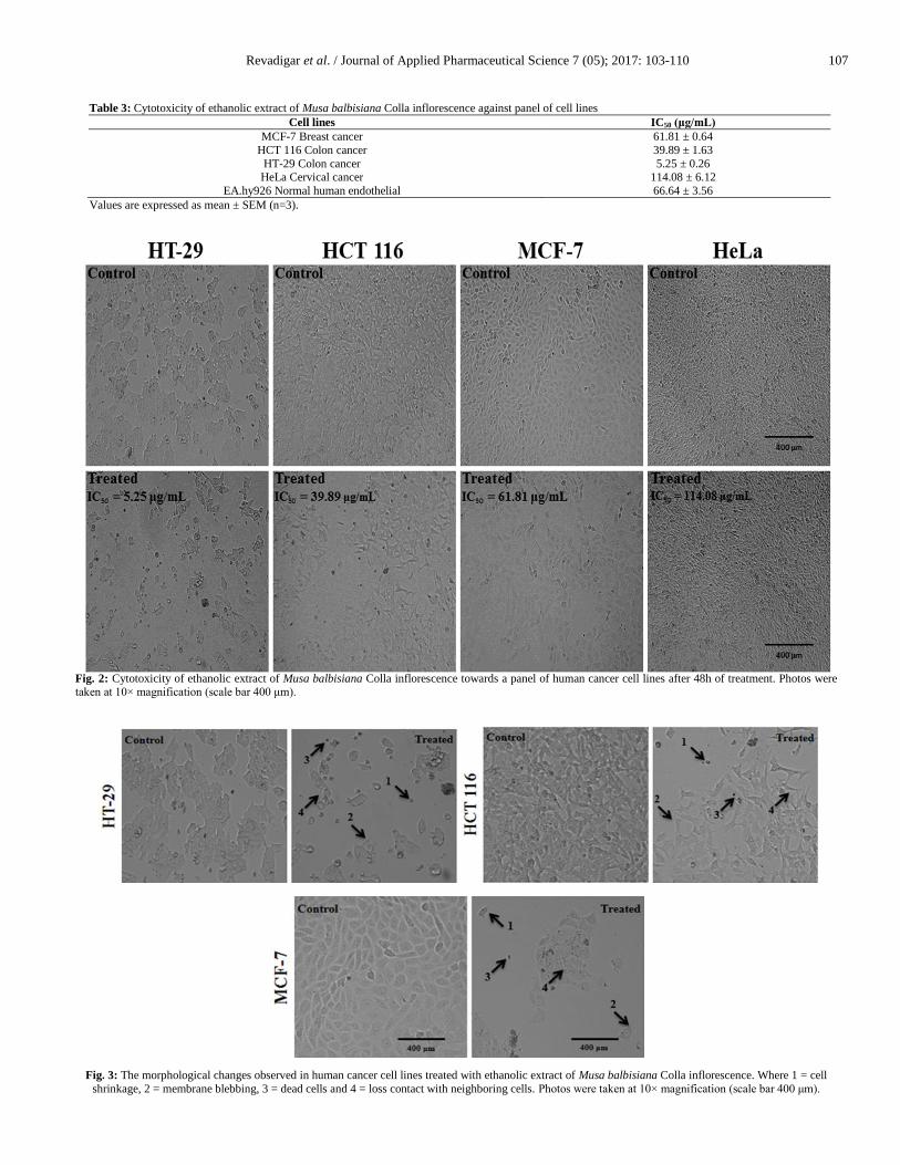
Table 3: Cytotoxicity of ethanolic extract of Musa balbisiana Colla inflorescence against panel of cell lines
Cell lines IC50 (μg/mL)
MCF-7 Breast cancer 61.81 ± 0.64
HCT 116 Colon cancer 39.89 ± 1.63
HT-29 Colon cancer 5.25 ± 0.26 HeLa Cervical cancer
EA.hy926 Normal human endothelial
114.08 ± 6.12
66.64 ± 3.56
Values are expressed as mean ± SEM (n=3).
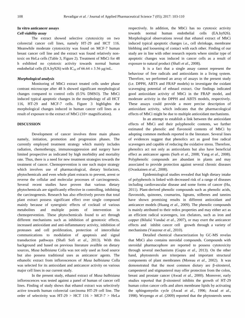
Fig. 2: Cytotoxicity of ethanolic extract of Musa balbisiana Colla inflorescence towards a panel of human cancer cell lines after 48h of treatment. Photos were taken at 10× magnification (scale bar 400 μm).
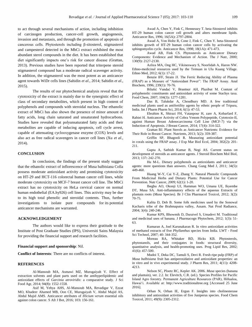
Fig. 3: The morphological changes observed in human cancer cell lines treated with ethanolic extract of Musa balbisiana Colla inflorescence. Where 1 = cell
shrinkage, 2 = membrane blebbing, 3 = dead cells and 4 = loss contact with neighboring cells. Photos were taken at 10× magnification (scale bar 400 μm).
108 Revadigar et al. / Journal of Applied Pharmaceutical Science 7 (05); 2017: 103-110
In vitro anticancer assays
Cell viability assay
The extract showed selective cytotoxicity on two
colorectal cancer cell lines, namely HT-29 and HCT 116.
Meanwhile moderate cytotoxicity was found on MCF-7 human
breast cancer cell line and the extract was found relatively non-
toxic on HeLa cells (Table 3, Figure 2). Treatment of MbCi for 48
h exhibited no cytotoxic activity towards normal human
endothelial cells (EA.hy926) with IC50 of 66.64 ± 3.56 μg/mL.
Morphological analysis
Monitoring of MbCi extract treated cells under phase
contrast microscope after 48 h showed significant morphological
changes compared to control cells (0.5% DMSO). The MbCi
induced typical apoptotic changes in the morphology of all HCT
116, HT-29 and MCF-7 cells. Figure 3 highlights the
morphological changes induced in human cancer cell lines as a
result of exposure to the extract of MbCi (10× magnification).
DISCUSSION
Development of cancer involves three main phases
namely, initiation, promotion and progression phases. The
currently employed treatment strategy which mainly includes
radiation, chemotherapy, immunosuppression and surgery have
limited prospective as indicated by high morbidity and mortality
rate. Thus, there is a need for new treatment strategies towards the
treatment of cancer. Chemoprevention is one such major strategy
which involves use of pharmacological, dietary biofactors,
phytochemicals and even whole plant extracts to prevent, arrest or
reverse the cellular and molecular processes of carcinogenesis.
Several recent studies have proven that various dietary
phytochemicals are significantly effective in controlling, inhibiting
the carcinogenesis. Research has also effectively proven that total
plant extract possess significant effect over single compound
mainly because of synergistic effects of cocktail of various
metabolites and multiple points of intervention in
chemoprevention. These phytochemicals found to act through
different mechanisms such as inhibition of genotoxic effects,
increased antioxidant and anti-inflammatory activity, inhibition of
proteases and cell proliferation, protection of intercellular
communications to modulation of apoptosis and signal
transduction pathways (Shafi Sofi et al., 2013). With this
background and based on previous literature availble on dietary
sources, Musa balbisiana Colla was not only used as food source
but also possess traditional uses as anticancer agents. The
ethanolic extract from infloroscences of Musa balbisiana Colla
was selected for its antioxidant and anticancer activity on various
major cell lines in our current study.
In the present study, ethanol extract of Musa balbisiana
infloroscences was tested against a panel of human of cancer cell
lines. Finding of study shows that ethanol extract was selectively
active towards human colorectal carcinoma HT-29 cell line. The
order of selectivity was HT-29 > HCT 116 > MCF-7 > HeLa
respectively. In addition, the MbCi has no cytotoxic activity
towards normal human endothelial cells (EA.hy926).
Morphological observations reveal that ethanol extract of MbCi
induced typical apoptotic changes i.e., cell shrinkage, membrane
blebbing and loosening of contact with each other. Finding of our
study are in line with other research reports where similar type of
apoptotic changes was induced in cancer cells as a result of
exposure to natural product (Shafi et al., 2008).
It is a fact that a single assay cannot represent the
behaviour of free radicals and antioxidants in a living system.
Therefore, we performed an array of assays in the present study
(i.e. DPPH, ABTS and FRAP models) to investigate the oxidant
scavenging potential of ethnaol extract. Our findings indicated
good antioxidant activity of MbCi in the FRAP model, and
moderate activity in the DPPH and ABTS models, respectively.
These assays could provide a more precise description of
antioxidant activity, which indicates that the pharmacological
effects of MbCi might be due to multiple antioxidant mechanisms.
In an attempt to establish a link between the antioxidant
effects of MbCi and their polyphenolic contents, we further
estimated the phenolic and flavonoid contents of MbCi by
adopting common methods reported in the literature. Several lines
of evidences suggest that phenolics act as good free radical
scavengers and capable of reducing the oxidative stress. Therefore,
phenolics act not only as antioxidants but also have beneficial
effect as anticancer agents (Surh et al., 2008; Yang et al., 2008).
Polyphenolic compounds are abundant in plants and may
associated to provide protection against several chronic diseases
(Ovaskainen et al., 2008).
Epidemiological studies revealed that high dietary intake
of polyphenols is linked with decreased risk of a range of diseases
including cardiovascular disease and some forms of cancer (Hu,
2011). Plant-derived phenolic compounds such as phenolic acids,
flavonoids, quinones, coumarins, lignans, stilbenes and tannins
have shown promising results in different antioxidant and
anticancer models (Huang et al., 2009). The phenolic compounds
is mainly attributed to their redox properties and may either act as
an efficient radical scavengers, ion chelators, such as iron and
copper (Bilušić Vundać et al., 2007), or may exert the anticancer
effects and inhibit cancer cell growth through a variety of
mechanisms (Vauzour et al., 2010).
Detailed chemical characterization by GC-MS revelas
that MbCi also contains steroidal compounds. Compounds with
steroidal pharmacophore are reported to possess cytotoxicity
through several mechanisms (Gupta et al., 2013). On the other
hand, phytosterols are triterpenes and important structural
components of plant membranes (Moreau et al., 2002). It was
demonstrated that the most common dietary are β-sitosterol,
campesterol and stigmasterol may offer protection from the colon,
breast and prostate cancer (Awad et al., 2000). Moreover, early
studies reported that β-sitosterol inhibits the growth of HT-29
human colon cancer cells and alters membrane lipids by activating
the sphingomyelin cycle (Awad et al., 1996; Awad et al.,
1998). Woyengo et al. (2009) reported that the phytosterols seem
Revadigar et al. / Journal of Applied Pharmaceutical Science 7 (05); 2017: 103-110 109
to act through several mechanisms of action, including inhibition
of carcinogen production, cancer-cell growth, angiogenesis,
invasion and metastasis, and through the promotion of apoptosis of
cancerous cells. Phytosterols including β-sitosterol, stigmasterol
and campesterol detected in the MbCi extract exhibited the most
abundant sterol compounds in the diet. It has been established that
diet significantly impacts one’s risk for cancer disease (Grattan,
2013). Previous studies have been reported that triterpene steroid
stigmasterol compound has a moderate radical scavenger activity.
In addition, the stigmasterol was the most potent as an anticancer
agent towards WiDr cells lines (Sahidin et al., 2014; Sahidin et al.,
2015).
The results of our phytochemical analysis reveal that the
cytotoxicity of the extract is mainly due to the synergistic effect of
class of secondary metabolites, which present in high content of
polyphenols and compounds with steroidal nucleus. The ethanolic
extract of MbCi has also shown the presence of minor amount of
fatty acids, long chain saturated and unsaturated hydrocarbons.
Studies have revealed that polyunsaturated fatty acids and their
metabolites are capable of inducing apoptosis, cell cycle arrest,
capable of attenuating cyclooxygenase enzyme (COX) levels and
also act as free radical scavengers in cancer cell lines (Xu et al.,
2014).
CONCLUSION
In conclusion, the findings of the present study suggest
that the ethanolic extract of inflorescence of Musa balbisiana Colla
possess moderate antioxidant activity and promising cytotoxicity
on HT-29 and HCT-116 colorectal human cancer cell lines, while
moderate cytotoxicity on MCF-7 breast cancer cell line. The MbCi
extract has no cytotoxicity on HeLa cervical cancer on normal
human endothelial (EA.hy926) cell lines. This activity may be due
to its high total phenolic and steroidal contents. Thus, further
investigations to isolate pure compounds for its potential
anticancer mechanisms are warranted.
ACKNOWLEDGEMENT
The authors would like to express their gratitude to the
Institute of Post Graduate Studies (IPS), Universiti Sains Malaysia
for providing the financial support and research facilities.
Financial support and sponsorship: Nil.
Conflict of Interests: There are no conflicts of interest.
REFERENCES
Al-Mansoub MA, Asmawi MZ, Murugaiyah V. Effect of
extraction solvents and plant parts used on the antihyperlipidemic and
antioxidant effects of Garcinia atroviridis: a comparative study. J Sci
Food Agr, 2014; 94(8): 1552-1558.
Asif M, Yehya AHS, Al-Mansoub MA, Revadigar V, Ezzat
MO, Khadeer Ahamed MB, Oon CE, Murugaiyah V, Abdul Majid AS,
Abdul Majid AMS. Anticancer attributes of Illicium verum essential oils
against colon cancer. S Afr J Bot, 2016; 103: 156-161.
Awad A, Chen Y, Fink C, Hennessey T. beta-Sitosterol inhibits
HT-29 human colon cancer cell growth and alters membrane lipids.
Anticancer Res, 1996; 16(5A): 2797-2804.
Awad A, Von Holtz R, Cone J, Fink C, Chen Y. beta-Sitosterol
inhibits growth of HT-29 human colon cancer cells by activating the
sphingomyelin cycle. Anticancer Res, 1998; 18(1A): 471-473.
Awad AB, Fink CS. Phytosterols as Anticancer Dietary
Components: Evidence and Mechanism of Action. The J Nutr, 2000;
130(9): 2127-2130.
Azliza MA, Ong HC, Vikineswary S, Noorlidah A, Haron NW.
Ethno-medicinal resources used by the Temuan in Ulu Kuang Village.
Ethno Med, 2012; 6(1): 17-22.
Benzie IFF, Strain JJ. The Ferric Reducing Ability of Plasma
(FRAP) as a Measure of “Antioxidant Power”: The FRAP Assay. Anal
Biochem, 1996; 239(1): 70-76.
Bilušić Vundać V, Brantner AH, Plazibat M. Content of
polyphenolic constituents and antioxidant activity of some Stachys taxa.
Food Chem, 2007; 104(3): 1277-1281.
Das B, Talukdar A, Choudhury MD. A few traditional
medicinal plants used as antifertility agents by ethnic people of Tripura,
India. Int J Pharm Pharm Sci, 2014; 6(1): 47-53.
Ebrahim K, Shirazi FH, Vatanpour H, zare A, Kobarfard F,
Rabiei H. Anticancer Activity of Cobra Venom Polypeptide, Cytotoxin-II,
against Human Breast Adenocarcinoma Cell Line (MCF-7) via the
Induction of Apoptosis. J Breast Cancer, 2014; 17(4): 314-322.
Grattan BJ. Plant Sterols as Anticancer Nutrients: Evidence for
Their Role in Breast Cancer. Nutrients, 2013; 5(2): 359-387.
Griffin SP, Bhagooli R. Measuring antioxidant potential
in corals using the FRAP assay. J Exp Mar Biol Ecol, 2004; 302(2): 201-
211.
Gupta A, Sathish Kumar B, Negi AS. Current status on
development of steroids as anticancer agents. J Steroid Biochem Mol Biol,
2013; 137: 242-270.
Hu M-L. Dietary polyphenols as antioxidants and anticancer
agents: more questions than answers. Chang Gung Med J, 2011; 34(5):
449-460.
Huang W-Y, Cai Y-Z, Zhang Y. Natural Phenolic Compounds
From Medicinal Herbs and Dietary Plants: Potential Use for Cancer
Prevention. Nutr Cancer, 2009; 62(1): 1-20.
Ibegbu AO, Okonji UJ, Hamman WO, Umana UE, Ikyembe
DT, Musa SA. Anti-inflammatory effects of the aqueous Extracts of
plantain roots (Musa Species). Br J Clin Pharmacol Toxicol, 2012; 3(2):
70-75.
Kalita D, Deb B. Some folk medicines used by the Sonowal
Kacharis tribe of the Brahmaputra valley, Assam. Nat Prod Radiance,
2004; 3(4): 240-246.
Kumar KPS, Bhowmik D, Duraivel S, Umadevi M. Traditional
and medicinal uses of banana. J Pharmacogn Phytochem, 2012; 1(3): 51-
63.
Kumaran A, Joel Karunakaran R. In vitro antioxidant activities
of methanol extracts of five Phyllanthus species from India. LWT - Food
Sci Technol, 2007; 40: 344-352.
Moreau RA, Whitaker BD, Hicks KB. Phytosterols,
phytostanols, and their conjugates in foods: structural diversity,
quantitative analysis, and health-promoting uses. Prog Lipid Res, 2002;
41(6): 457-500.
Mudoi T, Deka DC, Tamuli S, Devi R. Fresh ripe pulp (FRP) of
Musa balbisiana fruit has antiperoxidative and antioxidant properties: an
in vitro and in vivo experimental study. J Pharm Res, 2011; 4(11): 4208-
4213.
Nelson SC, Ploetz RC, Kepler AK. 2006. Musa species (banana
and plantain), ver. 2.2. In: Elevitch, C.R. (ed.). Species Profiles for Pacific
Island Agro forestry. Permanent Agriculture Resources (PAR), Hōlualoa,
Hawai‘i. Available at: http://www.traditionaltree.org [Accessed 25 June
2016].
Orhan N, Orhan IE, Ergun F. Insights into cholinesterase
inhibitory and antioxidant activities of five Juniperus species. Food Chem
Toxicol, 2011; 49(9): 2305-2312.
110 Revadigar et al. / Journal of Applied Pharmaceutical Science 7 (05); 2017: 103-110
Ovaskainen M-L, Törrönen R, Koponen JM, Sinkko H,
Hellström J, Reinivuo H, Mattila P. Dietary Intake and Major Food
Sources of Polyphenols in Finnish Adults. J Nutr, 2008; 138(3): 562-566.
Re R, Pellegrini N, Proteggente A, Pannala A, Yang M, Rice-
Evans C. Antioxidant activity applying an improved abts radical cation
decolorization assay. Free Rad Biol Med, 1999; 26: 1231-1237.
Sahidin I, Nohong, Sani A, Anggrenimanggau M, Sukohar A,
Widodo H, Baharum S. Radical scavenging activity of triterpene steroids
from stem of polygonum pulchrum BL. Int J Pharm Pharm Sci, 2014; 6(8):
350-354.
Sahidin I, Suwandi A, Nohong, Manggau MA. Profile of
anticancer and radical scavenging activities of steroids from stems of
Polygonum pulchrum. IJPSR, 2015; 6(5): 2178-2184.
Shafi G, Hasan TN, Syed NA. Methanolic Extract of Nigella
sativa Seeds is Potent Clonogenic Inhibitor of PC3 Cells. Int J Pharm,
2008; 4(6): 477-481.
Shafi Sofi M, Sateesh MK, Bashir M, Harish G, Lakshmeesha
TR, Vedashree S, Vedamurthy AB. Cytotoxic and pro-apoptotic effects of
Abrus precatorius L. on human metastatic breast cancer cell line, MDA-
MB-231. Cytotechnology, 2013; 65(3): 407-417.
Sulaiman SF, Yusoff NAM, Eldeen IM, Seow EM, Sajak AAB,
Supriatno, Ooi KL (). Correlation between total phenolic and mineral
contents with antioxidant activity of eight Malaysian bananas (Musa sp.). J
Food Comp Anal, 2011; 24(1): 1-10.
Surh Y-J, Kundu JK, Na H-K. Nrf2 as a Master Redox Switch
in Turning on the Cellular Signaling Involved in the Induction of
Cytoprotective Genes by Some Chemopreventive Phytochemicals. Planta
Med, 2008; 74(13): 1526-1539.
Vauzour D, Rodriguez-Mateos A, Corona G, Oruna-Concha
MJ, Spencer JPE. Polyphenols and Human Health: Prevention of Disease
and Mechanisms of Action. Nutrients, 2010; 2(11): 1106-1131.
Vilela C, Santos SAO, Villaverde JJ, Oliveira L, Nunes A,
Cordeiro N, Freire CSR, Silvestre AJD. Lipophilic phytochemicals from
banana fruits of several Musa species. Food Chem, 2014; 162: 247-252.
Vimala S, Adenan MI, Ahmad AR, Shahdan R. Nature’s Choice
to Wellness: Antioxidant Vegetables/Ulam, (Vol. 7). Kepong, Kuala
Lumpur: Forest Research Institute of Malaysia, 2003; 69-71.
Woyengo TA, Ramprasath VR, Jones PJH. Anticancer effects
of phytosterols. Eur J Clin Nutr, 2009; 63(7): 813-820.
Xu Y, Qian SY. Anti-cancer activities of ω-6 polyunsaturated
fatty acids. Biomed J, 2014; 37(3): 112-119.
Yang CS, Ju J, Lu G, Xiao H, Hao X, Sang S, Lambert JD.
Cancer prevention by tea and tea polyphenols. Asia Pac J Clin Nutr, 2008;
17(Suppl 1): 245-248.
How to cite this article:
Revadigar V, Al-Mansoub MA, Asif M, Hamdan MR, AbdulMajid
AMS, Asmawi MZ, Murugaiyah V. Anti-oxidative and cytotoxic
attributes of phenolic rich ethanol extract of Musa balbisiana Colla
inflorescence. J App Pharm Sci, 2017; 7 (05): 103-110.