Embed Size (px)
Citation preview

Chapter 16
Anti-Angiogenic Active Immunotherapy for Cancers:Dawn of a New Era?
Jianping Pan and Lihuang Zhang
Additional information is available at the end of the chapter
http://dx.doi.org/10.5772/55323
1. Introduction
1.1. Angiogenesis in health and disease
The formation of blood vessels occurs by two mechanisms [1]: vasculogenesis and angiogen‐esis. Vasculogenesis is the process during which blood vessels are formed de novo by in situdifferentiation of the primitive progenitor (i.e. angioblasts) into mature endothelial cells, whichwas thought to take place only during embryonic development. However, angiogenesis occursboth during embryonic development and postnatal life. Angiogenesis is defined as a processwhich gives rise to new blood vessels by proliferation and migration of preexisting differen‐tiated endothelial cells. In embryonic life, angiogenesis is a critical process that leads toformation of stable vasculature comprised of endothelial cells, mural cells (pericytes) andbasement membrane in the adult [2]. Vasculature in healthy adult is very stable with theexception of rare events such as cyclical growth of vessels in the ovarian corpus luteum orduring pregnancy, angiogenesis activities are rare in adult individuals [2]. In addition tonormal development, angiogenesis is known to be an important event in pathological condi‐tions such as tissue repair during wound healing and in the growth of tumors [3].
Tumor angiogenesis, the formation of new blood vessels supplying the tumor mass, plays acritical role in tumor growth, progression, persistence and metastasis, because the proliferationand metastasis of malignant tumors are dependent on the sufficient nutrition supplied by thenew vessels [4-6]. Many molecules have been demonstrated as positive regulators of angio‐genesis, including vascular endothelial growth factor (VEGF), acidic or basic fibroblast growthfactor (aFGF, bFGF), epidermal growth factor (EGF), transforming growth factor-α/β (TGF-α,TGF-β), placental growth factor (PlGF), angiopoietin, angiogenin, endoglin (CD105), prostate-specific membrane antigen (PSMA), the anthrax-toxin-receptor (ATR, TEM8), connective
© 2013 Pan and Zhang; licensee InTech. This is an open access article distributed under the terms of theCreative Commons Attribution License (http://creativecommons.org/licenses/by/3.0), which permitsunrestricted use, distribution, and reproduction in any medium, provided the original work is properly cited.

tissue growth factor (CTGF, CCN2), urokinase plasminogen activator (uPA), and severalothers [7-10]. However, VEGF-mediated signaling through its receptor VEGFR-2 is the keyrate-limiting step in tumor angiogenesis, and plays the most important role in neovasculari‐zation, development, and progression of various tumors including hematopoietic malignan‐cies [11, 12], breast cancer [13], bladder cancer [14], and renal cell cancer [15]. Importantly, ithas been found that tumor growth can be attenuated via the suppression of angiogenesis [7].
1.2. Anti-angiogenic strategies for cancer treatment
Given the role of angiogenesis in tumor growth and progression, therapeutic strategiestargeting tumor vascular endothelia rather than tumor cells have several merits in comparisonto conventional anti-tumor therapies [16, 17]: (a) Vascular endothelial cells have geneticallystable MHC expression on the surface, which will not be down-regulated, in contrast to thesurface of tumor cells [18]; (b) Effector cells or antibodies can reach targeted endothelial cellsmore readily than they can reach tumor cells [17]; (c) Treatment by targeting endothelial cellsis not restricted to specific tumor entities [16, 17, 19]; (d) As each tumor vessel supplieshundreds of tumor cells, the inhibition or diminishment of a large amount of tumor cells couldbe achieved merely by the comparatively limited impairment of neovascularized endothelialcells; as a consequence, the efficiency of targeting tumor blood vessel endothelium should behigher than that of targeting tumor cells themselves [15]. (e) Several specific anti-angiogenicagents, such as IFN-γ, have very low toxicity in some cases of drug combination-therapyregimens in both patients and animal models [16]. In recent years, the field of anti-angiogenictherapy for cancers has attracted much attention. In general, anti-angiogenic strategies can bedivided into the following five categories:
1. Passive immunotherapy: The use of antibody to neutralize angiogenesis positive regula‐tors such as VEGF. In 2003, the Food & Drug Administration (FDA) of the United Statesapproved Bevacizumab (Avastin®; Genentech Inc.), a humanized variant of VEGFneutralizing monoclonal antibody, as the first anti-angiogenic agent for combinatorialtreatment with standard of care in metastatic colorectal cancer [20] and subsequently fortreatment of patients with non-small-cell lung cancer [21] or metastatic breast cancer [22].The combinatorial treatment of Bevacizumab with conventional chemotherapy showedincreased therapeutic efficacy, for example in patients with metastatic colorectal cancer,the median survival time was extended by 4.7 months [20]. Besides combining withconventional chemotherapy, bevacizumab could combine with radiation therapy safelyand effectively [23].
2. Therapy with VEGF inhibitors: Since the approval of Bevacizumab in the clinical useby FDA, several VEGF inhibitors including small molecules targeting VEGF or itsreceptors came into different stages of clinical development. For example VEGF-TrapR1-
R2 (Aflibercept; Regeneron Inc.), a chimeric soluble receptor containing structuralelements from VEGFR1 and VEGFR2, has the ability to bind to and neutralize thecirculating VEGF [24]. VEGF-TrapR1-R2 has shown potent anti-tumor activity inpreclinical animal models and is currently in clinical trials [24]. In addition to VEGFinhibitors, several small molecule receptor tyrosine kinase inhibitors (RTKIs) target‐
Cancer Treatment - Conventional and Innovative Approaches364

ing VEGF and other signaling pathways have been developed. Some of the mostclinically relevant RTKIs are summarized in Table 1.
Compound Company Targets Indications
Sunitinib (SU11248) Pfizer VEGFRs, PDGFRB, CSF1R, c-Kit RCC, GIST
Pazopanib (Votrient) GSK VEGFRs, PDGFRs and c-kit RCC
Sorafenib (Bay 43- 9006;
Nexavar®)Bayer
VEGFR-2 and -3, PDGFR-b, Flt-3, c-kit, Raf
kinasesRCC, Inoperable HCC
Vendatanib (Caprelsa) AstraZenecaVEGFRs and EGFRs,
RET- tyrosine kinases
Late-stage Medullary
Thyroid Cancer
Cabozantinib (XL184) Exelixis VEGFRs Met Prostate Cancer
Tivozanib (AV-951,
KRN-951)
AVEO
PharmaceuticalVEGFRs RCC
Axitinib (AG-013736) Pfizer VEGFRs mRCC
Linifanib (ABT-869) Abbott VEGFRs, PDGFRB, CSF1R RCC, NSCLC
Table 1. VEGF RTKIs and their indications in caner patients (adopted from [2])
3. Therapy with angiogenesis negative regulators: Many negative regulators, such asangiostatin, endostatin, interferon-γ etc., are involved in the angiogenesis [25]. Thereforethe use of these agents to negatively regulate angiogenesis is another strategy for cancertreatment. For example, recombinant human endostatin has been approved in September2005 by the State Food and Drug Administration (SFDA), P. R. China. The phase III clinicaltrial of endostatin in China showed promising effects. Combination of endostatin and NPregimen (vinorelbine and cisplatin) significantly improved the therapeutic efficacy inpatients with advanced nonsmall-cell lung cancer and safely extended the median timeof tumor progression [26].
4. Therapy with vascular disrupting agents: A strategy directly induce vascular collapse.ASA404, a flavonoid compound, is one of the vascular disrupting agents that induceapoptosis of tumor associated endothelial cells, resulting in the inhibition of blood flow,causing hypoxia and necrosis in tumor mass. ASA404 is currently in advanced stage ofclinical trial in combination with standard of care in non-small-cell lung cancer [27].
5. Active immunotherapy: Active immunotherapy targeting tumor angiogenesis is a novelmodality for treatment of cancers which is based on several assumptions: a) Tumor-derived endothelial cells (ECs) possess characteristics distinct from those of normal tissue[18]. b) Specific immune responses against self-antigen can be elicited. c) Tumor growthcan be attenuated via suppression of angiogenesis [7]. The main aim of the active immu‐notherapy targeting tumor vessels is to break self immunological tolerance to the positiveregulators of angiogenesis, hereby inhibiting tumor angiogenesis and thus leading to theinhibition of tumor growth and metastasis. Anti-angiogenic active immunotherapies can
Anti-Angiogenic Active Immunotherapy for Cancers: Dawn of a New Era?http://dx.doi.org/10.5772/55323
365

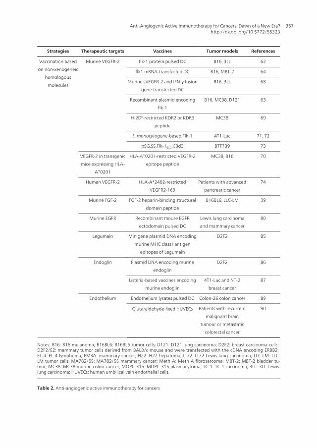
be divided into two categories: one is based on the immunological cross-reactionsmediated by vaccination with xenogeneic homologous molecules associated withangiogenesis, and the other targets non-xenogeneic homologous molecules. Therapeutictargets, vaccines and tumor models used in anti-angiogenic active immunotherapy forcancers are summarized in Table 2.
Strategies Therapeutic targets Vaccines Tumor models References
Vaccination based
on xenogeneic
homologous
molecules
Murine vascular
endothelial cells
Human umbilical vein endothelial
cells, human dermal microvascular
endothelial cells, and bovine
glomerular endothelial cells
Meth A, H22,
MA782/5S, FM3A, and
Lewis Lung carcinoma
29
Murine VEGF Recombinant plasmid encoding
VEGF of xenopus laevis
Meth A, H22 and
MA782/5S
32
pMAE5△5 vectors harboring
human VEGF 121 gene and
mutated human VEGF 121 gene
EL-4, B16, and TC-1 33
Canine VEGF Liposome-DNA adjuvant Soft tissue sarcoma 34
Murine VEGFR-2 Plasmid DNA encoding quail
VEGFR-2
Meth A, EL-4 and
MOPC-315,
36
Plasmid DNA encoding the C
terminal 37 amino acids of hCGβ,
5 different CTL epitopes from
human surviving and the 3rd and
4th extracellular domains of
VEGFR-2
LL/2 lung carcinoma 37
Bifidobacterium infantis expressing
human sKDR
Lewis lung cancer 38
Murine FGFR-1 cDNA encoding Xenopus FGFR-1 Meth A, H22, and
MA782/5S
41
Murine αvβ3 Plasmid DNA encoding the ligand-
binding domain of chicken
integrin β3
Meth A, H22, and
MA782/5S
43
Murine MMP-2 Plasmid DNA encoding chicken
MMP-2
LL/2, Meth A, H22 48
Murine RHAMM Plasmid encoding Xenopus
RHAMM
B16 49
Murine DLL4 Plasmid encoding human DLL4 D2F2/E2 55
Murine Angiomotin Plasmid encoding human
angiomotin
TUBO breast cancer 57, 61
Cancer Treatment - Conventional and Innovative Approaches366
Strategies Therapeutic targets Vaccines Tumor models References
Vaccination based
on non-xenogeneic
homologous
molecules
Murine VEGFR-2 flk-1 protein pulsed DC B16, 3LL 62
flk1 mRNA-transfected DC B16, MBT-2 64
Murine sVEGFR-2 and IFN-γ fusion
gene-transfected DC
B16, 3LL 68
Recombinant plasmid encoding
flk-1
B16, MC38, D121 63
H-2Db-restricted KDR2 or KDR3
peptide
MC38 69
L. monocytogene-based Flk-1 4T1-Luc 71, 72
pSG.SS.Flk-1ECD.C3d3 BTT739 73
VEGFR-2 in transgenic
mice expressing HLA-
A*0201
HLA-A*0201-restricted VEGFR-2
epitope peptide
MC38, B16 70
Human VEGFR-2 HLA-A*2402-restricted
VEGFR2-169
Patients with advanced
pancreatic cancer
74
Murine FGF-2 FGF-2 heparin-binding structural
domain peptide
B16BL6, LLC-LM 39
Murine EGFR Recombinant mouse EGFR
ectodomain pulsed DC
Lewis lung carcinoma
and mammary cancer
80
Legumain Minigene plasmid DNA encoding
murine MHC class I antigen
epitopes of Legumain
D2F2 85
Endoglin Plasmid DNA encoding murine
endoglin
D2F2 86
Listeria-based vaccines encoding
murine endoglin
4T1-Luc and NT-2
breast cancer
87
Endothelium Endothelium lysates pulsed DC Colon-26 colon cancer 89
Glutaraldehyde-fixed HUVECs Patients with recurrent
malignant brain
tumour or metastatic
colorectal cancer
90
Notes: B16: B16 melanoma; B16BL6: B16BL6 tumor cells; D121: D121 lung carcinoma; D2F2: breast carcinoma cells; D2F2/E2: mammary tumor cells derived from BALB/c mouse and were transfected with the cDNA encoding ERBB2; EL-4: EL-4 lymphoma; FM3A: mammary cancer; H22: H22 hepatoma; LL/2: LL/2 Lewis lung carcinoma; LLC-LM: LLC-LM tumor cells; MA782/5S: MA782/5S mammary cancer; Meth A: Meth A fibrosarcoma; MBT-2: MBT-2 bladder tu‐mor; MC38: MC38 murine colon cancer; MOPC-315: MOPC-315 plasmacytoma; TC-1: TC-1 carcinoma; 3LL: 3LL Lewis lung carcinoma; HUVECs: human umbilical vein endothelial cells.
Table 2. Anti-angiogenic active immunotherapy for cancers
Anti-Angiogenic Active Immunotherapy for Cancers: Dawn of a New Era?http://dx.doi.org/10.5772/55323
367
2. Anti-angiogenic active immunotherapy
2.1. Anti-angiogenic active immunotherapy based on xenogeneic homologous molecules
Homologous molecules in different species are formed as the result of evolution. Moleculeswith essential functions keep the stability of their molecular sequences, although somemoderate degree of evolution is essential for adaptation to different environments andphysiological requirements in different species. Many genes in the human and mouse genomeare similar (but not identical) to the corresponding genome sequences of the fruit fly Droso‐phila melanogaster and other non-vertebrates such as Xenopus laevis [28]. In consequence,effective immune response to self antigens associated with angiogenesis can thus be inducedby vaccination with xenogeneic homologous molecules.
2.1.1. Cell vaccine
Neovascular endothelial cells in tumor tissues express proteins not present or not detectablein normal vascular endothelial cells, such as αvβ3 integrin and receptors for certain angiogenicgrowth factors [18]. These proteins in murine vascular endothelial cells share homology tovarying degree with counterparts of other species including human [18]. Vaccination of micewith paraformaldehyde-fixed xenogeneic human and bovine proliferative vascular endothe‐lial cells, such as human umbilical vein endothelial cells, human dermal microvascularendothelial cells, and bovine glomerular endothelial cells, resulted in successful breaking ofthe immunological tolerance to autogeneic vascular endothelial cells in several murine tumormodels, such as Meth A fibrosarcoma, MA782/5S and FM3A mammary cancer, H22 hepatoma,and Lewis lung carcinoma, generating a protective and therapeutic anti-tumor immunologicalreaction [29]. Antibodies against the receptors associated with tumor angiogenesis generatedin mice immunized with the xenogeneic homologous proliferative vascular endothelial cellvaccines might inhibit the proliferation of endothelial cells in vivo, leading to the regressionof established tumor, and the prolonged survival of tumor-bearing mice [29]. Tumor angio‐genesis could be suppressed by the adoptive transfer of autoreactive immunoglobulinspurified from the immunized mouse, resulting in inhibition of tumor growth in mice [29].Autoantibody sediments were detected on ECs within tumor tissues in the immunized miceby immunohistochemical analysis [29]. Furthermore, Western blot analysis showed thatreactions between the extract from murine ECs and the serum from the immunized miceresulted in several positive bands, at least two of which, with the molecular weight of 220 kDaand 130 kDa, had similar molecular sizes to those of ligand-binding sites of known VEGFR2and αv integrin, respectively [29], although the authors did not provide direct evidence todemonstrate that the two positive bands aforementioned contained VEGFR2 and αv integrinrespectively. Immune cell subset depletion experiments showed that the production ofautoantibodies against tumor vascular ECs and the anti-tumor effect were dependent onCD4+ T lymphocytes [29].
In 2006, early-outgrowth progenitor endothelial cells (EO-EPCs) have been characterized onthe basis of their dendritic-like phenotypes (such as expression of HLA-DR, CD40, CD54,CD80, and CD86), phagocytotic and antigen-presenting functions, and endothelial markers
Cancer Treatment - Conventional and Innovative Approaches368

(such as VEGER2, von Willebrand factor, CD105) [30]. EO-EPCs also incorporated DiLDL andbound UEA-I, which are endothelial features, and additionally, they formed vascular-likestructures on Matrigel [30]. Thus, it might be a promising strategy toward anti-angiogeniccancer treatment to use EO-EPCs as cell vaccine to inhibit tumor angiogenesis, since such cellsmight function both as dendritic-like cells to augment anti-tumor immunity and as xenogeneicproliferative endothelial cells to break self-tolerance, thereby inducing profound anti-angio‐genic effects in vivo.
2.1.2. Non-cell vaccines
2.1.2.1. VEGF/VEGFR2
VEGF is a potent and crucial vasculogenic and angiogenic factor, which can induce endothelialcell proliferation, promote cell migration, and inhibit endothelial cell apoptosis [5, 6]. In mosttypes of cancers, VEGF is often present at elevated levels, and strategies aimed at blocking itsactivity usually lead to suppression of tumor angiogenesis and consequently tumor growthinhibition [31]. The amino acid sequence of VEGF in Xenopus laevis shares 75 % and 73 %homology with that of VEGF164 in mice and that of VEGF165 in humans, respectively [32].Recombinant eukaryotic expression plasmids harboring VEGF-encoding gene of mice andXenopus laevis, respectively, designated as MVEGF-P and XVEGF-P, have been constructed.Immunization of mice with XVEGF-P provoked protective and therapeutic anti-tumorimmunological effects in mouse tumor models with Meth A fibrosarcoma, MA782/5S mam‐mary cancer and H22 hepatoma [32]. Anti-VEGF specific autoantibody was detected in serumof mice vaccinated with XVEGF-P by Western blot and ELISA [32]. The VEGF levels in thetumor-bearing mice immunized with XVEGF-P was lower than that in the control groups [32].Furthermore, the frequency of anti-VEGF antibody-producing B cells in the spleen of miceimmunized with XVEGF-P was remarkably higher than that in the spleen of control groupswhere such B cells were undetectable [32]. VEGF-mediated proliferation of ECs could beinhibited in vitro by purified immunoglobulins from XVEGF-P-immunized mice. Adoptivetransfer of the purified immunoglobulins into non-immunized tumor-bearing mice could alsoinhibit tumor angiogenesis in vivo and generate anti-tumor effects [32]. Anti-CD4+ monoclonalantibody could obstruct the escalation of concentration of immunoglobulin IgG1 and IgG2 inserum and also block the anti-tumor effects of XVEGF-P DNA vaccines, indicating that CD4+
T lymphocytes were responsible for XVEGF-P-induced anti-tumor effects [32]. The possibilitythat the anti-tumor activity may result from nonspecifically augmented immune responsecould be ruled out by the findings that no increase in NK activity of spleen cells or in the levelof cytokines such as IFN-α, IFN-β, TNF-α, or β-chemokine in sera was found in immunizedmice [32]. Recently, it was reported that when immunized with human VEGF isoform 121 gene(hVEGF121) inserted into pMAE5△5 vector (pM-VEGF) and later challenged with melanomaor lung carcinoma tumor cells, a reduction of tumor growth and an increased survival oftumor-bearing C57BL/6 mice were observed because the hVEGF121 gene is highly homolo‐gous to its murine counterpart [33]. A decrease in tumor cell density around vessels and inmitotic figures, as well as an increase in apoptotic tumor cells were manifested by histopatho‐logical analyses of tumors from C57BL/6 mice immunized with hVEGF121 [33]. Spleen cells
Anti-Angiogenic Active Immunotherapy for Cancers: Dawn of a New Era?http://dx.doi.org/10.5772/55323
369

from mice immunized with pM-VEGF showed a significant enhanced cytotoxic activity againstVEGF-secreting tumor cells, including EL-4 lymphoma, B16-F10 melanoma, and TC-1carcinoma, as compared with those obtained from the mice immunized with the pMAE5△5‘‘empty’’ vector [33]. IFN-γ ELISPOT assay revealed a significant increase in the number ofspots in spleen cells from mice immunized with pM-VEGF [33]. Vaccination with a mutatedhVEGF121 gene inserted into the pMAE5△5 vector (pM-VEGFmut) produced similar in vitroand in vivo results, and remarkably reduced the number of spontaneous metastases in a murinemodel with Lewis lung carcinoma [33]. Serum VEGF levels decreased 8-fold in mice vaccinatedwith pM-VEGF or pM-VEGFmut as compared with those in pMAE5△5 treated mice [33]. Asignificant correlation was also found between the elevation of serum VEGF level and theincrease of the tumor dimensions [33]. However, antibody responses against theGSThVEGF121 fusion protein or GST alone used as capture antigens in ELISA were undetect‐able in animals vaccinated with pM-VEGF or pM-VEGFmut [33]. These findings indicate thathuman VEGF-harboring DNA vaccine can be employed for anti-angiogenic active immuno‐therapy for cancers in mice and direct cell cytotoxicity contributes to the overall anti-tumoreffects observed in immunized mice [33].
Previous studies in rodent tumor models have indicated that immunization against xenogeneicgrowth factors is more likely to induce effective anti-tumor responses than immunizationagainst the syngeneic growth factor [34]. In 2007, an investigation was conducted to assess thesafety and anti-tumor and anti-angiogenic effects of a xenogeneic VEGF vaccine in pet dogswith spontaneous cancer. Nine dogs with soft tissue sarcoma were immunized with arecombinant human VEGF vaccine over a 16-week period [34]. The xenogeneic VEGF vaccinewas well-tolerated by all dogs and resulted in induction of humoral responses against bothhuman and canine VEGF in animals that remained in the study long enough to receive multipleimmunizations [34]. Three of five multiply immunized dogs also experienced sustaineddecreases in circulating plasma VEGF concentrations and two dogs had a significant decreasein tumor microvessel density [34]. The overall tumor response (>50% decrease in tumorvolume) rate was 30% for all treated dogs in the study. Thus, it was concluded that a xenogeneicVEGF vaccine may be a safe and effective alternative means of controlling tumor growth andangiogenesis [34].
VEGF receptor-2 (VEGFR-2, also known as fetal liver kinse-1 (flk-1) in mouse and kinase-containing domain receptor (KDR) in human) is the main receptor responsible for theVEGF-mediated angiogenic activity [6]. The impairment of vasculogenesis and death ofembryo at day 8.5 were observed as the result of the targeted inactivation of flk-1 gene inmice [35]. Overexpression of KDR was found on activated endothelial cells of newly formedvessels [6]. It was discovered that the primary sequence of quail VEGFR-2 (qVEGFR-2) was67% and 70% identical at the amino acid level with mouse and human homologues (flk-1and KDR), respectively [36]. Immunotherapy with a vaccine based on quail homologousVEGFR-2 elicited protective and therapeutic anti-tumor immunity in both solid andhematopoietic tumor models in mice, such as LL/2 Lewis lung carcinoma, CT26 coloncarcinoma, Meth A fibrosarcoma, MOPC-315 plasmacytoma, and EL-4 lymphoma [36].Autoantibodies against flk-1 in the immunized mice were identified. Sera from qVEGFR-2-
Cancer Treatment - Conventional and Innovative Approaches370

immunized mice recognized not only recombinant qVEGFR-2, but also recombinant mouseVEGFR-2 (mVEGFR) in Western blot analysis [36]. In contrast, the sera isolated fromcontrols showed negative staining [36]. Sera from mice immunized with qVEGFR-2recognized a single band in flk-1-positive mouse SVEC4-10 endothelial cells and KDR-positive human umbilical vein endothelial cells, with the same size as recognized bycommercially available flk-1 or KDR antibodies [36]. Sera from qVEGFR-2-immunized micealso recognized recombinant protein qVEGFR-2 and mVEGFR-2 in ELISA [36]. DetectableIgG1 and IgG2b with significantly elevated concentration in sera were found to beresponsible for the immunoglobulin response to VEGFR-2 [36]. Anti-VEGFR-2 specificantibody-producing B cells were detected by ELISPOT. The number of anti-VEGFR-2antibody-producing B cells was elevated in the spleens of mice immunized with qVEGFR-2,compared with that in controls [36]. Deposition of immunoglobulins on endothelial cellswas found within tumors from qVEGFR-2-immunized mice, but not from controls [36].Adoptive transfer of the purified immunoglobulins from qVEGFR-2-immunized miceresulted in inhibition of VEGF-mediated endothelial cell proliferation and effectiveprotection against tumor growth [36]. Angiogenesis was markedly suppressed within thetumors, and the vascularization of alginate beads was also diminished [36]. Depletion ofCD4+ T lymphocyte could abrogate the anti-tumor activity and the production of autoanti‐bodies against flk-1 [36].
Very recently, a DNA vaccine designed by synergizing different tumor antigens with VEGFR2was constructed. A DNA fragment (HSV) encoding the C terminal 37 amino acids of humanchorionic gonadotropin β chain (hCGβ), 5 different HLA-restricted cytotoxic T lymphocyteepitopes from human survivin and the third and fourth extracellular domains of VEGFR2 wasinserted into the sequence between the luminal and transmembrane domain of humanlysosome-associated membrane protein-1 cDNA for the construction of a novel DNA vaccine(p-L/HSV) [37]. Vaccination of the mice with p-L/HSV elicited potent and long-lasting cellularand humoral immune responses to the specific antigens and showed a prominent anti-tumoreffect on the LL/2 lung carcinoma model in syngeneic C57BL/6 mice. In addition, the tumorvasculature was abrogated as observed by immunohistochemistry in p-L/HSV immunizedmice [37]. These data indicates that the strategies of combining anti-tumor with anti-angio‐genesis cooperate well. Such a study may shed new light on the designing of vaccine for cancerin the future.
Again in 2012, a Bifidobacterium infantis-based vaccine that express human extracellular domainof VEGFR2 (sKDR) was established [38]. Immunization of the mice with the Bifidobacteriuminfantis-based vaccine through caudal vein could significantly suppress the tumor growth andprolong the survival of the tumor-bearing mice. On the other hand, this immunization strategycould significantly increase the tumor necrosis, and obviously decrease microvessel densityand the blood flow signals in tumor [38].
2.1.2.2. FGFR-1
Fibroblast growth factor receptor-1 (FGFR-1) is expressed on endothelial cells and many typesof tumors [39, 40]. The Xenopus homologue of FGFR-1 is 80% and 74% identical at the amino
Anti-Angiogenic Active Immunotherapy for Cancers: Dawn of a New Era?http://dx.doi.org/10.5772/55323
371

acid level with mouse FGFR-1 and human FGFR-1, respectively [41]. Therefore, FGFR-1 maybe used as another ideal target for anti-angiogenesis therapy. Vaccination with XenopusFGFR-1 (pxFR1) provoked protective and therapeutic effects in three murine tumor models,including Meth A fibrosarcoma cells, H22 hepatoma cells, and MA782/5S mammary carcinoma[41]. FGFR-1-specific autoantibodies were detected in sera of pxFR1-immunized mice byWestern blot analysis, and the purified immunoglobulins effectively inhibited endothelial cellproliferation in vitro [41]. However, the immunoglobulins had no direct inhibitory effect onthe proliferation of above three tumor cell lines [41]. Adoptive transfer of sera or purifiedimmunoglobulin isolated from pxFR1-immunized mice into unimmunized mice providedeffective protection against tumor growth, while adsorption of sera or immunoglobulin withFGFR-1-positive endothelial cells before adoptive transfer could abrogate its anti-tumoractivity [41]. Autoantibodies deposited on the endothelial cells within tumor tissues andsignificantly suppressed intratumoral angiogenesis were found in pxFR1-immunized mice byhistological examination [41]. Furthermore, this anti-tumor activity and production of FGFR-1-specific autoantibodies were abrogated by depletion of CD4+ T lymphocytes, again pointingto their essential helper function for antibody production [41].
2.1.2.3. Integrins
Integrins are heterodimeric transmembrane proteins consisting of α and β subunits with largeextracellular domain and short cytoplasmic tail. They play very crucial roles in angiogenesisas the migration of endothelial cells is dependent on their adhesion to extracellular matrixproteins such as vitronectin [42]. αvβ3 is not generally found on blood vessels in normal tissues,but its expression is enhanced on newly developing blood vessels in human wound tissue,tumors, diabetic retinopathy, macular degeneration and rheumatoid arthritis, which impliesthat this integrin may play an important role in angiogenesis and development of neovascu‐larization [42]. This distributive characteristic also makes αvβ3 an attractive target for tumortherapy [42]. A plasmid DNA encoding the ligand-binding domain of chicken integrin β3 wasconstructed to test this assumption. Immunization with chicken homologous integrin β3-basedvaccine could elicit both protective and therapeutic anti-tumor immunity in murine tumormodels with Meth A fibrosarcoma, H22 hepatoma, or MA782/5S mammary carcinoma [43].Autoantibodies against integrin β3 in sera of the immunized mice were found by Western blotanalysis and ELISA [43]. The purified immunoglobulins could effectively inhibit endothelialcell proliferation in vitro, and adoptive transfer of the purified immunoglobulins into non-immunized mice could provide effective protection against tumor growth and markedlyinhibit tumor angiogenesis [43]. The anti-tumor activity and the production of integrin β3-specific autoantibodies were CD4+ T lymphocyte-dependent [43].
2.1.2.4. MMP
Angiogenesis is an invasive process, requiring proteolysis of the extracellular matrix [44].Inappropriate destruction of extracellular matrix components is involved in certain patholog‐ical conditions, including arteriosclerosis, rheumatoid arthritis, and tumor aggression andmetastasis [44]. The matrix metalloproteinases (MMPs), a family of extracellular endopepti‐
Cancer Treatment - Conventional and Innovative Approaches372

dases, can selectively degrade components of the extracellular matrix [44]. In vivo, elevatedstromal MMP-2 and MMP-9 activity is highly correlated with increased metastatic potentialin most malignant tumors [45]. Increased activity of MMPs appears to permit the tumor toremodel its surrounding microenvironment, to grow in a permissive space, and to promotethe development of supporting stroma, including angiogenesis [46]. Moreover, numerouspathological and clinical studies demonstrated that the MMPs were frequently overexpressedin various solid tumor cells and peritumoral stromal cells [46]. It was reported that theabrogation of MMP-2 alone resulted in the inhibition of the transition from the prevascular tothe vascular stage during tumor development and then of tumor growth [47]. Furthermore,the suppression of tumor-induced angiogenesis and of invasion and metastasis of tumor cellscould be observed in MMP-2-deficient mice [47]. These findings indicated that MMP-2 aloneplayed an important role in angiogenesis and tumor growth. Sequence comparison analysisshowed that the primary sequence of mouse MMP-2 at the amino acid level was 82% and 91%identical with chicken and human homologues, respectively [48]. It was reported that theplasmid DNA vaccination with chicken homologous MMP-2 (c-MMP-2)-based model antigencould induce both protective and therapeutic anti-tumor immunity in murine tumor modelswith LL/2 Lewis lung carcinoma, Meth A fibrosarcoma, and H22 hepatoma [48]. The elevationof MMP-2 in the sera of tumor-bearing mice was abrogated with the vaccination of c-MMP-2[48]. The autoimmune response against MMP-2 may be provoked in a cross-reaction by theimmunization with c-MMP-2, and the autoantibody targeting to MMP-2 was elevated andprobably responsible for the anti-tumor activity [48]. Moreover, gelatinase activity of MMP-2,including both latent MMP-2 and active MMP-2, derived from the above mentioned threemurine tumor models was apparently inhibited by the vaccination with c-MMP-2 [48].However, the vaccination did not inhibit the gelatinase activity of MMP-9 [48]. These findingsindicate that the activity of MMP-2 is impaired by immunization with c-MMP-2 in mice.Angiogenesis was apparently inhibited within tumors in immunized mice. The anti-tumoractivity and production of auto-antibodies against MMP-2 were abrogated by depletion ofCD4+ T lymphocytes [48].
2.1.2.5. xRHAMM
In 2010, Yang et al. [49] used a cross-reactive serological expression cloning (SEREX) strategy(CR-SEREX) to identify novel xenogenic angiogenesis- and tumor-associated antigens inoocytes of Xenopus laevis and found that Xenopus receptor for hyaluronic-acid-mediatedmotility (xRHAMM) was the most frequently clone among 78 CR-SEREX positive clones,suggesting that xRHAMM has the strongest immunogenic potential for xenogenic immuno‐therapy. It was demonstrated that expression of RHAMM is restricted to the testis, thymus,placenta, vascular endothelial cells, and various cancer cells, and RHAMM functions invascular endothelial cell migration, angiogenesis, and in hyaluronic-acid-induced cell mobility[50]. In order to examine the anti-angiogenic effects, a DNA vaccine based on xRHAMM(pcDNA3.1-xRHAMM) was constructed [49]. Intramuscular vaccination of the cationicliposome encapsulated pcDNA3.1-xRHAMM DNA effectively induced a protective anti-tumor immunity against local tumor and lung metastasis in B16 melanoma mouse models.Angiogenesis was inhibited and cell apoptosis was increased within tumors. Anti-tumor
Anti-Angiogenic Active Immunotherapy for Cancers: Dawn of a New Era?http://dx.doi.org/10.5772/55323
373

activity of xRHAMM was mediated by both the antigen-specific cellular and humoralresponses against RHAMM, as confirmed by the depletion of immune cell subsets in vivo.Furthermore, the anti-angiogenic and anti-tumor effects induced by vaccination of pcDNA3.1-xRHAMM were significantly stronger than that induced by vaccination of the correspondingautologous counterpart pcDNA3.1-mRHAMM [49].
2.1.2.6. DLL4
Notch signaling has recently emerged as a critical regulator of developmental and tumorangiogenesis. Notch signaling in both endothelial and smooth muscle cells appears to providecritical regulatory information to these cells downstream of the initiating signal induced byVEGF [51, 52]. Studies in humans and mice have demonstrated that Notch ligand delta-like 4(DLL4) is strongly expressed by the tumor vasculature and generally not by the tumor cellsthemselves. In various mouse models, strong DLL4 expression was observed in the majorityof tumor vessels, contrasting with significantly lower vascular expression in adjacent normaltissues [51]. In humans, DLL4 expression was analyzed in tumors from kidney, bladder, colon,brain and breast [53, 54]. Robust DLL4 expression was observed specifically in the tumorvasculature in all of these tumor types, whereas DLL4 expression was low to undetectable inthe vasculature of adjacent normal tissue. Furthermore, at least in the case of breast cancer, thedegree of DLL4 expression correlated with outcome: tumors with high DLL4 in the vasculatureprogressed more rapidly [54]. These findings suggest that DLL4 is an attractive new thera‐peutic anti-angiogenesis target. To generate the DLL4 plasmid vaccine, the cDNA encodinghuman DLL4 was cloned into the pVAX1 expression vector (DLL4 vaccine), which isspecifically designed for the development of DNA vaccines and approved for use in humans.Immunization of Balb/c mice with DLL4 vaccine could bring about a break in tolerance againstthe self-antigen, DLL4. Readily detectable titers of serum antibodies against DLL4 wereinduced. Moreover, immunization with DLL4-encoding plasmid DNA severely retarded thegrowth of orthotopically implanted D2F2/E2 mammary carcinomas in mice by induction of anon-productive angiogenic response. In addition to the promising therapeutic effects, noevidence for a delayed wound healing response, or for toxicity associated with pharmacolog‐ical blockade of DLL4 signaling, was observed in mice immunized with the DLL4 vaccine [55].
2.1.2.7. Angiomotin
Angiomotin (Amot), one of angiostatin receptors [56], is a membrane-associated proteinpresent on the endothelial cell surface of angiogenic tissues [57] characterized by conservedcoiledcoil and carboxy termini-PDZ domains [58]. A shorter Amot isoform (p80) confers ahyper-migratory and invasive phenotype in transfected cells [59] and induces endothelial cellmigration during angiogenesis [60]. The longer (p130) isoform localizes to tight junctions,regulates cell shape and appears to play a role in the later phase of angiogenesis [60]. It wasdemonstrated that increased Amot expression on tumor endothelia concomitant with theprogression from pre-neoplastic lesions to full-fledged carcinoma, therefore, plasmid vaccineencoding human p80 Amot (pAmot) was constructed [57]. Immunization of mice with pAmotcan overcome immune tolerance and induce a significant antibody response that mimic the
Cancer Treatment - Conventional and Innovative Approaches374

effect of angiostatin. These antibodies inhibit endothelial cell migration, block tumor cell- andbFGF-induced angiogenesis in the matrigel plug assay and prevent growth of transplantedtumors without impairing normal stromal or retina vessels [57]. Very recently, Arigoni et al.further showed that the pAmot-induced antibodies alter tumor vessel permeability andstructure. These combined effects of vaccine-induced anti-Amot antibodies lead to inhibitionof established clinically evident mammary tumors, massive tumor perivascular necrosis, andan effective tumor antigen presentation in a form of epitope spreading that induces an immuneresponse against other oncoantigens overexpressed by tumor cells [61]. Greater tumor vesselpermeability also markedly boosts the local accumulation of doxorubicin and enhances theanti-tumor effect of the drugs [61]. These data provide a rationale for the development of freshanticancer treatments based on anti-Amot vaccination in conjunction with chemotherapyregimens.
Taken together, it is obvious that vaccination with xenogeneic homologous molecules associ‐ated with angiogenesis, such as pro-angiogenic factors, integrins, MMP, could induce anti-tumor immunity and thus might be a feasible strategy for cancer therapy with potential clinicalapplications.
2.2. Anti-angiogenic active immunotherapies based on non-xenogeneic homologousmolecules
Given that vaccination with xenogeneic homologous molecules associated with tumorangiogenesis could effectively induce anti-tumor immunity, it can be assumed that vaccinesbased on non-xenogeneic homologous molecules, such as allogeneic homologues of some pro-angiogenic factors or other important molecules associated with angiogenesis, could alsosuccessfully induce specific and potent anti-tumor immunity. To date, several vaccines basedon non-xenogeneic homologous molecules were used in anti-angiogenic active immunother‐apy for tumors.
2.2.1. VEGFR-2
As has been discussed above, VEGF-mediated signaling pathway through VEGFR-2 is a rate-limiting step during tumor angiogenesis. Thus, VEGF/VEGFR-2 is still an ideal target in thenon-xenogeneic homologous molecules-based anti-angiogenic strategy. Immunization of micewith VEGF receptor-2 (flk-1)-pulsed dendritic cells (DC) can break self-tolerance to VEGFR-2,induce CTL and antibody responses to VEGFR-2 [62]. Significant inhibition of tumor growthand metastasis was observed in both melanoma and Lewis lung carcinoma metastasis murinemodels [62]. Oral administration of mice with DNA vaccines encoding murine VEGFR-2carried by attenuated Salmonella typhimurium could break the immune tolerance to VEGFR-2,induce CTL response to VEGFR-2, inhibit tumor cell-induced neoangiogenesis, and suppressthe formation of spontaneous and experimental pulmonary metastases, with slight impact onwounds healing and no influence on hematopoiesis and pregnancy [63]. Immunization of micewith flk1-encoding mRNA-transfected DC could induce specific CTL response to VEGFR-2,partially inhibit the tumor cell-induced neoangiogenesis, and suppress tumor growth andmetastasis in murine B16/F10.9 melanoma and MBT-2 bladder tumor models [64]. We studied
Anti-Angiogenic Active Immunotherapy for Cancers: Dawn of a New Era?http://dx.doi.org/10.5772/55323
375

the regulatory effects of IFN-γ on the differentiation and development of DC and found thatIFN-γ is an autocrine mediator for DC maturation [65]. IFN-γ gene transfection could promotedifferentiation, development, and functional maturation of DC [66]. IFN-γ gene-modified DChad increased capacity to induce Th1 type immune response, and intratumoral injection ofIFN-γ gene-modified DC in a murine model with pre-established B16 melanoma resulted inthe potentiation of the anti-tumor effect of DC [66]. On the other hand, it was demonstratedthat IFN-γ itself is also a negative regulator of neoangiogenesis [67]. In order to combine theanti-angiogenic immunotherapy with the cytokine immunotherapy, we constructed recombi‐nant plasmid expressing murine VEGFR-2 extracellular domain (sVEGFR-2) and IFN-γ fusionprotein, pcDNA3.1/sVEGFR-2-IFN-γ, and found that the fusion protein expressed by recombi‐nant plasmid shared biological activities of both sVEGFR-2 and IFN-γ [68]. Immunization ofmice with murine sVEGFR-2-IFN-γ fusion gene-transfected DC could significantly augmentthe CTL response to murine VEGFR-2 and pronouncedly inhibit tumor cell-induced angioge‐nensis and tumor metastasis in comparison with murine sVEGFR2 gene-transfected DC [68].
In 2006, three CTL epitope candidates, designated as KDR1, KDR2 and KDR3, respectively,from VEGFR-2 with high binding affinity to the H-2Db molecule were predicted by twocomputer programs: Bimas and SYFPEITH [69]. Two of them, KDR2 and KDR3, were fromthe extracellular domain; KDR1 was from the intracellular part of the receptor [69]. Immuni‐zation of mice with KDR2 or KDR3 peptide in combination with murine GM-CSF and agonistanti-mouse CD40 antibodies as adjuvant could break self-tolerance and induce specificimmune responses in C57BL/6 mice [69]. Furthermore, immunization of mice with these twopeptide epitopes elicited pronounced specific CTL responses to murine VEGFR-2, effectivelyinhibited VEGF-induced angiogenesis, and suppressed tumor growth in MC38 murine coloncancer model [69]. Similarly, the epitope peptides of human VEGFR-2 restricted by HLA-A*0201 and HLA-A*2402 were also identified by analyzing the binding affinities to thecorresponding HLA molecules [70]. Antigen based on the epitope peptide with high bindingaffinity to human HLA-A*0201 could successfully induce specific CTL response in vitro [70].Furthermore, transgenic mice expressing HLA-A*0201, A2/Kb, were generated, and thevascular endothelial cells in that mice could not only express human VEGFR-2 (KDR), but alsoexpress human MHC class Ι molecules [70]. After inoculation of A2/Kb with HLA-A*0201restricted VEGFR-2 epitope peptide, specific IFN-γ-expressed CTL was induced [70]. Immu‐nization of tumor-bearing A2/Kb transgenic mice with VEGFR-2 epitope peptide couldmarkedly inhibit tumor-induced angiogenesis, hereby inhibiting tumor growth in MC38 coloncancer and B16 melanoma models, and prolong survival of the tumor-bearing animals withoutfatal adverse effects [70]. To further study whether specific CTL response to KDR can be elicitedin human or not, KDR epitope peptide vaccines were used to stimulate peripheral bloodmononuclear cells derived from 6 cancer patients in vitro, and CTLs specific for the peptideepitope were successfully induced in all patients [70].
In comparison with the full-length protein, peptide vaccines like the aforementioned KDRepitope peptides can be easily synthesized in high purity and are less expensive. Moreover,immunization with such vaccines could avoid the potential dangers involving induction of aninfection by recombinant viruses or exposure to a latently allergenic exogenous protein.
Cancer Treatment - Conventional and Innovative Approaches376

In 2009, Seavey, et al developed Listeria monocytogens based VEGFR-2 vaccines that encode thepeptide of VEGFR-2 extracellular domain fused to the first 441 residues of the microbialadjuvant listeriolysin O (Lm-LLO-Flk-E1 and Lm-LLO-Flk-E2) and the peptide of VEGFR-2intracellular domain that also fused to LLO (Lm-LLO-Flk-I1), respectively [71, 72]. Immuni‐zation of the mice with the Listeria-based Flk1 vaccines elicited potent antitumor CTL respons‐es. Lm-LLO-Flk-1 was able to eradicate some established Her-2/neu+ breast tumors, reducemicrovascular density in the remaining tumors, protect against tumor rechallenge andexperimental metastases, and induce epitope spreading to various regions of the tumor-associated antigen Her-2/neu. Tumor eradication was found to be dependent on epitopespreading to Her-2/neu and was not solely due to the reduction of tumor vasculature [71]. Inan autochthonous model for Her-2/neu+ breast cancer, theses vaccines could significantlydelay tumor onset, while tumors that grew out overtime accumulated mutations in the Her-2/neu molecule near or within CTL epitopes [72]. Moreover, vaccine efficacy did not affectnormal wound healing nor have toxic side effects on pregnancy [71]. These data suggest thatan anti-angiogenesis vaccine can overcome tolerance to the host vasculature driving epitopespreading to an endogenous tumor protein and drive active tumor regression.
Recently, a DNA vaccine (pSG.SS.Flk-1ECD.C3d3) encoding Flk-1 extracellular domain and thecomplement fragment C3d fusion protein was constructed [73]. Vaccination of mice withpSG.SS.Flk-1ECD.C3d3 could also elicit Flk-1 specific antibody response, leading to suppressionof angiogenesis and tumor growth in bladder translational cell carcinoma mouse model,suggesting that C3d can be used as an adjuvant to enhance the immune response [73].
In 2010, Miyazawa, et al. [74] reported the results of phase I clinical trial combining of epitopepeptide for VEGFR-2 (VEGFR2-169) with gemcitabine for patients with advanced pancreaticcancer. 18 HLA-A*2402-positive patients with metastatic and unresectable pancreatic cancerwere enrolled in the trial. Gemcitabine was administered at a dose of 1000 mg/m2 on days 1,8, and 15 in a 28-day cycle. The VEGFR2-169 peptide was subcutaneously injected weekly ina dose-escalation manner (doses of 0.5, 1, and 2 mg/body, six patients/one cohort). Safety andimmunological parameters were assessed. No severe adverse effect of grade 4 or higher wasobserved. Of the 18 patients who completed at least one course of the treatment, 15 (83%)developed immunological reactions at the injection sites. VEGFR2-169 specific CTLs wereinduced in 11 (61%) of the 18 patients. The disease control rate was 67%, and the median overallsurvival time was 8.7 months. This combination therapy for pancreatic cancer patients wastolerable at all doses. Peptide-specific CTL could be induced by the VEGFR2-169 peptidevaccine at a high rate, even in combination with gemcitabine. Therefore, they suggested thatthe optimal dose for further clinical trials might be 2 mg/body or higher.
2.2.2. bFGF
Basic fibroblast growth factor (bFGF/FGF2) is an important proangiogenic factor, which issecreted by tumor cells and macrophages or released by extracelluar matrix, and functions inthe autocrine or paracrine manner. FGF2 can upregulate the expression of several dominantpro-angiogenic factors, such as VEGF [75], and activator of plasminogen [76], and inhibitapoptosis of endothelial cells by bcl-2 pathway [77]. bFGF exerts its biological activities
Anti-Angiogenic Active Immunotherapy for Cancers: Dawn of a New Era?http://dx.doi.org/10.5772/55323
377

through its binding to high affinity receptor, fibroblast growth factor receptor-1 (FGFR1). Itwas found that both peptide segments of synthetic human FGF2 heparin-binding structuraldomain and receptor-binding structural domain could inhibit the in vitro proliferation ofhuman umbilical vein endothelial cells [39]. Immunization of mice with vaccine based onheparin-binding structural domain peptide could induce production of anti-FGF2 specificantibody, which could hamper the binding of FGF2 to heparin sulphate, and inhibit tumor-induced angiogenesis in a gelatin sponge model and tumor growth in a tumor metastaticmodel [39]. Surprisingly, despite an immune response toward FGF2, this modality of treatmentdid not affect wound healing as shown by the fact that the treatment did not alter the meantime of wound healing [78]. It also did not affect fertility, because the vaccinated females werenot impaired in their ability to become pregnant, to support the growth and development oftheir embryos, and to deliver viable offspring when compared with control animals [78].Furthermore, histological analyses did not reveal any alterations in organogenesis in theseoffsprings [78]. Therefore, the authors concluded that although vaccination against FGF2induced a specific FGF2 antibody response and inhibited angiogenesis and tumor develop‐ment in a pathological setting, it did not adversely alter normal physiological events dependenton FGF2.
2.2.3. EGFR
Epidermal growth factor receptor (EGFR), a membrane surface sensor with tyrosine kinaseactivity, is widely distributed on the membrane of mammalian cells [79]. In the physiologicalcondition, EGFR exerts, through binding to ligands (epidermal growth factor, EGF), itsphysiological activities in regulation of cell division, proliferation and differentiation [79].Results from clinical studies show that high expression level of EGFR is frequently observedin non-small cell lung cancer, and has been implicated in aggressive biological behavior oftumor cells and poor prognosis of tumor patients [79]. Therefore, immunotherapy targetingEGFR should be another attractive approach to the treatment of EGFR-positive tumors. Inmurine tumor models with Lewis lung carcinoma and mammary cancer, immunization ofmice with DC pulsed with recombinant ectodomain of mouse EGFR (DC-edMER) inhibitedtumor angiogenesis, reduced tumor growth, and prolonged the survival of tumor-bearingmice [80]. Spleen cells isolated from DC-edMER-immunized mice showed a high frequency ofEGFR-specific antibody-producing cells [80]. Anti-EGFR specific antibody was markedlyelevated in sera of immunized mice and was shown to be effective against tumor growth byadoptive transfer [80]. Immunization with DC-edMER vaccine also elicited CTL responses [80].Depletion of CD4+ T lymphocytes could completely abrogate the anti-tumor activity andgeneration of EGFR-specific antibody responses, whereas depletion of CD8+ T lymphocytesshowed partial abrogation of the anti-tumor activity but antibody was still detected [80].Furthermore, tumor-induced angiogenesis was suppressed in DC-edMER-immunized miceor mice treated with antibody adoptive transfer [80]. These findings indicate that vaccinationwith DC-edMER can induce both humoral and cellular anti-tumor immunity, and may suggestnovel strategies for the treatment of EGFR-positive tumors through the induction of activeimmunity against EGFR [80].
Cancer Treatment - Conventional and Innovative Approaches378

2.2.4. Legumain
Tumor associated macrophages (TAMs) are well known to play a very important role in tumorangiogenesis and metastasis, as the abrogation of TAMs in tumor tissues effectively reducedseveral pro-tumor growth and angiogenesis factors, such as VEGF, TGF-β, TNF-α and MMP-9[81]. Thus, the suppression of TAMs in the tumor-microenvironment provides a novel strategyto inhibit tumor growth and dissemination by remodeling the tumor’s stroma. Legumain isan asparaginyl endopeptidase and a member of the C13 family of cystine proteases which wasfound to be highly upregulated in many murine and human tumor tissues and, furthermore,also overexpressed on TAMs in the murine tumor stroma, but absent or present at only verylow levels in all normal tissues from which such tumors arose [81-84]. Recently, several oralminigene vaccines against murine MHC class I antigen epitopes of Legumain were constructedbased on the binding predictions for these MHC class I molecules by the HLA peptide bindingpredictions program [85]. Expression vectors encoding these epitopes were designated aspLegu-H-2Dd and pLegu-H-2Kd respectively [85]. Oral administration of those vaccines bytransforming them into attenuated Salmonella typhimurium (Dam–, AroA–) resulted insignificant suppression of angiogenesis in tumor tissues of D2F2 breast carcinoma in syngeneicBALB/c mice [85]. The possible mechanism of angiogenic inhibition involved the induction ofa specific CTL response capable of killing Legumain positive cells, especially TAMs, which islikely to be responsible for anti-tumor angiogenesis [85]. Generally, the anti-angiogenic effectaided in the protection of BALB/c mice from lethal challenges with D2F2 breast tumor cells ina prophylactic setting [85].
2.2.5. Endoglin (CD105)
Endoglin, a 95 kDa cell surface protein expressed as a homodimer, functions as an accessoryprotein for kinase receptor complexes of the TGF-β superfamily and modulates TGF-βsignaling [8]. Expression of CD105 is correlated with vascular density and poor prognosis [8].Endoglin is over-expressed on proliferating endothelial cells in the breast tumor neovascula‐ture and thus offers a target for anti-angiogenic therapy [8]. It was reported that an oral murineendoglin-encoding DNA vaccine carried by double attenuated Salmonella typhimurium(dam–, AroA–) to a secondary lymphoid organ, i.e., Peyer’s patches, resulted in activation ofantigen-presenting dendritic cells, induction of immune responses mediated by CD8+ T cellsagainst endoglin-positive target cells, and suppression of angiogenesis and dissemination ofpulmonary metastases of D2F2 breast carcinoma cells presumably by eliminating proliferatingendothelial cells in the tumor vasculature, thus providing an promising strategy to therapiesfor breast cancer [86]. More recently, Wood et al. [87] developed Listeria-based vaccinesdirected against CD105, Lm-LLO-CD105A and Lm-LLO-CD105B. The region of CD105 in Lm-LLO-CD105A vaccine contains at least three predicted H-2Kd epitopes, while the region ofCD105 in Lm-LLO-CD105B contains at least two predicted H-2Kd epitopes. Immunization ofthe Listeria-based vaccines led to therapeutic responses against primary and metastatic tumorsin the 4T1-Luc and NT-2 mouse models of breast cancer. In a mouse model for autochthonousHer-2/neu-driven breast cancer, Lm-LLO-CD105A vaccination prevented tumor incidence in20% of mice by week 58 after birth while all control mice developed tumors by week 40. In
Anti-Angiogenic Active Immunotherapy for Cancers: Dawn of a New Era?http://dx.doi.org/10.5772/55323
379

comparison with previous Listeria-based vaccines (Lm-LLO-HMWMAA-C [88] and Lm-LLO-FLK-I1 and Lm-LLO-FLK-E2 [71] ) targeting tumor vasculature, Lm-LLO-CD105A and Lm-LLO-CD105B demonstrated equivalent or superior efficacy against two transplantable mousemodels of breast cancer. Mechanism analysis revealed that the anti-tumor therapeutic efficacyof Listeria-based CD105 vaccines was mediated by epitope spreading to endogenous tumorantigens and reduction in tumor vascularity [87]. These data suggest that CD105 therapeuticvaccines are highly effective in stimulating anti-angiogenesis and anti-tumor immuneresponses leading to therapeutic efficacy against primary and metastatic breast cancer.
2.2.6. Endothelial cell lysates-pulsed dendritic cells
Dendritic cells (DCs) are the most potent professional antigen-presenting cells, they playcrucial roles in the initiation of an immune response. DCs prepared from BALB/c mouse werepulsed with lysates of autologous or xenogeneic endothelium, and their anti-tumor effectswere tested in two syngeneic models of colon cancer [89]. Immunization of endothelium lysatespulsed DCs could induce a break in self tolerance against endothelial cells and mount both theendothelium-specific CTL response and antibody response, leading to significant inhibitionof tumor angiogenesis and the growth of subcutaneous tumors as well as pulmonary meta‐stases in mice. Furthermore, the decrease in the mean vascular density of tumors correlateswell with the extent of tumor inhibition [89]. Therefore, immunization of endothelium lysatespulsed DCs is also an effective modality of anti-angiogenic active immunotherapy for cancers,and should have important clinical implications for adjuvant cancer therapy.
2.2.7. Endothelial cell vaccine
In 2008, Okaji Y, et al. [90] reported a pilot phase I clinical study in which glutaraldehyde-fixed human umbilical vein endothelial cells (HUVECs) were used as the vaccine. Sixpatients with recurrent malignant brain tumour and three patients with metastaticcolorectal cancer were given intradermal injections of 5x10⁷ HUVECs/dose, first monthweekly, and then every 2 weeks (in total 230 vaccinations). ELISA and flow cytometryrevealed immunoglobulin response against HUVECs’ membrane antigens. ELISPOT and51Cr-release cytotoxicity assay revealed a specific cellular immune response againstHUVECs, which were lysed in an effectors:targets ratio-dependent manner. Gadolinium-contrasted MRI showed partial or complete tumour responses in three malignant braintumour patients. Except for a DTH-like skin reaction at the injection site, no adverse effectof vaccination was observed. These results suggested that the endothelial vaccine canovercome peripheral tolerance of self-angiogenic antigens in clinical settings, and there‐fore could be useful for adjuvant immunotherapy of cancer.
3. Concluding remarks
Recent research achievements have disclosed inspiring pragmatic perspectives of anti-angiogenic active immunotherapy for cancers. In comparison with application of angiogenic
Cancer Treatment - Conventional and Innovative Approaches380

inhibitors and angiogenic antibodies, anti-angiogenic active immunotherapy has its obviousmerits. Provided that a break of immunological tolerance to positive regulators of angiogenesisis successfully induced, the long-lasting immune response to angiogenesis-related moleculewill be present in the body, hereby providing long-lasting inhibitory effects on angiogenesis.Therefore, it is expected to be the more cost-effective strategy than angiogenic inhibitor or anti-angiogenic antibody therapy where continuous use of the drugs is needed.
Here we divided anti-angiogenic active immunotherapy into two categories: therapiesbased on vaccination with xenogeneic homologous molecules and with non-xenogeneichomologous molecules related to angiogenesis. Presently, it is difficult to point out whichone is better for clinical application because most of the outcomes reported to date werebased on pre-clinical animal experiments. As VEGF-mediated signaling through its receptorVEGFR-2 is the key rate-limiting step in tumor angiogenesis, and plays the most impor‐tant role in neovascularization, development, and progression of various tumors [6], as wellas human VEGFR2-169 peptide vaccination could effectively break peripheral self toler‐ance against VEGFR-2 in patients with metastatic and unresectable pancreatic cancer [74],anti-angiogenic active immunotherapy targeting VEGF or VEGFR-2 might be the mosteffective strategy among all these therapies. Moreover, considering the potential clinicalapplication of anti-angiogenic immunotherapy based on the specific antibodies raisedagainst a variety of angiogenesis-associated molecules in different tumor entities likeglioma, renal cell cancer, and breast cancer, etc, a promising clinical application of anti-angiogenic active immunotherapy alone or in combination with other anti-tumor strat‐egies could be expected. However, there exist as well caveats and deficiencies in thisstrategy. Firstly, in the early phase of tumor growth when the tumor diameter is less than2-3 mm, tumor cells simply depend on passive diffusion rather than blood perfusion toacquire enough oxygen and nutrition indispensable for growth. Therefore, anti-angiogen‐ic therapy against tumor in this early stage might be ineffective when applied alone.Secondly, although current anti-angiogenic active immunotherapy is focused on specifictargets, potential adverse effects might include impairment of wound healing and menstru‐al cycle. Furthermore, this approach has also limited application perspectives in childrenwith cancers. Therefore, along with recent developments in molecular biology andimmunology, future studies will focus on multiple approaches, such as series analysis ofgene expression to analyze the gene expression in normal endothelial cells and in prolifer‐ative endothelial cells, phage display technology to search for new endothelial cellreceptors, and proteomics to discover peptide segments or proteins regulating endothelialcell growth. These approaches are expected to discover more tumor-specific endothelial cellmarkers for the purpose of selecting specific targets for anti-angiogenic active immunother‐apy. In addition, further studies are also required to optimize protocols how to constructvaccines to effectively break self-tolerance and to induce efficient immune response. Withthese issues being solved continuously, anti-angiogenic active immunotherapy for cancerswill become more applicable and effective.
Anti-Angiogenic Active Immunotherapy for Cancers: Dawn of a New Era?http://dx.doi.org/10.5772/55323
381
Acknowledgements
This work was supported by a grant from the Science and Technology Bureau of Hangzhou,Zhejiang Province, P.R. China (No. 20120633B30).
Author details
Jianping Pan* and Lihuang Zhang
*Address all correspondence to: [email protected]
Department of Clinical Medicine, Zhejiang University City College School of Medicine,Hangzhou, P.R. China
References
[1] Arbab AS. Activation of alternative pathways of angiogenesis and involvement ofstem cells following anti-angiogenesis treatment in glioma. Histol Histopathol 2012;27: 549-557.
[2] Shojaei F. Anti-angiogenesis therapy in cancer: current challenges and future per‐spectives. Cancer Lett 2012; 320:130-137.
[3] Hiratsuka S. Vasculogenensis, angiogenesis and special features of tumor blood ves‐sels. Front Biosci 2011; 16:1413-1427.
[4] Blagosklonny MV. Antiangiogenic therapy and tumor progression. Cancer Cell 2004;5:13-17.
[5] Ferrara N. VEGF and the quest for tumor angiogenesis factors. Nat Rev Cancer 2002;2:795-803.
[6] Ferrara N, Gerber HP, LeCouter J. The biology of VEGF and its receptors. Nat Med2003; 6:669-676.
[7] Ferrara N, Kerbel RS. Angiogenesis as a therapeutic target. Nature 2005; 438:967-974.
[8] Hofmeister V, Schrama D, Becker JC, et al. Anti-cancer therapies targeting the tumorstroma. Cancer Immunol Immunother 2008; 57: 1-17.
[9] Shojaei F, Ferrara N. Antiangiogenesis to treat cancer and intraocular neovasculardisorders. Lab Invest 2007; 87:227-230.
[10] Veikkola T, Karkkainen M, Claesson-Welsh L, et al. Regulation of angiogenesis viaendothelial growth factor receptors. Cancer Rev 2000; 60:203-212.
Cancer Treatment - Conventional and Innovative Approaches382

[11] Caldini R, Barletta E, Del Rosso M, et al. FGF2-mediated upregulation of urokinase-type plasminogen activator expression requires a MAP-kinase dependent activationof poly (ADP-ribose) polymerase. J Cell Physiol 2005; 202: 125-134.
[12] Ferrajoli A, Manshouri T, Estrov Z, et al. High levels of vascular endothelial growthfactor receptor-2 correlate with shortened survival in chronic lymphocytic leukemia.Clin Cancer Res 2001; 7:795-799.
[13] Schneider BP, Sledge GW. Drug insight: VEGF as a therapeutic target for breast can‐cer. Nat Clin Pract Oncol 2007; 4:181-189.
[14] Kanda S, Miyata Y, Kanetake H. Current status and perspective of antiangiogenictherapy for cancer: urinary cancer. Int J Clin Oncol 2006; 11:90-107.
[15] Rini BI, Rathmell WK. Biological aspects and binding strategies of vascular endothe‐lial growth factor in renal cell carcinoma. Clin Cancer Res 2007; 13:741-746.
[16] Kerbel RS, Folkman J. Clinical translation of angiogenesis inhibitors. Nat Rev Cancer2002; 2:727-739.
[17] Shant K, Li CG. Targeting of vasculature in cancer and other angiogenic diseases.Trends Immunol 20001; 22:129-133.
[18] St Croix B, Rago C, Velculescu V, et al. Genes expressed in human tumor endotheli‐um. Science 2000; 289:1197-1202.
[19] Reisfeld RA, Niethammer AG, Luo Y, et al. DNA vaccines designed to inhibit tumorgrowth by suppression of angiogenesis. Int Arch Allergy Immunol 2004; 133:295-304.
[20] Hurwitz H, Fehrenbacher L, Novotny W, et al. Bevacizumab plus irinotecan, fluo‐rouracil, and leucovorin for metastatic colorectal cancer. N Engl J Med 2004;350:2335-2342.
[21] Sandler A, Gray R, Perry MC, et al. Paclitaxel-carboplatin alone or with bevacizumabfor non-small-cell lung cancer. N Engl J Med 2006; 355:2542-2550.
[22] Miller K, Wang M, Gralow J, et al. Paclitaxel plus bevacizumab versus paclitaxelalone for metastatic breast cancer. N Engl J Med 2007; 357:2666-2676.
[23] Willett CG, Duda DG, di Tomaso E, et al. Efficacy, safety, and biomarkers of neoadju‐vant bevacizumab, radiation therapy, and fluorouracil in rectal cancer: a multidisci‐plinary phase II study. J Clin Oncol 2009; 27:3020-3026.
[24] Holash J, Davis S, Papadopoulos N, et al. VEGF-Trap: a VEGF blocker with potentantitumor effects. Proc Natl Acad Sci USA 2002; 99:11393-11398.
[25] Kerbel RS. Tumor angiogenesis. N Engl J Med 2008; 358:2039-2049.
[26] Sun Y, Wang J, Liu Y, et al. Results of phase III trial of rh-endostatin (YH-16) in ad‐vanced non-small cell lung cancer (NSCLC) patients. J Clin Oncol 2005; 23:654S.
Anti-Angiogenic Active Immunotherapy for Cancers: Dawn of a New Era?http://dx.doi.org/10.5772/55323
383

[27] McKeage MJ, Von Pawel J, Reck M, et al. Randomised phase II study of ASA404combined with carboplatin and paclitaxel in previously untreated advanced non-small cell lung cancer. Br J Cancer 2008; 99:2006-2012.
[28] Kornberg TB, Krasnow MA. The Drosophila genome sequence: implications for biol‐ogy and medicine. Science 2000; 287: 2218-2220.
[29] Wei YQ, Wang QR, Zhao X, et al. Immunotherapy of tumors with xenogeneic endo‐thelial cells as a vaccine. Nat Med 2000; 6:1160-1166.
[30] Asakage M, Tsuno NH, Kitayama J, et al. Early-outgrowth of endothelial progenitorcells can function as antigen-presenting cells. Cancer Immunol Immunother 2006; 55:708-716.
[31] Kerbel RS. Tumor angiogenesis: past, present and the near future. Carcinogenesis2000; 21:505-515.
[32] Wei YQ, Huang MJ, Yang L, et al. Immunogene therapy of tumors with vaccinebased on xenopus homologous vascular endothelial growth factors as a model anti‐gen. Pro Natl Acad Sci USA 2001; 98:11545-11550.
[33] Bequet-Romero M, Ayala M, Acevedo BE, et al. Prophylactic naked DNA vaccinationwith the human vascular endothelial growth factor induces an anti-tumor responsein C57Bl/6 mice. Angiogenesis 2007; 10:23-34.
[34] Kamstock D, Elmslie R, Thamm D, et al. Evaluation of a xenogeneic VEGF vaccine indogs with soft tissue sarcoma. Cancer Immunol Immunother 2007; 56: 1299-1309.
[35] Shalaby F, Rossant J, Yamaguchi TP, et al. Failure of blood-island formation and vas‐culogenesis in Flk-1-deficient mice. Nature 1995; 376:62-66.
[36] Liu JY, Wei YQ, Yang L, et al. Immunotherapy of tumors with vaccine based on quailhomologous vascular endothelial growth factor receptor-2. Blood 2003;102:1815-1823.
[37] Wei Y, Sun Y, Song C, et al. Enhancement of DNA vaccine efficacy by targeting thexenogeneic human chorionic gonadotropin, survivin and vascular endothelialgrowth factor receptor 2 combined tumor antigen to the major histocompatibilitycomplex class II pathway. J Gene Med 2012; 14: 353-362.
[38] Li ZJ, Zhu H, Ma BY, et al. Inhibitory effect of Bifidobacterium infantis-mediatedsKDR prokaryotic expression system on angiogenesis and growth of Lewis lung can‐cer in mice. BMC Cancer 2012; 12:155.
[39] Plum SM, Holaday JW, Ruiz A, et al. Administration of a liposomal FGF-2 peptidevaccine leads to abrogation of FGF-2-mediated angiogenesis and tumor develop‐ment. Vaccine 2000; 19:1294-1303.
Cancer Treatment - Conventional and Innovative Approaches384
[40] Valesky M, Spang AJ, Fisher GW, et al. Noninvasive dynamic fluorescence imagingof human melanomas reveals that targeted inhibition of bFGF or FGFR-1 in melano‐ma cells blocks tumor growth by apoptosis. Mol Med 2002; 8: 103-112.
[41] He QM, Wei YQ, Tian L, et al. Inhibition of tumor growth with a vaccine based onxenogeneic homologous fibroblast growth factor receptor-1 in mice. J Biol Chem2003; 278:21831-21836.
[42] Gutheil JC, Campbell TN, Pierce PR, et al. Targeted antiangiogenic therapy for cancerusing Vitaxin: a humanized monoclonal antibody to the Integrin αvβ3. Clin CancerRes 2000; 6:3056-3061.
[43] Lou YY, Wei QY, Yang L, et al. Immunogene therapy of tumors with vaccine basedon the ligand binding domain of chick homologous integrin beta3. Immunol Invest2002; 31:51-69.
[44] William G, Stetler S. Matrix metalloproteinases in angiogenesis: a moving target fortherapeutic intervention. J Clin Invest 1999; 103:1237-1241.
[45] Liotta LA, Steeg PS, Stetler-Stevenson WG. Cancer metastasis and angiogenesis: animbalance of positive and negative regulation. Cell 1991; 64:327-336.
[46] Albini A, Melchiori A, Santi L, et al. Tumor cell invasion inhibited by TIMP-2. J NatlCancer Inst 1991; 83: 775-779.
[47] Itoh T, Tanioka M, Yoshida H, et al. Reduced angiogenesis and tumor progression ingelatinase A-deficient mice. Cancer Res 1998; 58:1048-1051.
[48] Su JM, Wei YQ, Tian L, et al. Active immunotherapy of cancer with vaccine on thebasis of chicken homologous matrix metalloproteinase-2. Cancer Res 2003;63:600-607.
[49] Yang HS, Zhang DM, Deng HX, et al.Antitumor and anti-angiogenesis immunity in‐duced by CR-SEREX-identified Xenopus RHAMM.Cancer Sci 2010; 101:862-868.
[50] Hardwick C, Hoare K, Owens R, et al. Molecular cloning of a novel hyaluronan re‐ceptor that mediates tumor cell motility. J Cell Biol 1992; 117:1343-1350.
[51] Kuhnert F, Kirshner JR, Thurston G. Dll4-Notch signaling as a therapeutic target intumor angiogenesis.Vasc Cell 2011; 3:20.
[52] Gu JW, Rizzo P, Pannuti A, et al. Notch signals in the endothelium and cancer ”stem-like” cells: opportunities for cancer therapy. Vascular Cell 2012; 4: 7.
[53] Ridgway J, Zhang G, Wu Y, et al. Inhibition of Dll4 signalling inhibits tumourgrowth by deregulating angiogenesis. Nature 2006; 444:1083-1087.
[54] Jubb AM, Soilleux EJ, Turley H, et al. Expression of vascular notch ligand delta-like 4and inflammatory markers in breast cancer. Am J Pathol 2010; 176:2019-2028.
Anti-Angiogenic Active Immunotherapy for Cancers: Dawn of a New Era?http://dx.doi.org/10.5772/55323
385
[55] Haller BK, Bråve A, Wallgard E, et al. Therapeutic efficacy of a DNA vaccine target‐ing the endothelial tip cell antigen delta-like ligand 4 in mammary carcinoma. Onco‐gene 2010; 29:4276-4286.
[56] Troyanovsky B, Levchenko T, Månsson G, et al. Angiomotin: an angiostatin bindingprotein that regulates endothelial cell migration and tube formation. J Cell Biol. 2001;152: 1247-1254.
[57] Holmgren L, Ambrosino E, Birot O, et al. A DNA vaccine targeting angiomotin in‐hibits angiogenesis and suppresses tumor growth. Proc Natl Acad Sci USA 2006; 103:9208-9213.
[58] Bratt A, Wilson WJ, Troyanovsky B, et al. Angiomotin belongs to a novel proteinfamily with conserved coiled-coil and PDZ binding domains. Gene 2002; 298:69-77.
[59] Levchenko T, Bratt A, Arbiser JL, et al. Angiomotin expression promotes heman‐gioendothelioma invasion. Oncogene 2004; 23:1469-1473.
[60] Ernkvist M, Birot O, Sinha I, et al. Differential roles of p80- and p130-angiomotin inthe switch between migration and stabilization of endothelial cells. Biochim BiophysActa 2008; 1783: 429-437.
[61] Arigoni M, Barutello G, Lanzardo S, et al. A vaccine targeting angiomotin induces anantibody response which alters tumor vessel permeability and hampers the growthof established tumors. Angiogenesis 2012; 15:305-316.
[62] Li Y, Wang MN, Li H, et al. Active immunization against the vascular endothelialgrowth factor receptor flk1 inhibits tumor angiogenesis and metastasis. J Exp Med2002; 195: 1575-1584.
[63] Nithammer AG, Xiang R, Becker JC, et al. A DNA vaccine against VEGF receptor 2prevents effective angiogenesis and inhibits tumor growth. Nat Med 2002;8:1369-1375.
[64] Nair S, Boczkowski D, Moeller B, et al. Synergy between tumor immunotherapy andantiangiogenic therapy. Blood 2003; 102: 964-971.
[65] Pan J, Zhang M, Wang J, et al. Interferon-gamma is an autocrine mediator for den‐dritic cell maturation. Immunol Lett 2004; 94:141-151.
[66] Pan J, Zhang M, Wang J, et al. Intratumoral injection of interferon-gamma gene-modified dendritic cells elicits potent antitumor effects: effective induction of tumor-specific CD8+ CTL response. J Cancer Res Clin Oncol 2005; 131:468-478.
[67] Qin Z, Blankenstein T. CD4+ T cell-mediated tumor rejection involves inhibition ofangiogenesis that is dependent on IFN gamma receptor expression by nonhemato‐poietic cells. Immunity 2000; 12:677-686.
Cancer Treatment - Conventional and Innovative Approaches386

[68] Pan J, Heiser A, Marget M, et al. Enhanced antimetastatic effect of fetal liver kinase 1extracellular domain and interferon-gamma fusion gene-modified dendritic cell vac‐cine. Gene Ther 2005; 12:742-750.
[69] Dong Y, Qian J, Ramy I, et al. Identification of H-2Db-specific CD8+ T-cell epitopesfrom mouse VEGFR2 that can inhibit angiogenesis and tumor growth. J Immunother2006; 29:32-40.
[70] Wada S, Tsunoda T, Baba T, et al. Rationale for antiangiogenic cancer therapy withvaccination using epitope peptides derived from human vascular endothelial growthfactor 2. Cancer Res 2005; 65:4939-4946.
[71] Seavey MM, Maciag PC, Al-Rawi N, et al. An anti-vascular endothelial growth factorreceptor 2/fetal liver kinase-1 Listeria monocytogenes anti-angiogenesis cancer vac‐cine for the treatment of primary and metastatic Her-2/neu+ breast tumors in amouse model. J Immunol 2009; 182(9):5537-5546.
[72] Seavey MM, Paterson Y. Anti-Angiogenesis immunotherapy induces epitope spread‐ing to Her-2/neu resulting in breast tumor immunoediting. Breast Cancer 2009;1:19-30.
[73] Liang PH, Zhang KQ, Xu GL, et al. Construction of a DNA vaccine encoding Flk-1extracellular domain and C3d fusion gene and investigation of its suppressing effecton tumor growth. Cancer Immunol Immunother 2010; 59:93-101.
[74] Miyazawa M, Ohsawa R, Tsunoda T, et al. Phase I clinical trial using peptide vaccinefor human vascular endothelial growth factor receptor 2 in combination with gemci‐tabine for patients with advanced pancreatic cancer. Cancer Sci 2008; 101:433-439.
[75] Tokuda H, Kozawa O, Uematsu T, et al. Basic fibroblast growth factor stimulates vas‐cular endothelial growth factor release in osteoblasts: divergent regulation byp44/p42 mitogen-activated protein kinase and p38 mitogen-activated protein kinase.J Bone Miner Res 2000; 15: 2371-2379.
[76] Dias S, Hattori K, Zhu Z, et al. Autocrine stimulation of VEGFR-2 activates humanleukemic cell growth and migration. J Clin Invest 2000; 106: 511-521.
[77] Karsan A, Yee E, Poirier GG, et al. Fibroblast growth factor-2 inhibits endothelial cellapoptosis by Bcl-2-dependent and independent mechanisms. Am J Pathol 1997;151:1775-1784.
[78] Plum SM, Vu HA, Mercer B, et al. Generation of a specific immunological responseto FGF-2 does not affect wound healing or reproduction. Immunopharmacol Immu‐notoxicol 2004; 26:29-41.
[79] Harari PM. Epidermal growth factor receptor inhibition strategies in oncology. En‐docr Relat Cancer 2004; 11: 689-708.
Anti-Angiogenic Active Immunotherapy for Cancers: Dawn of a New Era?http://dx.doi.org/10.5772/55323
387

[80] Hu B, Wei YQ, Tian L, et al. Active antitumor immunity elicited by vaccine based onrecombinant form of epidermal growth factor receptor. J Immunother 2005;28:236-244.
[81] Luo Y, Zhou H, Krueger J, et al. Targeting tumor-associated macrophages as a novelstrategy against breast cancer. J Clin Invest 2006; 116:2132-2141.
[82] Liu C, Sun C, Huang H, et al. Overexpression of legumain in tumors is significant forinvasion / metastasis and a candidate enzymatic target for prodrug therapy. CancerRes 2003; 63:2957-2964.
[83] Murthy RV, Arbman G, Gao J, et al. Legumain expression in relation to clinicopatho‐logic and biological variables in colorectal cancer. Clin Cancer Res 2005; 11:2293-2299.
[84] Oosterling SJ, van der Bij GJ, Meijer GA, et al. Macrophages direct tumor histologyand clinical outcome in a colon cancer model. J Pathol 2005; 207:147-155.
[85] Lewen S, Zhou H, Hu HD, et al. A Legumain-based minigene vaccine targets the tu‐mor stroma and suppresses breast cancer growth and angiogenesis. Cancer ImmunolImmunother 2008; 57:507-515.
[86] Lee SH, Mizutani N, Mizutani M, et al. Endoglin (CD105) is a target for an oral DNAvaccine against breast cancer. Cancer Immunol Immunother 2006; 55:1565-1574.
[87] Wood LM, Pan ZK, Guirnalda P, et al. Targeting tumor vasculature with novel Liste‐ria-based vaccines directed against CD105. Cancer Immunol Immunother 2011;60:931-942.
[88] Maciag PC, Seavey MM, Pan ZK, et al. Cancer immunotherapy targeting the highmolecular weight melanoma-associated antigen protein results in a broad antitumorresponse and reduction of pericytes in the tumor vasculature. Cancer Res 2008; 68:8066-8075.
[89] Yoneyama S, Okaji Y, Tsuno NH, et al. A study of dendritic and endothelial cell in‐teractions in colon cancer in a cell line and small mammal model. Eur J Surg Oncol2007; 33:1191-1198.
[90] Okaji Y, Tsuno NH, Tanaka M, et al. Pilot study of anti-angiogenic vaccine usingfixed whole endothelium in patients with progressive malignancy after failure ofconventional therapy. Eur J Cancer 2008; 44:383-390.
Cancer Treatment - Conventional and Innovative Approaches388