Embed Size (px)
Citation preview

Structure
Article
Anions Mediate Ligand Bindingin Adineta vaga Glutamate Receptor Ion ChannelsSuvendu Lomash,1 Sagar Chittori,1 Patrick Brown,2 and Mark L. Mayer1,*1Laboratory of Cellular and Molecular Neurophysiology, Porter Neuroscience Research Center, NICHD2Bioengineering and Physical Science Shared Resource, NIBIB
NIH, DHHS, Bethesda, MD 20892, USA*Correspondence: [email protected]
http://dx.doi.org/10.1016/j.str.2013.01.006
SUMMARY
AvGluR1, a glutamate receptor ion channel fromthe primitive eukaryote Adineta vaga, is activatedby alanine, cysteine, methionine, and phenylalanine,which produce lectin-sensitive desensitizing re-sponses like those to glutamate, aspartate, andserine. AvGluR1 LBD crystal structures reveal anunusual scheme for binding dissimilar ligands thatmay be utilized by distantly related odorant/chemo-sensory receptors. Arginine residues in domain 2coordinate the g-carboxyl group of glutamate,whereas in the alanine, methionine, and serinecomplexes a chloride ion acts as a surrogate ligand,replacing the g-carboxyl group. Removal of Cl�
lowers affinity for these ligands but not for glutamateor aspartate nor for phenylalanine, which occludesthe anion binding site and binds with low affinity.AvGluR1 LBD crystal structures and sedimentationanalysis also provide insights into the evolutionarylink between prokaryotic and eukaryotic iGluRs andreveal features unique to both classes, emphasizingthe need for additional structure-based studies oniGluR-ligand interactions.
INTRODUCTION
Genome sequencing projects provide a rich resource for anal-
ysis of ligand recognition and signal transduction mechanisms
in diverse families of membrane proteins. Such projects have
identified numerous proteins in prokaryotes (Ger et al., 2010),
invertebrate eukaryotes (Croset et al., 2010; Srivastava et al.,
2008), and plants (Chiu et al., 2002; Lam et al., 1998), which
have a modular domain organization characteristic of glutamate
receptor ion channels (iGluRs). This modular architecture likely
arose from gene fusion events between bacterial periplasmic
proteins and primitive ion channels, generating a Venus flytrap
receptor ligand binding domain (LBD), interrupted by a pore
loop ion channel motif with two membrane spanning segments,
as first reported for GluR0 from the cyanobacterium Synecho-
cystis (Chen et al., 1999). AMPA, kainate, and NMDA receptors,
which are widely expressed in the central nervous system of
vertebrates, together with structurally related proteins in plants,
414 Structure 21, 414–425, March 5, 2013 ª2013 Elsevier Ltd All righ
represent a second class of eukaryotic iGluRs. These more
complex receptors probably evolved from prokaryotic iGluRs
via additional gene fusion events, which attached a 380-residue
extracellular amino terminal domain (ATD) and a third transmem-
brane segment with a cytoplasmic carboxy terminal domain.
Despite great significance for understanding the role of iGluRs
in organisms that lack complex nervous systems, and for gaining
insight into how iGluRs evolved, the majority of iGluR-related
genes found in prokaryotes, primitive eukaryotes, and plants
remain virtually uncharacterized.
Recently, a glutamate receptor named AvGluR1 was identified
in the freshwater bdelloid rotifer Adineta vaga and was proposed
to be an evolutionary link between prokaryotic and eukaryotic
iGluR receptor classes (Janovjak et al., 2011). In common with
eukaryotic iGluRs, AvGluR1 has both an amino terminal domain
and three membrane spanning segments with the ‘‘SYTAN’’
motif characteristic of AMPA, kainate, and NMDA receptors.
On the other hand, like GluR0, the pore loop of AvGluR1 has a
K+ channel selectivity filter sequence, TXVGYG, although elec-
trophysiological experiments revealed permeability to both Na+
andK+ (Janovjak et al., 2011). In the present study, we character-
ized the ligand binding properties, structure, and assembly of
the AvGluR1 ligand binding domain (LBD). Analysis of AvGluR1
ligand selectivity unexpectedly revealed activation of ion channel
gating by the hydrophobic amino acids alanine, cysteine, methi-
onine, and phenylalanine, as well as by glutamate, aspartate,
and serine. To obtain mechanistic insight into how AvGluR1
binds such chemically diverse ligands, we solved six AvGluR1
ligand binding domain crystal structures for complexes with
diverse amino acids and discovered an unusual binding mecha-
nism in which chloride ions act as a surrogate carboxyl group
in the alanine, serine, and methionine complexes. A structure-
based phylogenetic analysis revealed that the AvGluR1 LBD
most closely resembles that of prokaryotic iGluRs. However,
bound glutamate adopts the same folded conformation as found
in NMDA, AMPA, and kainate receptors, not the extended con-
formation found in prokaryote iGluR structures. In AvGluR1
LBD dimers, subunit packing is the same as found in prokaryotic
iGluRs but with a low affinity for dimer assembly characteristic of
eukaryote iGluRs.
RESULTS AND DISCUSSION
Ligand Binding Profile of AvGluR1Prior studies revealed activation of AvGluR1 by glutamate,
aspartate, serine, and kainate (Janovjak et al., 2011). To further
ts reserved
Figure 1. AvGluR1 Ligand Binding Profile
(A) Saturation binding isotherm for [3H]L-Glu with
nonspecific binding measured in the presence of
20 mM alanine.
(B) Competitive displacement assays with 100 mM
concentrations of 20 genetically encoded amino
acids; the dashed horizontal line shows the mean
binding for 100 nM [3H]L-Glu.
(C) Equilibrium dose inhibition curves for dis-
placement of 100 nM [3H]L-Glu by various amino
acids.
(D) Equilibrium dose inhibition curves for AMPA,
kainate, and NMDA receptor ligands. Data points
are mean ± SEM of three observations for all
panels.
See also Table S1.
Structure
AvGluR1 LBD Structures and Assembly
investigate the selectivity of AvGluR1, we performed radioligand
binding assays, using the ligand binding domain of AvGluR1, S1
residues A433–K543 connected via a GT dipeptide linker to S2
residues L656–P788 (Stern-Bach et al., 1994), expressed as
a soluble protein in Escherichia coli. The AvGluR1 LBD apo
protein exhibited robust binding to [3H]L-glutamate, Kd 203 ±
18 nM (Figure 1A), similar to GluR0 from Synechocystis, Kd
193 nM (Chen et al., 1999), the GluA2 AMPA receptor, Kd
821 nM (Armstrong and Gouaux, 2000), and the GluK1 and
GluK2 kainate receptors, Kd 57 nM and 1.4 mM, respectively
(Mayer, 2005); by contrast, GluR0 from Nostoc punctiforme
binds glutamate with 125-fold lower affinity (Lee et al., 2008).
In displacement assays with 100 mMconcentrations of 20 genet-
ically coded amino acids, binding of 100 nM [3H]L-glutamate was
abolished by glutamate and aspartate and inhibited by >50%
for glutamine, asparagine, serine, and the hydrophobic amino
acids alanine, cysteine, methionine, and phenylalanine; histidine,
lysine, and arginine were inactive (Figure 1B). Concentration
displacement curves for the ten amino acids with highest affinity
(Figure 1C; Table S1 available online), and for 14 ligands that
have activity at AMPA, kainate, or NMDA receptors (Figure 1D;
Table S1), permitted quantitative comparisons between different
ligands. The sequence of Kd values, Glu 203 nM < Asp 875 nM <
Ala 9 mM < Met 15 mM < Ser 24 mM < Gln 37 mM < Cys 46 mM <
Asn 81 mM revealed that small hydrophobic amino acids were
surprisingly potent compared to glutamate, aspartate, and their
amides. Binding was stereoselective and affinity decreased 650-
fold for D-Glu (Kd 130 mM), 28-fold for D-Ser (Kd 700 mM), and 14-
fold for D-Asp (Kd 12 mM) compared to their L-stereoisomers.
Structure 21, 414–425, March 5, 2013
Amino acid sequence alignments re-
vealed slightly greater similarity of the
AvGluR1 LBD to kainate receptors
(22%–23% identity) compared to AMPA
receptors (18%–20% identity) and
NMDA receptors (16%–19% identity).
Related to this, the kainate-receptor-
preferring agonist 2S,4R-4-methyl gluta-
mate (SYM2081 Kd 49.5 mM) and the
GluK1-preferring antagonist UBP-310
(Kd 160 mM) bind with higher affinity than
other subtype selective compounds,
such as NMDA (Kd 9.9 mM), the NMDA
receptor antagonist AP5 (Kd 530 mM), and the nonselective
antagonist DNQX (Kd 250 mM). Prior measurements of ligand-
activated ion currents for AvGluR1 showed activation by AMPA
and kainate but not NMDA (Janovjak et al., 2011), but displace-
ment assays with [3H]L-glutamate revealed very low affinity for
both kainate (Kd 2.7 mM) and NMDA (Kd 9.9 mM), with higher
affinity binding of AMPA (Kd 130 mM) and the nonselective iGluR
agonist quisqualate (Kd 39 mM).
Activation of AvGluR1 byAlanine andOtherHydrophobicAmino AcidsTo test whether small hydrophobic amino acids activate ion
channel gating, we expressed full-length AvGluR1 in Xenopus
oocytes and applied ligands at a concentration 300 times the
Kd estimated from displacement assays with [3H]L-glutamate.
Large inward currents (5.1 ± 1.4 mA, mean ± SD, n = 9) were acti-
vated by 60 mM glutamate, with a 10%–90% rise time of 240 ±
67 ms, followed by complete desensitization well fit by a single
exponential of time constant 626 ± 255 ms (Figure 2A), consis-
tent with prior experiments (Janovjak et al., 2011); the time
constant of recovery from desensitization, measured using a
twin pulse protocol, was 26 s (Figures 2B and 2C). Similar
responses were recorded for 260 mM aspartate and 7.4 mM
serine, 85% ± 3% and 88% ± 6% of the amplitude of those to
glutamate. However, AvGluR1 was also activated by hydro-
phobic amino acids, all of which also evoked complete desensi-
tization (Figure 2A). The amplitude of responses for 2.8 mM
alanine and 14 mM cysteine was 85% ± 6% and 86% ± 8%
of those to glutamate, whereas for 4.5 mM methionine and
ª2013 Elsevier Ltd All rights reserved 415

Figure 2. Activation and Desensitization of AvGluR1 byHydrophobic
Amino Acids
(A) Responses to 60 mM glutamate and 2.8 mM alanine before and after
application of concanavalin A, 0.5 mg/ml 4 min; the onset of desensitization is
fit with single exponential functions of time constant 1.25 and 1.0 s for gluta-
mate and alanine, respectively.
(B) Recovery from desensitization evoked by 60 mMglutamatemeasured using
a twin pulse protocol.
(C) The rate of recovery was estimated from a single exponential function fit
to the ratio of the test/control pulse amplitude, time constant 25.7 s.
(D) Bar plot showing the amplitude of responses to acidic, polar, and hydro-
phobic amino acids applied at 300 times the Kd and normalized to the
response to glutamate recorded in the same oocyte; data points showmean ±
SEM (n = 6).
Structure
AvGluR1 LBD Structures and Assembly
63 mM phenylalanine, the amplitude was 64% ± 7% and 33% ±
6% (Figure 2D).
In mammalian iGluRs, the plant lectin concanavalin A
strongly attenuates desensitization for kainate receptors, with
only modest effects on AMPA receptors (Partin et al., 1993),
most likely by binding to N-linked glycosylated residues that
sterically inhibit conformational changes associated with desen-
sitization (Everts et al., 1999; Partin et al., 1993). Of interest, given
the greater sequence similarity of AvGluR1 to kainate versus
AMPA receptors, and the larger number of predicted N-linked
glycosylation sites for AvGluR1 compared to GluA2, desensitiza-
416 Structure 21, 414–425, March 5, 2013 ª2013 Elsevier Ltd All righ
tion was strongly attenuated following treatment with 0.5 mg/ml
concanavalin A for 4 min (Figure 2A).
The Structure of AvGluR1 Glutamate and AspartateComplexesThe results of binding assays and electrophysiological experi-
ments reveal that AvGluR1 ligand selectivity is different from
other iGluRs. To elucidate the molecular mechanism, we solved
AvGluR1 LBD crystal structures for complexes with glutamate,
aspartate, serine, alanine, methionine, and phenylalanine at
resolutions of 1.4–1.9 A (Table 1). The AvGluR1 glutamate and
aspartate complex LBD structures were nearly identical, with
a root-mean-square deviation (rmsd) of 0.29 A for 242 Ca atoms.
Omit maps reveal unambiguous electron density for the bound
amino acids and seven water molecules (Figure 3), trapped in
a roughly pyramidal shaped cavity of volume of 302 ± 1.9 A3,
comparable in size to that for GluK1 (305 ± 6 A3) but larger
than that for GluK2 (255 ± 15 A3) or GluA2 (218 ± 4 A3). Similar
to GluA2 and GluK2, the cavity for AvGluR1 has an overall posi-
tive charge; however, the AvGluR1 cavity is pinched off into
a series of smaller vestibules by the side chains of Thr679,
Arg676, Arg702, and Asp515, whereas in AMPA and kainate
receptors the cavities have a smoother surface. The a-carboxyl
groups of glutamate and aspartate form a bidentate salt bridge
with the guanidinium group of Arg522 in domain 1 and make
H-bonds with the main chain amide groups of Thr517 in domain
1 and Ala680 in domain 2. The glutamate and aspartate a-amino
groups are bound in a tetrahedral arrangement by the carboxyl
group of Asp720 in domain 2, by the Asp515 main chain
carbonyl, and by the Thr517 hydroxyl group in domain 1.
Although this mode of binding is observed in all known eukary-
otic and prokaryotic iGluRs, the bound glutamate ligand adopts
different conformations in the two receptor classes (Figure S1A).
In the Synechocystis andNostoc punctiforme prokaryotic iGluRs
the ligand adopts an extended conformation (c2 = 177�), and the
g-carboxyl group interacts with residues in domain 1; by
contrast, for eight representative eukaryotic iGluR LBDs (c2 =
�70� ± 6.5�), the ligand has undergone a 107� rotation such
that the g-carboxyl group projects toward and interacts with
a helix F in domain 2; in AvGluR1, glutamate adopts a similar
pose (c2 =�70�). However, a pair of arginine residues in domain
2, which are replaced by hydrophobic or polar residues in other
iGluRs (Figure S1B), generates an unusual binding mechanism
for the terminal carboxyl groups of glutamate and aspartate in
AvGluR1. The side chain of Arg676, in a loop preceding a helix
E, forms a salt bridge with the terminal carboxyl groups of both
acidic amino acids, whereas because of the different size and
geometry of these ligands, the side chain of Arg702 at the tip
of helix F forms a second salt bridge with the bound aspartate
but not with glutamate (Figure 3).
Water molecules trapped in the ligand binding cavity form
a network of hydrogen bonds that link domains 1 and 2 and
which mediate additional contacts of the bound glutamate and
aspartate ligands to AvGluR1. Water molecules W1 to W6 line
up against the base of the pyramidal shaped ligand bind-
ing cavity, with W7 occupying the vertex. The side chains of
Asp743 in the second interdomain b strand and Thr517 in
domain 1 are connected to the terminal carboxyl groups of gluta-
mate and aspartate via W1, W2, andW3. In a pocket adjacent to
ts reserved

Table 1. Data Collection and Refinement Statistics
Dataset L-Glu L-Asp L-Ser L-Ala L-Met L-Phe
Data Collection
Space group P21 P21 P21 P21 P21 P21
Unit cell a, b, c (A) 55.4, 101.0, 56.7 55.1, 100.4, 56.9 55.4, 100.3, 59.9 55.4, 100.7, 56.7 55.0, 100.5, 56.7 55.5, 100.2, 56.8
a = g, b 90, 116.5 90, 116.4 90, 117.4 90, 116.4 90, 116.2 90, 116.5
Number per a.u. 2 2 2 2 2 2
Wavelength (A) 1.0000 1.0000 1.0000 1.0000 1.0000 1.0000
Resolution (A)a 30–1.37 (1.39) 40–1.66 (1.69) 40–1.94 (1.97) 40–1.72 (1.75) 40–1.60 (1.63) 40–1.92 (1.95)
Unique observations 116,416 65,127 43,189 58,814 73,025 42,897
Mean redundancyb 3.8 (3.6) 3.8 (3.8) 3.8 (3.8) 3.9 (3.8) 3.7 (3.0) 3.8 (3.8)
Completeness (%)b 97.7 (95.2) 100 (99.9) 99.9 (98.7) 99.0 (98.0) 99.8 (97.5) 100 (100)
Rmergeb,c 0.043 (0.59) 0.055 (0.58) 0.050 (0.56) 0.061 (0.57) 0.044 (0.50) 0.065 (0.70)
I/s(I)b 26.6 (2.0) 22.3 (2.1) 27.4 (2.4) 23.1 (2.5) 26.2 (2.0) 21.6 (2.1)
Refinement
Resolution (A) 29.5–1.37 35.8–1.66 35.1–1.94 29.5–1.72 29.5–1.60 29.5–1.92
Protein atoms (AC)d 3,988 (310) 3,857 (89) 3,981 (209) 3,883 (115) 3,961 (171) 3,947 (135)
Ligand atoms 20 18 14 12 18 24
Cl�/glycerol atoms 4/0 4/0 7/12 6/0 7/0 4/0
Water atoms 689 435 262 564 516 291
Rwork/Rfree (%)e 13.8/16.9 15.7/18.0 14.7/18.3 15.8/19.5 15.0/17.7 15.5/18.7
Rmsd
Bond lengths (A) 0.014 0.010 0.012 0.011 0.010 0.011
Bond angles (�) 1.47 1.27 1.30 1.28 1.25 1.28
Mean B values (A2)
Protein overall 21.8 21.3 37.4 19.7 24.5 27.6
MC/SCf 19.8/23.6 18.8/23.9 34.5/40.2 17.1/22.5 22.2/26.8 24.6/30.8
Ligand 13.2 13.3 26.4 9.3 16.1 21.2
Cl ions/glycerol 23.3/– 22.1/– 40.7/44.6 24.1/– 25.1/– 25.6/–
Water 35.6 30.8 40.9 31.2 36.5 35.2
Ramachandran (%)g 98.3/0 98.3/0 98.1/0 98.3/0 98.6/0 98.1/0
PDB ID code 4IO2 4IO3 4IO4 4IO5 4IO6 4IO7aValues in parentheses indicate the low-resolution limit for the highest-resolution shell of data.bValues in parentheses indicate statistics for the highest-resolution shell of data.cRmerge = (Sj II � < II > j) / SI jIIj, where < II > is the mean II over symmetry-equivalent reflections.dAlternate conformations.eRwork = (S jjFoj � jFcjj) /S jFoj, where Fo and Fc denote observed and calculated structure factors, respectively; 5%of the reflections were set aside for
the calculation of the Rfree value.fMain chain/side chain.gPreferred/disallowed conformation.
Structure
AvGluR1 LBD Structures and Assembly
a helix E in domain 2,W5 andW6 play a structural role, linking the
main-chain amide of Arg676 with the side-chain hydroxyl group
of Thr679; in the glutamate complex, W4 connects the ligand
g-carboxyl group to this water network, whereas in the aspartate
complex W4moves 3.4 A to occupy a position isosteric with one
of the ligand g-carboxyl group oxygen atoms in the glutamate
complex. At the vertex of the cavity, W7 links the side-chain
hydroxyl group of Thr723 with the b- and g-carboxyl groups of
aspartate and glutamate; in the aspartate complex, W7 is also
connected via W4 to W8 in a new site created by an alternate
conformation of the Arg676 side chain.
To gain further insight into the underlying mechanisms of
ligand selectivity for AvGluR1, we performed docking experi-
ments, using the glutamate complex N, C, and Ca atoms as a
Structure 21,
template for least-squares superpositions of additional ligands,
followed by rotamer selection to obtain the best fit into an omit
map. Docking of AMPA, in the conformation found in the
GluA2 complex (1FTM), reveals a bad contact of the 5-methyl
group with the Asp515 side chain (Figure S2A), accounting for
its low affinity (Table S1). This clash is absent for the related
agonist quisqualate, the oxadiazolidine ring of which substitutes
for W7 and one of the glutamate g-carboxyl group oxygen
atoms. For kainate, which also binds with very low affinity, the
4-propenyl group clashes with Tyr497 and Arg702, whereas
the g-carboxyl group clashes with Thr679 (Figure S2B). Like-
wise, the 250-fold lower affinity of the potent kainate receptor
agonist 2S,4R-4-methyl glutamate for AvGluR1 can be ac-
counted for by bad contacts of the 4-methyl group with Arg676
414–425, March 5, 2013 ª2013 Elsevier Ltd All rights reserved 417

Figure 3. Mechanism of Binding of Gluta-
mate and Aspartate
(A) Stereoview of an electron density omit map
contoured at 5 s for glutamate and seven water
molecules trapped in the AvGluR1 ligand binding
cavity. H-bonds anchoring the ligand in the binding
site are represented as black dashed lines. Inter-
action of arginine residues Arg676 and Arg702 are
unique among iGluRs. The solvent accessible
volume of the ligand binding cavity colored by
electrostatic potential is shown as transparent
surface, highlighting the positive charge of the
cavity. For clarity, domain 1 residues 445–476 and
491–509 have been omitted. This includes Tyr497
that caps the binding site and forms H-bonds with
Arg702 and Asp515 side chains. The S1 and S2
segments are colored cyan and gold, respectively.
(B) Shows the equivalent view for the aspartate
complex for which W4 moves into the position
occupied by one of the g-carboxyl group oxygen
atoms in the glutamate complex; the Arg676 side
chain was modeled with two conformations.
See also Figures S1 and S2.
Structure
AvGluR1 LBD Structures and Assembly
and Arg702. Weak binding of NMDA probably originates from
a clash between the a-amino N-methyl group and Asp720. By
contrast, in NMDA receptors, the aspartate residue equivalent
to Asp720 forms a water-mediated contact with the a-amino
group, and this water is displaced to accommodate the N-methyl
group of NMDA.
Chloride Ions Act as Surrogate Ligand AtomsDocking experiments also revealed a steric clash with Thr679
for the b-carbon-branched amino acids valine, threonine, and
isoleucine, which bind with low affinity (Figures S2C and S2D),
but did not give insight into the mechanism underlying the higher
affinity binding of alanine, serine, and methionine (Table S1). To
address this we solved additional crystal structures for AvGluR1
alanine, serine, methionine, and phenylalanine complexes.
These revealed similar conformations and extents of domain
closure to the acidic amino acid complexes, rmsd 0.19, 0.34,
0.24, and 0.45 A for 242 Ca atoms superimposed on the gluta-
mate complex and identical interactions of the ligand a-amino
and a-carboxyl groups; likewise, the position of water molecules
W1, W2, W5, and W6 was conserved in all six structures (Fig-
ure 4; Figure S3). Strikingly, the volume of the ligand binding
pocket and AvGluR1 side-chain conformations were essentially
identical for all six structures, except for the phenylalanine com-
plex. Differences in ligand geometry and chemistry are instead
accommodated by the recruitment of Cl� ions as surrogates
for the g-carboxyl group of glutamate and by rearrangement of
solvent structure in the ligand binding pocket. In the alanine
complex W4 is displaced and a Cl� ion occupies a position
equivalent to one of the ligand g-carboxyl group oxygen atoms
in the glutamate complex; the anion is coordinated by the
Arg676 side chain, the main chain amide of Asp720, and by
W3 and W7 (Figure 4A). In the serine complex the Cl� ion is dis-
placed by 2.3 A and is coordinated by the side chains of both
418 Structure 21, 414–425, March 5, 2013 ª2013 Elsevier Ltd All righ
Arg702 and Arg676 and by the main-chain amide of Asp720,
whereas the ligand OH group forms H-bonds with W3 and
the side chains of Asp720 and Arg676. Water structure in the
binding cavity also differs in the alanine and serine complexes,
because of themovement ofW3, which now forms H-bonds link-
ing W2 and W5 (Figure 4B). Surprisingly, side-chain conforma-
tions, and the location of the Cl� ion in the methionine complex,
are essentially identical to those in the serine complex, with the
thiomethyl group accommodated by displacement of W3 and
W4 (Figure 4C). In the phenylalanine complex the bulky aromatic
ring pushes the Arg676 and Arg702 side chains away from the
binding site, displacing both the Cl� ion and W3 and W4, in-
creasing the volume of the ligand binding site cavity to 541 A3
(Figure 4D).
To test whether Cl� ions play a key role in the binding of neutral
and hydrophobic amino acids, as suggested by the crystal struc-
tures, the affinity for alanine, serine, methionine, glutamate, and
aspartate was measured by competitive displacement assays
with [3H]L-glutamate, with NaCl in the reaction buffer substituted
by an equimolar concentration of 4-(2-hydroxyethyl)piperazine-
1-ethanesulfonic acid (HEPES) titrated to pH 7.4 with NaOH.
HEPES was chosen on the basis of pilot experiments that tested
protein stability in Cl� free solutions with a range of anion substi-
tutes large enough not to occupy the Cl� binding site. Kd values
in Cl� versus HEPES were practically unaffected for glutamate,
0.27 ± 0.02 mM versus 0.30 ± 0.04 mM, and aspartate, 0.87 ±
0.08 mM versus 1.41 ± 0.12 mM (Figure 4E). By contrast, the
affinity for serine and alanine decreased 48- and 73-fold, respec-
tively, in the absence of Cl�, from 25 ± 1.9 mM to 1,170 ± 49 mM
for serine, and from 9.3 ± 1.2 mM to 677 ± 96 mM for alanine (Fig-
ure 4F). These results suggest that in the alanine and serine
complexes the Cl� ion acts as an essential countercharge for
the domain 2 binding site arginine residues, the guanidinium
groups of which are separated by only 3.0–3.6 A. Surprisingly,
ts reserved

Figure 4. Anions Mediate Binding of
Alanine, Serine, and Methionine
(A) Electron density omit map contoured at 5 s for
alanine, six water molecules, and a Cl� ion trapped
in the AvGluR1 ligand binding cavity; coloring and
water numbering is the same as for Figure 3.
(B) Shows the equivalent view for the serine
complex; note the different position of the Cl� ion
and displacement of W7.
(C) Equivalent view for the methionine complex.
(D) Equivalent view for the phenylalanine complex.
(E) Equilibrium dose inhibition curves for dis-
placement of 100 nM [3H]L-Glu by glutamate and
aspartate in the absence of Cl�; curves for control
responses, taken from Figure 1, are plotted as
dashed lines.
(F) Equilibrium dose inhibition curves for alanine,
serine, and methionine; note the large rightward
shift in in the absence of Cl�.See also Figures S2 and S3.
Structure
AvGluR1 LBD Structures and Assembly
the affinity for methionine decreased only 18-fold, from 15.1 ±
2.5 mM to 270 ± 52 mM inHEPES, although therewas strong elec-
tron density for a Cl� ion (Figure 4C). Modeling experiments
suggest that in the absence of Cl� the bound methionine residue
can adopt a different rotamer, positioning the thiomethyl group in
the anion binding site and acting as a barrier between the closely
positioned arginine side chains.
Molecular Architecture and Evolution of the AvGluR1LBDAvGluR1 was proposed to be an evolutionary link between
prokaryotic and eukaryotic iGluR receptor classes (Janovjak
et al., 2011), and thus we compared its structure to that of these
receptor classes. The AvGluR1 LBD crystal structures reveal
a two-domain closed-cleft clamshell typical of iGluRs, where
the S1 and S2 segments form part of both domain 1 and domain
2 (Figure 5A). A structure-based alignment with 14 other iGluR
LBD crystal structures from prokaryotic iGluRs, AMPA recep-
Structure 21, 414–425, March 5, 2013
tors, kainate receptors, and NMDA recep-
tors revealed that 80% of AvGluR1 LBD
Ca atoms can be superposed on other
iGluR LBD structures with an rmsd of
<2 A. Similar to bacterial iGluRs, the
AvGluR1 LBD has deletions compared
to AMPA, kainate, and NMDA receptors
and lacks both loop 2, which has been
shown to participate in interdomain
contacts that stabilize the closed-cleft
conformation of AMPA and kainate
receptor LBDs (Weston et al., 2006a), as
well as a helix G, shown in red for the
GluA2 LBD crystal structure (Figure 5A).
From this comparison, AvGluR1 appears
closer to bacterial receptors than verte-
brate iGluRs. However, in common with
vertebrate iGluRs, AvGluR1has adisulfide
bond between Cys733 and Cys787, link-
ing the loop following helix G in domain
2 with the C-terminal end of the LBD;
both Cys residues are absent in GluR0, suggesting that the
presence of this disulfide bond is evolutionarily linked to the
addition of a third membrane spanning segment in eukaryotic
iGluRs. Despite this, a structure-based phylogenetic analysis
reveals clustering of AvGluR1 with GluR0 LBDs from Synecho-
cystis and Nostoc punctiforme (Figure 5B).
Structure-based sequence alignments revealed six amino
acids that are conserved in all known bacterial and vertebrate
iGluRs but which do not play any direct role in ligand binding.
To investigate why these residues, which are scattered in linear
sequence (black boxes in Figures 5C and S1B) but conserved in
iGluRs with diverse ligand binding properties, we mapped them
back to the AvGluR1 crystal structure and found that they form
two distinct clusters that play a key role in organizing domain 1
as a rigid body primed to bind glutamate in a dock and lock
mechanism (Abele et al., 2000). The first cluster, composed of
Pro449, Gly469, Asp473, and Trp781, links a helix A with a helix
I (Figure 5D). The second cluster, composed of Phe528 and
ª2013 Elsevier Ltd All rights reserved 419

Figure 5. Crystal Structure of the AvGluR1 LBD Reveals Prokaryotic iGluR-like Features
(A) Ribbon diagrams showing a comparison of domain architecture for the glutamate complexes of AvGluR1 and eukaryotic iGluRs represented by GluA2;
coloring is the same as in Figure 3, with the GT linker drawn in gray. Secondary structure features conserved in eukaryotic iGluRs but absent in AvGluR1 are
colored red in the GluA2 structure.
(B) A phylogenetic tree based on additional structural alignments reveals clustering of AvGluR1 with bacterial iGluRs. QH indicates an overall structural similarity
score between structures.
(C) A structure-based sequence alignment for AvGluR1 with representative prokaryotic and eukaryotic iGluRs reveal highly conserved residues widely scattered
in linear sequence and not involved in ligand binding (black boxes); cyan and yellow coloring indicates a helices (and one 310 helix) and b strands, respectively.
(D) Structure of the cluster 1 core of conserved residues.
(E) Structure of the hydrophobic cluster of core 2 conserved residues.
See also Figure S1.
Structure
AvGluR1 LBD Structures and Assembly
Gly748, forms part of a conserved hydrophobic core that
positions Arg522 in an extended conformation ready to bind
glutamate (Figure 5E).
Our initial attempts at crystallization were hindered by inability
of thrombin to cleave the N-terminal His tag; this was overcome
by extending the native N-terminal sequence by four residues
420 Structure 21, 414–425, March 5, 2013 ª2013 Elsevier Ltd All righ
(ARLK) compared to GluA2 and GluK2 LBDs (Armstrong and
Gouaux, 2000; Mayer, 2005). In the AvGluR1 LBD structure the
side chain of Leu435 in the ARLK sequence is wedged in a
conserved hydrophobic pocket located between a helices A
and H and b strand 1 on the upper surface of domain 1 (Fig-
ure 5A). The trapping of Leu435 is of potential significance
ts reserved

Figure 6. Low Affinity for Dimer Assembly by the AvGluR1 LBD
(A) Sedimentation equilibrium scans for the AvGluR1 LBD, initial loading
concentration 43.5 mM, at rotor speeds of 12, 22, and 26 krpm, fit with a single
species model; the lower panel shows residuals from a global fit of nine data
sets: three loading concentrations, each run at three speeds.
(B) Sedimentation velocity profiles for seven loading concentrations varying
from 2 to 135 mM, reveals only a single species.
Structure
AvGluR1 LBD Structures and Assembly
because this region is involved in the allosteric regulation of
iGluR activation and desensitization, and although in the full-
length GluA2 structure this segment is solvent exposed, the
linker was shortened by deletion of six residues. Thus, it is
possible that in intact iGluRs the ATD-LBD linker adopts a
different conformation that buries Leu435, as observed in the
AvGluR1 LBD crystal structure.
Sedimentation Analysis of AvGluR1 LBD AssemblyGlutamate receptor ion channels are tetramers in which the
extracellular domains assemble as a dimer of dimers (Sobolev-
sky et al., 2009). A distinguishing feature of eukaryotic versus
prokaryotic iGluRs is a large difference in affinity for LBD dimer
assembly. In AMPA, kainate, and NMDA receptors the LBDs
interact very weakly in solution, with a monomer-dimer Kd >5–
10 mM (Furukawa et al., 2005; Sun et al., 2002; Weston et al.,
2006b), whereas the amino terminal domains form dimers at
submicromolar protein concentrations (Jin et al., 2009; Kumar
et al., 2011; Rossmann et al., 2011; Zhao et al., 2012); this strong
ATD interaction plays a key role in receptor biogenesis. By
contrast, for Synechocystis and Nostoc punctiforme iGluRs,
which do not have an amino terminal domain, the LBDs interact
more strongly, with a monomer-dimer Kd of 0.8 and 5 mM,
respectively (Lee et al., 2008; Mayer et al., 2001), suggesting
that for iGluRs, which lack an ATD, LBD interactions are required
for efficient receptor assembly. In this context, AvGluR1 stands
out because, although like other eukaryotic iGluRs AvGluR1
has a large amino terminal domain (Janovjak et al., 2011), the
results of a structure-based phylogenetic analysis reveals in-
stead that the LBD of AvGluR1 clusters with bacterial iGluRs
(Figure 5B). However, we found using sedimentation analysis
that there was no measureable self-association of the AvGluR1
LBD at protein concentrations of up to 135 mM (4 mg/ml), similar
to the behavior of other eukaryotic iGluRs. We initially performed
a sedimentation equilibrium experiment (Figure 6A), for which a
weight-average MW of 27,346 g/mol (95% CI 27,146�27,546)
was determined from a global analysis of nine data sets acquired
at three speeds (12,000, 22,000, and 26,000 rpm) for three
loading concentrations (5, 14, and 44 mM), in excellent agree-
ment with the predicted mass of 27,462 g/mol based on the
amino acid sequence. Globally fitting the sedimentation equilib-
rium data to a monomer-dimer association model, c2 value 6.96,
did not improve fit quality compared to a single-species model,
c2 value 6.87. To investigate higher protein concentrations, we
performed a sedimentation velocity experiment with loading
concentrations varying from 2 to 135 mM (Figure 6B). The iso-
therm of weighted average sedimentation coefficients did not
reveal any concentration dependent shift, with an average value
for Sw(S) of 2.53S. Thus, although the AvGluR1 LBD has prokary-
otic like structural features, its low-affinity oligomerization is like
that of other eukaryotic iGluRs.
AvGluR1 LBD Dimer Crystal StructuresDespite low affinity for self-association, AvGluR1 LBD amino
acid complexes crystallized as back-to-back dimers (Figure 7),
the canonical arrangement found in the full-length GluA2 struc-
ture (Sobolevsky et al., 2009). The AvGluR1 glutamate complex
dimer has a buried surface of 1,305 A2 per subunit (Figure S4A),
with 2-fold symmetric contacts between a helices C and H in the
Structure 21,
upper lobe forming most of the dimer contact surface but with
no contacts on the dimer axis of symmetry as found in other eu-
karyotic iGluR LBD dimer assemblies. However, different from
AMPA and kainate receptors, but similar to NMDA receptors
(Furukawa et al., 2005), a helix E in domain 2 forms intermolec-
ular contacts with a helix H in domain 1 of the dimer partner. By
comparing the angle between structurally equivalent pairs of
a helices, which form the domain 1 dimer contact surface, we
found that subunit orientation in the AvGluR1 dimer differed
from that of other eukaryotic iGluRs. For AvGluR1 the angle
between a helix H in the two subunits was 45� (Figure 7A); for
GluA2 the two subunits pivot away from each other such that
the angle between the equivalent a helix in the two subunits
increases to 61� (Figure 7B). As a result of the more ‘‘upright’’
poise of subunits in the AvGluR1 dimer assembly, the distance
between threonine Ca atoms in the GT linker that replaces the
ion channel segments increases from 40 A in the GluA2 dimer
to 49 A in the AvGluR1 dimer. Similar measurements for other
AMPA, kainate and NMDA receptor dimer assemblies gave
distances of 38, 39, 43, 38, and 36 A and angles of 59�, 61�,56�, 65�, and 50� for GluA3, GluA4, GluK1, GluK2, and the
414–425, March 5, 2013 ª2013 Elsevier Ltd All rights reserved 421
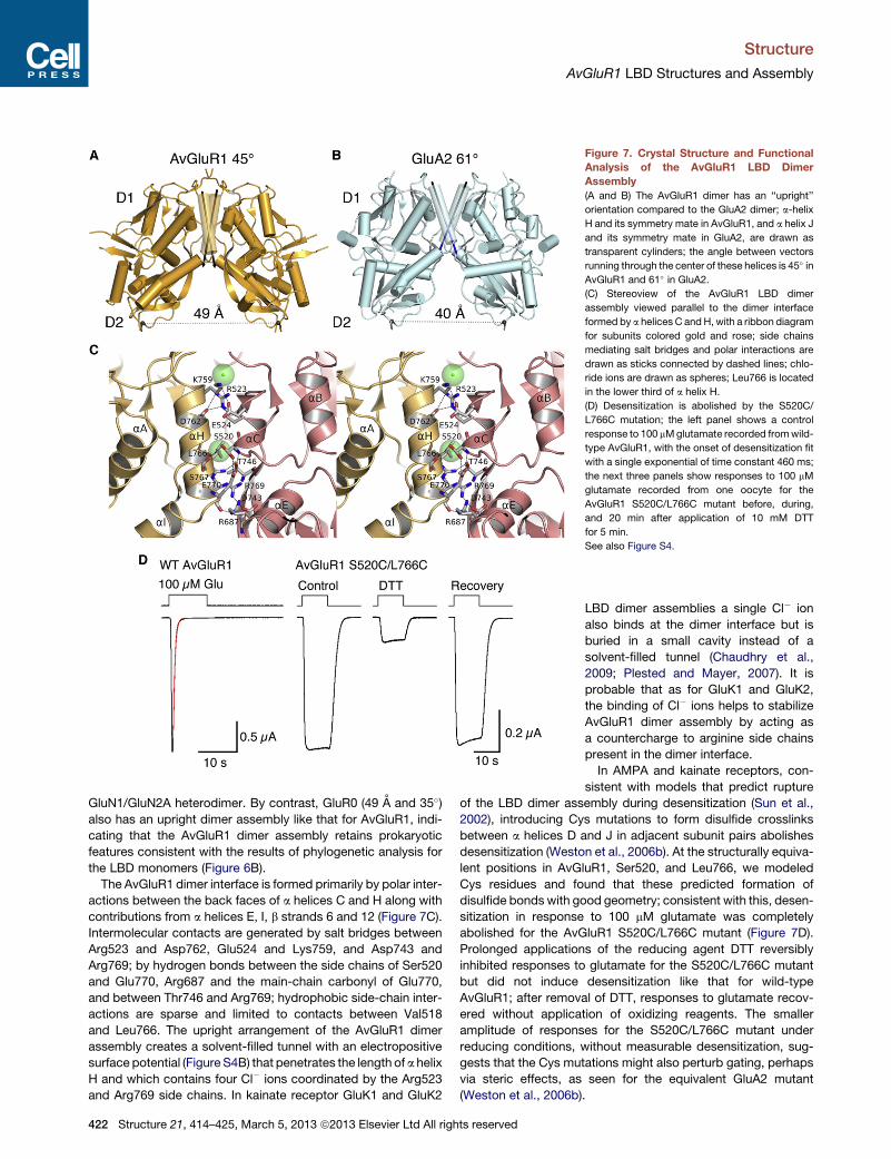
Figure 7. Crystal Structure and Functional
Analysis of the AvGluR1 LBD Dimer
Assembly
(A and B) The AvGluR1 dimer has an ‘‘upright’’
orientation compared to the GluA2 dimer; a-helix
H and its symmetry mate in AvGluR1, and a helix J
and its symmetry mate in GluA2, are drawn as
transparent cylinders; the angle between vectors
running through the center of these helices is 45� inAvGluR1 and 61� in GluA2.
(C) Stereoview of the AvGluR1 LBD dimer
assembly viewed parallel to the dimer interface
formed by a helices C andH, with a ribbon diagram
for subunits colored gold and rose; side chains
mediating salt bridges and polar interactions are
drawn as sticks connected by dashed lines; chlo-
ride ions are drawn as spheres; Leu766 is located
in the lower third of a helix H.
(D) Desensitization is abolished by the S520C/
L766C mutation; the left panel shows a control
response to 100 mMglutamate recorded fromwild-
type AvGluR1, with the onset of desensitization fit
with a single exponential of time constant 460 ms;
the next three panels show responses to 100 mM
glutamate recorded from one oocyte for the
AvGluR1 S520C/L766C mutant before, during,
and 20 min after application of 10 mM DTT
for 5 min.
See also Figure S4.
Structure
AvGluR1 LBD Structures and Assembly
GluN1/GluN2A heterodimer. By contrast, GluR0 (49 A and 35�)also has an upright dimer assembly like that for AvGluR1, indi-
cating that the AvGluR1 dimer assembly retains prokaryotic
features consistent with the results of phylogenetic analysis for
the LBD monomers (Figure 6B).
The AvGluR1 dimer interface is formed primarily by polar inter-
actions between the back faces of a helices C and H along with
contributions from a helices E, I, b strands 6 and 12 (Figure 7C).
Intermolecular contacts are generated by salt bridges between
Arg523 and Asp762, Glu524 and Lys759, and Asp743 and
Arg769; by hydrogen bonds between the side chains of Ser520
and Glu770, Arg687 and the main-chain carbonyl of Glu770,
and between Thr746 and Arg769; hydrophobic side-chain inter-
actions are sparse and limited to contacts between Val518
and Leu766. The upright arrangement of the AvGluR1 dimer
assembly creates a solvent-filled tunnel with an electropositive
surface potential (Figure S4B) that penetrates the length of a helix
H and which contains four Cl� ions coordinated by the Arg523
and Arg769 side chains. In kainate receptor GluK1 and GluK2
422 Structure 21, 414–425, March 5, 2013 ª2013 Elsevier Ltd All rights reserved
LBD dimer assemblies a single Cl� ion
also binds at the dimer interface but is
buried in a small cavity instead of a
solvent-filled tunnel (Chaudhry et al.,
2009; Plested and Mayer, 2007). It is
probable that as for GluK1 and GluK2,
the binding of Cl� ions helps to stabilize
AvGluR1 dimer assembly by acting as
a countercharge to arginine side chains
present in the dimer interface.
In AMPA and kainate receptors, con-
sistent with models that predict rupture
of the LBD dimer assembly during desensitization (Sun et al.,
2002), introducing Cys mutations to form disulfide crosslinks
between a helices D and J in adjacent subunit pairs abolishes
desensitization (Weston et al., 2006b). At the structurally equiva-
lent positions in AvGluR1, Ser520, and Leu766, we modeled
Cys residues and found that these predicted formation of
disulfide bonds with good geometry; consistent with this, desen-
sitization in response to 100 mM glutamate was completely
abolished for the AvGluR1 S520C/L766C mutant (Figure 7D).
Prolonged applications of the reducing agent DTT reversibly
inhibited responses to glutamate for the S520C/L766C mutant
but did not induce desensitization like that for wild-type
AvGluR1; after removal of DTT, responses to glutamate recov-
ered without application of oxidizing reagents. The smaller
amplitude of responses for the S520C/L766C mutant under
reducing conditions, without measurable desensitization, sug-
gests that the Cys mutations might also perturb gating, perhaps
via steric effects, as seen for the equivalent GluA2 mutant
(Weston et al., 2006b).

Structure
AvGluR1 LBD Structures and Assembly
In conclusion the ligand binding properties of AvGluR1 are
distinct and resemble neither those in eukaryotic glutamate
receptors nor their previously characterized prokaryotic precur-
sors. It is widely believed that the two domain Venus flytrap
structure of iGluR LBDs evolved from the large family of bacterial
periplasmic binding proteins whose members utilize the same
scaffold to selectively bind a wide variety of ligands, including
small oxyanions, mono- and oligo-saccharides, amino acids, oli-
gopeptides, polyamines, and vitamins. AvGluR1 represents an
unusual example in which the same protein molecule is able to
bind to chemically diverse amino acids, using ions as substitute
ligands. This raises the question as to why glutamate was
selected as a neurotransmitter among a wide spectrum of candi-
date amino acids and small molecules and if fine-tuning was
subsequently required during evolution to remove sensitivity to
other amino acids, as a necessary step for high-fidelity infor-
mation processing at synapses. Binding of the glutamate
g-carboxyl group by arginine residues in AvGluR1 has prece-
dent from the mechanism found in metabotropic glutamate
receptor (mGluR) GPCRs, for which Arg and Lys residues play
a similar role; however, in mGluRs these residues are located
in domain 1, not in domain 2 (Kunishima et al., 2000). Intriguingly,
the binding of (2S)-2-amino-4-phosphonobutanoic acid (AP4) to
mGluR4 requires Cl� ions (Kuang and Hampson, 2006), but
structural information on the anion binding site of mGluRs is
lacking. Homology models, based on mGluR1 (Kunishima
et al., 2000) and atrial natriuretic peptide receptor (Ogawa
et al., 2010) ligand binding domain crystal structures, suggest
the location of a putative anion binding site that is remote from
the glutamate g-carboxyl group and that has been proposed
to create a pocket for subtype selective ligands (Acher et al.,
2011). Consistent with its proposed role as an evolutionary inter-
mediate (Janovjak et al., 2011), structure-based phylogenetic
analysis revealed that the AvGluR1 LBD most closely resembles
prokaryotic iGluRs, although the bound glutamate ligand adopts
the same twisted conformation found in AMPA, kainate, and
NMDA receptors, instead of the extended conformation found
in prokaryotic iGluRs. An analysis of dimer structures also
reveals AvGluR1 LBD packing like that in prokaryotic iGluRs,
whereas analytical ultracentrifugation experiments revealed
instead a low affinity for dimer assembly like that for eukaryotic
iGluRs.
EXPERIMENTAL PROCEDURES
Construct Design and Protein Expression
Design of the AvGluR1 LBD construct was based on domain boundaries
demarcated previously for AMPA and kainate receptor LBDs, but with a four-
residue extension at the N terminus, and included S1 residues A433-K543
and S2 residues L656-P788 joined by a GT linker. These were isolated from
the full-length cDNA by overlap PCR, cloned into pET22b with an N-terminal
MH8SSGLVPRGS affinity tag and thrombin cleavage site, sequenced, and
expressed in E. coli Origami B(DE3) induced with 30 mM IPTG for 15 hr at
18�C. The soluble fraction from bacterial cell lysates was purified using
Ni-NTA chromatography, followed by thrombin cleavage, and ion exchange
chromatography using SP sepharose. Final yields were typically 8–10 mg
from 12 l cultures.
Ligand Binding Assays
Reactions were carried out in a ligand binding buffer (LBB) containing 150 mM
NaCl, 2 mMEDTA, 10mMHEPES (pH 7.0), and 10%glycerol. Apo protein was
Structure 21,
generated by extensive dialysis against LBB, with eight changes over 5 days,
for a total volume exchange of 1020. Reactions were set up on ice in 96-well
plates with 250 nM apo protein and 10 mg/ml BSA in 200 ml LBB per well
with [3H]L-Glu (50.6 Ci/mmol) diluted 1:20 with [1H]L-Glu; nonspecific binding
was measured in the presence of 20 mM alanine. Competing ligands were
added either at 100 mM or as a concentration series and incubated for
120 min prior to filtering through Millipore multiscreen IP filter plates prewet
with 50% ethanol and washed twice with ice-cold LBB, both before and after
filtration of the binding reactions. Plates were dried thoroughly, sealed with
clear adhesive plastic tape, incubated with 50 ml/well Optiphase Supermix
(PerkinElmer, Waltham, MA, USA) scintillation fluid, and counted on a liquid
scintillation counter (Wallac Trilux 1450 microbeta).
Electrophysiological Analysis
Two electrode voltage clamp recordings were performed using stage 5-6
Xenopus laevis oocytes injected with AvGluR1 cRNA, agarose cushion
electrodes filled with 3 M KCl (Schreibmayer et al., 1994) and a custom-
made recording chamber to allow rapid solution exchange, with ligands
applied using computer controlled valves as described previously (Panchenko
et al., 1999). The extracellular solution contained (in mM) 100 NaCl, 1 KCl,
2 CaCl2, 1 MgCl2 and 5 HEPES titrated to pH 7.4 with NaOH; in some exper-
iments the divalent ion concentration was changed to 10 mM CaCl2 and 2 mM
MgCl2.
Crystallization and Structure Determination
Crystallization experiments used protein dialyzed against 50 mMNaCl, 10 mM
TrisCl (pH 8.0), 1 mM EDTA, and either 2 mM L-Glu or 10 mM of either L-Asp,
L-Ala, L-Ser, or L-Met or 50 mM L-Phe. Trays were set up in hanging-drop
format at 20�C using a protein concentration of 5–10 mg/ml at a 1:1 or 1:2 pro-
tein:reservoir volume ratio, with a reservoir containing 0.1 M BisTris propane
(pH 6.5), 50 or 100 mM NaCitrate, and 17.5 to 20.0% PEG 3350. Diffraction
quality crystals were obtained by streak seeding, cryoprotected by serial
transfers to glycerol, final concentration 10%–15%, and flash frozen in liquid
nitrogen. Data were collected at APS beamline ID22, using 1 A radiation and
a MAR300 CCD detector. Diffraction data were indexed, scaled, and merged
using HKL2000 (Otwinowski and Minor, 1997). The AvGluR1 LBD glutamate
complex was solved by molecular replacement with Phaser (McCoy et al.,
2007) using a GluK2 monomer (1S50) as a search probe after deletion of resi-
dues 18–34 (loop 1), 63–72 (loop 2), and 254–259 (C terminus), with mutation of
all side chains to Ser; two molecules were located in the asymmetric unit with
rotation and translation Z scores of 6.3 and 5.7 for protomer 1 and 5.6 and 10.7
for protomer 2. The solution was successfully built using Phenix Autobuild
(Adams et al., 2010). The aspartate, alanine, methionine, and phenylalanine
complexes were solved by Fourier difference techniques, using the refined
coordinates for the AvGluR1 LBD glutamate complex stripped of alternate
conformations and heteroatoms as a starting model; molecular replacement
was required for the serine complex because of a difference in cell dimensions
(Table 1), with rotation and translation Z scores of 8.7 and 8.1 for protomer 1
and 10.8 and 17.9 for protomer 2. Iterative cycles of refinement and model
building were carried out using Phenix (Adams et al., 2010) and Coot (Emsley
et al., 2010), with either four TLS groups or for the L-Glu complex individual
anisotropic B-factors. For the glutamate and alanine complexes, the location
of Cl� ions was confirmed by calculation of anomalous difference Fourier
maps using data collected at 1.5418 A in the home lab. Models were validated
with Molprobity (Chen et al., 2010), with the following scores, where 100% is
the best among structures of comparable resolution: glutamate 0.95 (99%);
aspartate 0.96 (100%); serine 1.01 (100%); alanine 1.12 (99%); methionine
0.94 (100%); phenylalanine 0.98 (100%); cavity calculations were performed
using VOIDOO with a probe radius of 1.4 A on a 1.0 A grid (Kleywegt and
Jones, 1994); LSQMANwas used for superpositions (Kleywegt, 1996). Figures
were generated using PyMol (Schrodinger).
Structural Analysis
The pairwise structural alignment algorithm DALI was used for the identifica-
tion of AvGluR1 LBD homologs with known structure (Holm and Rosenstrom,
2010). Structure-based sequence alignments were generated based on
a progressive pairwise heuristic algorithm as implemented in MUSTANG
(Konagurthu et al., 2006). Structure-based phylogeny was calculated based
414–425, March 5, 2013 ª2013 Elsevier Ltd All rights reserved 423

Structure
AvGluR1 LBD Structures and Assembly
on the structural similarity score (QH) using theMultiSeqmodule of VMD (Hum-
phrey et al., 1996). The value of QH indicates an overall structural similarity
score between two structures and is calculated using the equation QH = a�1
[qaln + qgap], where a is the normalization that accounts the contribution from
both contacts between the aligned regions as well as between the residue
present in the aligned position and the gap region; qaln represents the fraction
of Ca-Ca distances that are similar between the two aligned structures; and
qgap introduces a penalty term to account for the presence of insertions with
the QH value decreasing with larger perturbations (O’Donoghue and Luthey-
Schulten, 2005).
Analytical Ultracentrifugation
Sedimentation velocity (SV) and equilibrium (SE) experiments were performed
in a ProteomeLab XL-I analytical ultracentrifuge (Beckman Coulter, Indianap-
olis, IN, USA). Samples were prepared by dilution of a concentrated protein
stock using reference buffer (20 mM phosphate buffer (pH 7.5), 150 mM
NaCl, 1 mM EDTA, and 2 mM L-glutamate) and loaded into cell housings
with either 3 or 12 mm double-sector charcoal-filled epon centerpieces and
sapphire windows. For SV, the evolution of the concentration gradient was
recorded using absorbance optics at wavelengths of 250 and 280 nm and
interference detection at a rotor speed of 50 krpm and temperature of 20�C.Data were analyzed in SEDFIT using a c(s) distribution model with maximum
entropy regularization (p = 0.68) and systematic noise decomposition, leaving
the meniscus position and weight-average frictional ratio as fitting parameters
(Schuck, 2003). The buffer density (1.006995 g/ml) and viscosity (1.0373 cP)
were measured using a DMA500M density meter or an AmVnmicroviscometer
from Anton Paar (Graz, Austria). For SE, the equilibrium concentration profiles
were recorded using interference detection at rotor speeds of 12, 22, and 26
krpm and a temperature of 10�C. Data were analyzed in SEDPHAT using
a single-species model or a monomer-dimer equilibrium association model
with mass conservation constraints (Vistica et al., 2004). The buffer density
(1.009043 g/ml) and viscosity (1.3316 cP) were measured using a DMA500M
density meter or an AmVn microviscometer from Anton Paar (Graz, Austria),
respectively.
ACCESSION NUMBERS
Atomic coordinates and structure factors have been deposited in the Protein
Data Bank (PDB) with the accession codes 4IO2, 4IO3, 4IO4, 4IO5, 4IO6,
and 4IO7 (Table 1).
SUPPLEMENTAL INFORMATION
Supplemental Information includes four figures and one table and can be
found with this article online at http://dx.doi.org/10.1016/j.str.2013.01.006.
ACKNOWLEDGMENTS
We thank Dr. E. Isacoff for the AvGluR1 cDNA, the Swartz lab (NINDS) for
oocytes, and Andi Balbo and Carla Glasser for technical assistance. Synchro-
tron diffraction data was collected at the Southeast Regional Collaborative
Access Team (SER-CAT) 22-ID beamline at the Advanced Photon Source
(APS), Argonne National Laboratory. Use of APS was supported by the U.S.
Department of Energy, Office of Science, Office of Basic Energy Sciences,
under contract no. W-31-109-Eng-38. This work was supported by the intra-
mural research program of NICHD, NIH, DHHS.
Received: November 21, 2012
Revised: January 10, 2013
Accepted: January 11, 2013
Published: February 21, 2013
REFERENCES
Abele, R., Keinanen, K., andMadden, D.R. (2000). Agonist-induced isomeriza-
tion in a glutamate receptor ligand-binding domain. A kinetic and mutagenetic
analysis. J. Biol. Chem. 275, 21355–21363.
424 Structure 21, 414–425, March 5, 2013 ª2013 Elsevier Ltd All righ
Acher, F.C., Selvam, C., Pin, J.P., Goudet, C., and Bertrand, H.O. (2011). A crit-
ical pocket close to the glutamate binding site of mGlu receptors opens new
possibilities for agonist design. Neuropharmacology 60, 102–107.
Adams, P.D., Afonine, P.V., Bunkoczi, G., Chen, V.B., Davis, I.W., Echols, N.,
Headd, J.J., Hung, L.W., Kapral, G.J., Grosse-Kunstleve, R.W., et al. (2010).
PHENIX: a comprehensive Python-based system for macromolecular struc-
ture solution. Acta Crystallogr. D Biol. Crystallogr. 66, 213–221.
Armstrong, N., and Gouaux, E. (2000). Mechanisms for activation and antag-
onism of an AMPA-sensitive glutamate receptor: crystal structures of the
GluR2 ligand binding core. Neuron 28, 165–181.
Chaudhry, C., Plested, A.J., Schuck, P., and Mayer, M.L. (2009). Energetics of
glutamate receptor ligand binding domain dimer assembly are modulated by
allosteric ions. Proc. Natl. Acad. Sci. USA 106, 12329–12334.
Chen, G.Q., Cui, C., Mayer, M.L., and Gouaux, E. (1999). Functional character-
ization of a potassium-selective prokaryotic glutamate receptor. Nature 402,
817–821.
Chen, V.B., Arendall, W.B., 3rd, Headd, J.J., Keedy, D.A., Immormino, R.M.,
Kapral, G.J., Murray, L.W., Richardson, J.S., and Richardson, D.C. (2010).
MolProbity: all-atom structure validation for macromolecular crystallography.
Acta Crystallogr. D Biol. Crystallogr. 66, 12–21.
Chiu, J.C., Brenner, E.D., DeSalle, R., Nitabach, M.N., Holmes, T.C., and
Coruzzi, G.M. (2002). Phylogenetic and expression analysis of the gluta-
mate-receptor-like gene family in Arabidopsis thaliana. Mol. Biol. Evol. 19,
1066–1082.
Croset, V., Rytz, R., Cummins, S.F., Budd, A., Brawand, D., Kaessmann, H.,
Gibson, T.J., and Benton, R. (2010). Ancient protostome origin of chemosen-
sory ionotropic glutamate receptors and the evolution of insect taste and
olfaction. PLoS Genet. 6, e1001064.
Emsley, P., Lohkamp, B., Scott, W.G., and Cowtan, K. (2010). Features and
development of Coot. Acta Crystallogr. D Biol. Crystallogr. 66, 486–501.
Everts, I., Petroski, R., Kizelsztein, P., Teichberg, V.I., Heinemann, S.F., and
Hollmann, M. (1999). Lectin-induced inhibition of desensitization of the kainate
receptor GluR6 depends on the activation state and can be mediated by
a single native or ectopic N-linked carbohydrate side chain. J. Neurosci. 19,
916–927.
Furukawa, H., Singh, S.K., Mancusso, R., and Gouaux, E. (2005). Subunit
arrangement and function in NMDA receptors. Nature 438, 185–192.
Ger, M.F., Rendon, G., Tilson, J.L., and Jakobsson, E. (2010). Domain-based
identification and analysis of glutamate receptor ion channels and their rela-
tives in prokaryotes. PLoS ONE 5, e12827.
Holm, L., and Rosenstrom, P. (2010). Dali server: conservation mapping in 3D.
Nucleic Acids Res. 38(Web Server issue), W545–W549.
Humphrey, W., Dalke, A., and Schulten, K. (1996). VMD: visual molecular
dynamics. J. Mol. Graph. 14, 33–38, 27–28.
Janovjak, H., Sandoz, G., and Isacoff, E.Y. (2011). A modern ionotropic
glutamate receptor with a K(+) selectivity signature sequence. Nat.
Commun. 2, 232.
Jin, R., Singh, S.K., Gu, S., Furukawa, H., Sobolevsky, A.I., Zhou, J., Jin, Y.,
and Gouaux, E. (2009). Crystal structure and association behaviour of the
GluR2 amino-terminal domain. EMBO J. 28, 1812–1823.
Kleywegt, G.J. (1996). Use of non-crystallographic symmetry in protein struc-
ture refinement. Acta Crystallogr. D Biol. Crystallogr. 52, 842–857.
Kleywegt, G.J., and Jones, T.A. (1994). Detection, delineation, measurement
and display of cavities in macromolecular structures. Acta Crystallogr. D
Biol. Crystallogr. 50, 178–185.
Konagurthu, A.S., Whisstock, J.C., Stuckey, P.J., and Lesk, A.M. (2006).
MUSTANG: a multiple structural alignment algorithm. Proteins 64, 559–574.
Kuang, D., and Hampson, D.R. (2006). Ion dependence of ligand binding
to metabotropic glutamate receptors. Biochem. Biophys. Res. Commun.
345, 1–6.
Kumar, J., Schuck, P., and Mayer, M.L. (2011). Structure and assembly mech-
anism for heteromeric kainate receptors. Neuron 71, 319–331.
ts reserved

Structure
AvGluR1 LBD Structures and Assembly
Kunishima, N., Shimada, Y., Tsuji, Y., Sato, T., Yamamoto, M., Kumasaka, T.,
Nakanishi, S., Jingami, H., and Morikawa, K. (2000). Structural basis of gluta-
mate recognition by a dimeric metabotropic glutamate receptor. Nature 407,
971–977.
Lam, H.M., Chiu, J., Hsieh, M.H., Meisel, L., Oliveira, I.C., Shin, M., and
Coruzzi, G. (1998). Glutamate-receptor genes in plants. Nature 396, 125–126.
Lee, J.H., Kang, G.B., Lim, H.H., Jin, K.S., Kim, S.H., Ree, M., Park, C.S., Kim,
S.J., and Eom, S.H. (2008). Crystal structure of the GluR0 ligand-binding core
from Nostoc punctiforme in complex with L-glutamate: structural dissection of
the ligand interaction and subunit interface. J. Mol. Biol. 376, 308–316.
Mayer, M.L. (2005). Crystal structures of the GluR5 and GluR6 ligand binding
cores: molecular mechanisms underlying kainate receptor selectivity. Neuron
45, 539–552.
Mayer, M.L., Olson, R., and Gouaux, E. (2001). Mechanisms for ligand binding
to GluR0 ion channels: crystal structures of the glutamate and serine com-
plexes and a closed apo state. J. Mol. Biol. 311, 815–836.
McCoy, A.J., Grosse-Kunstleve, R.W., Adams, P.D., Winn, M.D., Storoni, L.C.,
and Read, R.J. (2007). Phaser crystallographic software. J. Appl. Cryst. 40,
658–674.
O’Donoghue, P., and Luthey-Schulten, Z. (2005). Evolutionary profiles derived
from the QR factorization of multiple structural alignments gives an economy
of information. J. Mol. Biol. 346, 875–894.
Ogawa, H., Qiu, Y., Philo, J.S., Arakawa, T., Ogata, C.M., and Misono, K.S.
(2010). Reversibly bound chloride in the atrial natriuretic peptide receptor
hormone-binding domain: possible allosteric regulation and a conserved
structural motif for the chloride-binding site. Protein Sci. 19, 544–547.
Otwinowski, Z., and Minor, W. (1997). Processing of X-ray diffraction data
collected in oscillation mode. In Methods in Enzymology, C.W. Carter and
R.M. Sweets, eds. (Charlottesville: University of Virginia), pp. 307–326.
Panchenko, V.A., Glasser, C.R., Partin, K.M., and Mayer, M.L. (1999). Amino
acid substitutions in the pore of rat glutamate receptors at sites influencing
block by polyamines. J. Physiol. 520, 337–357.
Partin, K.M., Patneau, D.K., Winters, C.A., Mayer, M.L., and Buonanno, A.
(1993). Selective modulation of desensitization at AMPA versus kainate recep-
tors by cyclothiazide and concanavalin A. Neuron 11, 1069–1082.
Plested, A.J., and Mayer, M.L. (2007). Structure and mechanism of kainate
receptor modulation by anions. Neuron 53, 829–841.
Structure 21,
Rossmann, M., Sukumaran, M., Penn, A.C., Veprintsev, D.B., Babu, M.M., and
Greger, I.H. (2011). Subunit-selective N-terminal domain associations orga-
nize the formation of AMPA receptor heteromers. EMBO J. 30, 959–971.
Schreibmayer, W., Lester, H.A., and Dascal, N. (1994). Voltage clamping of
Xenopus laevis oocytes utilizing agarose-cushion electrodes. Pflugers Arch.
426, 453–458.
Schuck, P. (2003). On the analysis of protein self-association by sedimentation
velocity analytical ultracentrifugation. Anal. Biochem. 320, 104–124.
Sobolevsky, A.I., Rosconi, M.P., and Gouaux, E. (2009). X-ray structure,
symmetry and mechanism of an AMPA-subtype glutamate receptor. Nature
462, 745–756.
Srivastava, M., Begovic, E., Chapman, J., Putnam, N.H., Hellsten, U.,
Kawashima, T., Kuo, A., Mitros, T., Salamov, A., Carpenter, M.L., et al.
(2008). The Trichoplax genome and the nature of placozoans. Nature 454,
955–960.
Stern-Bach, Y., Bettler, B., Hartley, M., Sheppard, P.O., O’Hara, P.J., and
Heinemann, S.F. (1994). Agonist selectivity of glutamate receptors is specified
by two domains structurally related to bacterial amino acid-binding proteins.
Neuron 13, 1345–1357.
Sun, Y., Olson, R., Horning, M., Armstrong, N., Mayer, M., and Gouaux, E.
(2002). Mechanism of glutamate receptor desensitization. Nature 417,
245–253.
Vistica, J., Dam, J., Balbo, A., Yikilmaz, E., Mariuzza, R.A., Rouault, T.A., and
Schuck, P. (2004). Sedimentation equilibrium analysis of protein interactions
with global implicit mass conservation constraints and systematic noise
decomposition. Anal. Biochem. 326, 234–256.
Weston, M.C., Gertler, C., Mayer, M.L., and Rosenmund, C. (2006a).
Interdomain interactions in AMPA and kainate receptors regulate affinity for
glutamate. J. Neurosci. 26, 7650–7658.
Weston, M.C., Schuck, P., Ghosal, A., Rosenmund, C., and Mayer, M.L.
(2006b). Conformational restriction blocks glutamate receptor desensitization.
Nat. Struct. Mol. Biol. 13, 1120–1127.
Zhao, H., Berger, A.J., Brown, P.H., Kumar, J., Balbo, A., May, C.A., Casillas,
E., Jr., Laue, T.M., Patterson, G.H., Mayer, M.L., and Schuck, P. (2012).
Analysis of high-affinity assembly for AMPA receptor amino-terminal domains.
J. Gen. Physiol. 139, 371–388.
414–425, March 5, 2013 ª2013 Elsevier Ltd All rights reserved 425