Embed Size (px)
Citation preview

i
Animal Production Unit Activities Report 2007
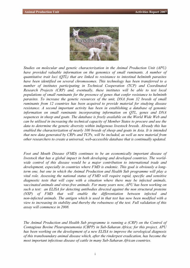
Studies on molecular and genetic characterization in the Animal Production Unit (APU) have provided valuable information on the genomics of small ruminants. A number of quantitative trait loci (QTL) that are linked to resistance to intestinal helminth parasites have been identified on several chromosomes. This technology has been transferred to a number of institutes participating in Technical Cooperation (TCP) and Coordinated Research Projects (CRP) and, eventually, these institutes will be able to test local populations of small ruminants for the presence of genes that confer resistance to helminth parasites. To increase the genetic resources of the unit, DNA from 32 breeds of small ruminants from 12 countries has been acquired to provide material for studying disease resistance. A second important activity has been in establishing a database of genomic information on small ruminants incorporating information on QTL, genes and DNA sequences in sheep and goats. The database is freely available on the World Wide Web and can be utilised in increasing the technical capacity of Member States to procure and use the data to determine the genetic diversity within indigenous livestock breeds. Already this has enabled the characterization of nearly 100 breeds of sheep and goats in Asia. It is intended that new data generated by CRPs and TCPs, will be included, as well as new material from other researchers to create a universal, web-accessible database that is continually updated. Foot and Mouth Disease (FMD) continues to be an economically important disease of livestock that has a global impact in both developing and developed countries. The world-wide control of this disease would be a major contribution to international trade and development, especially in countries where FMD is endemic. This goal is obviously a long-term one, but one in which the Animal Production and Health Sub programme will play a vital role. Assessing the national status of FMD will require rapid, specific and sensitive diagnostic tests that will cope with a situation where there may be infected animals, vaccinated animals and virus-free animals. For many years now, APU has been working on such a test: an ELISA for detecting antibodies directed against the non structural proteins (NSP) of FMD that will enable the differentiation between infected and non-infected animals. The antigen which is used in that test has now been modified with a view to increasing its stability and thereby the robustness of the test. Full validation of this assay will commence in 2008. The Animal Production and Health Sub programme is running a (CRP) on the Control of Contagious Bovine Pleuropneumonia (CBPP) in Sub-Saharan Africa; for this project, APU has been working on the development of a new ELISA to improve the serological diagnosis of this transboundary animal disease which, after the rinderpest eradication, has become the most important infectious disease of cattle in many Sub-Saharan African countries.

ii
Animal Production Unit Activities Report 2007
Goat Pox and Sheep Pox are responsible for economically important diseases in small ruminants. The unit is currently developing sensitive and specific diagnostic tests and molecular characterization techniques that will provide a more comprehensive assessment of the epidemiology of these diseases. The classical and real-time polymerase chain reaction (PCR) assays have been developed to enable the differentiation between Goat Pox Virus (GTPV) and Sheep Pox Virus (SPPV). Further validation will be undertaken in 2008, including laboratory and field trials in Africa to confirm the potential of the real-time PCR test. Sequencing and analysis of a specific capripox virus gene, the chemokine receptor gene, that is probably involved in virus pathogenicity, showed that SPPV and GTPV clustered separately, suggesting that this gene could be used to differentiate the two viruses. As part of the activity on the development of a marker vaccine and its companion test for the better control of Peste des Petit Ruminants (PPR) the work has been concentrated on the study of the interaction between the nucleoprotein (N) and the phosphoprotein (P) of the virus. ELISA tests have shown that N proteins can react with several peptides of P protein, and can be grouped as major and minor reactive domains. These different interactive sites of N protein are located in the variable area of the P protein. The APU is also involved in a project on Gene-based Technologies in Livestock Breeding: Characterization of Small Ruminant Genetic Resources in Asia, and a project on the Genomics of the Alpaca: Identification of Expressed Genes and Genetic Markers Associated with Productivity and Embryonic Mortality. On the animal health side, the Unit is involved in work on The Early and Sensitive Diagnosis and Control of Peste des Petits Ruminants and continues to support the work on Veterinary Surveillance of Rift Valley Fever.

iii
Animal Production Unit Activities Report 2007
Table of ContentsTable of ContentsTable of Contents Executive Summary 1. PROGRAMMATIC AND UNIT OBJECTIVES·············································································· 1 2. STAFF···························································································································································· 2 3. RESEARCH AND DEVELOPMENT ACTIVITIES······································································ 4
3.1. Animal Genetics ····································································································································· 4 3.1.1. Mapping the Sheep Genome for Determining Association with Helminth Resistance··················7 3.1.1.1 Helminth Resistance Genome Scan ··················································································· 7
3.1.1.2. Helminth Resistance Markers............................................................................................7 3.1.1.3. Creating a DNA Gene Bank at the IAEA Laboratories for Use by Member States ········ 11
3.2. Bioinformatics and Genomics ············································································································· 12 3.2.1. Development of a Real Time Database for Genetic Information (RT-db) on Small Ruminants · 12 3.2.2. Database of Genetic Resources (GR-db) for the DNA Bank and Gene Profiling…………………12
3.3. Animal Health ····································································································································· 13 3.3.1. Use of Non Structural Protein (NSP) of Foot and Mouth Disease Virus to Differentiate
Between Vaccinated and Infected Animals················································································· 13 3.3.1.1. Enhancement of Stability of NSP to Increase Robustness of the ELISA by Using
Recombinant Mutated Non-Structural FMDV Protein ················································ 14 3.3.1.2. Production and Purification of Recombinant FMDV NSP from the Baculovirus
Expression System·········································································································17 3.3.1.3. Validation of FMDV NSP-Based ELISA tests······································································ 18
3.4. The Control of Contagious Bovine Pleuropneumonia (CBPP) in Sub-Saharan Africa ················· 19 3.4.1. Expression and Purification of Recombinant CBPP LppQ Protein·············································· 19 3.4.2. Development of a Prototype iELISA to Detect Antibodies against CBPP in Cattle····················· 21
3.5. Study of the Interaction of N and P Proteins of Peste des Petit Ruminants (PPR) ······················· 22 3.5.1. Cloning the PPRV Gene into the Baculovirus Vector Genome and Expression of the
Recombinant in Insect Cells········································································································ 22 3.5.2. Mapping N Binding Site on P ······································································································ 23
3.6. Development of a PPR Specific Serological Diagnostic Test: Production of New Monoclonal Antibodies ·························································································································································· 26
3.7. Development of Capripox Virus Diagnostic Tests and Molecular Epidemiology of the Disease ·· 27 3.7.1. Development of Molecular-Based Diagnostic Tests for the Identification of Capripox Virus ···· 27
3.7.1.1. Classical PCR ················································································································27 3.7.1.2. Cloning the Chemokine Gene of Sheep and Goat Pox Viruses Molecular
Epidemiology ················································································································28 3.7.1.3. Real-Time PCR···············································································································29
3.8. Sequencing the Capripox Virus Genome ··························································································· 31 3.9. IAEA Reference Serum Bank ············································································································· 36
4. TRAINING ACTIVITIES ····················································································································· 37 4.1. Training Course on the Molecular Characterization of Small Ruminant Breeds, Aleppo, Syria·37 4.2. Fellowships············································································································································ 37 4.3. Internships ············································································································································ 38 4.4. Scientific Visitors·································································································································· 38
5. ACKNOWLEDGEMENTS··················································································································· 39 6. APPENDICES ··········································································································································· 40 6.1. Staff Publications ································································································································· 40 6.2. Staff Travel ··········································································································································· 41 6.3. External Collaborations and Partnerships ························································································ 42 6.4. Trainees, Fellows and Scientific Visitors···························································································· 44 6.5. Coordinated Research Projects and Technical Cooperation Projects············································· 45 6.6. Abbreviations ······································································································································· 46

iv
Animal Production Unit Activities Report 2007

1
Animal Production Unit Activities Report 2007
The vision and goal of the Animal Production and Health (APH) Sub-programme are to minimise risks to livestock in FAO and IAEA Member States in order to increase food security, to fight hunger and to improve the livelihoods of the poor in FAO and IAEA Member States. To achieve this objective, two strategies guide the activities of the Sub-programme: 1. Capacity building within regions and countries. Success in the control of highly infectious diseases relies on the capacity of early warning and early reaction, a capacity that is missing in many developing countries because of a lack of financial resources and also human and physical resources. Training of scientists is important for helping developing countries to manage different risks that are threatening their livestock production. 2. Promotion of applied research targeting areas that help alleviate risks for livestock in developing FAO and IAEA Member States. This involves promoting the transfer of technologies, in particular, nuclear and nuclear-related techniques to developing countries, promoting and implementing applied research projects for the development of improved diagnostic tests and vaccines, leading to better breeding strategies, and to improved farm management to optimize the use of animal feed resources and hence protect the environment. To implement its activities, the transfer of new technologies to developing countries, the APH Sub-programme, together with FAO and CSIRO, held two regional training courses in Egypt and Australia on the diagnosis of highly pathogenic avian influenza. The Animal Production Unit has worked very closely with the Section in all these activities.
1.PROGRAMMATIC AND UNIT OBJECTIVES

2
Animal Production Unit Activities Report 2007
2. STAFF
Name Title E-Mail Address Extension Erik BUSCH-PETERSEN
Laboratory Head [email protected] 28267
Kyoko MAKOVICKY Clerk [email protected] 28362
Name Title E-Mail Address Extension Adama DIALLO Unit Head,
Molecular Biologist [email protected] 28355
Massoud MALEK Molecular Geneticist [email protected] 28358
Eva Maria WINGER Senior Laboratory Technician
[email protected] 28302
Mamadou LELENTA Laboratory Technician [email protected] 28321
Ericka PESTANA DELGADO
Laboratory Technician [email protected] 28378
Sanne Charles BODJO Consultant [email protected] 28356
Caroline Melanie ADOMBI
Consultant [email protected] 28380
Charles Euloge LAMIEN
Consultant [email protected] 28314
FAO/IAEA Agriculture & Biotechnology Laboratory
Animal Production Unit
Name Title E-Mail Address Extension Gabriele VOIGT Director [email protected] 28200
IAEA Laboratories Seibersdorf

3
Animal Production Unit Activities Report 2007
Adama DIALLO
Massoud MALEK
Eva Maria WINGER
Mamadou LELENTA
Ericka PESTANA DELGADO
Sanne Charles BODJO Charles Euloge LAMIEN Caroline Melanie ADOMBI
4
Animal Production Unit Activities Report 2007
3. RESEARCH AND DEVELOPMENT ACTIVITIES
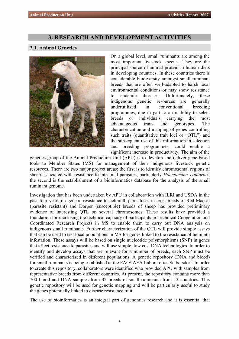
3.1. Animal Genetics On a global level, small ruminants are among the most important livestock species. They are the principal source of animal protein in human diets in developing countries. In these countries there is considerable biodiversity amongst small ruminant breeds that are often well-adapted to harsh local environmental conditions or may show resistance to endemic diseases. Unfortunately, these indigenous genetic resources are generally underutilized in conventional breeding programmes, due in part to an inability to select breeds or individuals carrying the most advantageous traits and genotypes. The characterization and mapping of genes controlling such traits (quantitative trait loci or “QTL”) and the subsequent use of this information in selection and breeding programmes, could enable a significant increase in productivity. The aim of the
genetics group of the Animal Production Unit (APU) is to develop and deliver gene-based tools to Member States (MS) for management of their indigenous livestock genetic resources. There are two major project areas: the first is to identify chromosomal regions of sheep associated with resistance to intestinal parasites, particularly Haemonchus contortus; the second is the establishment of a bioinformatics database for the analysis of the small ruminant genome. Investigation that has been undertaken by APU in collaboration with ILRI and USDA in the past four years on genetic resistance to helminth parasitoses in crossbreeds of Red Maasai (parasite resistant) and Dorper (susceptible) breeds of sheep has provided preliminary evidence of interesting QTL on several chromosomes. These results have provided a foundation for increasing the technical capacity of participants in Technical Cooperation and Coordinated Research Projects in MS to enable them to carry out DNA analysis on indigenous small ruminants. Further characterization of the QTL will provide simple assays that can be used to test local populations in MS for genes linked to the resistance of helminth infestation. These assays will be based on single nucleotide polymorphisms (SNP) in genes that affect resistance to parasites and will use simple, low cost DNA technologies. In order to identify and develop assays that are relevant for a number of breeds, each SNP must be verified and characterized in different populations. A genetic repository (DNA and blood) for small ruminants is being established at the FAO/IAEA Laboratories Seibersdorf. In order to create this repository, collaborators were identified who provided APU with samples from representative breeds from different countries. At present, the repository contains more than 700 blood and DNA samples from 32 breeds of small ruminants from 12 countries. This genetic repository will be used for genetic mapping and will be particularly useful to study the genes potentially linked to disease resistance trait. The use of bioinformatics is an integral part of genomics research and it is essential that

5
Animal Production Unit Activities Report 2007
national research institutes in MS have the required technology to routinely access genomic information databases. Part of the research programme is therefore targeted towards increasing the technical capacity of developing MS countries in the use of bioinformatics in national research institutes and to assist them accessing genomics information. There is a wealth of data on identification of QTL in the scientific literature, but the access to this information is often limited, due to various constraints (e.g. lack of library facilities). To improve the availability of genomic information on small ruminants, a web-accessible database for QTL, genes and general DNA sequences for sheep and goats has been created. The database will make available the pertinent results regarding genomic locations of QTL from all known studies on these species. Finally, through a CRP on the characterization of small ruminant genetic resources, MS in Asia were helped to characterize approximately 100 breeds of sheep and goats. The characterization included analysis of both phenotypic and genetic data, including the genotypes of microsatellites from the standard FAO/ISAG panel. The information will allow the participants to evaluate local biodiversity both within and across breeds. Currently, the genetics group at APU is building a web-accessible database for the CRP participants from which users will be able to view and download data from the CRP. The eventual goal is to make this a global resource, with results from other characterization studies being included together with data from new studies provided during the course of the CRP. Breeds with unique genetic characteristics are considered to be more valuable for conservation programmes and by comparing the allelic frequency at common loci across breeds, MS will be able to compare the genetic profiles of indigenous breeds with those from surrounding countries and around the world. These activities offer an exciting and novel way to increase the genomic information on sheep and goats and to disseminate this information on a wide scale. It is likely that genomic tools for disease resistance, wool and meat quality and other traits will become available in the future. The use of these genetic markers will help to increase the speed and efficiency of increased productive performance in a population and assist MS in breeding genetically superior stock. Making this genetic and genomic information available for application in small ruminant genetic programmes will help make genetic improvements via Marker Assisted Selection (MAS) or Introgression (MAI) a reality. An outline of the Genetics Resources Network upon which this programme is based is shown in the illustration below.

6
Animal Production Unit Activities Report 2007

7
Animal Production Unit Activities Report 2007
3.1.1. Mapping the Sheep Genome for Determining Association with Helminth Resistance
The activities described here aim to identify regions of the sheep chromosome that are associated with helminth resistance, based on molecular genome scan analysis. Genetic markers are identifiable DNA sequences that facilitate the study of inheritance of a trait or a gene. It is likely that the majority of anonymous genetic markers will have no effect on performance traits themselves, but such markers do make it possible to identify areas of the genome containing important genes. Genes closely linked to the marker will generally be inherited with it. Genetic markers will help trace regions of chromosomes from parents to offspring. To refine these findings further, a positional candidate gene approach was used, which is currently a successful method for identifying a gene that is associated with a particular trait. 3.1.1.1. Helminth Resistance Genome Scan APU, in collaboration with ILRI and the USDA, analysed data from genome scan studies in small ruminants (both linkage and QTL analysis) to plan for the future direction of the programme. The aim of the sheep genome mapping project is to analyze and evaluate a genome scan based on a cross between the parasite resistant Red Maasai and the susceptible Dorper breeds. Preliminary analyses have provided evidence for QTLs on several chromosomes. Further studies will take place and the work is expected to be completed by 2008. Although genome scans can identify chromosomal regions that contain QTLs associated with helminth resistance and related traits, this approach has only limited mapping resolution, therefore, following the genome scan, a candidate gene approach was used in order to more fully refine the identity of the potential QTL. 3.1.1.2. Helminth Resistance Markers The development of DNA-based markers has had a revolutionary impact on gene mapping and, more generally, on all animal genetics. DNA markers make it possible to exploit the entire diversity in DNA sequence that exists in any crossbreed. For this reason, high resolution genetic maps are being rapidly developed. Comparative mapping is based on the alignment of chromosomes using common molecular markers. This technology helps researchers to interpret the results from the genomic map of one species based on those obtained from another more extensively characterized species. Since there is a high level of synteny (physical co-localization of genetic loci on the same chromosome) between the genomes of different animal species, it is possible to extrapolate characterised information from one known map, which contains certain genes of interest, with another, in which only quantitative information (markers shared between those two maps) is available. Markers in this way operate as anchors that allow identification of regions on two different genomes that could share synteny. This powerful tool helps in the case of species such as sheep, where insufficient information is available, but molecular markers exist that are shared with cattle, in which the genome is more extensively characterised and mapped. This comparison permits the co-location of related traits from different maps and across different species. Comparative mapping shows that cattle chromosome 5 (BTA 5) shares regions of homology with sheep chromosome 3 (OAR3) (Figure 1a). This helps to identify positional candidate genes for QTL for chromosome 3 of sheep by using data for Chromosome 5 of cattle, where genome sequence information is

8
Animal Production Unit Activities Report 2007
fully available. The positional candidate gene approach will allow us to combine information about a gene’s chromosomal location for easier identification of a potential causative gene. It is assumed that candidate genes represent a large proportion of the QTL that has been previously identified from the helminth resistance genome scan. The positional candidate gene approach relies on a four stage process: 1) localizing the area of interest to a chromosomal sub region, which was based on literature review and preliminary results from the helminth resistance genome scan, 2) searching databases for attractive candidate genes within the sub region (149 genes), 3) testing the candidate
gene for causative mutations, and 4) developing simple, inexpensive, and robust nuclear-based tests that can be used by MS in their own laboratories. As explained above, comparative mapping was use to address this issue, and to fine map a chromosomal region (Figure 1a) and improve the ability to find genes responsible for helminth resistance. A total of 809 genes were selected based on the cattle BTA5 region sharing synteny with markers on the OAR3 QTL region associated with helminth resistance on previous scans (Figure 1b). The selected genes were then assessed for their putative gene function using the programme GeneCards (ht tp: / /www.genecards.org/index.shtml). In this step, gene function, protein ID, and biochemical pathways were annotated for all genes. After clearance, 149 genes involved in immune response pathways were selected as candidate genes for further analysis. The next objective was the development of a test to detect Single Nucleotide Polymorphisms (SNPs) associated with helminth resistance in
Figure 1a: QTL contour plot of OAR3.
BTA 5 Region Displayed: 0-76M bp Genes in Region: 809
OAR3 Number of Genes to Monitor: 149
Figure 1b: Comparative mapping between OAR3 and BTA5. QTL analysis in sheep showed that a region on OAR3 contain an important QTL associated with resistance to nematodes. Those flanking markers for each of the QTL peaks found in sheep were used to search for synteny in the cattle genome (Comparative Mapping). QTL markers were found to be located in BTA5. Genes contained on these areas were then used for primer design and SNP detection.

9
Animal Production Unit Activities Report 2007
Figure 2: Pooled samples from six different countries were used for PCR amplification and subsequent sequencing of amplified DNA for detection of SNP in the different populations.
sheep for the 149 selected genes as an input into the current CRP on small ruminant genetic characterization in Asia (CRP.D.3.10.25) (Figure 2).
The following steps were followed for the identification of SNPs in candidate genes: 1) selection of a candidate gene, 2) search of databases for known protein function of the candidate gene and selection of genes involved in immune responses, 3) designing primers from the known cattle sequences, 4) sequencing of the PCR products for gene verification, 5) amplification of pooled genomic DNA samples to find polymorphisms, 6) designing polymerase chain reaction restriction fragment length polymorphisms (PCR-RFLP) tests to allow amplification and analysis of large numbers of individuals, and 7) analysis of associations between traits of interest and genotypes for the selected candidate gene (Figures 3a, b).
Great Britain
Bangladesh
Kurdistan
Iran
Dorper
Red Massai
Peru
University of Teheran
10
Animal Production Unit Activities Report 2007
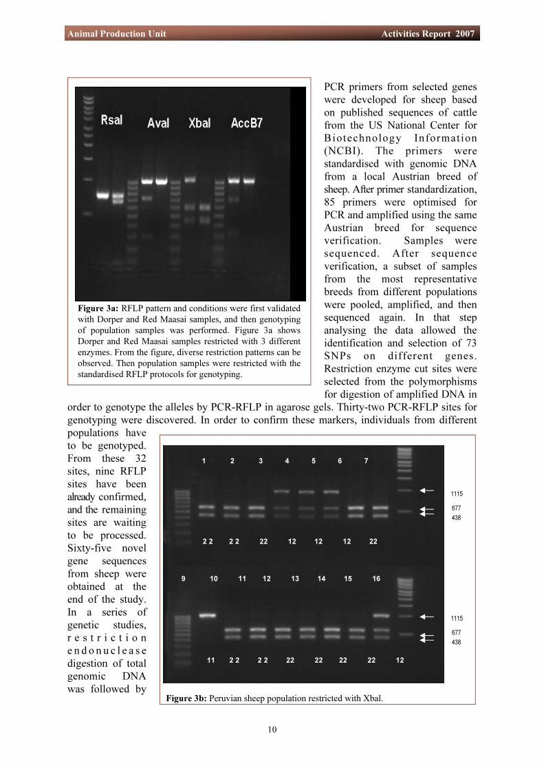
PCR primers from selected genes were developed for sheep based on published sequences of cattle from the US National Center for Biotechnology Information (NCBI). The primers were standardised with genomic DNA from a local Austrian breed of sheep. After primer standardization, 85 primers were optimised for PCR and amplified using the same Austrian breed for sequence verification. Samples were sequenced. After sequence verification, a subset of samples from the most representative breeds from different populations were pooled, amplified, and then sequenced again. In that step analysing the data allowed the identification and selection of 73 SNPs on different genes. Restriction enzyme cut sites were selected from the polymorphisms for digestion of amplified DNA in
order to genotype the alleles by PCR-RFLP in agarose gels. Thirty-two PCR-RFLP sites for genotyping were discovered. In order to confirm these markers, individuals from different populations have to be genotyped. From these 32 sites, nine RFLP sites have been already confirmed, and the remaining sites are waiting to be processed. Sixty-five novel gene sequences from sheep were obtained at the end of the study. In a series of genetic studies, r e s t r i c t i o n e n d o n u c l e a s e digestion of total genomic DNA was followed by
Figure 3a: RFLP pattern and conditions were first validated with Dorper and Red Maasai samples, and then genotyping of population samples was performed. Figure 3a shows Dorper and Red Maasai samples restricted with 3 different enzymes. From the figure, diverse restriction patterns can be observed. Then population samples were restricted with the standardised RFLP protocols for genotyping.
Figure 3b: Peruvian sheep population restricted with Xbal.
9 10 11 12 13 14 15 16
11 2 2 2 2 22 22 22 22 12
1 2 3 4 5 6 7
2 2 2 2 22 12 12 12 22
1115
677438
1115
677438

11
Animal Production Unit Activities Report 2007
hybridization with a labelled probe (short-lived radioisotopes) which reveals differently-sized hybridising fragments and is a form of polymorphism.
3.1.1.3. Creating a DNA Gene Bank at the Agency’s Laboratory for Use by Member States
An objective for several current and planned Technical and Coordinated Research Projects in the Animal Production and Health Programme is to transfer the technical capacity for DNA analysis and marker assisted selection to Member States (MS). In order to identify
Single Nucleotide Polymorphisms (SNP) for helminth resistance in sheep and to develop assays to detect these, each SNP needs to be verified in different populations. Consequently, it is important to develop a gene bank for small ruminants. Arrangements were made to acquire samples of blood and DNA from representative breeds from different populations. Different methods for transportation of blood and DNA samples to the FAO/IAEA Laboratory have been evaluated. Blood samples were sent in two ways; blood mixed with Magic buffer in a vacuette tube, and blood
deposited on Whatman FTA Classic cards (Figure 4). Both methodologies allow the long-term storage of the blood sample. Each MS was assigned with material for blood collection using both methodologies, and samples from indigenous breeds were isolated and stored. These data were entered into a Genetic Resource Database (GR-db) that was created specifically for this purpose. Presently, this bank contains over 700 samples representing 32 breeds of ruminant from 12 countries.
Figure 4: The two methods for transportation of blood samples to the FAO/IAEA Laboratory. On the left, collection of blood mixed with Magic buffer and on the right, blood collection using Whatman FTA Classic cards.

12
Animal Production Unit Activities Report 2007
3.2.2. Database of Genetic Resources (GR-db) for the DNA Bank and Gene Profiling
3.2. Bioinformatics and Genomics
APU has worked extensively on the development of an RT-db (Real-Time database) for Quantitative Trait Loci (QTL)/Genes/DNA Sequences and Genetic characterization in small ruminants. The database will make available the genomic locations of QTL from all known studies on small ruminants. It will allow users to view graphically the positions of the QTL, filtered according to a number of criteria, such as trait name, chromosome number, and statistical significance. RT-db will be the first step in providing the MS with up-to-date genomic information on small ruminants in a user-friendly format. The database is now available online and is fully functional.
3.2.1. Development of Real-Time Databases (RT-db) for Genetic Information on Small Ruminants
The DNA samples held at APU have all been assigned unique IDs and a Genetic Resources database (GR-db) has been created for this purpose (see right). In addition, all genes standardized, sequenced and genotyped have been included in the database (Figure 5).

13
Animal Production Unit Activities Report 2007
Figure 5: Example of an entry into the GR-db.
.
PCR and RFLP profile
Gene Profile
3.3. Animal Health 3.3.1. Use of Non Structural Protein (NSP) of Foot and Mouth Disease Virus to Differentiate Between Vaccinated and Infected Animals Foot and Mouth Disease (FMD) is a highly contagious viral disease of ruminants and swine, responsible for large economic loss in susceptible cloven-hoofed animals. As a transboundary disease FMD excludes infected countries from international trade. In many countries where FMD is endemic, it is controlled through routine vaccination using a killed vaccine, whereas in disease free countries the policy most commonly implemented is one of strict animal movement control and the slaughter of infected animals and contact animals whenever outbreaks occur. However, this “stamping out” policy is less and less accepted by the general public in developed countries. Discussions of a “vaccinate-to-live” policy for the control of FMD in Europe are an ongoing issue to minimize large scale culling of animals at risk. However, it is well known that animals vaccinated with this killed vaccine can become sub-clinically infected and transmit virulent FMDV. Thus in “vaccinate-to-live” policy, it is necessary to detect and remove all infected animals, whatever their vaccination status, in order to recover FMD-free country status. For differentiation from other diseases clinically similar to FMD, laboratory diagnosis is always required to confirm suspected FMD cases. Currently, FMD diagnosis is done by virus isolation, demonstration of FMD viral antigen or nucleic acid in samples and detection of anti-FMDV antibodies in the serum of animals. For this serological diagnosis, there is the classical virus neutralisation test (VNT) which is being replaced by enzyme linked immunosorbent assay (ELISA).

14
Animal Production Unit Activities Report 2007
3.3.1.1. Enhancement of Stability and Robustness of the ELISA by Using a Recombinant Mutated Non-Structural FMDV Protein
Figure 6b: Non-infected, negative control insect cells.
The advantage of ELISA over VNT is that it is faster, does not require cell cultures and can be performed with inactivated antigens, which are safer to work with. One of the current ELISAs is based on the use of the FMD virus non-structural polyprotein (NSP) 3AB or 3ABC as antigen. This NSP-based ELISA test can differentiate infected animals from those that have been vaccinated with purified killed vaccines, vaccines which do not contain the non-structural protein and therefore will not induce anti-NSP antibodies in recipient animals. In the past two years an indirect ELISA (iELISA) and a competitive ELISA (cELISA) have been developed at the APU, based on an FMD recombinant protein (3ABC NSP) produced in vitro in E.coli. These ELISAs allow the differentiation between truly infected animals and vaccinated or non-infected animals (DIVA). However, the FMDV 3ABC is a protease and induces its own self processing, a process that can affect the functional stability of the test. In order to develop a stable, more robust test, it was decided therefore to attempt to improve the stability of this antigen.
To enhance robustness of the FMDV NSP-based ELISA, the work focussed on increasing stability of the recombinant protein used in the test. For this purpose two DNAs ( 3ABC s i t e _ p r o _mu t , a n d 3ABC_mut_optG) corresponding to the FMDV 3ABC gene were synthesised but including 5 mutations in order to inactivate the active protease site of the protein and the four cleavage sites. The codons of one of the mutated genes were optimized for better protein p roduc t ion in insec t ce l l s
(3ABC_mut_optG). A third gene, corresponding to the normal gene, was also synthesized and r ep re sen t s t he w i ld - type (3ABC_WT_Ge) for a control. All three genes were introduced into the baculovirus vector genome for expression in insect cells SF21. Figures 6a and 6b illustrate insect cells transfected with the recombinant baculovirus DNA and control cells. The shape of the transfected cells (Figure 6a) is different from the non-transfected
cells (Figure 6b), indicating the cytopathic effect (cpe) of the baculovirus.
Figure 6a: Insect cells infected with an FMD synthetic gene, 3ABC site_pro_mut.

15
Animal Production Unit Activities Report 2007
The findings were confirmed by Indirect Immunofluorescence (IIF) using the monoclonal antibody 1E6, which is the competing antibody in the cELISA. Detection was done with an anti-mouse FITC-conjugated antibody. 2x10⁴ SF21 insect cells per well were used for infection (Figures 7a, b). Protein detection and analysis was done after infected cells protein separation by electrophoresis on SDS-PAGE and Western Blot. These fractioned proteins were stained directly with Coomassie Blue
(Figure 8) and thereafter blotted onto a nitrocellulose membrane for Western Blot (Figure 9). For the identification of the recombinant 3ABC NSP produced in insect cells and mapping of the B-epitopes recognized by the monoclonal antibody 1E6-11 Western blot analysis were done. The 3ABC NSP was detected with the same mAb (1E6-11) that is in use in the cELISA. The protein
was detected on the membrane by using the ECL kit according to the manufacturer’s manual. The results of the Western blot show clearly that the recombinant protein expressed from mutated synthetic gene 3ABC_mut_optG, which includes five mutations in order to inactivate the active protease site in the C part of the protein and the four cleavage sites, is not degraded by the proteolytic process. In addition, this gene is optimized for insect cells thereby ensuring optimal protein expression. In contrast, the wild-type gene, corresponding to the normal gene, shows many bands of degraded protein, together with a strong band at 50kDa, the size of the specific 3ABC NSP. The last three lanes show different recombinant protein bands from the same 3ABC_site_pro_mut gene. This gene has the same mutations as 3ABC mut_optG, but is not optimized for insect cells. The western blot detects many bands of degraded protein.
Figure 7a: IF staining of infected insect cells/synthetic gene 3ABC_mut_optG optimized for insect cells.
Figure 7b: IF staining of infected insect cells synthetic gene 3ABC_wild type.
1 2 3 4 5
50 kDa
Figure 8: SDS-gel stained with Coomassie Blue. Lane 1: Protein Molecular Weight Marker MagicMark XP; lane 2: 3ABC_mut_optG (synthetic gene optimized for insect cells); lane 3: SF21 non-infected cells; lane 4: empty; lane 5: 3ABC_WT_Ge (Wild-type).

16
Animal Production Unit Activities Report 2007
Another technique was performed to characterize the recombinant proteins that were produced using a radioactive isotope marker for detection purposes to increase sensitivity of detection. For that purpose recombinant baculovirus infected and non-infected-cells were grown in medium containing the radioactive isotope 35S labelled methionine. After 24 hours of labelling, the cells were lysed and the proteins were immunoprecipitated with an t i -FMDV monoc lona l antibody.
The immunoprecipitation products were analysed by electrophoresis on gel which was dried and s u b m i t t e d t o autoradiography. The results, presented in Figure 10, show a clear single band with synthetic gene 3ABC_mut_optG, while the wild-type shows again several bands of d e g r a d e d p r o t e i n (Figure 10, lane 4). This confirms the result obtained with the Western Blot analysis. (see above).
220 120
80 60 50 40
30
20
1 2 3 4 5 6 7
Figure 9: Western blot analysis. Lane 1: Protein Molecular Weight Marker MagicMark XP; lane 2: 3ABC_mut_optG (synthetic gene optimized for insect cells); lane 3: SF21 non-infected cells/negative control; lane 4: 3ABC_WT_Ge (Wild type); lane 5: 3ABC site pro mut (synthetic gene non-optimized for insect cells) sample 1; lane 6: 3ABC site pro mut - sample 2; lane 7: 3ABC site pro mut - sample 5.
50 kDa
1 2 3 4 5
Figure 10: Immunoprecipitation method: Lane 1: 3ABC_mut_optG (synthetic gene optimized for insect cells) without Mab 1E6-11; lane 2: SF21 non-infected cells/negative control without Mab 1E6-11; lane 3: 3ABC_mut_optG (synthetic gene optimized for insect cells) with Mab 1E6-11; lane 4: 3ABC_WT_Ge (Wild type) with Mab 1E6-11; lane 5: 3ABC site pro mut - sample 2.

17
Animal Production Unit Activities Report 2007
Table 1: Summary of conditions used to optimize the separation of recombinant FMDV NSP
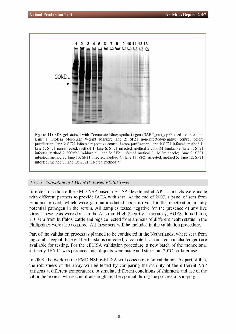
The NSPs, expressed in insect cells after infection with the recombinant baculovirus virus of each of the three synthetic FMDV NSP genes, were purified with a new automated purification system, which is based on the use of paramagnetic precharged nickel particles to bind poly-histidine tagged protein. All three synthetic genes were designed in a way to add six histidine residues at the C-terminal end of the recombinant proteins. Seven different buffer conditions were compared with each other. Starting with the buffers in the kit, they were modified by increasing the concentration of imidazole in order to improve conditions for purification of the recombinant histidine-tagged NSP. Best results were achieved with APU buffers at an Imidazole concentration of 1M for elution as shown in methods 2 and 4 using 1M Imidazole (Table 1). This procedure resulted in a semi-pure product with minor contaminants that was suitable for use in the cELISA. The table below shows the different parameters and buffer compositions that were used.
3.3.1.2. Production and Purification of Recombinant FMDV NSP from the Baculovirus Expression System
18
Animal Production Unit Activities Report 2007
3.3.1.3. Validation of FMD NSP-Based ELISA Tests
1 2 3 4 5 6 7 8 9 10 11 12 13
50kDa
Figure 11: SDS-gel stained with Coomassie Blue; synthetic gene 3ABC_mut_optG used for infection. Lane 1: Protein Molecular Weight Marker; lane 2: SF21 non-infected=negative control before purification; lane 3: SF21 infected = positive control before purification; lane 4: SF21 infected, method 1; lane 5: SF21 non-infected, method 1; lane 6: SF21 infected, method 2 250mM Imidazole; lane 7: SF21 infected method 2 500mM Imidazole; lane 8: SF21 infected method 2 1M Imidazole; lane 9: SF21 infected, method 3; lane 10: SF21 infected, method 4; lane 11: SF21 infected, method 5; lane 12: SF21 infected, method 6; lane 13: SF21 infected, method 7;
In order to validate the FMD NSP-based, cELISA developed at APU, contacts were made with different partners to provide IAEA with sera. At the end of 2007, a panel of sera from Ethiopia arrived, which were gamma-irradiated upon arrival for the inactivation of any potential pathogen in the serum. All samples tested negative for the presence of any live virus. These tests were done in the Austrian High Security Laboratory, AGES. In addition, 310 sera from buffalos, cattle and pigs collected from animals of different health status in the Philippines were also acquired. All these sera will be included in the validation procedure. Part of the validation process is planned to be conducted in the Netherlands, where sera from pigs and sheep of different health status (infected, vaccinated, vaccinated and challenged) are available for testing. For the cELISA validation procedure, a new batch of the monoclonal antibody 1E6-11 was produced and aliquots were made and stored at -20°C for later use. In 2008, the work on the FMD NSP c-ELISA will concentrate on validation. As part of this, the robustness of the assay will be tested by comparing the stability of the different NSP antigens at different temperatures, to simulate different conditions of shipment and use of the kit in the tropics, where conditions might not be optimal during the process of shipping.
19
Animal Production Unit Activities Report 2007
Therefore one focus of the work was the improvement of stability of the recombinant protein. This was addressed by including five mutations in order to inactivate the active protease site in the C part of the protein and the four cleavage sites. In addition the codons of one of the mutated genes were optimized for better protein production in insect cells (3ABC_mut_optG). These three proteins will be compared in a mid- and long-term stability study that will be initiated in 2008. The most stable product will be used for antigen production and use in the ELISA. 3.4. The Control of Contagious Bovine Pleuropneumonia (CBPP) in Sub-Saharan
Africa ¹
3.4.1. Expression and Purification of Recombinant CBPP LppQ Protein
CBPP is a highly contagious disease, caused by Mycoplasma mycoides subsp. mycoides. It was eradicated from many countries by the end of the 20th century but the disease persists in many parts of Africa while the situation in Asia is unclear². CBPP is a disease of major economic concern in affected countries because of the restrictions in cattle trade. Eradication of CBPP is problematic, because of the frequent occurrence in animals of sub-acute or asymptomatic infections and the persistence of infection in animals that remain as carriers after the clinical phase of the disease. Serological diagnostic tools are extremely important for the implementation of an effective disease control policy. The complement fixation test remains the prescribed test for international trade even though it has significant limitations regarding diagnostic sensitivity and specificity. The cELISA was designated as an alternative test by the OIE International Committee in May 2000 and as an OIE prescribed test for international trade in May 2004. In addition, an immunoblotting test has undergone evaluation and is reported to be highly specific and sensitive. The APU is involved in the development of an ELISA test using the lipoprotein LppQ, which is specific to Mycoplasma mycoides subsp. mycoides, as antigen. This recombinant protein was expressed in E.coli, purified and characterized in-house and then introduced as antigen to set up an iELISA for the diagnosis of antibodies against CBPP in cattle.
¹ CRP D3.20.24 ² Thiaucourt, F. (2004), Contagious Bovine Pleuropneumonia, Chapter 2.1.6, In “Manual Of Diagnostic Tests and Vaccines for Terrestrial Animals”, Organisation Mondiale de la Sante Animale, Paris, France
For the expression of the recombinant protein, the bacteria E.coli BL21 (DE3) harbouring a plasmid into which the LppQ protein gene has been cloned. These bacteria were provided by Professor Joachim Frey of the University of Berne, Switzerland. The construction of this recombinant protein was done by adding a poly-histidine tag at the N-terminal end of the protein to facilitate its purification. The bacteria were grown in the recommended medium and the production of the recombinant protein was induced by adding 1mM isopropylthiogalactoside (IPTG) to the culture medium. Three hours post-induction, the bacteria were harvested, washed with TES and submitted to protein purification. This was performed under denaturing conditions by adding 8M Urea and using the MagneHis Protein Purification system (Promega). This purification was optimized in APU by using the following conditions:

20
Animal Production Unit Activities Report 2007
- the wash buffer contained 1M Hepes, 20 mM Imidazole, 1M NaCl, 8M Urea and a cocktail of protease inhibitors, pH 7.5. - the elution buffer was of 500 mM Imidazole and 8 M Urea. Under these conditions two batches of antigen, #040 707 and #311 007, were produced. The purified product was submitted to electrophoresis by SDS-PAGE and Western blot analysis. Figure 12 shows the results when the purification kit is used without optimizing the buffer conditions. The protein is lost during the washing steps, while the elution does not contain the protein, either because it is lost during previous working steps and/or not entirely eluted from the beads. Figure 13 shows the results of purification of CBPP antigen under optimized buffer conditions and an optimized ratio between magnetic beads and lysate. Four different approaches were compared with each other, using 50 µl, 100 µl, 200 µl and 400 µl of his-tagged magnetic beads for 1ml lysate at 1 OD at 600 nm. Best reactions were achieved using 50 µL magnetic beads for 1 ml lysate. Therefore the remaining protein was purified using 50 µL beads for 1 ml lysate. This purified protein was used as antigen in the iELISA.
80kDa 60kDa 50kDa 40kDa
30kDa
20kDa
10kDa
~48kDa
~28kDa
1 2 3 4 5 6
Figure 12: LppQ protein before and after purification under non optimized condition, SDS-Page/gel after Coomassie Blue staining. lane 1: His tagged protein ladder; lane 2: E.coli-negative control; lane 3: LppQ expressed in E.coli – before purification; lane 4: 1st wash; lane 5: 2nd wash; lane 6: Elution 1.
50µL beads 100µL beads 200µL beads 400µL beads
1 2 3 4 5 6 7 8 1 2 3 4 5 6 7 8 1 2 3 4 5 6 7 8 1 2 3 4 5 6 7 8
Figure 13: LppQ protein before and after purification using different ratios between magnetic beads and E.coli lysate. lane 1: His tagged protein ladder; lane 2: E.coli-negative control; lane 3: LppQ expressed in E.coli – before purification; lane 4: 1st throughput; lane 5: 1st wash; lane 6: 2nd wash Lane 7: 1st elution; Lane 8: 2nd elution.

21
Animal Production Unit Activities Report 2007
Western Blot analysis was used for the identification of the recombinant LppQ antigen recognized by the polyclonal anti-LppQ antiserum. The protein was cleaved by proteases and only one fragment around 30 kDa was detected by Western Blot. The recombinant LppQ fragment was detected on the nitrocellulose membrane by using the ECL kit and according to the manufacturer’s instructions. The results are shown in Figure 14.
Figure 14: Western Blot analysis with recombinant purified LppQ protein reacting with polyclonal rabbit anti-serum, HRP A2074. Lane 1: Protein Molecular Weight Marker MagicMark XP; lane 2: E.coli negative control; Lane 3: LppQ protein-purified.
3.4.2. Development of a Prototype iELISA to Detect Antibodies against CBPP in Cattle
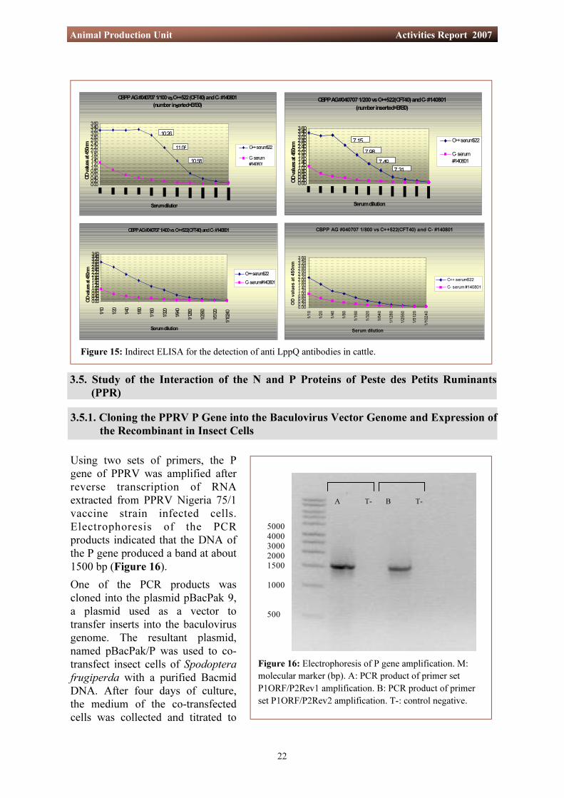
The purified LppQ protein was adjusted to 1mg/ml and coated in bicarbonate buffer pH 9.6 onto Immunolon 1B, Polysorp and Maxisorp ELISA plates. Coating step, serum incubation and conjugate (rabbit anti-bovine IgG (whole molecule)− horse radish peroxidase (HRP), Sigma 8917) incubation were done throughout at 37°C in an orbital ELISA shaker for 1 hour. Only the substrate incubation (TMB/H2O2) was done for 15 minutes at 37°C while shaking. Serum and HRP diluent buffer was prepared from 0.01M PBS containing 0.05% Tween 20 plus 5% skimmed milk. The best results were achieved with Immulon 1B and Polysorp plates. Maxisorp plates could not be used because of the unacceptably high background they produced. The results are shown in Figures 15a and b with antigen at dilutions of 1/100, 1/200, 1/400 and 1/800. The serum was applied in 2-fold dilutions. Commercial HRP from Sigma was used at 1/30 000. The reaction was stopped with 1M H3PO4 after 15 minutes of incubation at 37°C. Best results were achieved using Immulon 1B microtitre plates, an antigen dilution of 1/100 and HRP dilution 1/30 000 using test parameters described above. For 2008 it is planned to achieve a reduction in background and a final adjustment of the assay parameters. Background reduction is foreseen by testing a panel of commercial blocking buffers. This will enhance the Binding Ratio (B/B0) thus improving the discrimination between positive and negative samples. In addition a number of known negative and positive sera will be tested to provide information on the diagnostic sensitivity and specificity of the test.
20kD
30kD
40kD50kD
1 2 3
22
Animal Production Unit Activities Report 2007
3.5. Study of the Interaction of the N and P Proteins of Peste des Petits Ruminants (PPR)
3.5.1. Cloning the PPRV P Gene into the Baculovirus Vector Genome and Expression of the Recombinant in Insect Cells
CBPP AG #040707 1/100 vs C++522 (CFT40) and C- #140801
(number inserted=B/B0)
0.000.200.400.600.801.001.201.401.601.802.002.202.402.602.803.003.203.403.60
Serum dilution
OD va
lues a
t 450
nm C++ serum522
C- serum#140801
10.26
11.06
10.58
CBPP AG #040707 1/200 vs C++522(CFT40) and C- #140801(number inserted=B/B0)
0.000.200.400.600.801.001.201.401.601.802.002.202.402.602.803.003.203.403.60
Serum dilution
OD va
lues a
t 450
nm
C++ serum522
C- serum#140801
7.987.49
7.31
7.15
CBPP AG #040707 1/400 vs C++522(CFT40) and C- #140801
0.000.200.400.600.801.001.201.401.601.802.002.202.402.602.803.003.203.403.60
1/10
1/20
1/40
1/80
1/160
1/320
1/640
1/1280
1/2560
1/5120
1/10240
Serum dilution
OD va
lues a
t 450
nm C++ serum522C- serum #140801
CBPP AG #040707 1/800 vs C++522(CFT40) and C- #140801
0.000.200.400.600.801.001.201.401.601.802.002.202.402.602.803.003.203.403.60
1/10
1/20
1/40
1/80
1/16
0
1/32
0
1/64
0
1/12
80
1/25
60
1/51
20
1/10
240
Serum dilution
OD v
alue
s at
450
nm
C++ serum522C- serum #140801
Figure 15: Indirect ELISA for the detection of anti LppQ antibodies in cattle.
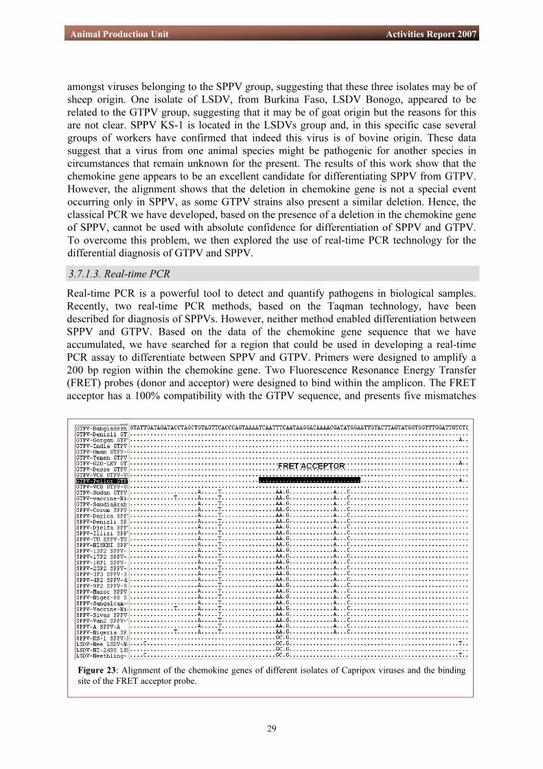
Figure 16: Electrophoresis of P gene amplification. M: molecular marker (bp). A: PCR product of primer set P1ORF/P2Rev1 amplification. B: PCR product of primer set P1ORF/P2Rev2 amplification. T-: control negative.
Using two sets of primers, the P gene of PPRV was amplified after reverse transcription of RNA extracted from PPRV Nigeria 75/1 vaccine strain infected cells. Electrophoresis of the PCR products indicated that the DNA of the P gene produced a band at about 1500 bp (Figure 16). One of the PCR products was cloned into the plasmid pBacPak 9, a plasmid used as a vector to transfer inserts into the baculovirus genome. The resultant plasmid, named pBacPak/P was used to co-transfect insect cells of Spodoptera frugiperda with a purified Bacmid DNA. After four days of culture, the medium of the co-transfected cells was collected and titrated to
5000 4000 3000 2000 1500 1000 500
A T- B T-

23
Animal Production Unit Activities Report 2007
Figure 17: P protein detection by Western Blot. M: protein molecular weight (Kda), SF21: non-infected control cells, 965: Clone expressing P protein.
3.5.2. Mapping N Binding Site on P It was decided also to study these interactions by mapping the binding site of N on P. The assay based on the protein capture by peptides was successfully used last year to study the N and M interactions. The same methodology was used to map the N binding site on P. For this study, fifty overlapping synthetic peptides covering the P protein were used to identify the amino acids of P which reacted with the N protein. Ninety-six-well NUNC Immobilizer amino microtitre plates were coated with 500 ng of each peptide for 4 hr at 37°C. Unbound material in the plate was then removed by washing three times with PBS-Tween 20 buffer (0.05%) washing buffer and the plates were incubated with N protein diluted in blocking buffer (PBS+ 0.5% Tween 20 + 2.5% skimmed milk) and incubated overnight at 4°C with continuous agitation. The plates were washed once more to remove unbound N protein and then incubated with anti-N monoclonal antibody (P4G5) for 1 hr at 37°C with agitation. Unbound P4G5 was removed by washing three times with washing buffer. The bound protein was detected by using anti-mouse immunoglobulin conjugated to horseradish peroxidase diluted 1:1000 in blocking buffer. One hundred µl of this diluted conjugate was added to the plate and incubated for 1 hr at 37°C with constant agitation. The plates were washed and 100 µl of SureBlue Tetramethylbenzidine peroxidase substrate was added to each well and colour reaction was developed for 5 min at 37°C. The reaction was stopped with 100 µl of 1M H2SO4 and the optical density (OD) readings were taken at 490 nm by a computer-interfaced ELISA plate reader (Figure 18).
detect the presence of virus. Five colonies of virus were collected, expanded and tested for the presence of the recombinant P protein. For this, infected cells were lysed at three days post-infection. The total proteins were analysed by gel electrophoresis (SDS-PAGE), and transferred onto a 0.2 µm PVDF membrane by Western Blot technique. The presence of the P protein was visualised by using a monoclonal antibody (mAb) specific for the P protein of the measles virus (MV) and which had been shown previously to cross react with the P proteins of PPRV, canine distemper virus (CDV) and rinderpest virus (RPV). Figure 17, shows that the recombinant baculovirus/PPRV P clone expresses the PPRV P protein in infected cells. In 2008, this recombinant virus will be used in co-infection with the recombinant baculovirus PPRV full length and deleted N proteins to study the interactions of N and P.
Measles Anti-P Monoclonal antibody
965
80 60 50
40
30
20
SF21 M

24
Animal Production Unit Activities Report 2007
The ELISA test showed that the N protein can interact with several peptides of the P protein. Based on the optical density readings, these peptides can be grouped into major and minor interactive domains. The major interaction domain corresponds to the peptides TVTECSSISGATQAVPESRW from amino-acid 241 to 260. Several minor interactive domains are distributed through the sequence of P protein from N-terminus to C-terminus. The different interactive sites of N protein on P protein are located in a variable area of the P protein according to the comparison of the sequence in Morbillivirus group (Figure 19). This result may explain why homologous N and P work better in a minigenome system than heterologous N and P between Morbilliviruses.
Figure 18: Interaction between N protein and peptides of P protein. The degree of binding to the N protein by different peptides (numbered) is indicated by the peaks in optical density.

25
Animal Production Unit Activities Report 2007
Figure 19: Alignment of the P protein amino-acid sequences in the Morbillivirus group and identification of the areas interacting with the N protein.

26
Animal Production Unit Activities Report 2007
All ELISA tests developed so far for the serological diagnosis of Morbillivirus infections are based on the use of either the whole virus nucleoprotein (N) or the haemagglutinin (H) as target antigens. The advantage of the nucleoprotein is that it is the most abundant viral protein and the majority of antibodies produced by the host against the virus are directed against this protein. Therefore, a test based on this antigen is expected to be very sensitive. Unfortunately, the two PPR N-based cELISA assays show cross-reactions with rinderpest sera. Another PPR cELISA in use and based on the detection of anti-haemagglutinin antibody also shows cross reactivity. However, a similar test that is used for the diagnosis of rinderpest has proven to be highly specific. With this success in mind, it was decided to produce more anti-PPR H protein monoclonal antibodies (mAb) and explore their use in developing a specific PPR cELISA. To accomplish this, a pCineo plasmid with the cloned PPRV H gene cDNA insert, constructed at CIRAD, was sent to APU to immunize three mice. After three boosts, only one mouse was found to be producing anti-H antibodies according to the results of an indirect fluorescent antibody test. This mouse was used to produce hybridomas after fusion of its spleen cells with myeloma cells. From that fusion, only two clones were identified that were producing anti-PPR H antibodies. In order to increase the number of hybridomas, another strategy for immunizing the mice was adopted. This involved co-inoculation of the pCineo/PPR-H plasmid with another pCineo with an immunostimulant gene, the GM-CSF. Forty micrograms of both plasmids were inoculated into the spleens of three mice. Five days after immunization, one of the mice was euthanized and its spleen cells were collected and fused with myeloma cells. Thirty-six hybridomas were obtained. The screening of the culture medium of those cells by ELISA have shown that six, or as many as nine, of them were producing anti-HPPRV mAb (Figure 20). These results, obtained only five days after a single inoculation of the plasmids, are encouraging and further studies will be undertaken to better characterize these mAb. The two remaining mice will be boosted and their spleen cells used for the production of other hybridomas.
3.6. Development of a PPR-Specific Serological Diagnostic Test: Production of New Monoclonal Antibodies
Figure 20: Screening of anti-PPRV H protein mAbs.
0
0,1
0,2
0,3
0,4
0,5
0,6
0,7
0,8
0,9
1
A1 A2 A4 A7 A9 A10
A11
A12 B1 B4 B5 B6 B7 B8 B10
B11
B12 C2 C6 C7 B2 B9 C1 C3 C8 C9 A3 A6 B3 C4 C5
MAB
B11
MAB
F3
P4G5
anti-
N
OD
Hybridomas
Screening of Hybridomas: Fusion from mouse immunisation with Hppr1. Coating the plate with Vero cells lysat intected with PPRV vaccine. 2. Washing the plate and blocking with PBS-T+5% Milk at 37 C.3. Add 100 ul surpernatant of the hybridomas.4. detection of Mab with anti-mouse.The MAB B11, MABF3 are anti H produced during a first fusion. The Mab P4G5 is an anti PPRV nucleoprotein

27
Animal Production Unit Activities Report 2007
3.7. Development of Capripox Virus Diagnostic Tests and Molecular Epidemiology of the Disease
Goat poxvirus (GTPV) and sheep poxvirus (SPPV) are responsible for economically important diseases in goats and sheep. These diseases are included the World Organization for Animal Health’s (OIE) list of notifiable diseases. Together with Lumpy Skin Disease Virus (LSDV), GTPV and SPPV form the genus Capripoxvirus (CaPV) of the Poxviridae family. SPPV and GTPV are endemic in Africa north of the equator, the Middle East, Central Asia and the Indian Subcontinent where they are associated with serious losses in productivity. Diseases caused by the two viruses are indistinguishable clinically and the various strains, including LSDV, cannot be differentiated serologically. There are many conflicting reports on whether SPPV can induce disease in goats and GTPV in sheep. Since 2005, in collaboration with partners in France and Africa, APU has been embarked on a project whose aims are to develop specific diagnostic tests for capripox, a safer vaccine and molecular and nuclear-based tools to apply in studies to enable better understanding of the epidemiology of these viruses and thereby make it possible to devise appropriate strategies for controlling the diseases. For the work at APU, several Capripoxvirus samples from sheep and goats were obtained from Africa (Algeria, Mali, Cameroon and Sudan), Turkey and the UK. This was made possible through an agreement between the IAEA and the Austrian Agency for Health and Food Safety (AGES) who received, stored and handled all these infectious materials at the High Security Laboratory of the Institute for Veterinary Disease Control. 3.7.1. Development of Molecular-Based Diagnostic Tests for the Identification of
Capripox Virus Differentiating CaPVs is important because the same virus may infect goat, sheep or cattle, but with possible differences in pathogenicity according to the origin of the virus. For example, goat strains are more pathogenic in goat compared with their effect in sheep and vice versa. Currently, the few molecular methods used for strain differentiation that have been published are based on restriction enzyme digestion of either the full virus genome or an amplified DNA product. One of the objectives of the project is to develop a more simple molecular-based method to differentiate GTPV from SPPV. 3.7.1.1. Classical PCR Inspection of the sequences available in the public gene sequence database on the alignment of the G protein-coupled receptor gene (known as the chemokine gene) revealed a 21 bp deletion in SPPV sequences in comparison with those of GTPV. It was therefore expected that the presence of this deletion could be utilised in a classical PCR to differentiate SPPV from GTPV. Primers were designed to amplify a region containing the deletion to give an amplicon of 209 bp for GTPV and 188 bp for SPPV. Indeed, as shown in Figure 21, gene amplification using these primers did enable distinction of CaPV strains possessing the deletion in their chemokine gene (21a) from those without it (Figure 21b).

28
Animal Production Unit Activities Report 2007
However, when this PCR was evaluated using the panel of 25 isolates received from our counterparts, some isolates from clinically diseased goats, putatively identified as GTPVs, were found to present the same amplicon (188 bp) as that seen in SPPVs (Figure 21b). Therefore, it is possible that some goat poxviruses may also have a deletion in their chemokine gene. To further validate this hypothesis, we proceeded to amplify, clone and sequence genes of several different CaPVs.
Figure 21: Classical PCR for GTPV and SPPV; a: SPPV and GTPV PCR differing in their length due to a deletion in SPPV chemokine gene; b: GTPV presenting similar amplicon in length with SPPV.
3.7.1.2. Cloning the Chemokine Gene of Sheep and Goat Pox Viruses: Molecular Epidemiology
Primers were designed and optimized to amplify the full chemokine gene of CaPVs. The PCR fragments obtained were then cloned into a Promega pGEM-T vector system. Ligation products were transformed in bacteria Top 10 competent cells. The recombinant plasmids were purified and the sequence of the insert was determined using a cycle sequencing reaction. The data o b t a i n e d w e r e compared with those available from our CIRAD partner in France. They were a n a l y z e d u s i n g appropriate software to determine the genetic relationship between the different viruses. A phylogenetic tree that wa s c on s t r u c t e d showed that SPPV, GTPV and LSDV clustered separately ( F i g u r e 2 2 ) . Nevertheless, GTPV Sudan, GTPV Saudi Arabia and GTPV vaccine Nigeria were found to be located
a) b)
GTPV-Bangladesh GTPV-India GTPV-Oman
GTPV-Denizli GTPV-Gorgan GTPV-G20-LKV (AY077836) GTPV-Pellor (AY077835) GTPV-Yemen GTPV-VC8 GTPV-VC6 LSDV-Benogo-3A GTPV-Desse I
GTPV
LSDV-Neethling-LW-1959 LSDV-Nee (AF336128S1)
LSDV-Niger-ibetecene LSDV-Niger-Tougounous LSDV-NI-2490 (AF325528) LSDV-Banfora LSDV-NW-LW (AF409137) LSDV-Vaccine-Nigeria SPPV-KS-1(S78201)
LSDV
SPPV-Sangalcam-88 SPPV-Darica SPPV-Denizli SPPV-Corum SPPV-Sivas GTPV-Sudan SPPV-Van2 SPPV-NISKHI SPPV-NISKHI (AY077834) SPPV-A GTPV-SaudiaArabia SPPV-3P3 SPPV-18P1 SPPV-Maroc GTPV-vaccine-Nigeria SPPV-Nigeria SPPV-Vaccine-Nigeria SPPV-Djelfa SPPV-Niger-88 SPPV-4P2 SPPV-TU SPPV-TU (AY077832) SPPV-Illizi SPPV-9P2 SPPV-23P2 SPPV-17P2 SPPV-13P2
SPPV
Deer-poxvirus-W-848-83 Swine-poxvirus(L21931)
Camel-poxvirus-M-96
77
76
60
56
43100
86
6447
100
100
100
9699
99
99
89
100
62
0.05
Figure 22: Phylogenetic tree generated from the alignment of the chemokine genes of 46 Capripox viruses. In addition to the sequence data from CIRAD, eight CaPV sequences available in the public database were added to the analysis. Also, examples of Deer poxvirus, Swine poxvirus and Camel poxvirus sequences were used as out groups.
29
Animal Production Unit Activities Report 2007
Real-time PCR is a powerful tool to detect and quantify pathogens in biological samples. Recently, two real-time PCR methods, based on the Taqman technology, have been described for diagnosis of SPPVs. However, neither method enabled differentiation between SPPV and GTPV. Based on the data of the chemokine gene sequence that we have accumulated, we have searched for a region that could be used in developing a real-time PCR assay to differentiate between SPPV and GTPV. Primers were designed to amplify a 200 bp region within the chemokine gene. Two Fluorescence Resonance Energy Transfer (FRET) probes (donor and acceptor) were designed to bind within the amplicon. The FRET acceptor has a 100% compatibility with the GTPV sequence, and presents five mismatches
amongst viruses belonging to the SPPV group, suggesting that these three isolates may be of sheep origin. One isolate of LSDV, from Burkina Faso, LSDV Bonogo, appeared to be related to the GTPV group, suggesting that it may be of goat origin but the reasons for this are not clear. SPPV KS-1 is located in the LSDVs group and, in this specific case several groups of workers have confirmed that indeed this virus is of bovine origin. These data suggest that a virus from one animal species might be pathogenic for another species in circumstances that remain unknown for the present. The results of this work show that the chemokine gene appears to be an excellent candidate for differentiating SPPV from GTPV. However, the alignment shows that the deletion in chemokine gene is not a special event occurring only in SPPV, as some GTPV strains also present a similar deletion. Hence, the classical PCR we have developed, based on the presence of a deletion in the chemokine gene of SPPV, cannot be used with absolute confidence for differentiation of SPPV and GTPV. To overcome this problem, we then explored the use of real-time PCR technology for the differential diagnosis of GTPV and SPPV. 3.7.1.3. Real-time PCR
Figure 23: Alignment of the chemokine genes of different isolates of Capripox viruses and the binding site of the FRET acceptor probe.

30
Animal Production Unit Activities Report 2007
with the SPPV sequence (Figure 23). The donor was labelled at the 3` end with 6'-Carboxy-Fluorescein (6-FAM). The acceptor was labelled at the 5` end with Cy5 and its 3` end was blocked with a phosphate moiety. Preliminary evaluations of this FRET pair failed to yield good fluorescence levels. Therefore we have substituted the normal nucleotides with Locked Nucleic Acid (LNA) at different positions of the FRET donor. Using this approach, a good FRET pair was obtained and optimized for the detection and differentiation of SPPV from GTPV (Figure 24).
Figure 24: Detection of sheep poxvirus and goat poxvirus by FRET real-time PCR. The amplification plots were realized on 10 fold dilutions (107 to 103 copies for sheep poxvirus and 107 to 102 copies for goat poxvirus), assayed in duplicate from plasmid containing the respective chemokine genes.
This real-time PCR readily distinguished individuals of the SPPV group from those of the GTPV using the melting curve Tm analyses after the PCR. The Tm for SPPV is 52°C and that of the GTPV is 69°C (Figure 25).
Figure 25: Melting curve analysis of SPPV and GTPV using FRET real-time PCR.
31
Animal Production Unit Activities Report 2007
T h e a n a l y t i c a l sensitivity of the method for SPPV and GTPV detection was e s t a b l i s h e d b y amplifying samples of different dilutions (107
to 10-2 molecules/µL) of solutions containing plasmids with the SPPV and the GTPV chemokine genes, respectively, as inserts. The assay could detect 25 copies and 250 copies/reaction tube for GTPV and SPPV respectively. The lower sensitivity for SPPV is probably due to the five mismatches that exist between its sequence and those of the FRET acceptor probes. These mismatches were selected to facilitate differentiation between the two viruses. The efficiency, as indicated by the slope of the standard curved obtained by plotting the Cycle Threshold (Ct) values against the Log10 of the known input copy number, is between -3.4 and -3.6 (Figure 26). The next step is to validate this real-time PCR using a panel of field isolates and samples from experimentally-infected animals.
3.8. Sequencing the Capripox Virus Genome
Figure 26: Linearity of the FRET assay. The standard curve is generated by plotting the Cycle Threshold measured in duplicate against the Log of the input copy number.
Goat poxvirus: y = -3.492x + 37.351; R2 = 0.994
Sheep poxvirus: y = -3.602x + 39.116; R2 = 0.999
10
15
20
25
30
35
1 2 3 4 5 6 7 8Log Number of copies
C T Goat poxvirusSheep poxvirus
In endemic areas, the control of SPPV and GTPV is attempted by vaccination campaigns using live, attenuated virus. However, vaccine failures and adverse effects have been reported on several occasions, indicating the need to develop safer and more effective capripox vaccines. Studies with other poxviruses have enabled the identification of genes responsible for pathogenicity. Rational deletion of pathogenic genes, as for example has been achieved in the case of vaccinia, has led to the attenuation of virus infectivity and virulence. In order to understand the molecular basis of capripox virus pathogenicity, and also the host species-specificity, it was decided to sequence the genomes of different capripox virus strains. Later, these viruses will be tested in animals in Africa to determine their virulence in vivo. Using the data obtained from these studies, as well as the information already available in literature, it may be possible to develop a method for the safe and efficient preparation of a capripox vaccine. The capripox virus sequencing work of this project started in 2007 with a goat strain isolated in Turkey, the Denizli strain. This virus was grown in sheep embryonic skin cells (ESH–L). After purification of the virus on linear sucrose gradients (24 to 40% and 28 to 60%), its DNA was extracted and sequenced. The data obtained were assembled into a continuous sequence of 148 103 bp. This was then compared with the sequence of another GTPV available in Genbank and the possible protein coding genes were annotated. Comparison of
32
Animal Production Unit Activities Report 2007
Fifty-three frame shift mutations occurred in GTPV Denizli, leading to the truncation of thirteen genes by 3 to 82 aa at the C terminus end, two genes by 183 aa and 88 aa at N terminus. Two genes were found to have 44 aa and 128 aa insertion at the C terminus in GTPV Denizli. Seventeen genes are fragmented into two or three smaller ORFs (Table 3). Two of the fragmented genes are well conserved within the poxvirus family one being the DNA polymerase gene. Two frameshift mutations occur in this gene resulting in its fragmentation into three smaller ORFs. When considering the nucleotide sequences, the fragment of the GTPV Denizli containing the three smaller ORFs of the DNA polymerase gene has a 99.8% similarity with its GTPV Pellor homologue (3030 vs 3036 nucleotides). The second gene is the equivalent of the DNA binding protein in GTPV Denzli. This gene is fragmented into two smaller ORFs because of two frameshift mutations. Because the DNA polymerase and the DNA binding proteins are know to be well conserved, we will further clone and sequence these two genes from GTPV Denizli in order to confirm whether these frameshift mutations are artifacts or a special future of GTPV Denizli. In the sequencing project, we are planning to sequence nine other CaPV genomes in 2008. Together with the genome sequences available in the gene bank, this will provide more consistent data and lead to better understanding of the genes involved in pathogenicity and host specificity of CaPVs.
Table 2: Characteristics of Genomes of Goat poxvirus Denizli and Goat poxvirus Pellor
the GTPV Denizli genome sequence with that of GTPV Pellor available in the gene bank shows that the two viruses share a 99.8% similarity at the nucleotide level over the length of their genomes. Only 238 genomic changes, including 175 single nucleotide substitutions were found. The nucleotide composition is 74.7% A+T which is identical to that of the GTPV Pellor. In this virus, 150 Open Reading Frames (ORF) were found and annotated as putative protein genes (Table 2). Out of these ORF, 87 are identical in the two viruses. Twenty two genes, seven genes, and six genes present more than 99%, 96 to 99% and 80 to 95% similarity, respectively.
Genome characteristics Goat pox Denizli Goat pox Pellor Length 148 103 149 599 A+T content 74.72% 74.69% Number of ORF 150 150 ORF Size 28aa to 2007aa 28aa to 2007aa

33
Animal Production Unit Activities Report 2007
Goat poxvirus Denzili
Goat poxvirus Pellor (AY077835)
ORF Function prediction and comparison vs Goat poxvirus Pellor
Position (Length aa) ORF Length (aa) Function Type of Change Blastp Score Length (aa % identity) E value
2105>1938(55) GTPV_gp005 355 Hypothetical protein 2 Fragments 123 55/55 (100%) 3.90E-27 3005>2625(126) GTPV_gp005 Hypothetical protein 217 107/107 (100%) 2.00E-55 3893>3432(153) GTPV_gp006 275 Putative soluble interferon gamma receptor Del 40 aa, Del 82 aa C terminal 285 135/135 (100%) 4.50E-76 6926>6291(211) GTPV_gp010 211 Ankyrin repeat protein R40K 438 (210/211) 99.5% 9.00E-122 8739>8275(154) GTPV_gp012 161 Putative interleukin-18 binding protein I7V, R148E, S151V; L152S; R153E; K154S; Del: 155E,
156S,157K,158N,159V,160I 161E 315 (148/150) 98.7% 4.10E-85
9045>8764(93) GTPV_gp013 96 EGF-like growth factor T93K, Del:H94, K95, I96 196 (92/92) 100.0% 3.60E-49 9567>9244(107) GTPV_gp014 176 Putative integral membrane protein Del 69 aa C terminal 209 (104/104) 100.0% 4.20E-53 11777>10089(562) GTPV_gp016 562 Kelch-like protein Q321E 1157 (561/562) 99.8% 0 12258>11842(138) GTPV_gp017 321 Ribonucleotide reductase small subunit 2 Fragments 281 (138/138) 100.0% 1.10E-74 12809>12390(139) GTPV_gp017 Ribonucleotide reductase small subunit 281 (138/138) 100.0% 1.10E-74 13473>13153(106) GTPV_gp019 108 Hypothetical protein DeLY2;N73 213 (106/108) 98.1% 3.80E-54 14622>13972(216) GTPV_gp021 216 Hypothetical protein L123F 436 (215/216) 99.5% 4.70E-121 18098>16863(411) GTPV_gp023 639 Putative EEV maturation protein 2 Fragments 826 (411/411) 100.0% 0 18783>18250(177) GTPV_gp023 Putative EEV maturation protein 362 (177/177) 100.0% 4.00E-99 19044>18790(84) GTPV_gp024 370 Putative palmitylated virion envelope
protein 2 Fragments 176 (84/84) 100.0% 5.20E-43
19901>19698(67) GTPV_gp024 Putative palmitylated virion envelope protein
140 (65/65) 100.0% 2.40E-32 21010>20615(131) GTPV_gp026 219 Hypothetical protein Del 88 aa N terminal 272 (131/131) 100.0% 5.20E-72 26588>27721(377) GTPV_gp032 377 Hypothetical protein K108Q, T109N 762 (375/377) 99.5% 0 27731>29377(548) GTPV_gp033 566 Hypothetical protein Q546T; I547N; I548Y; Del: L549, T550, N551, N552,
D553, K554, K555, Y556, I557, L558, E559, I560, I561, T562, S563, G564, R565, S566 C Terminal
1092 (545/545) 100.0% 0
31286>30234(350) GTPV_gp035 1010 DNA polymerase 3 Fragments 698 (350/350) 100.0% 0 31635>31480(51) GTPV_gp035 DNA polymerase 105 (51/51) 100.0% 1.10E-21 33269>31680(529) GTPV_gp035 DNA polymerase 1075 (528/528) 100.0% 0 36020>33966(684) GTPV_gp038 684 Hypothetical protein I68V, T221N 1354 (682/684) 99.7% 0 36299>36126(57) GTPV_gp039 314 DNA-binding virion core protein 2 Fragments 113 (57/57) 100.0% 3.10E-24 37071>36931(46) GTPV_gp039 DNA-binding virion core protein 86 (43/44) 97.7% 4.00E-16 38124>37294(276) GTPV_gp041 276 Putative DNA-binding phosphoprotein I196V 531 (275/276) 99.6% 2.30E-149 38754>38425(109) GTPV_gp043 394 Hypothetical protein 2 Fragments 221 (109/109) 100.0% 1.10E-56
Table 3: Comparison of ORF differing between Goat poxvirus Denizli and Goat poxvirus Pellor

34
Animal Production Unit Activities Report 2007
43b 39607>39341(88) GTPV_gp043 Hypothetical protein 175 (85/85) 100.0% 6.70E-43 46a 43158>42934(74) GTPV_gp046 595 Putative metalloprotease 2 Fragments 152 (74/74) 100.0% 8.10E-36 46b 44720>43227(497) GTPV_gp046 Putative metalloprotease 990 (493/493) 100.0% 0 50a 46058>46615(185) GTPV_gp050 437 Hypothetical protein 2 Fragments 355 (179/180) 99.4% 9.50E-97 50b 46629>47372(247) GTPV_gp050 Hypothetical protein 503 (247/247) 100.0% 5.40E-141 55a 50063>50296(77) GTPV_gp055 336 Putative myristylated protein 2 Fragments 117 (57/68) 83.8% 2.80E-25 55b 50341>51072(243) GTPV_gp055 Putative myristylated protein 511 (243/243) 100.0% 1.10E-143 58 53083>52127(318) GTPV_gp058 318 Hypothetical protein K187N 638 (317/318) 99.7% 0 63 55315>55920(201) GTPV_gp063 196 Putative host range protein Ins D72, C198, I199, F200, F201 397 (196/197) 99.5% 1.60E-109 67a 57938>60454(838) GTPV_gp067 1285 RNA polymerase subunit RPO147
[Sheeppox virus] 2 Fragments 1671 (834/835) 99.9% 0
67b 60396>61796(466) GTPV_gp067 RNA polymerase subunit RPO147 [Sheeppox virus]
904 (454/460) 98.7% 0 71 66294>63898(798) GTPV_gp071 798 RNA polymerase-associated protein,
RAP94 P588S 1587 (797/798) 99.9% 0
72 66436>67134(232) GTPV_gp072 240 Late transcription factor VLTF-4 N168D, DeL:D96;98;100;102, N97;99;101;103 455 (231/240) 96.2% 8.70E-127 75 68622>71150(842) GTPV_gp075 842 mRNA capping enzyme large subunit D5G, L719S 1681 (840/842) 99.8% 0 80a 75371>76891(506) GTPV_gp080 635 Putative early transcription factor small
subunit 2 Fragments 997 (502/502) 100.0% 0
80b 77056>77277(73) GTPV_gp080 Putative early transcription factor small subunit
151 (73/73) 100.0% 1.40E-35 81 77302>77568(88) GTPV_gp081 163 RNA polymerase subunit Del N81, G82, D83, I84, V85, M86S; E87G; L90N; V91I;
V92E; H93D; -70aa C terminall 168 (80/80) 100.0% 1.10E-40
88 84917>84234(227) GTPV_gp088 227 Putative late transcription factor N217D 458 (226/227) 99.6% 1.00E-127 92 87749>88267(172) GTPV_gp092 167 RNA polymerase subunit Ins: E22, K169,H170,H171, S172; N164K; K165Q;
T166N; D167R 314 (163/168) 97.0% 1.50E-84
94 91542>89407(711) GTPV_gp094 714 Early transcription factor VETFl G14D; N703K; I704Y; I705Y; Q706P; T707N; T708N; V709G; S710K; R711S; Del: M712, T713, N714 C terminal
1400 (701/702) 99.9% 0
97a 94003>92717(428) GTPV_gp097 904 Putative virion core protein 2 Fragments 840 (428/428) 100.0% 0 97b 95432>94008(474) GTPV_gp097 Putative virion core protein 956 (474/474) 100.0% 0 99 96970>96455(171) GTPV_gp099 190 Putative virion core protein Y166S; V170N; N171I, Del: K172, R173, G174, M175,
Y176, K177, V178, E179, T180, A181, D182, D183, S184, I185, D186, D187, G188, M189, D190 C terminal
333 (168/170) 98.8% 1.80E-90
100 97234>97031(67) GTPV_gp100 67 Putative IMV membrane protein G3S 134 (66/67) 98.5% 1.30E-30 104 99174>98173(333) GTPV_gp103 377 Putative myristylated membrane protein D333A; Del 44 aa C Terminal 706 (332/332) 100.0% 0 105a 99591>99190(133) GTPV_gp104 194 Phosphorylated IMV membrane protein 2 Fragments 258 (132/133) 99.2% 1.00E-67 105b 99773>99588(61) GTPV_gp104 Phosphorylated IMV membrane protein 122 (57/57) 100.0% 6.90E-27 106 99788>101230(480) GTPV_gp105 480 Putative DNA helicase transcriptional
elongation factor C72Y 949 (479/480) 99.8% 0
110 103043>103966(307) GTPV_gp109 179 Hypothetical protein M81S; E82G; L85N; N172K; H173P; L174F; I176N; I117N; M178N; D179G, +128 aa C terminal
353 (172/175) 98.3% 7.90E-96 111 104123>104731(202) GTPV_gp110 385 Putative intermediate transcription factor
subunit Del 183 aa N terminal 397 (202/202) 100.0% 2.70E-109
112 104762>108232(1156) GTPV_gp111 1156 RNA polymerase subunit S597F 2320 (1155/1156) 99.9% 0
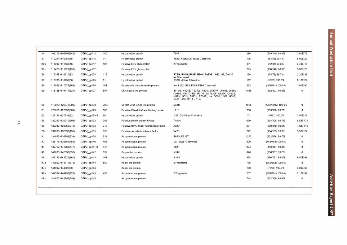
35
Animal Production Unit Activities Report 2007
114 109115>108693(140) GTPV_gp113 140 Hypothetical protein T68P 286 (139/140) 99.3% 3.60E-76 117 110221>110051(56) GTPV_gp115 74 Hypothetical protein Y52S; K55N; Del 18 aa C terminal 106 (54/56) 96.4% 5.00E-22 119a 111299>111439(46) GTPV_gp117 197 Putative EEV glycoprotein 2 Fragments 87 (42/45) 93.3% 2.40E-16 119b 111471>111929(152) GTPV_gp117 Putative EEV glycoprotein 295 (138/140) 98.6% 5.80E-79 126 116359>116610(83) GTPV_gp124 116 Hypothetical protein R78G; R80G; R84E; H86N; DelG81, N82, I83; Del 30
aa C terminal 160 (78/79) 98.7% 2.20E-38
127 116780>116950(56) GTPV_gp125 81 Hypothetical protein R56D; -25 aa C terminal 112 (55/55) 100.0% 9.10E-24 128 117082>117579(165) GTPV_gp126 161 Superoxide dismutase-like protein Ins: L162, I163, F164, F165 C terminal 333 (161/161) 100.0% 1.60E-90 129 118128>119711(527) GTPV_gp127 557 DNA ligase-like protein M191I; Y504S; T505Q, I512V; L513W; V514E; L515I;
Q516S, N517G; R518R, Y519S, I520F, Q521K, Q522S, M523I, G524, F525H; R526T;; Ins: G506, V507, G508, N509, I510, G511; - 31aa
1015 (502/503) 99.8% 0
130 119922>125945(2007) GTPV_gp128 2007 Variola virus B22R-like protein Q54H 4028 (2006/2007) 100.0% 0 131 126015>127097(360) GTPV_gp129 360 Putative IFN-alpha/beta binding protein L11F 738 (359/360) 99.7% 0 132 127136>127234(32) GTPV_gp129.5 90 Hypothetical protein I32F; Del 58 aa C terminal 70 (31/31) 100.0% 3.90E-11 135 129293>130210(305) GTPV_gp132 305 Putative ser/thr protein kinase T154A 630 (304/305) 99.7% 3.30E-179 136 130240>130962(240) GTPV_gp133 240 Putative RING finger host range protein A22V 501 (239/240) 99.6% 1.20E-140 138 131690>132097(135) GTPV_gp135 135 Putative secreted virulence factor V47S 272 (134/135) 99.3% 5.30E-72 141 134855>136759(634) GTPV_gp138 634 Ankyrin repeat protein R68G; K633T 1273 (632/634) 99.7% 0 143 138175>139584(469) GTPV_gp140 498 Ankyrin repeat protein Del 29aa C terminal 920 (463/463) 100.0% 0 144 139717>141060(447) GTPV_gp141.2 447 Ankyrin repeat protein Y82F 904 (446/447) 99.8% 0 145 141083>142096(337) GTPV_gp142 337 Serpin-like protein N74K 676 (336/337) 99.7% 0 146 142136>142621(161) GTPV_gp143 161 Hypothetical protein K10N 334 (160/161) 99.4% 8.80E-91 147a 142662>143774(370) GTPV_gp144 552 Kelch-like protein 2 Fragments 736 (362/362) 100.0% 0 147b 144092>144322(76) GTPV_gp144 Kelch-like protein 160 (76/76) 100.0% 3.00E-38 148a 144394>144756(120) GTPV_gp145 453 Ankyrin repeat protein 2 Fragments 207 (101/101) 100.0% 2.10E-52 148b 144677>145756(359) GTPV_gp145 Ankyrin repeat protein 714 (352/356) 98.9% 0

36
Animal Production Unit Activities Report 2007
3.9. IAEA Reference Serum Bank The successful development and standardization of serological diagnostic tests requires the availability of standardized, equilibrated and validated reference serum samples of different animal species whose origin and infection status provide the means to identify causative agents of infectious disease. The APU is building up and maintaining an important resource of internationally accepted reference sera for such applications, providing material essential for internal quality control when standardizing serological assays. These sera are initially provided by Member States and, in addition, are made available to MS as standards in the development of assays, to act as reference reagents both internally in diagnostic testing to establish quality control and to examine differences in the performance of different assays within and between laboratories. The APU serum bank currently comprises gamma-irradiated infected cattle sera against the following serotypes:
• Foot and Mouth Disease Virus Type SAT 1 Strain KNP 196/91/1 • Foot and Mouth Disease Virus Type SAT 1 Strain Nig 5/81 ZP02 • Foot and Mouth Disease Virus Type SAT 2 Strain KNP 19/88 • Foot and Mouth Disease Virus Type SAT 2 UGA 2/2002 • Foot and Mouth Disease Virus Type SAT 3 KNP 10/90 • Foot and Mouth Disease Virus Type O SAR 11/00/0 • Foot and Mouth Disease Virus Type Asia 1 05/96 • Foot and Mouth Disease Virus Type A GAM 52/98/A • Foot and Mouth Disease Virus Type A Cruzeiro • Peste des Petits Ruminants 100 samples
All sera have been irradiated and tested and are non-infectious. In future it is intended to acquire more samples, in order to enable a more comprehensive source of defined material.

37
Animal Production Unit Activities Report 2007
4. TRAINING ACTIVITIES
4.1. Training Course on the Molecular Characterization of Small Ruminant Breeds, Aleppo, Syria
This course, run jointly from 1-12 April 2007 by ICARDA/ILRI/FAO-IAEA, was attended by 16 participants from local and Central and West Africa and North Africa (CWANA) regions. The course aimed at building capacity in developing countries in Asia to use modern molecular methods and bioinformatics to characterize and use the available genetic advantages in indigenous small ruminants, enabling optimum management of these natural resources. It developed methodologies, generated information and formulated decision support systems for defining phenotypic and molecular genetic diversity, using microsatellite DNA marker and related technologies, and enabled the development and implementation of national, international and regional strategies for optimum use, improvement and conservation of small ruminant genetic resource in Asia. The training programme included both lectures and laboratory sessions. A presentation also covered the activities of IAEA to increase awareness of its current participation in the field of livestock breeding and genetics. A manual entitled “A practical approach to microsatellite genotyping with special reference to livestock population genetics” was prepared for the training course by the three groups and used during the course. One CRP, two regional TCPs and eight national TCPs are currently involved in the management of animal genetic resources.
4.2. Fellowships Ms Ivona Dimitrova
Ms Dimitrova is from Bulgaria, and was sponsored by the IAEA Technical Cooperation Programme (TCP). She stayed at the APU for three months, during which she was involved in the discovery of new SNPs in candidate genes in sheep.
Ms Ana Gabriela Murguia Quintana Ms Murguia is an assistant researcher in the field of molecular biology and genetics at the
University of Peruana Cayetano Heredia (UPCH) in Peru; she joined the APU for a 2-month fellowship. Her work was sponsored by the IAEA TCP. During her stay Ms Murguia was involved on the identification of expressed genes and genetic markers associated with productivity and mortality of embryos in Alpaca. She received training on site during her stay, using various tools such as real-time PCR, the use of public databases for candidate gene search and assisted also with routine experiments in APU.

38
Animal Production Unit Activities Report 2007
Ms Irene Guendel Ms Guendel was a cost-free Costa Rican intern for 8 months. She was trained on the development of new candidate genes involved in parasite resistance in sheep and the use of various tools for identifying putative genes involved in immune responses. She also received bioinformatics training and was directly involved in the development of the GC-db database.
Ms Myriam Soltani Ms Soltani was awarded an internship to work on Peste des Petits Ruminants, cloning the P gene of the virus and expressing the recombinant P protein. She also mapped the binding site of N on P.
4.3. Internships
4.4. Scientific Visitors
Mr Armando Hung Chaparro Mr Chaparro, Dean of the Faculty of Science at University of Peruana Cayetano Heredia (UPCH) in Peru; and Group leader of the Laboratory of Molecular Biology at the Veterinary Faculty, visited APU for scientific discussions for a period of two weeks.

39
Animal Production Unit Activities Report 2007
5. ACKNOWLEDGEMENTS
APU gratefully acknowledges the assistance of Dr Roland Silber and his colleagues at the Austrian Agency for Health and Food Safety (AGES) in screening samples imported from Africa and Asia, for potentially harmful pathogenic organisms and for enabling staff of APU to work on exotic pathogens in secure isolation premises in the high security laboratory.

40
Animal Production Unit Activities Report 2007
6. APPENDICES
6.1. Staff Publications Bodjo, SC, O Kwiatek, A Diallo, E Albina and G Libeau. Mapping and structural analysis of B-cell epitopes on Morbillivirus nucleoprotein N-terminus. J. Gen. Virol., 2007, 88: 1231 - 1242 Couacy-Hymann, E, SC Bodjo, MY Koffi, T Danho. Observations on rinderpest and rinderpest-like diseases throughout West and Central African countries during rinderpest eradication projects. Res. Vet. Sci., 2007, 83: 282 – 285 Couacy-Hymann, E, SC Bodjo, T Danho, G Libeau, A Diallo. Evaluation of the virulence of some strains of Peste des Petits Ruminants Virus (PPRV) in experimentally infected West African Dwarf Goats. Vet. J., 2007, 173: 180 -185 Couacy-Hymann, E, SC Bodjo, T Danho, MY Koffi, G Libeau, A Diallo. Early detection of viral excretion from experimentally infected goats with peste-des-petits-ruminants virus. Prev. Vet. Med., 2007, 78: 85 -88 Diallo, A, G Viljoen. Genus Capripoxvirus. In: Poxviruses, Birkhäuser Advances in Infectious Diseases, A Mercer, A Schmidt and O Weber (Eds), SpringerLink, 2007 Diallo, A, C Minet, C Le Goff, G Berhe, E Albina, G Libeau and T Barrett. The Threat of Peste des Petits Ruminants: Progress in Vaccine Development for Disease Control. Vaccine, 2007: 25, 5591 – 5597 Kwiatek, O, C Minet, C Grillet, C Hurard, E Carlsson, B Karimov, E Albina, A Diallo, G Libeau. Peste des Petits Ruminants (PPR) Outbreak in Tajikistan. J. Comp. Path., 2007, 136: 1111 – 1119 Lubroth, J, MM Rweyemamu, G Viljoen, A Diallo, W Amanfu. Veterinary vaccines and their use in developing countries. Rev. sci. tech. Off. int. Epiz., 2007, 26: 179 -201

41
Animal Production Unit Activities Report 2007
6.2. Staff Travel Staff Member Destination Period Purpose of Travel Bodjo, Sanne Charles
CIRAD, Montpellier, France
13-19 January To study PPR virus protein interactions by confocal microscopy.
Diallo, Adama FAO Headquarters, Rome, Italy
31 January – 1 February For discussions on collaboration in animal disease, reproduction and genetics.
Nairobi, Kenya 22-23 February To participate in the PACE meeting.
Asmara, Eritrea 26 February – 1 March To attend the 17th conference of the OIE Regional Commission for Africa.
Djibouti, Republic of Djibouti
27-30 March To participate in the 8th Executive Committee meeting of ALIVE.
Addis Ababa, Ethiopia
16-20 April To participate in the annual meeting of the MARKVAC project (Development of PPR and Rinderpest Marker Vaccine) .
Beijing, China 1-7 July To present a paper on Peste des Petits Ruminant.
Rome, Italy 25-26 September To attend the Global Rinderpest Eradication Programme (GREP) Consultative Group Meeting.
CIRAD, Montpellier, France
25 – 26 October To discuss work on two collaborative projects which are funded by the EC and the French Ministry for Foreign Affairs with partners at CIRAD.
ICARDA, Aleppo, Syria
29-31 October To participate in the first meeting of the EC-funded project on Avian Influenza (Conflutech).
Malek, Massoud
ICARDA, Aleppo, Syria
7-12 April To attend a training course and present a lecture entitled "Genetic characterization of small ruminants".

42
Animal Production Unit Activities Report 2007
6.3. External Collaboration and Partnerships
Institution Topic Austrian Agency for Health and Food Safety (AGES), Vienna, Austria
Collaborative studies in the high security laboratories that APU is allowed to use for work on live viruses, including Peste des Petits Ruminants and Capripox.
Centre International en Recherche Agronomique pour le Développement (CIRAD), Montpellier, France
Partner in the Wellcome Trust supported research, ‘Peste des Petits Ruminants (PPR) a serious and emerging plague of small ruminants in Developing countries : Epidemiology, transmission, host range and pathogenicity of the virus in West Africa’ and the project funded by the Fonds de Solidarité Prioritaire, ‘Renforcement de cinq laboratoires de recherche vétérinaire en Afrique pour la surveillance et le contrôle de maladies animales’ for work on capripox viruses.
Institute of Animal Health (IAH), Pirbright, UK
Partner in the Wellcome Trust supported research, ‘Peste des Petits Ruminants (PPR) a serious and emerging plague of small ruminants in Developing countries : Epidemiology, transmission, host range and pathogenicity of the virus in West Africa’. Also provides isolates of capripox viruses for studies in APU.
Institut National de Médecine Vétérinaire (INMV), Algeria
Partner in the Wellcome Trust supported research, ‘Peste des Petits Ruminants (PPR) a serious and emerging plague of small ruminants in Developing countries : Epidemiology, transmission, host range and pathogenicity of the virus in West Africa’ and the project funded by the Fonds de Solidarité Prioritaire, ‘Renforcement de cinq laboratoires de recherche vétérinaire en Afrique pour la surveillance et le contrôle de maladies animales’ for work on capripox viruses.
Laboratoire Central Vétérinaire (LCV), Bamako, Mali
Partner in the Wellcome Trust supported research, ‘Peste des Petits Ruminants (PPR) a serious and emerging plague of small ruminants in Developing countries : Epidemiology, transmission, host range and pathogenicity of the virus in West Africa’ and the project funded by the Fonds de Solidarité Prioritaire, ‘Renforcement de cinq laboratoires de recherche vétérinaire en Afrique pour la surveillance et le contrôle de maladies animales’ for work on capripox viruses. This institute will also carry out experimental studies in sheep and goats using isolates of capripox characterized and provided by APU.

43
Animal Production Unit Activities Report 2007
Institution Topic Laboratoire National d'Appui au Développement Agricole (LANADA), Bingerville, Côte d'Ivoire
Partner in the Wellcome Trust supported research, ‘Peste des Petits Ruminants (PPR) a serious and emerging plague of small ruminants in Developing countries : Epidemiology, transmission, host range and pathogenicity of the virus in West Africa’
Laboratoire National Vétérinaire (LANAVET), Garoua, Cameroun
Partner in the project funded by the Fonds de Solidarité Prioritaire, ‘Renforcement de cinq laboratoires de recherche vétérinaire en Afrique pour la surveillance et le contrôle de maladies animales’ for work on capripox viruses.
National Animal Research Health Center (NAHRC), Sabeta, Ethiopia
Partner in studies funded by the Fonds de Solidarité Prioritaire, ‘Renforcement de cinq laboratoires de recherche vétérinaire en Afrique pour la surveillance et le contrôle de maladies animales’ for work on capripox viruses. This institue will also carry out experimental studies in sheep and goats using isolates of capripox characterized and provided by APU.
University of Ankara, Ankara, Turkey In collaboration with APU, the university has provided isolates of capripox viruses from cattle, sheep and goats for characterization.

44
Animal Production Unit Activities Report 2007
6.4. Trainees, Fellows and Scientific Visitors
Name Project Months/Days From To Trainees Guendel, Ms I 7 months 2007-4-1 2007-10-31 Soltani, Ms M 7 months 2007-6-18 2007-12-31 Fellows Murguia Quintana, Ms AG PER/5029 3 months 2007-10-15 2007-12-15 Dimitrova, Ms I RER/0016 3 months 2007-4-2 2007-7-2 Scientific Visitors Hung Chaparro, Mr A PER/5029 12 days 2007-9-10 2007-9-21

45
Animal Production Unit Activities Report 2007
6.5. Coordinated Research Projects and Technical Cooperation Projects
CRP Title Scientific Secretary Gene-based Technologies in Livestock Breeding: Characterization of Small Ruminant Genetic Resources in Asia (2005-2009)
Malek Massoud
TCP Title Technical Officer Genomics of the Alpaca: Identification of Expressed Genes and Genetic Markers Associated with Productivity and Embryonic Mortality (2005-2009)
Malek Massoud

46
Animal Production Unit Activities Report 2007
6.6. Abbreviations 6-FAM 6'-Carboxy-Fluorescein AGES Austrian Agency for Health and Food Safety APU Animal Production Unit CBPP Contagious Bovine Pleuropneumonia CIRAD French Agricultural Research Centre for International Development CRP Co-ordinated Research Project CSIRO Commonwealth Scientific and Industrial Research Organisation CWANA Central and West Africa and North Africa DIVA Differentiation Between Infected and Vaccinated Animals DNA Deoxyribonucleic Acid ELISA Enzyme Linked Immunosorbent Assay FAO Food and Agricultural Organization of the United Nations, Rome, Italy FMDV Foot and Mouth Disease Virus FRET Fluorescence Resonance Energy Transfer GR-db Genetic Resource Database GTPV Goat Pox Virus HRP Horseradish Peroxidase IAEA International Atomic Energy Agency ICARDA International Center for Agricultural Research in the Dry Areas, Aleppo, Syrian Arab Republic IIF Indirect Immunofluorescence ILRI International Livestock Research Institute, Nairobi, Kenya IPTG Isopropylthiogalactoside ISAG International Society of Animal Genetics LANADA Laboratoire Central de Pathologie Animale de Bingerville, Cote d’Ivoire LCV Laboratoire Central Veterinaire, Bamako, Mali LNA Locked Nucleic Acid LSDV Lumpy Skin Disease Virus MAI Marker Assisted Introgression MAS Marker Assisted Selection MS Member States NCBI National Center for Biotechnology Information, USA NSP Non Structural Protein OIE World Organization for Animal Health, Paris, France PAGE Polyacrylamide Gel Electrophoresis PCR Polymerase Chain Reaction PPR Peste des Petits Ruminant QTL Quantitative Trait Loci RFLP Restriction Fragment Length Polymorphism RT-db Real-Time Database SDS Sodium Dodecyl Sulphate SNP Single Nucleotide Polymorphisms SPPV Sheep Pox Virus TCP Technical Cooperation Project TMB Tetramethylbenzidene USDA United States Department of Agriculture


http://www-naweb.iaea.org/nafa/aph/index.html