Embed Size (px)
Citation preview
Clinical Science (2013) 124, 443–456 (Printed in Great Britain) doi: 10.1042/CS20120461
Angiotensin-(1–7): beyond the cardio-renalactionsDanielle G. PASSOS-SILVA∗, Thiago VERANO-BRAGA∗† and Robson A. S. SANTOS∗
∗Department of Physiology and Biophysics, National Institute of Science and Technology in Nanobiopharmaceutics (INCT-Nanobiofar), Institute ofBiological Sciences, Federal University of Minas Gerais, Belo Horizonte (MG), Brazil†Department of Biochemistry and Molecular Biology, University of Southern Denmark, Odense, Denmark
AbstractIt is well known that the RAS (renin–angiotensin system) plays a key role in the modulation of many functions in thebody. AngII (angiotensin II) acting on AT1R (type 1 AngII receptor) has a central role in mediating most of the actionsof the RAS. However, over the past 10 years, several studies have presented evidence for the existence of a newarm of the RAS, namely the ACE (angiotensin-converting enzyme) 2/Ang-(1–7) [angiotensin-(1–7)]/Mas axis.Ang-(1–7) can be produced from AngI or AngII via endo- or carboxy-peptidases respectively. ACE2 appears to play acentral role in Ang-(1–7) formation. As described for AngII, Ang-(1–7) also has a broad range of effects in differentorgans and tissues which goes beyond its initially described cardiovascular and renal actions. Those effects aremediated by Mas and can counter-regulate most of the deleterious effects of AngII. The interaction Ang-(1–7)/Masregulates different signalling pathways, such as PI3K (phosphoinositide 3-kinase)/AKT and ERK (extracellular-signal-regulated kinase) pathways and involves downstream effectors such as NO, FOXO1 (forkhead box O1) andCOX-2 (cyclo-oxygenase-2). Through these mechanisms, Ang-(1–7) is able to improve pathological conditionsincluding fibrosis and inflammation in organs such as lungs, liver and kidney. In addition, this heptapeptide haspositive effects on metabolism, increasing the glucose uptake and lipolysis while decreasing insulin resistance anddyslipidaemia. Ang-(1–7) is also able to improve cerebroprotection against ischaemic stroke, besides its effects onlearning and memory. The reproductive system can also be affected by Ang-(1–7) treatment, with enhancedovulation, spermatogenesis and sexual steroids synthesis. Finally, Ang-(1–7) is considered a potential anti-cancertreatment since it is able to inhibit cell proliferation and angiogenesis. Thus the ACE2/Ang-(1–7)/Mas pathwayseems to be involved in many physiological and pathophysiological processes in several systems and organsespecially by opposing the detrimental effects of inappropriate overactivation of the ACE/AngII/AT1Raxis.
Key words: angiotensin-(1–7), angiotensin-converting enayme 2 (ACE2), Mas receptor, renin–angiotensin system (RAS)
INTRODUCTION
The textbook-like view of the RAS (renin–angiotensin system)as a linear limited proteolysis pathway toward the production ofa single active end product, AngII (angiotensin II), has changeddramatically over the past 10 years with the identification of novelcomponents [ACE (angiotensin-converting enzyme) 2, Mas andrenin/pro-renin receptor] and actions of this system [1–6]. Amajor shift in our understanding of the RAS was the proposition
Abbreviations: ACE, angiotensin-converting enzyme; AGT, angiotensinogen; AIA, antigen-induced arthritis; Ang-(1–7), angiotensin-(1–7); Ang-(1–9), angiotensin-(1–9); AngI etc.,angiotensin I etc.; AP2, adipose lipid-binding protein 2; ApoE, apolipoprotein E; AT1R, type 1 AngII receptor; AT2R, type 2 AngII receptor; BDL, bile-duct ligation; COX, cyclo-oxygenase;CTGF, connective tissue growth factor; ERK, extracellular-signal-regulated kinase; FOXO1, forkhead box O1; GnRH, gonadotropin-releasing hormone; HAEC, human aortic endothelialcell; HDAC1, histone deacetylase 1; IL, interleukin; JNK, c-Jun N-terminal kinase; LPS, lipopolysaccharide; LTP, long-term potentiation; MAPK, mitogen-activated protein kinase; MMP,metalloproteinase; mTOR, mammalian target of rapamycin; NEFA, non-esterified ‘free’ fatty acid; NEP, neutral endopeptidase; NF-κB, nuclear factor κB; NOS, NO synthase; eNOS,endothelial NOS; iNOS, inducible NOS; nNOS, neuronal NOS; PEP, prolyl-endopeptidase; PI3K, phosphoinositide 3-kinase; PI3KC2A, class II PI3Kα; PlGF, placental growth factor;PPARγ , peroxisome-proliferator-activated receptor γ ; PRAS40, proline-rich Akt substrate of 40 kDa; RAS, renin–angiotensin system; RASIP1, Ras-interacting protein 1; TGF,transforming growth factor; TGR, transgenic; TNF, tumour necrosis factor; TOP, thimet oligopeptidase; VEGF, vascular endothelial growth factor.
Correspondence: Dr Robson A.S. Santos (email [email protected] or [email protected]).
of the concept of the RAS as a dual axis system: one axis repres-ented by ACE/AngII/AT1R (type 1 AngII receptor) and the otherby ACE2/Ang-(1–7) [angiotensin-(1–7)]/Mas [7,8]. The identi-fication of the renin/pro-renin receptor [4] and its physiologicalactions, which go beyond its role in the RAS [9], and more re-cently of the other ACE2 product Ang-(1–9) [angiotensin-(1–9)]as a biologically active member of the RAS [10,11], suggestthat we are still far from understanding the complexity of thisfascinating system.
www.clinsci.org 443
D. G. Passos-Silva, T. Verano-Braga and R. A. S. Santos
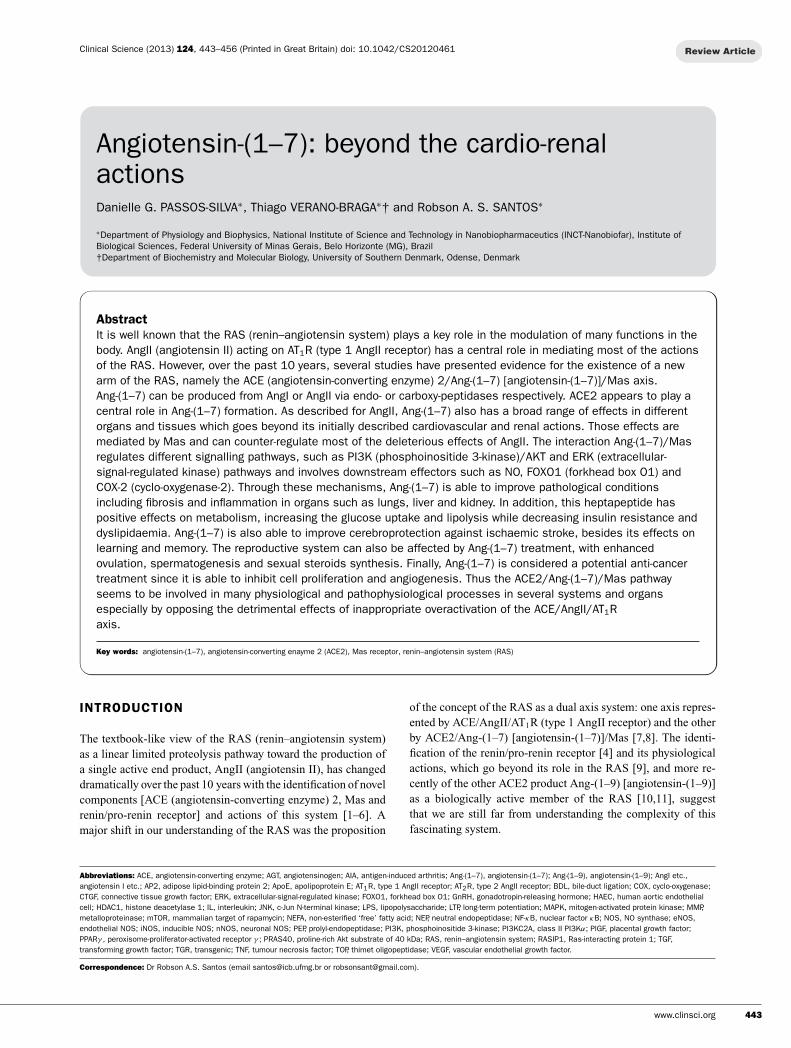
Figure 1 Simplified view of the different pathways of Ang-(1–7) formation and the receptors for Ang-(1–7) and AngII
The starting point in the canonical and non-canonical en-zymatic pathways of the RAS is the glycoprotein AGT (an-giotensinogen), which is produced and secreted into the circu-lation by the liver. Most organs, including the brain, vasculature,kidney, heart, and lungs, can also produce AGT. The limited pro-teolysis cascade starts with renin, which plays a central role inthe formation of the inactive decapeptide AngI by cleavage ofAGT. AngI (angiotensin I) can be cleaved by ACE or chymase toform the octapeptide AngII, and by many other peptidases, espe-cially PEP (prolyl-endopeptidase), NEP (neutral endopeptidase)and TOP (thymet oligopeptidase), which can generate Ang-(1–7) by the cleavage of its Pro7-Phe8 bond. Ang-(1–7) can alsobe formed by hydrolysis of AngII via ACE2 [12]. Furthermore,this heptapeptide can be generated via the hydrolysis of AngI byACE2 to form Ang-(1–9), which is subsequently cleaved by NEPor ACE [13]. However, it is important to stress that this pathwayseems to be catalytically less efficient than the ones mentionedabove [13]. A simplified view of the enzymatic pathways involvedin the formation of Ang-(1–7) is shown in Figure 1.
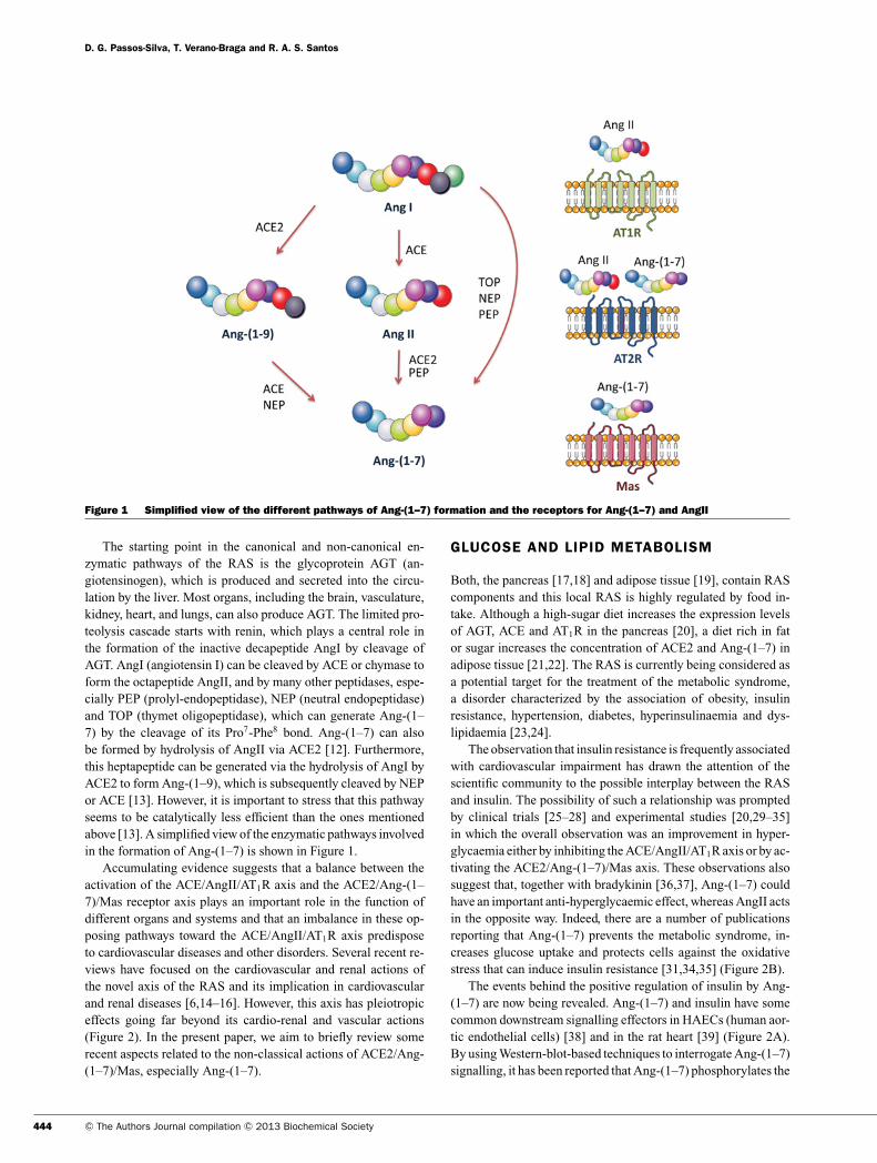
Accumulating evidence suggests that a balance between theactivation of the ACE/AngII/AT1R axis and the ACE2/Ang-(1–7)/Mas receptor axis plays an important role in the function ofdifferent organs and systems and that an imbalance in these op-posing pathways toward the ACE/AngII/AT1R axis predisposeto cardiovascular diseases and other disorders. Several recent re-views have focused on the cardiovascular and renal actions ofthe novel axis of the RAS and its implication in cardiovascularand renal diseases [6,14–16]. However, this axis has pleiotropiceffects going far beyond its cardio-renal and vascular actions(Figure 2). In the present paper, we aim to briefly review somerecent aspects related to the non-classical actions of ACE2/Ang-(1–7)/Mas, especially Ang-(1–7).
GLUCOSE AND LIPID METABOLISM
Both, the pancreas [17,18] and adipose tissue [19], contain RAScomponents and this local RAS is highly regulated by food in-take. Although a high-sugar diet increases the expression levelsof AGT, ACE and AT1R in the pancreas [20], a diet rich in fator sugar increases the concentration of ACE2 and Ang-(1–7) inadipose tissue [21,22]. The RAS is currently being considered asa potential target for the treatment of the metabolic syndrome,a disorder characterized by the association of obesity, insulinresistance, hypertension, diabetes, hyperinsulinaemia and dys-lipidaemia [23,24].
The observation that insulin resistance is frequently associatedwith cardiovascular impairment has drawn the attention of thescientific community to the possible interplay between the RASand insulin. The possibility of such a relationship was promptedby clinical trials [25–28] and experimental studies [20,29–35]in which the overall observation was an improvement in hyper-glycaemia either by inhibiting the ACE/AngII/AT1R axis or by ac-tivating the ACE2/Ang-(1–7)/Mas axis. These observations alsosuggest that, together with bradykinin [36,37], Ang-(1–7) couldhave an important anti-hyperglycaemic effect, whereas AngII actsin the opposite way. Indeed, there are a number of publicationsreporting that Ang-(1–7) prevents the metabolic syndrome, in-creases glucose uptake and protects cells against the oxidativestress that can induce insulin resistance [31,34,35] (Figure 2B).
The events behind the positive regulation of insulin by Ang-(1–7) are now being revealed. Ang-(1–7) and insulin have somecommon downstream signalling effectors in HAECs (human aor-tic endothelial cells) [38] and in the rat heart [39] (Figure 2A).By using Western-blot-based techniques to interrogate Ang-(1–7)signalling, it has been reported that Ang-(1–7) phosphorylates the
444 C© The Authors Journal compilation C© 2013 Biochemical Society
Angiotensin-(1–7): beyond the cardio-renal actions
Figure 2 Main non-cardiovascular effects of Ang-(1–7) via Mas in different tissues and processes(A) Signalling molecules involved in the action of Ang-(1–7). The molecules in green and red indicate activation or inhibitionby Ang-(1–7)/Mas respectively. The green arrows indicate activation or positive effects, and the red lines indicate inhibitionof the process. (B) The main non-cardiovascular consequences of Ang-(1–7) on each process.
www.clinsci.org 445
D. G. Passos-Silva, T. Verano-Braga and R. A. S. Santos
insulin downstream effectors PI3K (phosphoinositide 3-kinase)and AKT via the Mas receptor in HAECs [38,39], and IRS1 (in-sulin receptor substrate 1) and JAK2 (Janus kinase 2) via the AT1Rin rat hearts [39]. Moreover, Ang-(1–7)/Mas negatively regulatesAngII/AT1R signalling in HAECs by promoting dephosphoryla-tion of c-Src and ERK (extracellular-signal-regulated kinase)1/2, and inhibition of NADPH oxidase activity [40]. Recently,Munoz and co-workers [41] used FFRs (fructose-fed rats), amodel of the metabolic syndrome, to study whether Ang-(1–7)positively modulates insulin signalling via Mas and attenuates theinhibitory effect of AngII on this signal transduction pathway. In-deed, the authors observed that, in a Mas-dependent manner, thedownstream effectors of insulin including AKT, GSK-3β (glyco-gen synthase kinase-3β) and AS160 (AKT substrate of 160 kDa)were positively modulated by Ang-(1–7) in insulin-target tissues(skeletal muscle, liver and adipose tissue). In the same way, theinhibitory effect of AngII on these effectors was also attenuatedby Ang-(1–7) through Mas [41].
Western-blot-based studies have several advantages includ-ing high sensitivity and specificity. However, one needs to rely oncommercially available specific antibodies with proven quality.Moreover, this type of technique is hypothesis-driven. In otherwords, the downstream effectors of a specific signalling cas-cade need to have been defined previously. Therefore it is notpossible to identify unknown targets of signalling networks byWestern blotting. In order to overcome these technical limita-tions and to gain more insights into the Mas signalling path-way, we recently applied an MS-based approach to study theHAEC phosphoproteome treated with Ang-(1–7) [42]. In thatstudy, we were able to detect 79 phosphoproteins that hadtheir phosphorylation state significantly changed over the ob-served time frame (0–20 min). Among these phosphoproteins,eight known downstream effectors of the insulin signal trans-duction cascade had their phosphorylation levels differentiallyregulated by Ang-(1–7): AKT1 (RAC-α serine/threonine pro-tein kinase), PRAS40/AKT1S1 (proline-rich Akt substrate of40 kDa/proline-rich AKT1 substrate 1), CAV1 (caveolin-1 iso-form α), FOXO1 (forkhead box O1), MAPK1 (mitogen-activatedprotein kinase 1)/ERK2, PXN (paxillin isoform 1), PI3KC2A(class II PI3Kα) and VIM (vimentin) [42]. Ser124 on the AKT1was phosphorylated after 3 min of Ang-(1–7) treatment. Thephosphorylation of this residue, together with the phosphoryla-tion of Thr308 and Ser473, activates this kinase [43,44]. This find-ing is in agreement with previous publications reporting thatAKT is rapidly activated by Ang-(1–7) [38,39,45]. The class IPI3K plays an important role in the Ang-(1–7) signalling andit is an upstream kinase that mediates AKT phosphorylationin HAECs and rat hearts [38,39]. However, our phosphopro-teome study revealed that PI3KC2A also plays a role in thissignal transduction cascade, as Mas activation induced the phos-phorylation of Ser338 on this kinase [42]. Diverging from class IPI3K, PI3KC2A is not an upstream kinase of the AKT signallingbranch [46,47]. Interestingly, this kinase induces translocationof GLUT4 (glucose transporter 4) to the plasma membrane inresponse to insulin stimulation [47]. PRAS40 regulates insulin-induced mTOR (mammalian target of rapamycin) activity. WhenThr246 on PRAS40 is dephosphorylated, it binds to the mTOR
complex to inhibit it. On the other hand, AKT1 can phosphorylatePRAS40 to induce its dissociation from mTOR, activating thiscomplex [48,49]. The transcriptional factor FOXO1 is anothercomponent of the insulin signalling pathway that is a downstreamtarget of AKT1 [50]. Once activated, AKT1 translocates to thenucleus and phosphorylates FOXO1 at Thr24, Ser256 and Ser319,inactivating this transcriptional factor [50]. Strikingly, Ang-(1–7)induced the dephosphorylation of Thr246 on PRAS40 and Ser256
on FOXO1. These data are consistent with mTOR inactivationand FOXO1 activation. Indeed, we have also demonstrated thatAng-(1–7) induces FOXO1 activation with its translocation to thenucleus [42]. These findings suggest that Ang-(1–7) signalling isa highly controlled system, possibly with more regulatory inputsthan previously suspected. Concerning the AKT/FOXO relation-ship, the possibility that Ang-(1–7) modulates the equilibriumbetween FOXO1 activation, which is predominant during thefasting period, and AKT activation, which predominates afterfeeding [51], should be explored in future studies (Figure 3).
Lipid metabolism is also regulated by Ang-(1–7) (Figure 2).When treated with this heptapeptide, rats with diabetic cardio-myopathy [32] and diabetic nephropathy [52] have a significantreduction in dyslipidaemia in a Mas-dependent way (Figure 2b).Moreover, Mas-knockout mice on the FVB/N background haveimpaired lipid metabolism, leading to dyslipidaemia, lower gluc-ose tolerance and insulin sensitivity, hyperinsulinaemia, hyper-leptinaemia, lower adiponectin secretion, decreased glucose up-take and increased abdominal fat mass when compared with thewild-type phenotype [34]. On the other hand, TGR (transgenic)animals with increased plasma levels of Ang-(1–7) had reducedfat mass, decreased triacylglycerols (triglycerides) and choles-terol levels, despite normal food intake [53]. In addition, theexpression levels of adiponectin and AP2 (adipose lipid-bindingprotein 2) were increased, whereas there was a remarkable de-crease in AGT expression in TGR animals. Adiponectin is a keyadipokine that regulates insulin sensitivity and tissue inflamma-tion, and its plasma level is inversely proportional to body fatcontent. AP2 is an important protein in the adipose tissue meta-bolism involved in fatty acid esterification [53]. In the same way,ACE-knockout mice had reduced fat mass due to an increasein lipid metabolism and energy expenditure as a consequenceof higher expression levels of key genes involved in the hydro-lysis of lipids into NEFAs (non-esterified ‘free’ fatty acids) [LPL(lipoprotein lipase)], translocation of fatty acids to the mitochon-dria [CPT-1 (carnitine palmitoyltransferase-1)], and β-oxidationinside mitochondria and peroxisomes [LCAD (long-chain acyl-CoA dehydrogenase)] [54].
Recently, two groups have shed more light on the molecularmechanisms behind the beneficial modulation of lipid metabol-ism by Ang-(1–7). Mario et al. [55] demonstrated that the expres-sion of the PPARγ (peroxisome-proliferator-activated receptorγ ) is compromised in Mas− / − mice (Figure 2A). The tran-scription factor PPARγ is believed to have a beneficial effect ininsulin-resistant patients, as its activation leads to the expressionof target genes involved in fatty acid metabolism, triacylglycerolstorage and reduction in plasma NEFA supply [56,57]. In the sameway, Oh et al. [58] reported that Ang-(1–7) stimulates lipolysis viaMas. Interestingly, these authors observed that an AKT inhibitor
446 C© The Authors Journal compilation C© 2013 Biochemical Society
Angiotensin-(1–7): beyond the cardio-renal actions
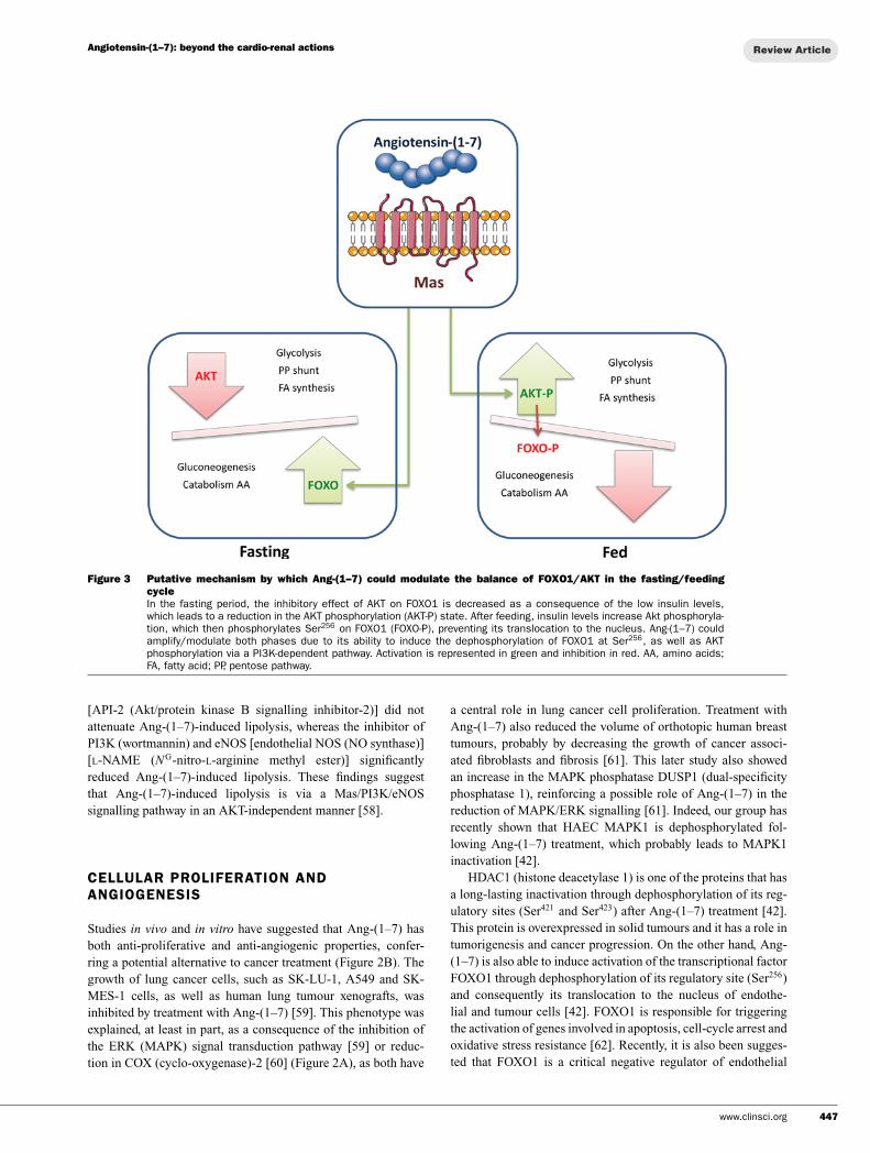
Figure 3 Putative mechanism by which Ang-(1–7) could modulate the balance of FOXO1/AKT in the fasting/feedingcycleIn the fasting period, the inhibitory effect of AKT on FOXO1 is decreased as a consequence of the low insulin levels,which leads to a reduction in the AKT phosphorylation (AKT-P) state. After feeding, insulin levels increase Akt phosphoryla-tion, which then phosphorylates Ser256 on FOXO1 (FOXO-P), preventing its translocation to the nucleus. Ang-(1–7) couldamplify/modulate both phases due to its ability to induce the dephosphorylation of FOXO1 at Ser256, as well as AKTphosphorylation via a PI3K-dependent pathway. Activation is represented in green and inhibition in red. AA, amino acids;FA, fatty acid; PP, pentose pathway.
[API-2 (Akt/protein kinase B signalling inhibitor-2)] did notattenuate Ang-(1–7)-induced lipolysis, whereas the inhibitor ofPI3K (wortmannin) and eNOS [endothelial NOS (NO synthase)][L-NAME (NG-nitro-L-arginine methyl ester)] significantlyreduced Ang-(1–7)-induced lipolysis. These findings suggestthat Ang-(1–7)-induced lipolysis is via a Mas/PI3K/eNOSsignalling pathway in an AKT-independent manner [58].
CELLULAR PROLIFERATION ANDANGIOGENESIS
Studies in vivo and in vitro have suggested that Ang-(1–7) hasboth anti-proliferative and anti-angiogenic properties, confer-ring a potential alternative to cancer treatment (Figure 2B). Thegrowth of lung cancer cells, such as SK-LU-1, A549 and SK-MES-1 cells, as well as human lung tumour xenografts, wasinhibited by treatment with Ang-(1–7) [59]. This phenotype wasexplained, at least in part, as a consequence of the inhibition ofthe ERK (MAPK) signal transduction pathway [59] or reduc-tion in COX (cyclo-oxygenase)-2 [60] (Figure 2A), as both have
a central role in lung cancer cell proliferation. Treatment withAng-(1–7) also reduced the volume of orthotopic human breasttumours, probably by decreasing the growth of cancer associ-ated fibroblasts and fibrosis [61]. This later study also showedan increase in the MAPK phosphatase DUSP1 (dual-specificityphosphatase 1), reinforcing a possible role of Ang-(1–7) in thereduction of MAPK/ERK signalling [61]. Indeed, our group hasrecently shown that HAEC MAPK1 is dephosphorylated fol-lowing Ang-(1–7) treatment, which probably leads to MAPK1inactivation [42].
HDAC1 (histone deacetylase 1) is one of the proteins that hasa long-lasting inactivation through dephosphorylation of its reg-ulatory sites (Ser421 and Ser423) after Ang-(1–7) treatment [42].This protein is overexpressed in solid tumours and it has a role intumorigenesis and cancer progression. On the other hand, Ang-(1–7) is also able to induce activation of the transcriptional factorFOXO1 through dephosphorylation of its regulatory site (Ser256)and consequently its translocation to the nucleus of endothe-lial and tumour cells [42]. FOXO1 is responsible for triggeringthe activation of genes involved in apoptosis, cell-cycle arrest andoxidative stress resistance [62]. Recently, it is also been sugges-ted that FOXO1 is a critical negative regulator of endothelial
www.clinsci.org 447
D. G. Passos-Silva, T. Verano-Braga and R. A. S. Santos
angiogenic behaviour [63], indicating a possible pathway bywhich Ang-(1–7) can inhibit angiogenesis and, overall, tumori-genesis. RASIP1 (Ras-interacting protein 1) also seems to be adownstream effector of Ang-(1–7), as it was phosphorylated onSer331 after 3 min of Ang-(1–7) treatment [42]. RASIP1 acts inblood vessel morphogenesis; however, it is yet to be establishedwhether this phosphorylation modulates the activity of this pro-tein [64]. The role of Ang-(1–7) in angiogenesis inhibition wasshown in a mouse sponge model of angiogenesis [65]. Daily injec-tions of Ang-(1–7) significantly inhibited angiogenesis, probablythrough interaction with Mas, since its antagonist A779 blockedthis effect [65]. On the other hand, the AT1R and AT2R (type 2AngII receptor) do not appear to be involved in this mechanism,since their antagonists did not affect the Ang-(1–7) effect [65].The mechanism of the anti-angiogenic effect of Ang-(1–7) inmice is related to NO release [65]. Anton et al. [66] also showedin vitro inhibition of angiogenesis by Ang-(1–7) in the tube form-ation assay using endothelial cells. This inhibition was also ab-olished by A779 [66]. Similarly, tube formation of A549 tumourcells was also inhibited after Ang-(1–7) treatment [67]. A reduc-tion in vessel density in Ang-(1–7)-treated human A549 lung tu-mours was also observed. The mechanism of the anti-angiogenicproperty of Ang-(1–7) in this condition seems to involve VEGF(vascular endothelial growth factor)-A, a primary pro-angiogenicprotein, which was reduced after treatment with this heptapeptide[67]. In addition, Ang-(1–7) decreased proliferation markers andintratumoral vessel densities of a xenograft prostate tumour inmice [68]. The treatment also reduced angiogenic factors suchVEGF and PlGF (placental growth factor), and increased thesoluble fraction of VEGF receptor 1 (sFlt-1), preventing the trig-gering of the angiogenesis signalling pathway [68]. Data fromin vivo and in vitro studies are in line with the results obtained inpatients with advanced cancer that were treated with Ang-(1–7) inPhase I clinical trials [69]. Patients treated with Ang-(1–7) hadreduced circulating levels of PIGF, a placental growth factor in-volved in angiogenesis. In this trial, treatment with Ang-(1–7) ledto disease stabilization [69]. Finally, recent results have shownthat Ang-(1–7) treatment also has anti-metastatic properties inlung and prostate cancer cells [70]. Ang-(1–7)-treated A549human lung adenocarcinoma cells reduced the expression andactivity of the MMPs (metalloproteinases) MMP-2 and MMP-9, which are involved in migration and invasion of cells. Themechanism for this effect includes the inhibition of PI3K/AKT,p38 and JNK (c-Jun N-terminal kianse) signalling pathways [71](Figure 2A). Therefore separate studies indicate that Ang-(1–7)is a potential candidate for cancer treatment as it presents bothanti-proliferative and anti-angiogenic properties.
REPRODUCTION
Several tissues of the reproductive system express a local RAS.Besides its vascular actions, the local RAS plays an importantrole during spermatogenesis, follicle maturation and ovulation,endometrium function and pregnancy [72–80] (Figure 2).
In female mammals, the ACE2/Ang-(1–7)/Mas axis hasbeen detected in the ovary, endometrium and placenta [72–76].The ACE2/Ang-(1–7)/Mas axis is highly regulated by GnRH(gonadotropin-releasing hormone) in the rat ovary [72]. Thishormone induces the expression of ACE2, leading to increasedlevels of Ang-(1–7). Furthermore, GnRH positively regulates theexpression of Mas [72]. In the same way, the ACE2/Ang-(1–7)/Mas axis also seems to play a role in the ovulation processin cattle. Following 24 h of GnRH stimulation, Ang-(1–7) levelsincrease in the follicular fluid, probably due to ACE2, NEP andPEP up-regulation in granulosa cells [81]. In addition, Ang-(1–7)increases during pro-oestrus and oestrus, and after eCG (equinechorionic gonadotropin) treatment [76,82]. Furthermore, perfu-sion of Ang-(1–7) in immature rat ovaries leads to increasingprogesterone and oestradiol synthesis in a Mas-dependent man-ner [76]. The evidence for a role of the ACE2/Ang-(1–7)/Masaxis in the female reproductive system was recently reinforced bythe observation that LH (luteinizing hormone) up-regulates theACE2/Ang-(1–7)/Mas axis and that Ang-(1–7) promotes mei-otic resumption, possibly as a gonadotrophin intermediate [83].Therefore there is substantial evidence suggesting an importantrole for the ACE2/Ang-(1–7)/Mas axis in the ovulation process.
The ACE2/Ang-(1–7)/Mas axis is also found in the uterus. Al-though the expression of ACE2 is constant during the menstrualcycle, the levels of Ang-(1–7) and Mas in the glandular endomet-rium increase during the mid- and late secretory phase [74]. Thislocal RAS may be regulated by sex steroids as increased expres-sion of local renin following progesterone stimulation has beenreported previously [77]. Recently, Brosnihan et al. [84] reportedthat Ang-(1–7) is present in the luminal and glandular epithelialcells of pseudopregnant ovariectomized rats submitted to hor-monal treatment. Ang-(1–7) levels were found to be reduced inthe decidualized horn when compared with the non-decidualizedhorn, and was irregular in the luminal epithelium, although per-sistent in the glandular epithelium [84]. In another study usingovariectomized rats, Vaz-Silva et al. [85] also evaluated the tissuedistribution of Ang-(1–7) in the uterus, but compared hormone-treated animals with non-treated ones. The authors observed thatAng-(1–7) levels were significantly decreased in the glandularepithelium and the circular myometrium of hormonally treatedanimals, suggesting that Ang-(1–7) production is negatively mod-ulated by steroids in the glandular compartment. It is important tomention that Ang-(1–7) levels did not change in the luminal epi-thelium, endometrial stroma and serosa following the hormonaltreatment [85]. In the same way, Mas expression did not changein all uterine tissues of hormone-treated animals [85]. Neveset al. [86] studied the uterine tissue distribution of Ang-(1–7) andACE2 during gestation in Sprague–Dawley rats. During earlypregnancy, Ang-(1–7) and ACE2 were found in the luminal andglandular epithelial cells and in the primary and secondary de-cidual zone, whereas, during late gestation, these RAS compon-ents were detected in the labyrinth placenta and amniotic andyolk sac epithelium [86].
The placenta has a pivotal role in the reproductive tract as itallows nutrients uptake and waste elimination by the fetus via themother circulation, as well as gas exchange. The local RAS playsan important role in the placentation, leading to angiogenesis and
448 C© The Authors Journal compilation C© 2013 Biochemical Society
Angiotensin-(1–7): beyond the cardio-renal actions
embryo development by modulating trophoblast proliferation andinvasion [75,79,80]. In the human placenta, ACE2 is markedlyup-regulated during early gestation (6–16 weeks) when comparedwith term gestation (37–41 weeks), whereas ACE is differentiallyexpressed in the term placenta. Moreover, the localization of theseisoforms in the early gestation placentae is also divergent; whileACE is only found in the fetal endothelium of the placental villi,ACE2 is predominantly detected in the syncytiotrophoblast andvillous stroma [75]. Decreased Mas expression in the placenta hasbeen observed in pre-eclampsia, a condition in which the plasmaconcentration of Ang-(1–7) is also lower than that present innormotensive pregnant women [87,88].
The male human tract and the testis express all components ofthe classical RAS [89,90]. Regarding the ACE2/Ang-(1–7)/Masaxis, its components have been detected in the testis of rats [91],mice [92], and humans [93]. In humans, Ang-(1–7) and Mas aremainly found in the interstitial compartment and in the cytoplasmof the Leydig cells, and are also detected with less intensity inthe seminiferous tubules [93]. As the Leydig cells are involvedin the synthesis of sex steroid hormones (e.g. testosterone), thepresence of Ang-(1–7) and Mas indicates that the ACE2/Ang-(1–7)/Mas axis may modulate the production of testosterone by thehuman testis. Even though it has been reported that Mas-deficientmice remain fertile [94], Leal et al. [78] found that Mas− / − miceexhibited a significant reduction in testis weight, although thetotal number of Sertoli and Leydig cells were comparable in bothwild-type and knockout animals. Moreover, in Mas− / − anim-als, the authors observed a significant number of apoptotic cellsduring meiosis, giant cells and vacuoles in the seminiferous epi-thelium, and a striking reduction in daily sperm production dueto disturbed spermatogenesis [78]. Accordingly, humans withsevere spermatogenesis impairment have lower levels of ACE2,Ang-(1–7) and Mas when compared with fertile subjects [93].Taken together, these data clearly indicate that the ACE2/Ang-(1–7)/Mas axis modulates spermatogenesis.
FIBROSIS
Growing evidence suggests a protective role of Ang-(1–7) againstfibrosis. This pathological condition is characterized by the ex-cessive deposition of extracellular matrix such as collagen, byfibroblasts [95]. The proliferation of circulating fibrocytes, pre-cursors of mature fibroblasts, is described to be involved in theprogression of tissue fibrosis and can be a predictor of myocar-dial fibrosis [96]. Recently, a study showed that Ang-(1–7) causesapoptosis of these cells, inhibits proliferation and diminishesthe secretion of collagen, leading to a regression of the car-diac fibrosis [97]. Indeed, Ang-(1–7) protects against myocar-dial fibrosis, counter-regulating the effects of AngII, as has beenshown by different studies reviewed recently by Oudit and Pen-ninger [98]. Moreover, the deletion of its receptor Mas in miceresulted in a pro-fibrotic profile of the extracellular matrix pro-teins [99], whereas rhACE2 (recombinant human ACE2) admin-istration inhibited the development of cardiac fibrosis, loweringthe levels of AngII and increasing Ang-(1–7) [100].
Besides its cardiac function, Ang-(1–7) can also reducefibrosis in different organs, such as lung, kidney and liver (Fig-ure 2). Indeed, a study using an experimental model of liverfibrosis, the BDL (bile-duct ligation) rat model, showed that Ang-(1–7) administration improved different aspects of fibrosis, suchas reduction in collagen and hydroxyproline content and dimin-ishment of gene expression of collagen IA1, α-SMA (smoothmuscle actin), VEGF and CTGF (connective tissue growth factor)[101] (Figure 2A). A different study using the same modeldemonstrated that incubation with the Mas antagonist A779 in-creased the collagen deposition in the liver of BDL rats [102].Other factors involved in fibrosis such as TGF (transforminggrowth factor)-β1 and hydroxyproline were also enhanced afterA779 treatment [102]. In addition, genetic deletion of ACE2worsened fibrosis in a chronic liver injury model, as demon-strated by increased fibrosis markers such as collagen and TNF(tumour necrosis factor)-α [103]. On the other hand, the admin-istration of recombinant ACE2 attenuated fibrosis in mice [103].In addition, data obtained in a rat fibrosis model showed elev-ated ACE2 activity in chronically injured liver and consequentlyenhanced Ang-(1–7) plasma levels [104,105]. In agreement, thelevels of circulating Ang-(1–7) are also increased in human liverdiseases, such as hepatitis C [104].
In order to demonstrate a possible action of Ang-(1–7) inpulmonary fibrosis, studies used rats treated with bleomycinto induce the development of fibrosis in the lung. Using thismodel, it was demonstrated that overexpression of an Ang-(1–7)-producing fusion protein or ACE2, using lentivirus-packagedAng-(1–7) administration into lungs of rats, decreased collagencontent and pro-inflammatory cytokines [106]. In addition, Masblockade with A779 abolished this effect, indicating that Mas me-diates this action [106]. Furthermore, pre-incubation with Ang-(1–7) prevents the AngII- or bleomycin-induced apoptosis ofalveolar epithelial cells, a distinct feature of pulmonary fibrosis,as measured by caspase 3 and 9 activation and nuclear frag-mentation [107]. The anti-apoptotic effect of Ang-(1–7) in thesecells involved inhibition of JNK phosphorylation [107] (Fig-ure 2A). These observations suggest activation of the ACE2/Ang-(1–7)/Mas axis as a potential strategy for antifibrotic therapy.
INFLAMMATION
The classical RAS component AngII via the AT1R contributesto the inflammatory process, increasing the expression of pro-inflammatory cytokines, chemokines and cell adhesion molecules[108–111]. In contrast, recent studies have shown that Ang-(1–7), through interaction with Mas, has an anti-inflammatory action(Figure 2). Mouse peritoneal macrophages treated with Ang-(1–7) had a reduction in pro-inflammatory cytokines expression, in-cluding TNF-α and IL (interleukin)-6 after LPS (lipopolysacchar-ide) stimulation [112] (Figure 2A). Indeed, the Mas transcriptsincreased 8-fold after LPS exposure and the Mas antagonist A779abrogated the anti-inflammatory effect, indicating that Mas medi-ates the Ang-(1–7) anti-inflammatory effect in these cells [112].The molecular mechanism of this effect involves c-Src, which is
www.clinsci.org 449
D. G. Passos-Silva, T. Verano-Braga and R. A. S. Santos
known to modulate inflammation, as it is dephosphorylated afterAng-(1–7) treatment as shown by Western blot analysis [112].Another study using two experimental models, AIA (antigen-induced arthritis) in mice and AdIA (adjuvant-induced arthritis)in rats, also showed an anti-inflammatory effect following Ang-(1–7) treatment. Arthritic joints express Mas, and treatment withAng-(1–7) or the orally active Mas agonist AVE0991 amelioratesarthritis in both experimental models [113]. Indeed, AIA-inducedneutrophil accumulation was reduced, as well as hypernocicep-tion and the production of pro-inflammatory cytokines, such asTNF-α and IL-1b, and the chemokine CXCL1 (CXC chemokineligand 1). Leucocyte rolling and adhesion were also reduced afterAVE0991 treatment. Moreover, the knockout of Mas in mice in-creased different inflammatory features in arthritis, indicatingan endogenous property of Mas in modulating the inflammatoryprocess [113]. Thus Ang-(1–7) leads to an overall reduction in in-flammation in models of arthritis, supporting a novel therapeuticapproach for arthritis [113].
Another inflammatory condition that has been reported tobe affected by Ang-(1–7) is atherosclerosis, which is charac-terized by endothelial dysfunction, vascular inflammation andthe accumulation of lipids within the intima of the vessel wall[114]. Chronic treatment with Ang-(1–7) of the atheroscleroticmodel ApoE (apolipoprotein E)− / − knockout mice improved en-dothelial function and attenuated the lesion progression as shownby a reduction in fatty deposits (Oil Red O) and intima/mediaratio [115]. The same model treated with the Ang-(1–7) agon-ist AVE 0991 also ameliorated atherogenesis. This is probably aconsequence of a decrease in inflammation as Ang-(1–7) inhib-ited NADPH oxidase expression, which is involved in productionof ROS (reactive oxygen species), and diminished the express-ion of co-stimulatory molecules on antigen-presenting cells, bothinvolved in inflammation development [116]. In addition, over-expression of ACE2 reduced atherosclerosis in ApoE− / − mice,with less macrophage infiltration and lipid deposition [117] andstabilized the atherosclerotic plaques [118].
The effects of Ang-(1–7) have also recently been evaluated ininflammatory conditions of the respiratory tract. A mouse modelof allergic asthma challenged with ovalbumin and treated withAng-(1–7) resulted in attenuation of ovalbumin-induced perivas-cular and peribronchial inflammation and fibrosis [119]. One pos-sible mechanism of the anti-inflammatory property of Ang-(1–7)in the airways is through the ERK and NF-κB (nuclear factor κB)signalling pathways, as Ang-(1–7) diminished the phosphoryla-tion of ERK1 and IκBα (inhibitor of NF-κB) induced by ovalbu-min [119] (Figure 2A). These effects are mediated by Mas, as itsantagonist A779 abolished the majority of these outcomes [119].In addition, ACE2 and Ang-(1–7) levels were diminished afterintratracheal administration of LPS in rats, a model of ARDS(acute respiratory distress syndrome) [120]. However, after treat-ment with a protease-resistant form of Ang-(1–7), the lung injuryscores diminished and lung function was improved through atten-uation of inflammation [120]. Thus these results indicate that thelocal levels of Ang-(1–7) are crucial in avoiding the developmentand progression of this syndrome [120].
Taken together, these studies have shown that Ang-(1–7) hasanti-inflammatory properties that can be useful in the treatment of
different inflammatory conditions such as arthritis, atherosclero-sis and asthma. However, in the kidney, conflicting results havebeen observed ([121]; reviewed in [122]).
CEREBROPROTECTION, LEARNING ANDMEMORY
Ang-(1–7) is the main angiotensin peptide in the CNS (centralnervous system) and is expressed in different regions of the brain,including the hypothalamus, hippocampus, amygdala and manyothers [123]. Besides its effects as a critical neuromodulator ofcardiac baroreflex mechanisms ([124]; reviewed in [125]), Ang-(1–7) influences different non-cardiovascular functions in thebrain such as learning, memory and cerebroprotection againstischaemia (Figure 2). Indeed, early in 1992, Holy et al. [126]showed that administration of Ang-(1–7) had psychotropic ef-fects.
The hippocampus is one of the regions with the highest ex-pression of Ang-(1–7) [91]. The knockout or blockage of Masleads to a deficit in object recognition memory, indicating thatthe Ang-(1–7)/Mas axis is an important modulator of learningand memory [127]. One of the possible molecular pathways in-volved in this mechanism is the synthesis of NO, a crucial factorfor object recognition memory that is modulated by Ang-(1–7). Indeed, Yang et al. [128] have shown that Ang-(1–7), viaMas activation, increases NO levels through nNOS (neuronalNOS) in catecholaminergic neurons [128]. Besides that, Ang-(1–7) through its interaction with Mas is also able to increase LTP(long-term potentiation) in the hippocampus and amygdala [129].LTP is the basis of learning and memory [129] (Figure 2B) andit is increased by Ang-(1–7) in the amydgala through changes inCOX-2 and NO [130] (Figure 2A).
Ang-(1–7) may also confer protection against cerebralischaemic stroke. Central administration of Ang-(1–7) up-regulates eNOS expression and consequently increases NO re-lease in ischaemic tissues in rats [131]. Interestingly, ACE2 over-expression also increased NOS (both eNOS and nNOS) and NOlevels in cerebrospinal fluid of mice [132]. During the early stagesof cerebral ischaemia, eNOS-derived NO is beneficial as it pro-motes collateral circulation and microvascular flow [133]. How-ever, the induction of iNOS (inducible NOS) expression in neur-ons produces toxic levels of NO, contributing to neuronal deathelicited by cerebral ischaemia ([134]; reviewed in [135]). A recentstudy using an endothelin-induced model of cerebral ischaemiashowed that central administration of Ang-(1–7) or an ACE2 ac-tivator diminished the expression of iNOS (Figure 2B), leadingto a decrease in cerebral infarct size and behavioural deficits(Figure 2A). Blockade of Mas with A779 reversed this pheno-type [136]. Thus Ang-(1–7) appears to modulate the NO releasefrom different sources, contributing to improving the protectionagainst cerebral ischaemia. Another recent study added a newmechanism for the cerebroprotective action of Ang-(1–7) [137].Ang-(1–7) infusion into the cerebroventricular region attenuatedneurological deficits and diminished infarct volume. This effectwas mediated by its anti-inflammatory effects through a reduction
450 C© The Authors Journal compilation C© 2013 Biochemical Society
Angiotensin-(1–7): beyond the cardio-renal actions
of oxidative stress and pro-inflammatory cytokines, in addition toNF-κB suppression [137] (Figure 2B). Therefore Ang-(1–7) is apotential viable treatment to prevent individuals with a high riskof stroke suffering greater damage caused by ischaemia. How-ever, as pointed out by Jiang et al. [137], new studies are neededto establish whether this peptide can improve cerebroprotectionin a post-ischaemia scenario.
ADDITIONAL MECHANISMS
Ang-(1–7) has been detected in glial cells of human retina [138]and its receptor Mas has been described in different parts of theeye, such as the retina and cilliary body, in rats [139]. Interest-ingly, intravitreal treatment with Ang-(1–7) in rabbits diminishedthe intraocular pressure without modifying aqueous humour out-flow [140], indicating a possible direct action/function of thispeptide in the eye. In addition, intraocular administration of AAV(adeno-associated virus)-mediated gene delivery of ACE2 orAng-(1–7) to diabetic rats and mice diminished diabetes-inducedretinal vascular leakage, infiltrating inflammatory cells and oxid-ative damage, conferring protection against diabetic retinopathy[141].
Ang-(1–7) also seems to be involved in several other pro-cesses. For example, this heptapeptide increases the jejunal ab-sorption of water when administered to rats. This effect occursthrough interaction with the Mas receptor and it is mediated byNO and COX [142]. Ang-(1–7) also plays a role in the regulationof haematopoiesis and progenitor cells, an action that was recentlyreviewed by Durik et al. [143]. Baykan et al. [144] evaluated theeffects of Ang-(1–7) on skin ischaemia induced by subcutaneousadministration of nicotine in female Sprague–Dawley rats. Theauthors observed that the group treated with Ang-(1–7) had a de-crease in the ischaemic area due to increased angiogenesis [144].Finally, Ang-(1–7) is highly expressed in the gastric mucosa andthis heptapeptide has a potent gastroprotective effect (reviewedin [145]).
CONCLUSIONS AND PERSPECTIVES
As observed for AngII, the effects of Ang-(1–7) in the body arenot restricted to the cardiovascular or renal system. Althoughit is difficult in many instances to differentiate between cardi-ovascular and non-cardiovascular actions, it is clear that Ang-(1–7) can influence many organs and systems. With advancesin our understanding related to its signalling mechanisms, novelinsights about the overall physiological and pathophysiologicalrole of Ang-(1–7) will soon be available. In general, despitesome controversial findings in the kidney [122], ACE2/Ang-(1–7)/Mas seems to exert protective effects in several systems andorgans, opposing the detrimental effects of the overactivation ofthe ACE/AngII/AT1R axis. What are the mechanisms govern-ing the fine tuning of the equilibrium between these two RASaxes? When and how do the angiotensin-mediated actions via theAT2R take place in this balance? What is the role of Ang-(1–
9)-mediated effects in the overall function of the RAS [10,11]?These are just a few questions waiting for enlightenment in orderto advance our knowledge of this fascinating system.
REFERENCES
1 Schiavone, M. T., Santos, R. A., Brosnihan, K. B., Khosla, M. C.and Ferrario, C. M. (1988) Release of vasopressin from the rathypothalamo-neurohypophysial system by angiotensin-(1–7)heptapeptide. Proc. Natl. Acad. Sci. U.S.A. 85, 4095–4098
2 Santos, R. A., Simoes e Silva, A. C., Maric, C., Silva, D. M.,Machado, R. P., de Buhr, I., Heringer-Walther, S., Pinheiro, S. V.,Lopes, M. T., Bader, M., Mendes, E. P., Lemos, V. S.,Campagnole-Santos, M. J., Schultheiss, H. P., Speth, R. andWalther, T. (2003) Angiotensin-(1–7) is an endogenous ligandfor the G protein-coupled receptor Mas. Proc. Natl. Acad. Sci.U.S.A. 100, 8258–8263
3 Donoghue, M., Hsieh, F., Baronas, E., Godbout, K., Gosselin,M., Stagliano, N., Donovan, M., Woolf, B., Robison, K.,Jeyaseelan, R., Breitbart, R. E. and Acton, S. (2000) A novelangiotensin-converting enzyme-related carboxypeptidase (ACE2)converts angiotensin I to angiotensin 1–9. Circ. Res. 87, E1–E9
4 Nguyen, G., Delarue, F., Berrou, J., Rondeau, E. and Sraer, J. D.(1996) Specific receptor binding of renin on human mesangialcells in culture increases plasminogen activator inhibitor-1antigen. Kidney Int. 50, 1897–1903
5 Ferrario, C. M. (2011) ACE2: more of Ang-(1–7) or less Ang II?Curr. Opin. Nephrol. Hypertens. 20, 1–6
6 Rabelo, L. A., Alenina, N. and Bader, M. (2011)ACE2-angiotensin-(1–7)-Mas axis and oxidative stress incardiovascular disease. Hypertens. Res. 34, 154–160
7 Ferreira, A. J. and Santos, R. A. (2005) Cardiovascular actionsof angiotensin-(1–7). Braz. J. Med. Biol. Res. 38, 499–507
8 Ferrario, C. M. and Varagic, J. (2010) The Ang-(1–7)/ACE2/masaxis in the regulation of nephron function. Am. J. Physiol. Renal.Physiol. 298, F1297–F1305
9 Bader, M. (2007) The second life of the (pro)renin receptor.JRAAS 8, 205–208
10 Ocaranza, M. P. and Jalil, J. E. (2012) Protective Role of theACE2/Ang-(1–9) Axis in Cardiovascular Remodeling. Int. J.Hypertens. 2012, 594361
11 Flores-Munoz, M., Work, L. M., Douglas, K., Denby, L.,Dominiczak, A. F., Graham, D. and Nicklin, S. A. (2012)Angiotensin-(1–9) attenuates cardiac fibrosis in thestroke-prone spontaneously hypertensive rat via theangiotensin type 2 receptor. Hypertension 59, 300–307
12 Ferrario, C. M. and Iyer, S. N. (1998) Angiotensin-(1–7): abioactive fragment of the renin-angiotensin system. Regul.Pept. 78, 13–18
13 Rice, G. I., Thomas, D. A., Grant, P. J., Turner, A. J. and Hooper,N. M. (2004) Evaluation of angiotensin-converting enzyme(ACE), its homologue ACE2 and neprilysin in angiotensinpeptide metabolism. Biochem. J. 383, 45–51
14 Iwata, M., Cowling, R. T., Yeo, S. J. and Greenberg, B. (2011)Targeting the ACE2-Ang-(1–7) pathway in cardiac fibroblasts totreat cardiac remodeling and heart failure. J. Mol. Cell. Cardiol.51, 542–547
15 Tikellis, C., Bernardi, S. and Burns, W. C. (2011)Angiotensin-converting enzyme 2 is a key modulator of therenin-angiotensin system in cardiovascular and renal disease.Curr. Opin. Nephrol. Hypertens. 20, 62–68
16 Castro-Chaves, P., Cerqueira, R., Pintalhao, M. andLeite-Moreira, A. F. (2010) New pathways of therenin-angiotensin system: the role of ACE2 in cardiovascularpathophysiology and therapy. Expert Opin. Ther. Targets 14,485–496
www.clinsci.org 451
D. G. Passos-Silva, T. Verano-Braga and R. A. S. Santos
17 Leung, P. S., Chan, H. C., Fu, L. X. and Wong, P. Y. (1997)Localization of angiotensin II receptor subtypes AT1 and AT2 inthe pancreas of rodents. J. Endocrinol. 153, 269–274
18 Leung, P. S., Chan, W. P., Wong, T. P. and Sernia, C. (1999)Expression and localization of the renin-angiotensin system inthe rat pancreas. J. Endocrinol. 160, 13–19
19 Schling, P., Mallow, H., Trindl, A. and Loffler, G. (1999) Evidencefor a local renin angiotensin system in primary cultured humanpreadipocytes. Int. J. Obes. Relat. Metab. Disord. 23, 336–341
20 Lupi, R., Del Guerra, S., Bugliani, M., Boggi, U., Mosca, F., Torri,S., Del Prato, S. and Marchetti, P. (2006) The direct effects ofthe angiotensin-converting enzyme inhibitors, zofenoprilat andenalaprilat, on isolated human pancreatic islets. Eur. J.Endocrinol. 154, 355–361
21 Gupte, M., Boustany-Kari, C. M., Bharadwaj, K., Police, S.,Thatcher, S., Gong, M. C., English, V. L. and Cassis, L. A.(2008) ACE2 is expressed in mouse adipocytes and regulatedby a high-fat diet. Am. J. Physiol. Regul. Integr. Comp. Physiol.295, R781–R788
22 Coelho, M. S., Lopes, K. L., Freitas Rde, A., de Oliveira-Sales,E. B., Bergasmaschi, C. T., Campos, R. R., Casarini, D. E.,Carmona, A. K., Araujo Mda, S., Heimann, J. C. and Dolnikoff,M. S. (2010) High sucrose intake in rats is associated withincreased ACE2 and angiotensin-(1–7) levels in the adiposetissue. Regul. Pept. 162, 61–67
23 Grundy, S. M., Brewer, Jr, H. B., Cleeman, J. I., Smith, Jr, S. C.and Lenfant, C. (2004) Definition of metabolic syndrome:Report of the National Heart, Lung and BloodInstitute/American Heart Association conference on scientificissues related to definition. Circulation 109, 433–438
24 Reaven, G. M. (1988) Role of insulin resistance in humandisease. Diabetes 37, 1595–1607
25 Hansson, L., Lindholm, L. H., Niskanen, L., Lanke, J., Hedner,T., Niklason, A., Luomanmaki, K., Dahlof, B., de Faire, U.,Morlin, C. et al. (1999) Effect of angiotensin-converting-enzymeinhibition compared with conventional therapy on cardiovascularmorbidity and mortality in hypertension: the CaptoprilPrevention Project (CAPPP) randomised trial. Lancet 353,611–616
26 Brenner, B. M., Cooper, M. E., de Zeeuw, D., Keane, W. F.,Mitch, W. E., Parving, H. H., Remuzzi, G., Snapinn, S. M.,Zhang, Z. and Shahinfar, S. (2001) Effects of losartan on renaland cardiovascular outcomes in patients with type 2 diabetesand nephropathy. N. Engl. J. Med. 345, 861–869
27 Yusuf, S., Gerstein, H., Hoogwerf, B., Pogue, J., Bosch, J.,Wolffenbuttel, B. H. and Zinman, B. (2001) Ramipril and thedevelopment of diabetes. JAMA, J. Am. Med. Assoc. 286,1882–1885
28 Dahlof, B., Devereux, R. B., Kjeldsen, S. E., Julius, S., Beevers,G., de Faire, U., Fyhrquist, F., Ibsen, H., Kristiansson, K.,Lederballe-Pedersen, O. et al. (2002) Cardiovascular morbidityand mortality in the Losartan Intervention For Endpointreduction in hypertension study (LIFE): a randomised trialagainst atenolol. Lancet 359, 995–1003
29 Oliveira, M. A., Carvalho, M. H., Nigro, D., Passaglia Rde, C.and Fortes, Z. B. (2002) Angiotensin-(1–7) and bradykinininteraction in diabetes mellitus: in vivo study. Peptides 23,1449–1455
30 Furuhashi, M., Ura, N., Takizawa, H., Yoshida, D., Moniwa, N.,Murakami, H., Higashiura, K. and Shimamoto, K. (2004)Blockade of the renin-angiotensin system decreases adipocytesize with improvement in insulin sensitivity. J. Hypertens. 22,1977–1982
31 Giani, J. F., Mayer, M. A., Munoz, M. C., Silberman, E. A., Hocht,C., Taira, C. A., Gironacci, M. M., Turyn, D. and Dominici, F. P.(2009) Chronic infusion of angiotensin-(1–7) improves insulinresistance and hypertension induced by a high-fructose diet inrats. Am. J. Physiol. Endocrinol. Metab. 296, E262–E271
32 Singh, K., Singh, T. and Sharma, P. L. (2011) Beneficial effectsof angiotensin (1–7) in diabetic rats with cardiomyopathy. Ther.Adv. Cardiovasc. Dis. 5, 159–167
33 Bindom, S. M., Hans, C. P., Xia, H., Boulares, A. H. andLazartigues, E. (2010) Angiotensin I-converting enzyme type 2(ACE2) gene therapy improves glycemic control in diabetic mice.Diabetes 59, 2540–2548
34 Santos, S. H., Fernandes, L. R., Mario, E. G., Ferreira, A. V.,Porto, L. C., Alvarez-Leite, J. I., Botion, L. M., Bader, M.,Alenina, N. and Santos, R. A. (2008) Mas deficiency in FVB/Nmice produces marked changes in lipid and glycemicmetabolism. Diabetes 57, 340–347
35 Liu, C., Lv, X. H., Li, H. X., Cao, X., Zhang, F., Wang, L., Yu, M.and Yang, J. K. (2011) Angiotensin-(1–7) suppresses oxidativestress and improves glucose uptake via Mas receptor inadipocytes. Acta Diabetol. 49, 291–299
36 Beard, K. M., Lu, H., Ho, K. and Fantus, I. G. (2006) Bradykininaugments insulin-stimulated glucose transport in rat adipocytesvia endothelial nitric oxide synthase-mediated inhibition of JunNH2-terminal kinase. Diabetes 55, 2678–2687
37 Henriksen, E. J., Jacob, S., Augustin, H. J. and Dietze, G. J.(1996) Glucose transport activity in insulin-resistant ratmuscle. Effects of angiotensin-converting enzyme inhibitors andbradykinin antagonism. Diabetes 45 (Suppl. 1), S125–S128
38 Sampaio, W. O., Souza dos Santos, R. A., Faria-Silva, R., daMata Machado, L. T., Schiffrin, E. L. and Touyz, R. M. (2007)Angiotensin-(1–7) through receptor Mas mediates endothelialnitric oxide synthase activation via Akt-dependent pathways.Hypertension 49, 185–192
39 Giani, J. F., Gironacci, M. M., Munoz, M. C., Pena, C., Turyn, D.and Dominici, F. P. (2007) Angiotensin-(1 7) stimulates thephosphorylation of JAK2, IRS-1 and Akt in rat heart in vivo: roleof the AT1 and Mas receptors. Am. J. Physiol. Heart. Circ.Physiol. 293, H1154–H1163
40 Sampaio, W. O., Henrique de Castro, C., Santos, R. A.,Schiffrin, E. L. and Touyz, R. M. (2007) Angiotensin-(1–7)counterregulates angiotensin II signaling in human endothelialcells. Hypertension 50, 1093–1098
41 Munoz, M. C., Giani, J. F., Burghi, V., Mayer, M. A., Carranza, A.,Taira, C. A. and Dominici, F. P. (2012) The Mas receptormediates modulation of insulin signaling by angiotensin-(1–7).Regul. Pept. 177, 1–11
42 Verano-Braga, T., Schwammle, V., Sylvester, M., Passos-Silva,D. G., Peluso, A. A., Etelvino, G. M., Santos, R. A. andRoepstorff, P. (2012) Time-resolved quantitativephosphoproteomics: new insights into angiotensin-(1–7)signaling networks in human endothelial cells. J. ProteomeRes. 11, 3370–3381
43 Alessi, D. R., andjelkovic, M., Caudwell, B., Cron, P., Morrice, N.,Cohen, P. and Hemmings, B. A. (1996) Mechanism of activationof protein kinase B by insulin and IGF-1. EMBO J. 15,6541–6551
44 Bellacosa, A., Chan, T. O., Ahmed, N. N., Datta, K., Malstrom,S., Stokoe, D., McCormick, F., Feng, J. and Tsichlis, P. (1998)Akt activation by growth factors is a multiple-step process: therole of the PH domain. Oncogene 17, 313–325
45 Munoz, M. C., Giani, J. F. and Dominici, F. P. (2010)Angiotensin-(1–7) stimulates the phosphorylation of Akt in ratextracardiac tissues in vivo via receptor Mas. Regul. Pept. 161,1–7
46 Domin, J., Pages, F., Volinia, S., Rittenhouse, S. E., Zvelebil,M. J., Stein, R. C. and Waterfield, M. D. (1997) Cloning of ahuman phosphoinositide 3-kinase with a C2 domain thatdisplays reduced sensitivity to the inhibitor wortmannin.Biochem. J. 326, 139–147
47 Falasca, M., Hughes, W. E., Dominguez, V., Sala, G., Fostira, F.,Fang, M. Q., Cazzolli, R., Shepherd, P. R., James, D. E. andMaffucci, T. (2007) The role of phosphoinositide 3-kinaseC2alpha in insulin signaling. J. Biol. Chem. 282, 28226–28236
452 C© The Authors Journal compilation C© 2013 Biochemical Society
Angiotensin-(1–7): beyond the cardio-renal actions
48 Kovacina, K. S., Park, G. Y., Bae, S. S., Guzzetta, A. W.,Schaefer, E., Birnbaum, M. J. and Roth, R. A. (2003)Identification of a proline-rich Akt substrate as a 14–3–3binding partner. J. Biol. Chem. 278, 10189–10194
49 Vander Haar, E., Lee, S. I., Bandhakavi, S., Griffin, T. J. andKim, D. H. (2007) Insulin signalling to mTOR mediated by theAkt/PKB substrate PRAS40. Nat. Cell Biol. 9, 316–323
50 Brunet, A., Bonni, A., Zigmond, M. J., Lin, M. Z., Juo, P., Hu,L. S., anderson, M. J., Arden, K. C., Blenis, J. and Greenberg,M. E. (1999) Akt promotes cell survival by phosphorylating andinhibiting a Forkhead transcription factor. Cell 96, 857–868
51 Nakashima, K., Yakabe, Y., Yamazaki, M. and Abe, H. (2006)Effects of fasting and refeeding on expression of atrogin-1 andAkt/FOXO signaling pathway in skeletal muscle of chicks.Biosci. Biotechnol. Biochem. 70, 2775–2778
52 Singh, T., Singh, K. and Sharma, P. L. (2010) Ameliorativepotential of angiotensin1–7/Mas receptor axis instreptozotocin-induced diabetic nephropathy in rats. MethodsFind. Exp. Clin. Pharmacol. 32, 19–25
53 Santos, S. H., Braga, J. F., Mario, E. G., Porto, L. C.,Rodrigues-Machado Mda, G., Murari, A., Botion, L. M., Alenina,N., Bader, M. and Santos, R. A. (2010) Improved lipid andglucose metabolism in transgenic rats with increasedcirculating angiotensin-(1–7). Arterioscler. Thromb. Vasc. Biol.30, 953–961
54 Jayasooriya, A. P., Mathai, M. L., Walker, L. L., Begg, D. P.,Denton, D. A., Cameron-Smith, D., Egan, G. F., McKinley, M. J.,Rodger, P. D., Sinclair, A. J. et al. (2008) Mice lackingangiotensin-converting enzyme have increased energyexpenditure, with reduced fat mass and improved glucoseclearance. Proc. Natl. Acad. Sci. U.S.A. 105, 6531–6536
55 Mario, E. G., Santos, S. H., Ferreira, A. V., Bader, M., Santos,R. A. and Botion, L. M. (2012) Angiotensin-(1–7) Mas-receptordeficiency decreases peroxisome proliferator-activated receptorγ expression in adipocytes. Peptides 33, 174–177
56 Sharma, A. M. and Staels, B. (2007) Review: Peroxisomeproliferator-activated receptor γ and adiposetissue–understanding obesity-related changes in regulation oflipid and glucose metabolism. J. Clin. Endocrinol. Metab. 92,386–395
57 Benson, S. C., Pershadsingh, H. A., Ho, C. I., Chittiboyina, A.,Desai, P., Pravenec, M., Qi, N., Wang, J., Avery, M. A. and Kurtz,T. W. (2004) Identification of telmisartan as a uniqueangiotensin II receptor antagonist with selectivePPARgamma-modulating activity. Hypertension 43, 993–1002
58 Oh, Y. B., Kim, J. H., Park, B. M., Park, B. H. and Kim, S. H.(2012) Captopril intake decreases body weight gain viaangiotensin-(1–7). Peptides 37, 79–85
59 Gallagher, P. E. and Tallant, E. A. (2004) Inhibition of humanlung cancer cell growth by angiotensin-(1–7). Carcinogenesis25, 2045–2052
60 Menon, J., Soto-Pantoja, D. R., Callahan, M. F., Cline, J. M.,Ferrario, C. M., Tallant, E. A. and Gallagher, P. E. (2007)Angiotensin-(1–7) inhibits growth of human lungadenocarcinoma xenografts in nude mice through a reduction incyclooxygenase-2. Cancer Res. 67, 2809–2815
61 Cook, K. L., Metheny-Barlow, L. J., Tallant, E. A. and Gallagher,P. E. (2010) Angiotensin-(1–7) reduces fibrosis in orthotopicbreast tumors. Cancer Res. 70, 8319–8328
62 Calnan, D. R. and Brunet, A. (2008) The FoxO code. Oncogene27, 2276–2288
63 Oellerich, M. F. and Potente, M. (2012) FOXOs and sirtuins invascular growth, maintenance and aging. Circ. Res. 110,1238–1251
64 Xu, K., Chong, D. C., Rankin, S. A., Zorn, A. M. and Cleaver, O.(2009) Rasip1 is required for endothelial cell motility,angiogenesis and vessel formation. Dev. Biol. 329, 269–279
65 Machado, R. D., Santos, R. A. and Andrade, S. P. (2001)Mechanisms of angiotensin-(1–7)-induced inhibition ofangiogenesis. Am. J. Physiol. Regul. Integr. Comp. Physiol. 280,R994–R1000
66 Anton, L., Merrill, D. C., Neves, L. A. and Brosnihan, K. B.(2007) Angiotensin-(1–7) inhibits in vitro endothelial cell tubeformation in human umbilical vein endothelial cells through theAT(1–7) receptor. Endocrine 32, 212–218
67 Soto-Pantoja, D. R., Menon, J., Gallagher, P. E. and Tallant, E. A.(2009) Angiotensin-(1–7) inhibits tumor angiogenesis in humanlung cancer xenografts with a reduction in vascular endothelialgrowth factor. Mol. Cancer. Ther. 8, 1676–1683
68 Krishnan, B., Torti, F. M., Gallagher, P. E. and Tallant, E. A.(2012) Angiotensin-(1–7) reduces proliferation andangiogenesis of human prostate cancer xenografts with adecrease in angiogenic factors and an increase in sFlt-1.Prostate, doi:10.1002/pros.22540
69 Petty, W. J., Miller, A. A., McCoy, T. P., Gallagher, P. E., Tallant,E. A. and Torti, F. M. (2009) Phase I and pharmacokinetic studyof angiotensin-(1–7), an endogenous antiangiogenic hormone.Clin. Cancer Res. 15, 7398–7404
70 Krishnan, B., Smith, T. L., Dubey, P., Zapadka, M. E., Torti, F. M.,Willingham, M. C., Tallant, E. A. and Gallagher, P. E. (2012)Angiotensin-(1–7) attenuates metastatic prostate cancer andreduces osteoclastogenesis. Prostate,doi:10.1002/pros.22542
71 Ni, L., Feng, Y., Wan, H., Ma, Q., Fan, L., Qian, Y., Li, Q., Xiang,Y. and Gao, B. (2012) Angiotensin-(1–7) inhibits the migrationand invasion of A549 human lung adenocarcinoma cellsthrough inactivation of the PI3K/Akt and MAPK signalingpathways. Oncol. Rep. 27, 783–790
72 Pereira, V. M., Reis, F. M., Santos, R. A., Cassali, G. D., Santos,S. H., Honorato-Sampaio, K. and dos Reis, A. M. (2009)Gonadotropin stimulation increases the expression ofangiotensin-(1–7) and MAS receptor in the rat ovary. Reprod.Sci. 16, 1165–1174
73 Reis, F. M., Bouissou, D. R., Pereira, V. M., Camargos, A. F., dosReis, A. M. and Santos, R. A. (2011) Angiotensin-(1–7), itsreceptor Mas and the angiotensin-converting enzyme type 2 areexpressed in the human ovary. Fertil. Steril. 95, 176–181
74 Vaz-Silva, J., Carneiro, M. M., Ferreira, M. C., Pinheiro, S. V.,Silva, D. A., Silva-Filho, A. L., Witz, C. A., Reis, A. M., Santos,R. A. and Reis, F. M. (2009) The vasoactive peptideangiotensin-(1–7), its receptor Mas and theangiotensin-converting enzyme type 2 are expressed in thehuman endometrium. Reprod. Sci. 16, 247–256
75 Pringle, K. G., Tadros, M. A., Callister, R. J. and Lumbers, E. R.(2011) The expression and localization of the human placentalprorenin/renin-angiotensin system throughout pregnancy: rolesin trophoblast invasion and angiogenesis? Placenta 32,956–962
76 Costa, A. P., Fagundes-Moura, C. R., Pereira, V. M., Silva, L. F.,Vieira, M. A., Santos, R. A. and Dos Reis, A. M. (2003)Angiotensin-(1–7): a novel peptide in the ovary. Endocrinology144, 1942–1948
77 Shah, D. M., Higuchi, K., Inagami, T. and Osteen, K. G. (1991)Effect of progesterone on renin secretion in endometrialstromal, chorionic trophoblast and mesenchymal monolayercultures. Am. J. Obstet. Gynecol. 164, 1145–1150
78 Leal, M. C., Pinheiro, S. V., Ferreira, A. J., Santos, R. A.,Bordoni, L. S., Alenina, N., Bader, M. and Franca, L. R. (2009)The role of angiotensin-(1–7) receptor Mas in spermatogenesisin mice and rats. J. Anat. 214, 736–743
79 Araki-Taguchi, M., Nomura, S., Ino, K., Sumigama, S.,Yamamoto, E., Kotani-Ito, T., Hayakawa, H., Kajiyama, H.,Shibata, K., Itakura, A. and Kikkawa, F. (2008) Angiotensin IImimics the hypoxic effect on regulating trophoblast proliferationand differentiation in human placental explant cultures. LifeSci. 82, 59–67
www.clinsci.org 453
D. G. Passos-Silva, T. Verano-Braga and R. A. S. Santos
80 Xia, Y., Wen, H. Y. and Kellems, R. E. (2002) Angiotensin IIinhibits human trophoblast invasion through AT1 receptoractivation. J. Biol. Chem. 277, 24601–24608
81 Tonellotto dos Santos, J., Ferreira, R., Gasperin, B. G.,Siqueira, L. C., de Oliveira, J. F., Santos, R. A., Reis, A. M. andGoncalves, P. B. (2012) Molecular characterization andregulation of the angiotensin-converting enzymetype 2/angiotensin-(1–7)/MAS receptor axis during theovulation process in cattle. JRAAS 13, 91–98
82 Goncalves, P. B., Ferreira, R., Gasperin, B. and Oliveira, J. F.(2012) Role of angiotensin in ovarian follicular developmentand ovulation in mammals: a review of recent advances.Reproduction 143, 11–20
83 Honorato-Sampaio, K., Pereira, V. M., Santos, R. A. and Reis,A. M. (2012) Evidence that angiotensin-(1–7) is an intermediateof gonadotrophin-induced oocyte maturation in the ratpreovulatory follicle. Exp. Physiol. 97, 642–650
84 Brosnihan, K. B., Bharadwaj, M. S., Yamaleyeva, L. M. andNeves, L. A. (2012) Decidualized pseudopregnant rat uterusshows marked reduction in Ang II and Ang-(1–7) levels.Placenta 33, 17–23
85 Vaz-Silva, J., Tavares, R. L., Ferreira, M. C., Honorato-Sampaio,K., Cavallo, I. K., Santos, R. A., Dos Reis, A. M. and Reis, F. M.(2012) Tissue specific localization of angiotensin-(1–7) and itsreceptor Mas in the uterus of ovariectomized rats. J. Mol.Histol. 43, 597–602
86 Neves, L. A., Stovall, K., Joyner, J., Valdes, G., Gallagher, P. E.,Ferrario, C. M., Merrill, D. C. and Brosnihan, K. B. (2008) ACE2and Ang-(1–7) in the rat uterus during early and late gestation.Am. J. Physiol. Regul. Integr. Comp. Physiol. 294, R151–R161
87 Velloso, E. P., Vieira, R., Cabral, A. C., Kalapothakis, E. andSantos, R. A. (2007) Reduced plasma levels ofangiotensin-(1–7) and renin activity in preeclamptic patients areassociated with the angiotensin I-converting enzymedeletion/deletion genotype. Braz. J. Med. Biol. Res. 40,583–590
88 Anton, L., Merrill, D. C., Neves, L. A., Stovall, K., Gallagher,P. E., Diz, D. I., Moorefield, C., Gruver, C., Ferrario, C. M. andBrosnihan, K. B. (2008) Activation of local chorionic villiangiotensin II levels but not angiotensin (1–7) in preeclampsia.Hypertension 51, 1066–1072
89 Pandey, K. N., Maki, M. and Inagami, T. (1984) Detection ofrenin mRNA in mouse testis by hybridization with renin cDNAprobe. Biochem. Biophys. Res. Commun. 125, 662–667
90 Deschepper, C. F., Mellon, S. H., Cumin, F., Baxter, J. D. andGanong, W. F. (1986) Analysis by immunocytochemistry and insitu hybridization of renin and its mRNA in kidney, testis,adrenal and pituitary of the rat. Proc. Natl. Acad. Sci. U.S.A. 83,7552–7556
91 Metzger, R., Bader, M., Ludwig, T., Berberich, C., Bunnemann,B. and Ganten, D. (1995) Expression of the mouse and rat masproto-oncogene in the brain and peripheral tissues. FEBS Lett.357, 27–32
92 Alenina, N., Baranova, T., Smirnow, E., Bader, M., Lippoldt, A.,Patkin, E. and Walther, T. (2002) Cell type-specific expression ofthe Mas proto-oncogene in testis. J. Histochem. Cytochem. 50,691–696
93 Reis, A. B., Araujo, F. C., Pereira, V. M., Dos Reis, A. M.,Santos, R. A. and Reis, F. M. (2010) Angiotensin (1–7) and itsreceptor Mas are expressed in the human testis: implicationsfor male infertility. J. Mol. Histol. 41, 75–80
94 Walther, T., Balschun, D., Voigt, J. P., Fink, H., Zuschratter, W.,Birchmeier, C., Ganten, D. and Bader, M. (1998) Sustained longterm potentiation and anxiety in mice lacking the Masprotooncogene. J. Biol. Chem. 273, 11867–11873
95 Ghosh, A. K. and Vaughan, D. E. (2012) Fibrosis: is it acoactivator disease? Front. Biosci. 4, 1556–1570
96 Krenning, G., Zeisberg, E. M. and Kalluri, R. (2010) The originof fibroblasts and mechanism of cardiac fibrosis. J. Cell.Physiol. 225, 631–637
97 Wang, K., Hu, X., Du, C., Tu, S., Zhang, F. and Xie, X. (2012)Angiotensin-(1–7) suppresses the number and function of thecirculating fibrocytes by upregulating endothelial nitric oxidesynthase expression. Mol. Cell. Biochem. 365, 19–27
98 Oudit, G. Y. and Penninger, J. M. (2011) Recombinant humanangiotensin-converting enzyme 2 as a new renin-angiotensinsystem peptidase for heart failure therapy. Curr. Heart FailureRep. 8, 176–183
99 Santos, R. A., Castro, C. H., Gava, E., Pinheiro, S. V., Almeida,A. P., Paula, R. D., Cruz, J. S., Ramos, A. S., Rosa, K. T.,Irigoyen, M. C. et al. (2006) Impairment of in vitro and in vivoheart function in angiotensin-(1–7) receptor MAS knockoutmice. Hypertension 47, 996–1002
100 Zhong, J., Basu, R., Guo, D., Chow, F. L., Byrns, S., Schuster,M., Loibner, H., Wang, X. H., Penninger, J. M., Kassiri, Z. andOudit, G. Y. (2010) Angiotensin-converting enzyme 2suppresses pathological hypertrophy, myocardial fibrosis andcardiac dysfunction. Circulation 122, 717–728
101 Lubel, J. S., Herath, C. B., Tchongue, J., Grace, J., Jia, Z.,Spencer, K., Casley, D., Crowley, P., Sievert, W., Burrell, L. M.and Angus, P. W. (2009) Angiotensin-(1–7), an alternativemetabolite of the renin-angiotensin system, is up-regulated inhuman liver disease and has antifibrotic activity in thebile-duct-ligated rat. Clin. Sci. 117, 375–386
102 Pereira, R. M., Dos Santos, R. A., Teixeira, M. M., Leite, V. H.,Costa, L. P., da Costa Dias, F. L., Barcelos, L. S., Collares, G. B.and Simoes e Silva, A. C. (2007) The renin-angiotensin systemin a rat model of hepatic fibrosis: evidence for a protective roleof angiotensin-(1–7). J. Hepatol. 46, 674–681
103 Osterreicher, C. H., Taura, K., De Minicis, S., Seki, E.,Penz-Osterreicher, M., Kodama, Y., Kluwe, J., Schuster, M.,Oudit, G. Y., Penninger, J. M. and Brenner, D. A. (2009)Angiotensin-converting-enzyme 2 inhibits liver fibrosis in mice.Hepatology 50, 929–938
104 Paizis, G., Tikellis, C., Cooper, M. E., Schembri, J. M., Lew,R. A., Smith, A. I., Shaw, T., Warner, F. J., Zuilli, A., Burrell, L. M.and Angus, P. W. (2005) Chronic liver injury in rats and humansupregulates the novel enzyme angiotensin converting enzyme2. Gut 54, 1790–1796
105 Herath, C. B., Warner, F. J., Lubel, J. S., Dean, R. G., Jia, Z.,Lew, R. A., Smith, A. I., Burrell, L. M. and Angus, P. W. (2007)Upregulation of hepatic angiotensin-converting enzyme 2(ACE2) and angiotensin-(1–7) levels in experimental biliaryfibrosis. J. Hepatol. 47, 387–395
106 Shenoy, V., Ferreira, A. J., Qi, Y., Fraga-Silva, R. A., Diez-Freire,C., Dooies, A., Jun, J. Y., Sriramula, S., Mariappan, N., Pourang,D. et al. (2010) The angiotensin-converting enzyme2/angiogenesis-(1–7)/Mas axis confers cardiopulmonaryprotection against lung fibrosis and pulmonary hypertension.Am. J. Respir. Crit. Care Med. 182, 1065–1072
107 Uhal, B. D., Li, X., Xue, A., Gao, X. and Abdul-Hafez, A. (2011)Regulation of alveolar epithelial cell survival by theACE-2/angiotensin 1–7/Mas axis. Am. J. Physiol. Lung Cell.Mol. Physiol. 301, L269–L274
108 Ruiz-Ortega, M., Esteban, V., Ruperez, M., Sanchez-Lopez, E.,Rodriguez-Vita, J., Carvajal, G. and Egido, J. (2006) Renal andvascular hypertension-induced inflammation: role of angiotensinII. Curr. Opin. Nephrol. Hypertens. 15, 159–166
109 Ruiz-Ortega, M., Ruperez, M., Esteban, V., Rodriguez-Vita, J.,Sanchez-Lopez, E., Carvajal, G. and Egido, J. (2006)Angiotensin II: a key factor in the inflammatory and fibroticresponse in kidney diseases. Nephrol. Dial. Transplant. 21,16–20
110 Suzuki, Y., Ruiz-Ortega, M., Lorenzo, O., Ruperez, M., Esteban,V. and Egido, J. (2003) Inflammation and angiotensin II. Int. J.Biochem. Cell Biol. 35, 881–900
111 Marchesi, C., Paradis, P. and Schiffrin, E. L. (2008) Role of therenin-angiotensin system in vascular inflammation. TrendsPharmacol. Sci. 29, 367–374
454 C© The Authors Journal compilation C© 2013 Biochemical Society
Angiotensin-(1–7): beyond the cardio-renal actions
112 Souza, L. L. and Costa-Neto, C. M. (2012) Angiotensin-(1–7)decreases LPS-induced inflammatory response inmacrophages. J. Cell. Physiol. 227, 2117–2122
113 da Silveira, K. D., Coelho, F. M., Vieira, A. T., Sachs, D.,Barroso, L. C., Costa, V. V., Bretas, T. L., Bader, M., de Sousa,L. P., da Silva, T. A. et al. (2010) Anti-inflammatory effects of theactivation of the angiotensin-(1–7) receptor, MAS, inexperimental models of arthritis. J. Immunol. 185,5569–5576
114 Corrado, E., Rizzo, M., Coppola, G., Fattouch, K., Novo, G.,Marturana, I., Ferrara, F. and Novo, S. (2010) An update on therole of markers of inflammation in atherosclerosis. J.Atheroscler. Thromb. 17, 1–11
115 Tesanovic, S., Vinh, A., Gaspari, T. A., Casley, D. and Widdop,R. E. (2010) Vasoprotective and atheroprotective effects ofangiotensin (1–7) in apolipoprotein E-deficient mice.Arterioscler. Thromb. Vasc. Biol. 30, 1606–1613
116 Jawien, J., Toton-Zuranska, J., Gajda, M., Niepsuj, A., Gebska,A., Kus, K., Suski, M., Pyka-Fosciak, G., Nowak, B., Guzik, T. J.et al. (2012) Angiotensin-(1–7) receptor Mas agonistameliorates progress of atherosclerosis in apoE-knockout mice.J. Physiol. Pharmacol. 63, 77–85
117 Lovren, F., Pan, Y., Quan, A., Teoh, H., Wang, G., Shukla, P. C.,Levitt, K. S., Oudit, G. Y., Al-Omran, M., Stewart, D. J. et al.(2008) Angiotensin converting enzyme-2 confers endothelialprotection and attenuates atherosclerosis. Am. J. Physiol.Heart. Circ. Physiol. 295, H1377–H1384
118 Dong, B., Zhang, C., Feng, J. B., Zhao, Y. X., Li, S. Y., Yang, Y. P.,Dong, Q. L., Deng, B. P., Zhu, L., Yu, Q. T. et al. (2008)Overexpression of ACE2 enhances plaque stability in a rabbitmodel of atherosclerosis. Arterioscler. Thromb. Vasc. Biol. 28,1270–1276
119 El-Hashim, A. Z., Renno, W. M., Raghupathy, R., Abduo, H. T.,Akhtar, S. and Benter, I. F. (2012) Angiotensin-(1–7) inhibitsallergic inflammation, via the MAS1 receptor, throughsuppression of ERK1/2- and NF-κB-dependent pathways. Br. J.Pharmacol. 166, 1964–1976
120 Wosten-van Asperen, R. M., Lutter, R., Specht, P. A., Moll, G. N.,van Woensel, J. B., van der Loos, C. M., van Goor, H., Kamilic,J., Florquin, S. and Bos, A. P. (2011) Acute respiratory distresssyndrome leads to reduced ratio of ACE/ACE2 activities and isprevented by angiotensin-(1–7) or an angiotensin II receptorantagonist. J. Pathol. 225, 618–627
121 Esteban, V., Heringer-Walther, S., Sterner-Kock, A., de Bruin, R.,van den Engel, S., Wang, Y., Mezzano, S., Egido, J.,Schultheiss, H. P., Ruiz-Ortega, M. and Walther, T. (2009)Angiotensin-(1–7) and the g protein-coupled receptor MAS arekey players in renal inflammation. PLoS ONE 4, e5406
122 Zimmerman, D. and Burns, K. D. (2012) Angiotensin-(1–7) inkidney disease: a review of the controversies. Clin. Sci. 123,333–346
123 Becker, L. K., Etelvino, G. M., Walther, T., Santos, R. A. andCampagnole-Santos, M. J. (2007) Immunofluorescencelocalization of the receptor Mas in cardiovascular-related areasof the rat brain. Am. J. Physiol. Heart Circ. Physiol. 293,H1416–H1424
124 Campagnole-Santos, M. J., Heringer, S. B., Batista, E. N.,Khosla, M. C. and Santos, R. A. (1992) Differentialbaroreceptor reflex modulation by centrally infused angiotensinpeptides. Am. J. Physiol. 263, R89–R94
125 Xu, P., Sriramula, S. and Lazartigues, E. (2011)ACE2/Ang-(1–7)/Mas pathway in the brain: the axis of good.Am. J. Physiol. Regul. Integr. Comp. Physiol. 300,R804–R817
126 Holy, Z., Braszko, J., Kupryszewski, G., Witczuk, B. andWisniewski, K. (1992) Angiotensin II–derived peptides devoid ofphenylalanine in position 8 have full psychotropic activity of theparent hormone. J. Physiol. Pharmacol. 43, 183–192
127 Lazaroni, T. L., Raslan, A. C., Fontes, W. R., de Oliveira, M. L.,Bader, M., Alenina, N., Moraes, M. F., Dos Santos, R. A. andPereira, G. S. (2012) Angiotensin-(1–7)/Mas axis integrity isrequired for the expression of object recognition memory.Neurobiol. Learn. Mem. 97, 113–123
128 Yang, R. F., Yin, J. X., Li, Y. L., Zimmerman, M. C. and Schultz,H. D. (2011) Angiotensin-(1–7) increases neuronal potassiumcurrent via a nitric oxide-dependent mechanism. Am. J. Physiol.Cell. Physiol. 300, C58–C64
129 Hellner, K., Walther, T., Schubert, M. and Albrecht, D. (2005)Angiotensin-(1–7) enhances LTP in the hippocampus throughthe G-protein-coupled receptor Mas. Mol. Cell. Neurosci. 29,427–435
130 Albrecht, D. (2007) Angiotensin-(1–7)-induced plasticitychanges in the lateral amygdala are mediated by COX-2 and NO.Learn. Mem. 14, 177–184
131 Zhang, Y., Lu, J., Shi, J., Lin, X., Dong, J., Zhang, S., Liu, Y. andTong, Q. (2008) Central administration of angiotensin-(1–7)stimulates nitric oxide release and upregulates the endothelialnitric oxide synthase expression following focal cerebralischemia/reperfusion in rats. Neuropeptides 42,593–600
132 Feng, Y., Xia, H., Cai, Y., Halabi, C. M., Becker, L. K., Santos,R. A., Speth, R. C., Sigmund, C. D. and Lazartigues, E. (2010)Brain-selective overexpression of human angiotensin-convertingenzyme type 2 attenuates neurogenic hypertension. Circ. Res.106, 373–382
133 Veltkamp, R., Rajapakse, N., Robins, G., Puskar, M., Shimizu,K. and Busija, D. (2002) Transient focal ischemia increasesendothelial nitric oxide synthase in cerebral blood vessels.Stroke 33, 2704–2710
134 Nakashima, M. N., Yamashita, K., Kataoka, Y., Yamashita, Y. S.and Niwa, M. (1995) Time course of nitric oxide synthaseactivity in neuronal, glial and endothelial cells of rat striatumfollowing focal cerebral ischemia. Cell. Mol. Neurobiol. 15,341–349
135 Moro, M. A., Cardenas, A., Hurtado, O., Leza, J. C. andLizasoain, I. (2004) Role of nitric oxide after brain ischaemia.Cell Calcium 36, 265–275
136 Mecca, A. P., Regenhardt, R. W., O’Connor, T. E., Joseph, J. P.,Raizada, M. K., Katovich, M. J. and Sumners, C. (2011)Cerebroprotection by angiotensin-(1–7) in endothelin-1-inducedischaemic stroke. Exp. Physiol. 96, 1084–1096
137 Jiang, T., Gao, L., Guo, J., Lu, J., Wang, Y. and Zhang, Y. (2012)Suppressing inflammation by inhibiting NF-κB pathwaycontributes to the neuroprotection of angiotensin-(1–7) in ratswith permanent cerebral ischemia. Br. J. Pharmacol. 167,1520–1532
138 Senanayake, P., Drazba, J., Shadrach, K., Milsted, A.,Rungger-Brandle, E., Nishiyama, K., Miura, S., Karnik, S.,Sears, J. E. and Hollyfield, J. G. (2007) Angiotensin II and itsreceptor subtypes in the human retina. Invest. Ophthalmol.Visual Sci. 48, 3301–3311
139 Vaajanen, A., Lakkisto, P., Virtanen, I., Kankuri, E., Oksala, O.,Vapaatalo, H. and Tikkanen, I. (2010) Angiotensin receptors inthe eyes of arterial hypertensive rats. Acta Ophthalmol. 88,431–438
140 Vaajanen, A., Vapaatalo, H., Kautiainen, H. and Oksala, O.(2008) Angiotensin (1–7) reduces intraocular pressure in thenormotensive rabbit eye. Invest. Ophthalmol. Visual Sci. 49,2557–2562
141 Verma, A., Shan, Z., Lei, B., Yuan, L., Liu, X., Nakagawa, T.,Grant, M. B., Lewin, A. S., Hauswirth, W. W., Raizada, M. K. andLi, Q. (2012) ACE2 and Ang-(1–7) confer protection againstdevelopment of diabetic retinopathy. Mol. Ther. 20,28–36
142 Borges, E. L., Cabral, B. M., Braga, A. A., Neves, M. J., Santos,R. A. and Rogana, E. (2002) Effect of angiotensin-(1–7) onjejunal absorption of water in rats. Peptides 23, 51–56
www.clinsci.org 455
D. G. Passos-Silva, T. Verano-Braga and R. A. S. Santos
143 Durik, M., Seva Pessoa, B. and Roks, A. J. (2012) Therenin-angiotensin system, bone marrow and progenitor cells.Clin. Sci. 123, 205–223
144 Baykan, H., Gunay, G. K., Ozyazgan, I. and Soyuer, I. (2012) Theeffect of angiotensin (1–7) on survival of random pattern skinflaps with nicotine-induced ischemia in rats. Ann. Plast. Surg.68, 88–93
145 Brzozowski, T., Ptak-Belowska, A., Kwiecien, S.,Krzysiek-Maczka, G., Strzalka, M., Drozdowicz, D., Pajdo, R.,Olszanecki, R., Korbut, R., Konturek, S. J. and Pawlik, W. W.(2012) Novel concept in the mechanism of injury and protectionof gastric mucosa: role of renin-angiotensin system and activemetabolites of angiotensin. Curr. Med. Chem. 19,55–62
Received 28 August 2012/17 October 2012; accepted 19 October 2012
Published on the Internet 7 December 2012, doi: 10.1042/CS20120461
456 C© The Authors Journal compilation C© 2013 Biochemical Society


![9 r angi [recovered]](https://img.pdfslide.us/doc/110x75/557e1078d8b42a16408b54f4/9-r-angi-recovered-55848eba5666f.jpg)