Embed Size (px)
Citation preview
JPET #86553
ANGIOPOIETIN-2 CAUSES INFLAMMATION IN VIVO BY PROMOTING VASCULAR LEAKAGE
Fiorentina Roviezzo, Stelios Tsigkos, Anastasia Kotanidou, Mariarosaria Bucci,
Vincenzo Brancaleone, Giuseppe Cirino, and Andreas Papapetropoulos
Department of Experimental Pharmacology, Faculty of Pharmacy, University of
Naples-Federico II, Naples Italy (FR, MB, VB, GC) ; “George P. Livanos-Marianthi
Simou” Laboratories, Department of Critical Care and Pulmonary Services,
Evangelismos Hospital, University of Athens, Athens, Greece (ST, AK, AP) and
Laboratory for Molecular Pharmacology, School of Pharmacy, University of Patras,
Patras, Greece (AP)
1
JPET Fast Forward. Published on May 3, 2005 as DOI:10.1124/jpet.105.086553
Copyright 2005 by the American Society for Pharmacology and Experimental Therapeutics.
This article has not been copyedited and formatted. The final version may differ from this version.JPET Fast Forward. Published on May 3, 2005 as DOI: 10.1124/jpet.105.086553
at ASPE
T Journals on July 8, 2020
jpet.aspetjournals.orgD
ownloaded from
JPET #86553
Running Title: Ang-2 promotes vascular leakage
Corresponding author: Andreas Papapetropoulos, Ph.D.; Laboratory of Molecular
Pharmacology, Department of Pharmacy, University of Patras, Patras, Greece 26504
FAX +30-210-7219417, Tel: +30-2610-969337; E-mail: [email protected]
Text pages 24
Tables: 0
Figures: 5
References: 39
Abstract: 219 words
Introduction: 450 words
Discussion: 1454 words
Abbrevitions: angiopoietin (Ang), endothelial cell (EC), myeloperoxidase (MPO),
nitric oxide (NO), prostaglandin (PG), vascular endothelial growth factor (VEGF)
2
This article has not been copyedited and formatted. The final version may differ from this version.JPET Fast Forward. Published on May 3, 2005 as DOI: 10.1124/jpet.105.086553
at ASPE
T Journals on July 8, 2020
jpet.aspetjournals.orgD
ownloaded from
JPET #86553
ABSTRACT
Angiopoietins (Ang) are endothelium-selective ligands that exert their actions through
the Tie-2 receptor. It is widely accepted that Ang-1 promotes structural integrity of
blood vessels and exhibits anti-inflammatory properties. In contrast, the role of Ang-2
remains less clear, as it has been shown to behave as a Tie-2 agonist or antagonist
under different experimental conditions. To define the role of Ang-2 in acute
inflammation we studied the effects of recombinant Ang-2 administration in vivo.
Here we show that Ang-2, but not Ang-1, induces edema formation in the mouse paw
in a dose-dependent manner: the edema appears fast peaking at 30 min and resolves
within 4hr. The effect of Ang-2 is blocked by co-administration with a soluble form
of the Tie-2 receptor or Ang-1. Nitric oxide and PGE2 levels in mouse paw following
injection of Ang-2 remained unaltered, suggesting that the action of Ang-2 does not
involve these mediators. In addition, Ang-2 exerted a weak stimulatory effect on
leukocyte migration in the mouse paw. Similarly, Ang-2 injected into the mouse air
pouch produced only a modest effect on cell extravasation that peaked at 30 min.
However, when cell migration was elicited using zymosan, Ang-2 significantly
inhibited leukocyte migration. We conclude that Ang-2 by itself stimulates the
extravasation of cell-poor fluid, but in presence of ongoing inflammation reduces
cellular infiltration in tissues.
3
This article has not been copyedited and formatted. The final version may differ from this version.JPET Fast Forward. Published on May 3, 2005 as DOI: 10.1124/jpet.105.086553
at ASPE
T Journals on July 8, 2020
jpet.aspetjournals.orgD
ownloaded from
JPET #86553
INTRODUCTION
The acute inflammatory response is associated with an increase in vascular
permeability and cellular infiltration (Nathan, 2002). Both the extravasation of fluid
and proteins and the accumulation of leukocytes at the inflammatory site contribute to
the edema formation. Several mediators involved in inflammation have been
identified over the years: histamine, serotonin, bradykinin and prostaglandins (PG), to
name a few, trigger an increase in vascular permeability, while cytokines promote the
expression of molecules responsible for rolling, firm adhesion and diapedesis of
circulating white blood cells(Malik and Lo, 1996; Nathan, 2002). The list of
inflammatory mediators also includes a number of growth factors, like vascular
endothelial growth factor (VEGF) that increases both vascular permeability and
leukocyte infiltration(Dvorak et al., 1995; Kim et al., 2001a).
Although VEGF was initially identified as a factor that induces vascular permeability,
it has been most studied in the context of angiogenesis. VEGF is a receptor tyrosine
kinase ligand that stimulates endothelial cell proliferation, migration and promotes
endothelial cell (EC) organization into vessel structures(Ferrara et al., 2003; Zachary,
2003). As angiogenesis and inflammation are two tightly linked processes, the search
for factors that modify the inflammatory response among angiogenic growth factors
seemed natural. The newly discovered growth factor angiopoietin-1 has been shown
to not only promote vessel stabilization during angiogenesis, but to also inhibit
vascular permeability and to exert anti-inflammatory effects(Davis et al., 1996;
Gamble et al., 2000; Thurston et al., 1999). Ang-1 belongs to a family of proteins that
bind to the Tie-2 receptors on EC and is a Tie-2 agonist(Davis et al., 1996). The
second member of the Ang family, Ang-2, can inhibit Ang-1-induced Tie-2 receptor
4
This article has not been copyedited and formatted. The final version may differ from this version.JPET Fast Forward. Published on May 3, 2005 as DOI: 10.1124/jpet.105.086553
at ASPE
T Journals on July 8, 2020
jpet.aspetjournals.orgD
ownloaded from
JPET #86553
phosphorylation on endothelial cells, but stimulates phosphorylation of ectopically
expressed Tie-2 receptor in transfected fibroblasts(Maisonpierre et al., 1997). In
addition, using high concentrations or prolonged incubation times of EC with Ang-2
lead to the phospohrylation of Tie-2 receptors on some types of EC(Kim et al., 2000;
Teichert-Kuliszewska et al., 2001); Ang-2 is, thus, referred to as context-dependent
antagonist. The least studied angiopoietin so far is Ang-4. Ang-4 has been shown to
evoke an agonist response upon binding to the Tie2 receptor, sharing many of the
actions of Ang-1 on cultured cells (Valenzuela et al., 1999; Lee et al., 2004).
If Ang-2 truly acts as a Tie2 antagonist on the endothelium, one would expect this
angiopoietin to promote vascular permeability and aggravate inflammation; however,
the action of Ang-2 in these phenomena has not been examined to date. Due to the
conflicting results on the action of Ang-2 on Tie-2 receptor activation in vitro, valid
conclusions regarding the ability of Ang-2 to affect vascular leakage can only be
drawn using in vivo models. To this end, we used two in vivo inflammatory mouse
models of acute inflammation, namely the mouse hind paw and air pouch. We have
found that Ang-2 administered alone promotes vascular leakage that is characterized
by restricted migration of leukocytes, while its acts as an inhibitor of zymosan-
induced cell migration.
5
This article has not been copyedited and formatted. The final version may differ from this version.JPET Fast Forward. Published on May 3, 2005 as DOI: 10.1124/jpet.105.086553
at ASPE
T Journals on July 8, 2020
jpet.aspetjournals.orgD
ownloaded from
JPET #86553
MATERIALS AND METHODS
Mouse paw oedema
All studies were performed in accordance with European Union regulations for the
handling and use of laboratory animals and approved from the local committee. Male
Swiss mice (CD-1; Charles River, Italy) weighing 30±2 g were divided into groups
(n= 8 each group) and lightly anaesthetized with enflurane 4% mixed with O2, 0.5 l
min–1, N2O 0.5 l min–1. Each group of animals received subplantar administration of
50 µl of saline that contained either bovine serum albumin (0.1%, vehicle),
angiopoietin-1 (30-300 ng), angiopoietin-2 (300 ng), angiopoietin-4 (300 ng), or
VEGF (1-10 ng). In a separate set of experiments a soluble form of the Tie-2 receptor
(3000 ng; 1:10 ratio with Ang-2) was co-administered with angiopoietin-2 in the same
final volume (50µl). In order to verify a possible interplay between VEGF, Ang 1 and
Ang-2 mice were injected with the combination of VEGF plus Ang-1 or VEGF plus
Ang-2. The volume was measured by using a hydroplethysmometer specially
modified for small volumes (Ugo Basile, Milan, Italy) immediately before subplantar
injection, and 0.5, 1, 2, 3 and 4 h thereafter. The assessment of paw volume was
performed always in double blind and by the same operator. The increase in paw
volume was calculated by subtracting the initial paw volume (basal) to the paw
volume measured at each time point.
Mouse air pouch
To generate air pouches, mice were anesthetized (ketamine/xylazine) on day 0 and
received an injection of 5 ml of sterile air into the back (n=6 each group). 3 days later
the patency of the pouch formed was maintained by injecting 2.5 ml of sterile air at
6
This article has not been copyedited and formatted. The final version may differ from this version.JPET Fast Forward. Published on May 3, 2005 as DOI: 10.1124/jpet.105.086553
at ASPE
T Journals on July 8, 2020
jpet.aspetjournals.orgD
ownloaded from
JPET #86553
the same site. On day 6, 1 ml of vehicle (BSA plus saline) or Ang-2 (300ng) was
injected into the air pouch. Mice were sacrificed 0.5, 1 and 2h following injection of
either Ang-2 or vehicle by cervical dislocation and the exudates in the pouch were
collected by gently washing the pouch with 1ml of sterile saline. The liquid collected
was centrifuged and the pellet obtained resuspended in 500µl of saline. Leukocyte
counts were performed by diluting an aliquot of the cell suspension in Turk’s solution
using a microscope. The person scoring the samples was unaware of the treatment.
In another set of experiments mice that had the pouch formed, as described above,
were injected on day 6 with zymosan (1 ml of 1% w/v). Ang-2 was administered in
the pouch 30 min prior to injection of zymosan. Four hours later, animals were
sacrificed and leukocyte number determined as described above.
MPO measurement
Mice were killed with carbon dioxide at 0.5, 1 and 2h after Ang-2 administration, and
the paws were weighed, cut and homogenized in 1 ml of
hexadecyltrimethylammonium bromide (HTAB) buffer containing 5 g HTAB in 1L
potassium phosphate buffer 50 mM, pH 6.0 using a Polytron homogenizer (two cycles
of 10 s at maximum speed). After centrifugation at 10,000 r.p.m. for 2 min,
supernatant fractions were assayed for MPO activity, as an estimate of the presence of
neutrophils in the tissues. Briefly, samples (20 µl) were mixed with phosphate buffer
(180 µl) containing 1 mM O-dianisidine dyhydrochloride and 0.001% hydrogen
peroxide in a microtiter plate. Absorbance was measured at 450 nm, performing three
readings at 30-s intervals. Calculation of units of MPO was based on the fact that 1 U
MPO equals 1 µmol H2O2 generated per min and that 1 µmol H2O2 gives a change in
absorbance of 1.13 x 10⁻² (change in absorbance=nm min–1).
7
This article has not been copyedited and formatted. The final version may differ from this version.JPET Fast Forward. Published on May 3, 2005 as DOI: 10.1124/jpet.105.086553
at ASPE
T Journals on July 8, 2020
jpet.aspetjournals.orgD
ownloaded from
JPET #86553
NOx and PGE2 exudate levels
Mice from different groups were killed with carbon dioxide 0.5 1, 2h after Ang-2
administration. Paws were cut and centrifuged at 4000 r.p.m. for 30 min. Exudates
(supernatants) were collected with 100 µl of saline and were used for NOx (nitrite plus
nitrate) and PGE2 quantification. To determine NOx levels, proteins were removed
from the exudates with ZnSO4 30%. Supernatants and a standard curve of sodium
nitrate were incubated in a microplate with cadmium for 1 h to convert NO3– to NO2
–.
After centrifugation at 14,000 r.p.m. for 15 min, total nitrite (NOx) content was
determined fluorometrically in microtiter plates using a standard curve of sodium
nitrite. NO concentration in the samples was calculated by using the internal standard
curve. PGE2 levels were determined in deproteinized exudates by radioimmunoassay.
Drugs and reagents
Bradford reagent was from Bio-Rad (Bio-Rad Laboratories, Segrate, Milan, Italy).
[3H-PGE2] was from NEN Du Pont (Milan, Italy). Recombinant human angiopoietins,
VEGF1-165 and soluble Tie2 were purchased from R&D Systems (Minneapolis, MN).
The purity for each of the recombinant growth factor or protein used is as follows:
Ang-1 > 90%, Ang-2 >97%, Ang-4>85%, VEGF1-165 >97% and soluble Tie2>90% as
determined by SDS-PAGE and visualized by silver staining. The antibody against
PGE2 was kindly given by Professor Ciabattoni, Chieti University, Italy. All other
reagents and compounds used were obtained from Sigma-Aldrich (Milan, Italy).
Statistical analysis
8
This article has not been copyedited and formatted. The final version may differ from this version.JPET Fast Forward. Published on May 3, 2005 as DOI: 10.1124/jpet.105.086553
at ASPE
T Journals on July 8, 2020
jpet.aspetjournals.orgD
ownloaded from
JPET #86553
Data were expressed as mean±SEM. The level of statistical significance was
determined by one-way analysis of variance (ANOVA) followed by Bonferroni post
test for multiple comparisons, using the GraphPad Prism software.
9
This article has not been copyedited and formatted. The final version may differ from this version.JPET Fast Forward. Published on May 3, 2005 as DOI: 10.1124/jpet.105.086553
at ASPE
T Journals on July 8, 2020
jpet.aspetjournals.orgD
ownloaded from
JPET #86553
RESULTS
Angiopoietin-2 induces edema formation in vivo
Given that Ang-2 blocks phosphorylation of Tie2 by Ang-1 on vascular endothelial
cells exerting a destabilizing effect and that transgenic overexpression of Ang-2 in
vivo results in discontinuous and leaky vessels(Maisonpierre et al., 1997), we
postulated that acute administration of recombinant Ang-2 protein in vivo could alter
endothelial integrity and increase vascular leakage. To determine the ability of Ang-2
to stimulate edema formation, we assessed changes in paw volume after subplantar
injection of Ang-2. Ang-2 administration in the hind paw of CD1 Swiss mice resulted
in a dose- and time-dependent increase in paw volume, which was fast in onset,
peaking at 30 min, and lasting at least 3 hr (Fig.1A). To determine if Tie2 agonists
have a similar effect to that observed with Ang-2 on edema formation, we used 300ng
/ paw (equivalent to the maximal dose of Ang-2 used) of Ang-1 or Ang-4. Unlike
Ang-2, Ang-1 did not trigger edema formation in the hind paw, while Ang-4 caused
an inflammatory response that peaked at 1h (Fig.1B).
Soluble Tie2 and Angiopoietin-1 inhibit Angiopoietin-2-induced edema
Angiopoietins bind a common receptor on the EC surface, the Tie2
receptor(Yancopoulos et al., 2000). In order to verify that the effects of Ang-2 were
exerted through this receptor, we repeated the Ang-2 injections mixed with 10-fold
excess of a soluble form of Tie2 (Tie2/Fc, 3000ng). While Tie2/Fc alone did not have
any effect, co-injection with Ang-2 abrogated changes in paw volume stimulated by
the latter (Fig. 2A). To determine the ability of Ang-1 to protect against Ang-2-
induced vascular leakage, mice were co-injected with an Ang-1/Ang-2 mixture at 1:1
ratio. Under these conditions Ang-1 abolished Ang-2-stimulated edema (Fig.2B).
10
This article has not been copyedited and formatted. The final version may differ from this version.JPET Fast Forward. Published on May 3, 2005 as DOI: 10.1124/jpet.105.086553
at ASPE
T Journals on July 8, 2020
jpet.aspetjournals.orgD
ownloaded from
JPET #86553
Ang2-induced edema is independent of NO and PGE2 generation and is characterized
by only minor cellular infiltration
To determine the mechanisms mediating Ang-2 edema we measured nitric oxide
(NO) and prostaglandin E2 (common mediators produced by endothelial cells that
alter EC permeability) in vehicle- and Ang-2-treated tissues (Figs3A & B). After
subplantar injections of the dose of Ang-2 that caused maximal edema, no significant
increase in both mediators was noted after 0.5, 1 or 2h. To investigate if the edema
consisted mainly of fluid or was also leukocyte-rich, we measured tissue
myeloperoxidase (MPO) activity, an enzyme found in high amounts in phagocytes.
Treatment of mice with Ang-2 resulted in only a small increase in MPO activity in the
paw that was significant 2hr post-injection (Fig.3C). Histological sections from
animals at several different time points after Ang-2 treatment, confirmed that the
edema observed in response to Ang-2 consisted mainly of fluid with only very few
infiltrating cells (data not shown).
To better evaluate the effect of Ang-2 on cellular infiltration, we switched to a model
that allows quantitation of the number of cells migrated, the air-pouch model.
Injection of Ang-2 alone into the pouch triggered the migration of a few cells (mainly
neutrophils), similarly to what was observed in the hind paw. However, when Ang-2
was given in the presence of ongoing inflammation, it reduced the number of
infiltrating cells in response to zymosan by about 50% (Fig.4). In the same model,
Ang-1 had no effect on cell migration.
11
This article has not been copyedited and formatted. The final version may differ from this version.JPET Fast Forward. Published on May 3, 2005 as DOI: 10.1124/jpet.105.086553
at ASPE
T Journals on July 8, 2020
jpet.aspetjournals.orgD
ownloaded from
JPET #86553
Effect of Angs on VEGF-induced increase in paw volume
VEGF is a well documented permeability-inducing growth factor(Dvorak et al.,
1995). Because of its interaction with the angiopoietins during angiogenesis, we
sought to determine whether Angs modify VEGF-induced vascular permeability.
VEGF produced a time- and dose- dependent increase in paw volume (Fig5A), which
exhibited similar kinetics with Ang-2, reaching a maximun 0.5h after the injection and
residing after 4 hr. We proceeded to investigate whether co-injection of these two
factors would have an additive effect. Administration of a submaximal Ang-2 dose
(30ng) along with a submaximal VEGF dose (3ng) resulted in additive edema
formation after 0.5h (Fig.5B). In contrast, co-administration of maximal Ang2 and
VEGF doses resulted in an increase in paw volume that was not greater than the
increase observed when either growth factor was used alone (Fig.5C). When Ang-1
was used in combination with VEGF it prolonged the duration of edema, without
affecting the maximal response obtained in the presence of VEGF (Fig.5D).
12
This article has not been copyedited and formatted. The final version may differ from this version.JPET Fast Forward. Published on May 3, 2005 as DOI: 10.1124/jpet.105.086553
at ASPE
T Journals on July 8, 2020
jpet.aspetjournals.orgD
ownloaded from

JPET #86553
DISCUSSION
The angiopoietin family of proteins includes 4 different angiopoietins, termed Ang1
to Ang-4(Yancopoulos et al., 2000). Ang-1 is vital for neovascularization during
development, as Ang-1 deficient mice exhibit embryonic lethality(Suri et al., 1996).
Ang-1 lacks growth-stimulating properties, but promotes EC sprouting, migration and
survival(Koblizek et al., 1998; Papapetropoulos et al., 2000; Witzenbichler et al.,
1998). Ang-4 shares many of the properties of Ang-1 and together with Ang-1 are
classified as agonists of the Tie2 receptor based on their ability to promote Tie2
autophosphorylation(LEE et al., 2004; Valenzuela et al., 1999). Ang-2 on the other
hand, exhibits context-dependent behaviour as it can inhibit or stimulate Tie-2
receptor phosphorylation under different conditions(Kim et al., 2000; Maisonpierre et
al., 1997; Papapetropoulos et al., 2000). This is also reflected in the biological
responses brought about by Ang-2: for example, Ang-2 has been shown to both
promote EC migration (Mochizuki et al., 2002) and inhibit Ang-1-stimulated EC
migration(Witzenbichler et al., 1998). More recently, based on observations on the
ability of Ang-1 to rescue the phenotype of Ang-2 knockouts(Gale et al., 2002), Gale
et al. have suggested that Ang-2 in vivo acts a Tie-2 antagonist on vascular EC, while
it acts as a Tie-2 agonist on lymphatic vessels. The varying behaviour of Ang-2 in the
different experimental systems in vitro and its differential effects on vascular vs
lymphatic endothelium make it hard to predict the actions of this growth factor in vivo
on vascular leakage. To determine the role of Ang-2 in vascular leakage, we used
recombinant Ang-2 and measured edema formation in the mouse hind paw. Unlike
what had been reported for Ang-2 in vitro, where it has no effect on vascular
permeability(Wang et al., 2004), we observed that Ang-2 promoted edema formation
in a time and dose-dependent manner. Edema formation in response to Ang-2 was
13
This article has not been copyedited and formatted. The final version may differ from this version.JPET Fast Forward. Published on May 3, 2005 as DOI: 10.1124/jpet.105.086553
at ASPE
T Journals on July 8, 2020
jpet.aspetjournals.orgD
ownloaded from
JPET #86553
prevented by neutralizing its action using a soluble form of the Tie2 receptor. It
should be mentioned that targeted disruption of the Ang-2 locus results in ascites
formation and lethality soon after birth in mice (Gale et al., 2002). This observation is
not in conflict with the present data, since the increased leakage in Ang-2 knockout
mice is due to defects in lymphatic patterning during development.
One of the cardinal features of inflammation is the leukocyte migration into tissues.
To determine if Ang-2 in addition to promoting fluid passage also stimulates
transmigration of circulating leukocytes, we measured tissue MPO activity and cell
number after exposure to Ang-2 in the mouse paw and the air-pouch model,
respectively. We observed that Ang-2 promoted a small, but significant increase in
neutrophil accumulation in tissues. Ang-2 could promote neutrophil margination by
acting either on the endothelium or on the leukocytes themselves. A recent report by
Lemieux et al(Lemieux et al., 2005)demonstrated that Ang-2 promotes a rapid
translocation of P-selectin on the EC surface and stimulates neutrophil adherence to
the endothelium. In the same report, neutrophils were also shown to express
functional Tie2 receptors; Tie2 activation by Ang-1 or Ang-2 stimulates PAF
synthesis, while treatment with a combination of Ang-1 and Ang-2 also triggers the
functional up-regulation of the β2 integrin complex to facilitate binding of PMN to
EC. Interestingly, in the present study, Ang-2 blocked zymosan-induced leukocyte
infiltration in the air pouch, indicating that in the presence of this inflammatory
stimulus Ang-2 reduces excessive leukocyte mobilization. Our data taken together
indicate that Ang-2 when used by itself is efficient in promoting vascular leakage and
has a small effect on leukocyte trafficking; however, in the presence of ongoing
inflammation, it inhibits leukocyte migration.
14
This article has not been copyedited and formatted. The final version may differ from this version.JPET Fast Forward. Published on May 3, 2005 as DOI: 10.1124/jpet.105.086553
at ASPE
T Journals on July 8, 2020
jpet.aspetjournals.orgD
ownloaded from
JPET #86553
Although some observations have suggested that NO reduces permeability, recent in
vivo observations and in vitro studies using microvascular endothelial cells are
consistent with a permeability-promoting effect of NO(Yuan, 2002). We have
confirmed in the mouse paw that NO derived from the endothelium is critical for
vascular leakage during acute inflammation using eNOS knockout mice(Bucci et al.,
2005). A link between Tie-2 receptor activation and NO has also been proposed: NO
was reported to be increased following Ang-1 exposure and to contribute to the
angiogenic actions of Ang-1(Babaei et al., 2003; Chen et al., 2004). However, it
should also be mentioned that we have been unable to detect NO release from
cultured HUVEC, as measured by its surrogate marker cGMP(Papapetropoulos et al.,
1999). In the present study no change in NOx in response to Ang-2 administration
was noted, suggesting that vascular leakage in response to Ang-2 does not result from
increased NO production. To further investigate the mechanism through which Ang-2
promotes vascular leakage, we measured the levels of PGE2 in tissue homogenates of
vehicle- and Ang-2-treated mice. The data obtained ruled out the possibility that this
autacoid mediates the action of Ang-2.
The working hypothesis in the field of angiogenesis is currently that neovessel
formation requires the temporal and spatial integration of signals originating from
both the Tie-2 and VEGF receptors. When the need for new blood vessels arises,
Ang-2 expression is up-regulated blocking the vessel-stabilizing action of Ang-1; this
allows loosening of existent vascular structures, which in turn enables VEGF (and
other growth factors) to promote EC migration, proliferation and organization of EC
into networks(Gale and Yancopoulos, 1999). Once the new vessels have been formed
15
This article has not been copyedited and formatted. The final version may differ from this version.JPET Fast Forward. Published on May 3, 2005 as DOI: 10.1124/jpet.105.086553
at ASPE
T Journals on July 8, 2020
jpet.aspetjournals.orgD
ownloaded from
JPET #86553
a concomitant rise in Ang-1 levels and a drop in Ang-2 levels is observed, securing
the structure of the newly formed vasculature(Gale and Yancopoulos, 1999; Holash et
al., 1999). Due to the importance of the interaction of VEGF and the Angs in
angiogenesis we tested the effect of Ang-2 on the VEGF-induced increase in vascular
permeability. Similarly to what has been shown in other vascular beds and species,
VEGF administration promoted dose-dependent edema formation in the mouse paw.
This edema had similar kinetics and was of a comparable magnitude to the one
observed with Ang-2. When submaximal doses of both Ang-2 and VEGF were used
there was an additive effect of the two growth factors after 0.5h, while when maximal
Ang-2 and VEGF doses were used no additivity was observed, suggesting that Ang-2
and VEGF could be acting through similar pathways. A recent review reported 46
different signalling pathways that can be activated by VEGF in cultured endothelial
cells(Zachary and Gliki, 2001). However, only PLC and MAPK cascades have been
shown to mediate the increase in vascular permeability stimulated by VEGF in
microvessel preparations(Bates and Harper, 2002). We observed that Ang-2
stimulated ERK1/2 phoshporylation (data not shown); experiments are under way to
determine if the MAPK pathway mediates the increase in permeability brought about
by Ang-2.
Ang-1, in addition to being important for angiogenesis also possesses anti-
inflammatory properties. Genetic overexpression or overexpression following
infection with adenovirus carrying the Ang-1 gene protects the vasculature from
VEGF- and irritant-induced leakage(Thurston et al., 2000; Thurston et al., 1999). In
addition, Ang-1 blocks the increase in permeability brought about by a variety of
agents(Gamble et al., 2000; Pizurki et al., 2003), inhibits endothelial IL-8
16
This article has not been copyedited and formatted. The final version may differ from this version.JPET Fast Forward. Published on May 3, 2005 as DOI: 10.1124/jpet.105.086553
at ASPE
T Journals on July 8, 2020
jpet.aspetjournals.orgD
ownloaded from
JPET #86553
production(Pizurki et al., 2003), blocks VEGF-induced expression of adhesion
molecules and reduces leukocyte adhesion and transmigration in vitro(Gamble et al.,
2000; Kim et al., 2001b; Pizurki et al., 2003). The inhibition in VEGF-induced
permeability afforded by Ang-1 in vitro has been attributed to reduction in PKCβ
activation, inhibition of dissociation of β-catenin from VE-cadherin and stabilization
of EC junctional complexes (Gamble et al., 2000; Li et al., 2004; Wang et al., 2004).
A different PKC isoform (PKCζ) has been implicated in the inhibitory action of Ang-
1 on thrombin-induced permeability(Li et al., 2004). Contrary to what we expected,
administration of recombinant Ang-1 did not inhibit VEGF-induced vascular
permeability, but instead delayed edema resolution. The discrepancy between in vivo
and in vitro results can be easily explained by the presence of additional types of cells
that also expresses angiopoietin receptors and contribute/modify the Ang-1 response.
The prolonged action of VEGF on permeability in the presence of Ang-1 could result
from the activation of the neutrophil Tie2 receptor, leading to increased adherence of
neutrophils to the endothelium and PAF release (Lemieux et al., 2005). On the other
hand, the low levels of locally injected Ang-1 compared to the levels achieved after
overexpression, as well as the fact that different vascular beds were studied could
account for the differences between the present report and earlier in vivo studies. In
any case, our observations suggest that Ang-1 is not a universal inhibitor of EC
permeability, as previously thought.
In conclusion, we have demonstrated that Ang-2 can act as a modulator of the
inflammatory response by promoting vascular leakage. However, it does not exhibit
the full features of a classic inflammatory substance, as it mainly stimulates fluid
passage without strongly promoting leukocyte migration. This effect of Ang-2 on
17
This article has not been copyedited and formatted. The final version may differ from this version.JPET Fast Forward. Published on May 3, 2005 as DOI: 10.1124/jpet.105.086553
at ASPE
T Journals on July 8, 2020
jpet.aspetjournals.orgD
ownloaded from

JPET #86553
endothelial barrier function could be relevant for phenomena related to both
angiogenesis and inflammation.
18
This article has not been copyedited and formatted. The final version may differ from this version.JPET Fast Forward. Published on May 3, 2005 as DOI: 10.1124/jpet.105.086553
at ASPE
T Journals on July 8, 2020
jpet.aspetjournals.orgD
ownloaded from
JPET #86553
REFERENCES
Babaei S, Teichert-Kuliszewska K, Zhang Q, Jones N, Dumont DJ, and Stewart DJ
(2003) Angiogenic Actions of Angiopoietin-1 Require Endothelium-Derived Nitric
Oxide. Am J Pathol 162:1927-1936.
Bates DO, and Harper SJ (2002) Regulation of vascular permeability by vascular
endothelial growth factors. Vascul Pharmacol 39:225-237.
Bucci M, Roviezzo F, Posadas I, Yu J, Parente L, Sessa WC, Ignarro LJ, and Cirino G
(2005) Endothelial nitric oxide synthase activation is critical for vascular leakage
during acute inflammation in vivo. Proc Natl Acad Sci USA 102:904-908.
Chen JX, Lawrence ML, Cunningham G, Christman BW, and Meyrick B (2004)
HSP90 and Akt modulate Ang-1-induced angiogenesis via NO in coronary artery
endothelium. J Appl Physiol 96:612-620.
Davis S, Aldrich TH, Jones PF, Acheson A, Compton DL, Jain V, Ryan TE, Bruno J,
Radziejewski C, and Maisonpierre et a (1996) Isolation of angiopoietin-1, a ligand for
the TIE2 receptor, by secretion-trap expression cloning. Cell 87:1161-1169.
Dvorak HF, Brown LF, Detmar M, and Dvorak AM (1995) Vascular permeability
factor/vascular endothelial growth factor, microvascular hyperpermeability, and
angiogenesis. Am J Pathol 146:1029-1039.
19
This article has not been copyedited and formatted. The final version may differ from this version.JPET Fast Forward. Published on May 3, 2005 as DOI: 10.1124/jpet.105.086553
at ASPE
T Journals on July 8, 2020
jpet.aspetjournals.orgD
ownloaded from
JPET #86553
Ferrara N, Gerber H-P, and LeCouter J (2003) The biology of VEGF and its
receptors. Nature Med 9:669-676.
Gale N, Thurston G, Hackett S, Renard R, Wang Q, McClain J, Martin C, Witte C,
Witte M, Jackson D, Suri C, Campochiaro P, Wiegand S, and Yancopoulos G (2002)
Angiopoietin-2 is required for postnatal angiogenesis and lymphatic patterning, and
only the latter role is rescued by angiopoietin-1. Dev Cell 3:411-423.
Gale NW, and Yancopoulos GD (1999) Growth factors acting via endothelial cell-
specific receptor tyrosine kinases: VEGFs, angiopoietins, and ephrins in vascular
development. Genes Dev 13:1055-1066.
Gamble J, Drew J, Tresize L, Underwood A, Parsons M, Kasminkas L, Rudge J,
Yancopoulos G, and Vadas M (2000) Angiopoietin-1 is an anti-permeability and anti-
inflammatory agent in vitro and targets cells junctions. Circ Res 87:603-607.
Holash J, Maisonpierre PC, Compton D, Boland P, Alexander CR, Zagzag D,
Yancopoulos GD, and Wiegand SJ (1999) Vessel cooption, regression, and growth in
tumors mediated by angiopoietins and VEGF. Science 284:1994-1998.
Kim I, Kim JH, Moon SO, Kwak HJ, Kim NG, and Koh GY (2000) Angiopoietin-2 at
high concentration can enhance endothelial cell survival through the
phosphatidylinositol 3'-kinase/Akt signal transduction pathway. Oncogene 19:4549-
4552.
20
This article has not been copyedited and formatted. The final version may differ from this version.JPET Fast Forward. Published on May 3, 2005 as DOI: 10.1124/jpet.105.086553
at ASPE
T Journals on July 8, 2020
jpet.aspetjournals.orgD
ownloaded from

JPET #86553
Kim I, Moon SO, Kim SH, Kim HJ, Koh YS, and Koh GY (2001a) Vascular
endothelial growth factor expression of intercellular adhesion molecule 1 (ICAM-1),
vascular cell adhesion molecule 1 (VCAM-1), and E-selectin through nuclear factor-
kappa B activation in endothelial cells. J Biol Chem 276:7614-7620.
Kim I, Moon SO, Park SK, Chae SW, and Koh GY (2001b) Angiopoietin-1 reduces
VEGF-stimulated leukocyte adhesion to endothelial cells by reducing ICAM-1,
VCAM-1, and E-selectin expression. Circ Res 89:477-479.
Koblizek TI, Weiss C, Yancopoulos GD, Deutsch U, and Risau W (1998)
Angiopoietin-1 induces sprouting angiogenesis in vitro. Curr Biol 8:529-532.
Lee HI, Cho C-H, Hwang S-J, Choi H-H, Kim K-T, Ahn SY, Kim J-H, Oh J-L, Lee
GM, and Koh GY (2004) Biological characterization of angiopoietin-3 and
angiopoietin-4. FASEB J. 18:1200-1208.
Lemieux C, Maliba R, Favier J, Theoret J-F, Merhi Y, and Sirois MG (2005)
Angiopoietins can directly activate endothelial cells and neutrophils to promote
proinflammatory responses. Blood 105: 1523-1530.
Li X, Hahn CN, Parsons M, Drew J, Vadas MA, and Gamble JR (2004) Role of
protein kinase Czeta in thrombin-induced endothelial permeability changes: inhibition
by angiopoietin-1. Blood 104:1716-1724.
21
This article has not been copyedited and formatted. The final version may differ from this version.JPET Fast Forward. Published on May 3, 2005 as DOI: 10.1124/jpet.105.086553
at ASPE
T Journals on July 8, 2020
jpet.aspetjournals.orgD
ownloaded from
JPET #86553
Maisonpierre PC, Suri C, Jones PF, Bartunkova S, Wiegand SJ, Radziejewski C,
Compton D, McClain J, Aldrich TH, Papadopoulos N, Daly TJ, Davis S, Sato TN,
and Yancopoulos GD (1997) Angiopoietin-2, a natural antagonist for Tie2 that
disrupts in vivo angiogenesis. Science 277:55-60.
Malik A, and Lo S (1996) Vascular endothelial adhesion molecules and tissue
inflammation. Pharmacol Rev 48:213-229.
Mochizuki Y, Nakamura T, Kanetake H, and Kanda S (2002) Angiopoietin 2
stimulates migration and tube-like structure formation of murine brain capillary
endothelial cells through c-Fes and c-Fyn. J Cell Sci 115:175-183.
Nathan C (2002) Points of control in inflammation. Nature 420:846-852.
Papapetropoulos A, Fulton D, Mahboubi K, Kalb RG, O_Connor DS, Li F, Altieri
DC, and Sessa WC (2000) Angiopoietin-1 inhibits endothelial cell apoptosis via the
Akt/survivin pathway. J Biol Chem 275:9102-9405.
Papapetropoulos A, Garcia-Cardena G, Dengler TJ, Maisonpierre PC, Yancopoulos
GD, and Sessa WC (1999) Direct actions of angiopoietin-1 on human endothelium:
evidence for network stabilization, cell survival, and interaction with other angiogenic
growth factors. Lab Invest 79:213-223.
22
This article has not been copyedited and formatted. The final version may differ from this version.JPET Fast Forward. Published on May 3, 2005 as DOI: 10.1124/jpet.105.086553
at ASPE
T Journals on July 8, 2020
jpet.aspetjournals.orgD
ownloaded from
JPET #86553
Pizurki L, Zhou Z, Glynos K, Roussos C, and Papapetropoulos A (2003)
Angiopoietin-1 inhibits endothelial permeability, neutrophil adherence and IL-8
production. Br J Pharmacol 139:329-336.
Suri C, Jones PF, Patan S, Bartunkova S, Maisonpierre PC, Davis S, Sato TN, and
Yancopoulos GD (1996) Requisite role of angiopoietin-1, a ligand for the TIE2
receptor, during embryonic angiogenesis. Cell 87:1171-1180.
Teichert-Kuliszewska K, Maisonpierre PC, Jones N, Campbell AI, Master Z, Bendeck
MP, Alitalo K, Dumont DJ, Yancopoulos GD, and Stewart DJ (2001) Biological
action of angiopoietin-2 in a fibrin matrix model of angiogenesis is associated with
activation of Tie2. Cardiovasc Res 49:659-670.
Thurston G, Rudge JS, Ioffe E, Zhou H, Ross L, Croll SD, Glazer N, Holash J,
McDonald DM, and Yancopoulos GD (2000) Angiopoietin-1 protects the adult
vasculature against plasma leakage. Nature Med 6:460-463.
Thurston G, Suri C, Smith K, McClain J, Sato TN, Yancopoulos GD, and McDonald
DM (1999) Leakage-resistant blood vessels in mice transgenically overexpressing
angiopoietin-1. Science 286:2511-2514.
Valenzuela DM, Griffiths JA, Rojas J, Aldrich TH, Jones PF, Zhou H, McClain J,
Copeland NG, Gilbert DJ, and Jenkins et a (1999) Angiopoietins 3 and 4: diverging
gene counterparts in mice and humans. Proc Natl Acad Sci USA 96:1904-1909.
23
This article has not been copyedited and formatted. The final version may differ from this version.JPET Fast Forward. Published on May 3, 2005 as DOI: 10.1124/jpet.105.086553
at ASPE
T Journals on July 8, 2020
jpet.aspetjournals.orgD
ownloaded from
JPET #86553
Wang Y, Pampou S, Fujikawa K, and Varticovski L (2004) Opposing effect of
angiopoietin-1 on VEGF-mediated disruption of endothelial cell-cell interactions
requires activation of PKC beta. J Cell Physiol 198:53-61.
Witzenbichler B, Maisonpierre PC, Jones P, Yancopoulos GD, and Isner JM (1998)
Chemotactic properties of angiopoietin-1 and -2, ligands for the endothelial-specific
receptor tyrosine kinase Tie2. J Biol Chem 273:18514-18521.
Yancopoulos GD, Davis S, Gale NW, Rudge JS, Wiegand SJ, and Holash J (2000)
Vascular-specific growth factors and blood vessel formation. Nature 407:242-248.
Yuan SY (2002) Protein kinase signaling in the modulation of microvascular
permeability. Vascul Pharmacol 39:213-223.
Zachary I (2003) VEGF signalling: integration and multi-tasking in endothelial cell
biology. Biochem Soc Trans. 31:1171-1177.
Zachary I, and Gliki G (2001) Signaling transduction mechanisms mediating
biological actions of the vascular endothelial growth factor family. Cardiovasc Res
49:568-581.
24
This article has not been copyedited and formatted. The final version may differ from this version.JPET Fast Forward. Published on May 3, 2005 as DOI: 10.1124/jpet.105.086553
at ASPE
T Journals on July 8, 2020
jpet.aspetjournals.orgD
ownloaded from
JPET #86553
Footnote: This work was supported by grants from the Greek Secretariat of Research
and Technology (PENED 2001).
AP and GC contributed equally to this work
Reprint requests: Andreas Papapetropoulos, Ph.D.; Laboratory of Molecular
Pharmacology, Department of Pharmacy, University of Patras, Patras, Greece 26504
FAX +30-210-7219417, Tel: +30-2610-969337; E-mail: [email protected]
25
This article has not been copyedited and formatted. The final version may differ from this version.JPET Fast Forward. Published on May 3, 2005 as DOI: 10.1124/jpet.105.086553
at ASPE
T Journals on July 8, 2020
jpet.aspetjournals.orgD
ownloaded from
JPET #86553
FIGURE LEGENDS
Figure 1. Angiopoietin-2, but not Angiopoietin-1, promotes edema formation. A.
CD1 mice were injected subplantarly with Ang-2 (30-300ng/paw) and edema
formation measured at the indicated times; B. Angiopoietins were administered at
300ng/paw and edema measured up to 4hrs. Values are means+SEM; n=8 mice;
*p<0.05; **p<0.01 ***p<0.001 vs vehicle
Figure 2. Soluble Tie2 and Angiopoietin-1 prevent Angiopoietin-2 stimulated
edema. A. Ang-2 (300ng) was administered either alone or in combination with 10-
fold excess of soluble Tie2 (Tie2/Fc; 3000ng). Values are means+SEM; n=8 animals;
*p<0.05, **p<0.01, ***p<0.001 vs vehicle; # p<0.05 and ## p<0.01 vs Ang-2; B.
Ang-2 or Ang-1 (300ng each) were administered alone or in combination and edema
formation measured at the indicated time. Values are means+SEM; n=8 mice; ** p<
0.01 ,*** p<0.001 vs vehicle, ## p<0.01 vs Ang-2
Figure 3. MPO, NOx and PGE2 production after Angiopoietin-2 administration.
Mouse paws were injected with vehicle or Ang-2 (300ng); PGE2 levels (A), NOx
levels (B) and MPO activity (C) were measured in the exudates after 0.5, 1 or 2h.
Values are means+SEM; n= 6 mice, ** p<0.01 vs vehicle
Figure 4. Effects of Angiopoieitn-2 on leukocyte migration in the air-pouch
model. A. Angiopoietin-2 (300ng), Angiopoietin-1 (300ng) or soluble Tie-2 (3000
ng) were administered into the air pouch and leukocyte migration was evaluated after
30 min. B. Ang-2 (300ng) was administered locally in the pouch 0.5h prior to
26
This article has not been copyedited and formatted. The final version may differ from this version.JPET Fast Forward. Published on May 3, 2005 as DOI: 10.1124/jpet.105.086553
at ASPE
T Journals on July 8, 2020
jpet.aspetjournals.orgD
ownloaded from
JPET #86553
zymosan administration. Values are means+SEM; n=6 animals ** p<0.01 ***
p<0.001 vs vehicle.
Figure 5. Interaction of VEGF and Angs in edema formation A. VEGF (1-10
ng/paw) was administered subplantarly and edema formation measured at the
indicated times. Values are means+SEM; n=6 animals; *p<0.05; **p<0.01
***p<0.001 vs vehicle; B. Mice were injected subplantarly with Ang-2 (30ng) alone,
VEGF (3ng) alone or a combination Ang-2 (30ng) and VEGF (3ng) prior to
evaluation of edema formation. Values are means+SEM; n=8 animals each group;
*p<0.05 vs Ang-2; # <0.05, ### <0.001 vs VEGF; C. Mice were injected subplantarly
with Ang-2 (300ng) alone, VEGF (10ng) alone or a combination Ang-2 (300ng) and
VEGF (10ng) and edema formation determined. ** p<0.01, ***p<0.001 vs Ang-2; #
<0.05 vs VEGF; D. Angiopoietin-1 (300 ng) or VEGF (1ng) were injected alone or in
combination and edema formation measured at the indicated times. *p<0.05; **
p<0.01, ***p<0.001 vs Ang-1; ## <0.001 vs VEGF
27
This article has not been copyedited and formatted. The final version may differ from this version.JPET Fast Forward. Published on May 3, 2005 as DOI: 10.1124/jpet.105.086553
at ASPE
T Journals on July 8, 2020
jpet.aspetjournals.orgD
ownloaded from
Fig.1
A
B
incr
ease
paw
volu
me
(ml)
time (h)0.5 1.0 2.0 3.0 4.0
0.00
0.02
0.04
0.06
0.08 vehicle30 ng100 ng300 ng
incr
ease
paw
volu
me
(ml)
time (h)
0.5 1.0 2.0 3.0 4.00.00
0.02
0.04
0.06
0.08vehicleAng-2Ang-1Ang-4
****
***
***
***
****
**
***
***
****
**
*
JPET # 86553
This article has not been copyedited and form
atted. The final version m
ay differ from this version.
JPET
Fast Forward. Published on M
ay 3, 2005 as DO
I: 10.1124/jpet.105.086553 at ASPET Journals on July 8, 2020 jpet.aspetjournals.org Downloaded from
Fig.2
A
B
incr
ease
paw
volu
me
(ml)
0.5 1.0 2.0 3.0 4.00.00
0.02
0.04
0.06
0.08Ang-2Ang-1Ang-1+Ang-2
vehicle
***
*** **
######
###
0.5 1.0 2.0 3.0 4.00.00
0.02
0.04
0.06
0.08
vehicleTie-2/FcTie-2 /Fc +Ang-2Ang-2
incr
ease
paw
volu
me
(ml) ***
***
**
*###
### ### #
JPET # 86553
This article has not been copyedited and form
atted. The final version m
ay differ from this version.
JPET
Fast Forward. Published on M
ay 3, 2005 as DO
I: 10.1124/jpet.105.086553 at ASPET Journals on July 8, 2020 jpet.aspetjournals.org Downloaded from
Fig.3
0.5 1.0 2.00
25
50
75
100
nmol
NO
x/ml e
xuda
te
0.5 1.0 2.00
5
10
15
20
25
30
Time (h)
ngPG
E2/m
l exu
date
0.5 1.0 2.00
1
2
3
4
5vehicleAng-2
U.M
PO/m
g tis
sue
**
A
B
C
JPET # 86553
This article has not been copyedited and form
atted. The final version m
ay differ from this version.
JPET
Fast Forward. Published on M
ay 3, 2005 as DO
I: 10.1124/jpet.105.086553 at ASPET Journals on July 8, 2020 jpet.aspetjournals.org Downloaded from
Fig.4
vehicle Ang-1 Ang-2 Tie-20
100
200
300
400
500
600
cells
103 /m
l ***
zymosanvehicle Ang-1 Ang-2 Tie-2
0.0
2.5
5.0
7.5
10.0
cells
106 /m
l
**
A
B
JPET # 86553
This article has not been copyedited and form
atted. The final version m
ay differ from this version.
JPET
Fast Forward. Published on M
ay 3, 2005 as DO
I: 10.1124/jpet.105.086553 at ASPET Journals on July 8, 2020 jpet.aspetjournals.org Downloaded from
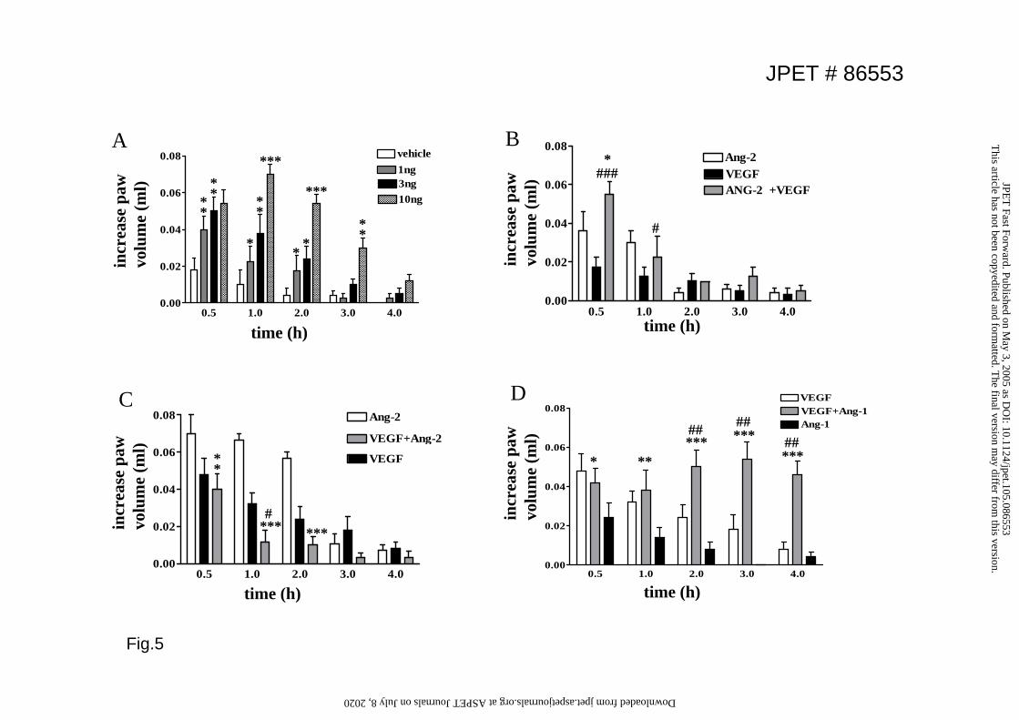
Fig.5
0.5 1.0 2.0 3.0 4.00.00
0.02
0.04
0.06
0.08 Ang-2
VEGF+Ang-2
VEGF
incr
ease
paw
vo
lum
e (m
l)
time (h)
incr
ease
paw
vo
lum
e (m
l)
time (h)0.5 1.0 2.0 3.0 4.0
0.00
0.02
0.04
0.06
0.08 vehicle1ng3ng10ng
*
***
***
**
***#
***
**
**
**
**
**
A
C
0.5 1.0 2.0 3.0 4.00.00
0.02
0.04
0.06
0.08Ang-2VEGFANG-2 +VEGF
incr
ease
paw
vo
lum
e (m
l)
time (h)
*###
#
B
incr
ease
paw
vo
lum
e (m
l)
time (h)
* *****## ***
##
0.5 1.0 2.0 3.0 4.00.00
0.02
0.04
0.06
0.08VEGFVEGF+Ang-1Ang-1
***##
D
JPET # 86553
This article has not been copyedited and form
atted. The final version m
ay differ from this version.
JPET
Fast Forward. Published on M
ay 3, 2005 as DO
I: 10.1124/jpet.105.086553 at ASPET Journals on July 8, 2020 jpet.aspetjournals.org Downloaded from


![INDEX [jpet.aspetjournals.org]jpet.aspetjournals.org/content/jpet/230/3/local/back-matter.pdf · histrionicotoxin effects (frogs), 619 ... distribution kinetics ana- ... and myocardium](https://img.pdfslide.us/doc/110x75/5b7ac0067f8b9ae1328d73ab/index-jpet-jpet-histrionicotoxin-effects-frogs-619-distribution-kinetics.jpg)


![INDEX [jpet.aspetjournals.org]jpet.aspetjournals.org/content/jpet/234/3/local/back...effect, 708 Blockade, reticuloendothelial, enzyme-al-bumin conjugates, chronic adininis-tration](https://img.pdfslide.us/doc/110x75/60757ab7f966210d5e51d2f2/index-jpet-jpet-effect-708-blockade-reticuloendothelial-enzyme-al-bumin.jpg)