Embed Size (px)
Citation preview

A NEW PRESENTATION OF COMBINATION SYNDROMEF Ahmad, N. Yunus, F McCord. A New Presentationof Combination Syndrome. Annal Dent Univ Malaya2008; 15(2): 94-99.
ABSTRACTThis article reviews the concept of CombinationSyndrome and presents a clinical case of a patientwith a modern variation to this clinical scenario': Theclinical procedures involved in the provision of amaxillary complete denture against a mandibularimplant-supported complete fixed prosthesis isdescribed with some suggestions on how to optimisethe treatment outcome for the patient.
Key words: dental implants, prostheses, 2}StCenturyCombination Syndrome; lingualised occlusion
Introduction
Combination Syndrome was originally termed byKelly (1) in 1972 to describe the clinical scenario ofa complete maxillary denture opposed by six or eightanterior mandibular teeth (Figure I). In addition, amandibular removable partial denture was typicallypresent, to restore the missing posterior mandibularteeth (Figure 2)
Kelly stated that patients in this categorypresented with 4 salient clinical findings:
• Fibrous (flabby) anterior maxillary ridges. Heconsidered that the effect of occlusal forces bythe mandibular teeth on the maxillary dentureanteriorly caused resorption of bone whichwas replaced by fibrous tissue.
• Relatively enlarged tuberosities which heconsidered to have "grown".
• Increased resorption of the residualmandibular ridge.
• Inflammation of the hard palate, often withpapillary hyperplasia present.
Whether these explanations are necessarily accurateor not is almost of no consequence, as the clinicianis, ultimately, faced with clinical difficulties which areof sufficient difficulty to present problems if anacceptable return to and of function are to beachieved.
In essence, the first three of these problem areasare essentially of a clinical complexity as to requiretreatment by a dental practitioner with someexperience in Removable Prosthodontist, notnecessarily a specialist in Prosthodontics.
Case Report
F. AhmadI, N. Yunus1, F. McCord2
1Department of Prosthetic DentistryFaculty of Dentistry, University of Malaya50603 Kuala Lumpur, MalaysiaTe~ 603-79674881Fax: 603-79674535Email: [email protected]
2Department of Restorative DentistryGlasgow Dental Hospital and School378, Sauchiehall StreetGlasgow, G23JZ
Correspondence author: Norsiah Yunus
Treatment of papillary hyperplasia should betreatable by a general dental practitioner using acombination of tissue conditioner and oral hygienemeasures, perhaps with appropriate antifungaltherapy in common with other types of denture-induced papillary inflammation.
The practising dentist is therefore faced withthree areas of difficulty which relate to:
I. Recording the definitive impression of themaxillary arch.
II. Restoration of the mandibular arch.
III. Recording appropriate intermaxillaryrelations
I. Recording the definitive impression of themaxillary arch.
When a markedly displaceable tissue is presentin the anterior maxillary ridge, then the problem isessentially one of support; the displaceable anterior(fibrous or flabby) ridge being more susceptible todisplacement / distortion during function. For thisreason, a minimally displacive impression (i.e. usingminimum pressure) technique is recommended asfollows (2):
o After recording the primary impression,make a 2mm spaced, non-perforated customtray.
o After securing a peripheral seal with tracingcompound, record the impression inmedium- bodied polyvinyl siloxane (PVS)impression material.
o Remove the area of the tray, including thePVS impression (Figure 3), re-insert the trayand inject light-bodied PVS into the areacorresponding to the fibrous ridge. The final

Figure I: Edentulous maxilla and partially dentatemandible of a patient without the prostheses in position.
Figure 2: Maxillary complete denture with mandibularKennedy class I removable partial denture.
Figure 3: Maxillary impression after modificationshowing the window made at around the region
of flabby area.
impression should record the flabby ridgewith minimal displacement (Figure 4).
o After placing the definitive impression in theappropriate disinfectant solution, pour themaster cast.
A New Presentation of Combination Syndrome 95
II. Restoring the mandibular arch.The restoration of missing posterior mandibular
teeth with a Kennedy class I type of removablepartial denture was widely adopted as a favouredtreatment option. In recent times, the need to restoremissing molar teeth, in appropriate cases, has beenquestioned by colleagues from Nijmegen (3) andhence the shortened dental arch philosophy evolvedwhereby it was shown that acceptable oral(masticatory) function may be maintained in reduceddentitions; this philosophy is now practiseduniversally. Shortened dental arch principles mayalso be used to avoid the biomechanical problemsinherent in the provision of Kennedy class Iremovable partial denture. The use of resin-bondedor conventional cantilevered (Figure 5) bridges toextend short arches opposing complete dentures hasbeen recommended (2) as an alternative to aconventional Kennedy I partial denture.
Whichever course of treatment is used, it shouldbe done at the same time as the replacementmaxillary complete denture ..
Figure 4: Definitive impression with the light-body PVS(arrows) recording the anterior flabby ridge.
Figure 5: Shortened dental arch showing cantilever fixedprosthesis to replace mandibular second premolar
on each side.
96 Annals of Dentistry, University of Malaya, Vol. 15 No.2 2008
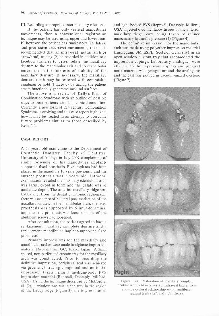
Figure 6: (a) Restoration of maxillary completedenture with gold overlays (b) Intraoral lateral view
showing occlusal relationship with mandibularnatural teeth (Left and right views).
and light-bodied PVS (Reprosil, Dentsply, Milford,USA) injected over the flabby tissues of the anteriormaxillary ridge, care being taken to reduceunnecessary hydraulic pressure (4) (Figure 4).
The definitive impression for the mandibulararch was made using polyether impression material(Impregum, 3M ESPE, Seefeld, Germany) in anopen window custom tray that accomodated theimpression copings. Laboratory analogues wereattached to the impression copings and gingivalmask material was syringed around the analogues;and the cast was poured in vacuum-mixed diestone(Figure 7).
CASE REPORT
III. Recording appropriate intermaxillary relations.If the patient has only vertical mandibular
movements, then a conventional registrationtechnique may be used using upper and lower rims.If, however, the patient has ruminatory (i.e. lateraland protrusive excursive) movements, then it isrecommended that an intra-oral (gothic arch orarrowhead) tracing (2) be recorded in addition to afacebow transfer to better relate the maxillarydenture to the mandibular axis and to mandibularmovement in the interests of stability of themaxillary denture. If necessary, the maxillarydenture teeth may be restored with composite,amalgam or gold (Figure 6) by having the patientcreate functionally-generated occlusal surfaces.
The above is a review of Kelly's form ofCombination Syndrome with an outline of possibleways to treat patients with this clinical condition.Currently, a new form of 2}Stcentury CombinationSyndrome is evolving and this case report highlightshow it may be treated in an attempt to overcomefuture problems similar to those described byKelly (I).
A 63 years old man came to the Department ofProsthetic Dentistry, Faculty of Dentistry,University of Malaya in July 2007 complaining ofslight looseness of his mandibular implant-supported fixed prosthesis. Five implants had beenplaced in the mandible 10 years previously and thecurrent prosthesis was 2 years old. Intraoralexamination revealed the maxillary edentulous archwas large, ovoid in form and the palate was ofmoderate depth. The anterior maxillary ridge wasflabby and, from the dental panaromic radiograph,there was evidence of bilateral pneumatisation of themaxillary sinuses. In the mandibular arch, the fixedprosthesis was supported by 5 intra-foraminalimplants; the prosthesis was loose as some of theabutment screws had loosened.
After consultation, the patient agreed to have areplacement maxillary complete denture and areplacement mandibular implant-supported fixedprosthesis.
Primary impressions for the maxillary andmandibular arches were made in alginate impressionmaterial (Aroma Fine, GC, Tokyo, Japan). A 2mmspaced, non-perforated custom tray for the maxillaryarch was constructed. Prior to recording thedefinitive impression, peripheral seal was achievedvia greenstick tracing compound and an initialimpression taken using a medium-body PVSimpression material (Reprosil, Dentsply, Milford,USA). Using the technique described by McCord etal. (2), a window was cut in the tray in the regionof the flabby ridge (Figure 3), the .tray re-inserted

A New Presentation of Combination Syndrome 97
Figure 10: Sectionedframeworkwhichwas replaced onthe implants and joined intraoraiJywith pattern resin.
The casts were mounted on a semiadjustablearticulator and the denture teeth were set-up withlingualized occlusion as this has been shown to bethe preferred occlusal form where stability of one orboth prostheses is compromised (5). The waxdentures were tried-in with the mandibular basesecured by the two impression copings and posts(Figure 8) and both prostheses assessed forocclusion, appearance and phonetics.
The resin framework for the mandibularprosthesis was fabricated using an autopolymerisingresin (GC Pattern Resin, Tokyo, Japan) on plasticUCLA type abutments (Renew Biocare Corp., Cal,USA). A full contour waxing on the resin frameworkwas completed (Figure 9) using the index of themandibular trial wax-up. The wax was cut back andthe assembly was cast in semi-precious alloy. Theframework was evaluated for fit in the mouth andas a rocking movement was detected, the frameworkwas sectioned and replaced on the implants. The gapwas filled with autopolymerising resin (GC PatternResin, Tokyo, Japan - Figure 10) and the frameworkwas soldered and retried in the mouth. Once theaccuracy of fit was satisfactory, the centric relationrecord was verified using reinforced wax (Aluwax,Mich., USA).
The casts were remounted on the articulator andporcelain was applied to the framework (Figure 11).A cobalt-chromium palatal baseplate was fabricatedfor the maxillary complete denture (Figure 12). Theteeth set-up of the complete denture was againchecked and adjusted for lingualized occlusionbefore processing (Figure 13).
At insertion stage, the mandibular implant-supported bridge were screwed in and the occlusionwith the maxillary complete denture was adjusted.Articulating paper was used for occlusal adjustmentin centric and lateral excursions. The patient wasinstructed on oral and denture hygiene. The patientwas reviewed at one week post-insertion (Figure 13).The importance of regular review visits wasemphasized to the patient because of thecomplicated design of mandibular prosthesis and thepotential for Combination syndrome as, although
Figure 9: Occlusalviewof full contour wax-up showingaccessholes for screw-retainedmandibular prosthesis.
Figure 7: Mandibular working stone cast with soft tissueproduced from gingivalmask around the implant
analogues.
Figure 8: Two impressioncopings and posts wereused tostabilize the mandibular wax denture during teeth try-in
(the posts were shorten before the occlusionwaschecked).
The intermaxillary relations were recorded usingthe conventional method as in complete dentureconstruction except that, for this case, themandibular record base was stabilized byincorporating two impression posts into the recordbase. Stabilisation of the record base afforded easierrecording of the jaw relationship and this isparticularly helpful in cases where the mandibularridge is severely resorbed. Facebow registration andcentric relation record were made.
98 Annals of Dentistry, University of Malaya, Vol. 15 No.2 2008
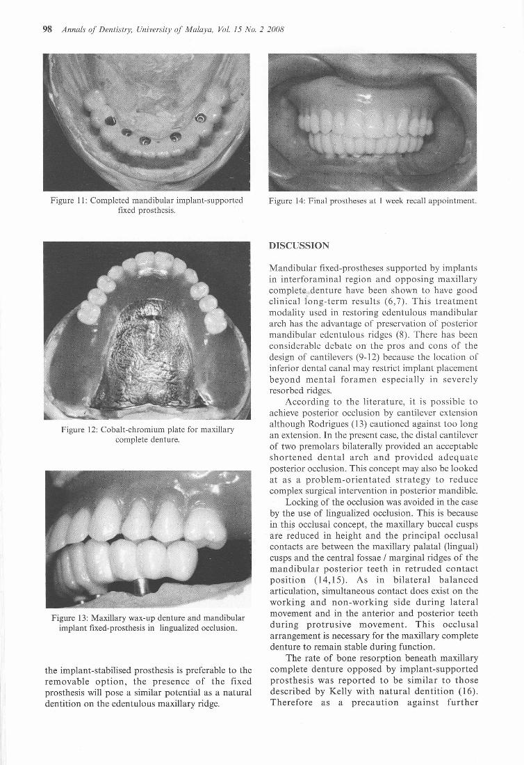
Figure II: Completed mandibular implant-supportedfixed prosthesis.
Figure 12: Cobalt-chromium plate for maxillarycomplete denture.
Figure 13: Maxillary wax-up denture and mandibularimplant fixed-prosthesis in lingualized occlusion.
the implant-stabilised prosthesis is preferable to theremovable option, the presence of the fixedprosthesis will pose a similar potential as a naturaldentition on the edentulous maxillary ridge.
Figure 14: Final prostheses at 1 week recall appointment.
DISCUSSION
Mandibular fixed-prostheses supported by implantsin interforaminal region and opposing maxillarycomplete- denture have been shown to have goodclinical long-term results (6,7). This treatmentmodality used in restoring edentulous mandibulararch has the advantage of preservation of posteriormandibular edentulous ridges (8). There has beenconsiderable debate on the pros and cons of thedesign of cantilevers (9-12) because the location ofinferior dental canal may restrict implant placementbeyond men tal foramen especially in severelyresorbed ridges.
According to the literature, it is possible toachieve posterior occlusion by cantilever extensionalthough Rodrigues (13) cautioned against too longan extension. In the present case, the distal cantileverof two premolars bilaterally provided an acceptableshortened dental arch and provided adequateposterior occlusion. This concept may also be lookedat as a problem-orientated strategy to reducecomplex surgical intervention in posterior mandible.
Locking of the occlusion was avoided in the caseby the use of lingualized occlusion. This is becausein this occlusal concept, the maxillary buccal cuspsare reduced in height and the principal occlusalcontacts are between the maxillary palatal (lingual)cusps and the central fossae / marginal ridges of themandibular posterior teeth in retruded contactposi tion (14,15). As in bilateral balancedarticulation, simultaneous contact does exist on theworking and non-working side during lateralmovement and in the anterior and posterior teethduring protrusive movement. This occlusalarrangement is necessary for the maxillary completedenture to remain stable during function.
The rate of bone resorption beneath maxillarycomplete denture opposed by implant-supportedprosthesis was reported to be similar to thosedescribed by Kelly with natural dentition (16).Therefore as a precaution against further

deterioration in the anterior maxillary ridge, noanterior tooth contact in maximum occlusion wasplanned for this patient. This follows therecommendation made by Lang and Razzoog (5)when providing patients with mandibular implant-supported fixed prosthesis.
CONCLUSION
The concept of Combination Syndrome is reviewedand the implications that current techniques maylead to a re-emergence of this condition areconsidered. In consequence, some clinical proceduresare highlighted, such as the impression technique,prosthesis design and occlusal scheme appropriate inthe rehabilitation of this type of patient.
REFERENCES
I. Kelly E. Changes caused by a mandibularremovable partial denture opposing a maxillarycomplete denture. 1 Prosthet Dent. 1972; 27:140-50.
2. McCord 1F, Smith P, Grey, N. Treatment ofEdentulous Patients; Churchill Livingstone 2004.
3. Witter 01, van Elteren P, Kayser AF. Migrationof teeth in shortened dental arches. 1 OralRehabi!. 1987; 14: 321-9.
4. AI-Ahmad A, Masri R, Driscoll CF, vonFraunhofer 1, Romberg E. Pressure generated ona simulated mandibular oral analog byimpression materials in custom trays of differentdesign. 1 Prosthodont. 2006; 15: 95-101.
5. Lang BR and Razzoog ME. LingualisedOcclusion: Tooth molds and an Occlusal schemefor edentulous implant patients. Implant Dent.1992; I: 204-211.
6. Lindquist LW, Carlsson GE, 1emt T. Aprospective IS-year follow-up study ofmandibular fixed prostheses supported byosseointegrated implants. Clinical results andmarginal bone loss. Clin Oral Implants Res.1996; 7: 329-36.
A New Presentation of Combination Syndrome 99
7. Wennerberg A, Carlsson GE, 1emt T. Influenceof occlusal factors on treatment outcome: astudy of 109 consecutive patients withmandibular implant-supported fixed prosthesesopposing maxillary complete dentures. Int 1Prosthodont. 2001; 14: 550-5.
8. Sennerby L, Carlsson GE, Bergman B,Warfvinge 1. Mandibular bone resorption inpatients treated with tissue-integrated prosthesesand in complete-denture wearers. Acta OdontolScand: 1988; 46: 135-40.
9. Adell R, Lekholm D, Rockier B, Branemark PI.A IS-year study of osseointegrated implants inthe treatment of the edentulous jaw. Int 1 OralSurg. 1981; 10: 387-416.
10. Branemark PI, Hansson BO, Adell R, Breine D,Lindstrom 1, Hallen 0, Ohman A.Osseointegrated implants in the treatment of theedentulous jaw. Experience from a 10-yearperiod. Scand 1 Plast Reconstr Surg Supp!. 1977;16: 1-132.
II. T Albrektsson, A multicenter report ofosseointegrated oral implants. 1 Prosthet Dent;1988: 60. 75-84.
12. Brosky ME, Korioth TW, Hodges 11. Theanterior cantilever in the implant-supportedscrew-retained mandibular prosthesis. 1 ProsthetDent. 2003; 89: 244-9.
13. Rodriguez AM, Aquilino SA, Lund PS.Cantilever and implant biomechanics: a reviewof the literature, 1 Prosthodont 1994; 3: 114-8.
14. Lang BR. Complete denture occlusion. DentClin North Am. 2004; 48: 641-65.
IS. Garcia LT, Bohnenkamp OM. Lingualizedocclusion: an occlusal solution for edentulouspatients. Pract Proced Aesthet Dent. 2005; 17: 5.
16. Barber HD, Scott RF, Maxson BB, Fonseca R1.Evaluation of anterior maxillary alveolar ridge
6$ resorption when opposed by thetransmandibularimplant. 1 Oral MaxillofacSurg. 1990; 48: 1283-7.