Embed Size (px)
Citation preview
Anemia-2: Overview and Select Cases
Marc Zumberg Associate Professor
Division of Hematology/Oncology
Initial Laboratory • Complete blood count with indices
– MCV-indication of RBC size – RDW-indication of RBC size variation
• Examination of the peripheral blood smear
• Reticulocyte count-measurement of newly
produced young rbc’s • Stool guiac
Additional Labs • Vitamin levels--FE battery, Vit-b12, folate • LDH, bilirubin, haptoglobin • Coomb’s test • Hemoglobin electrophoresis • SPEP • Creatinine • TSH • urinalysis • Consider bone marrow examination
Anemia: Morphology Classification
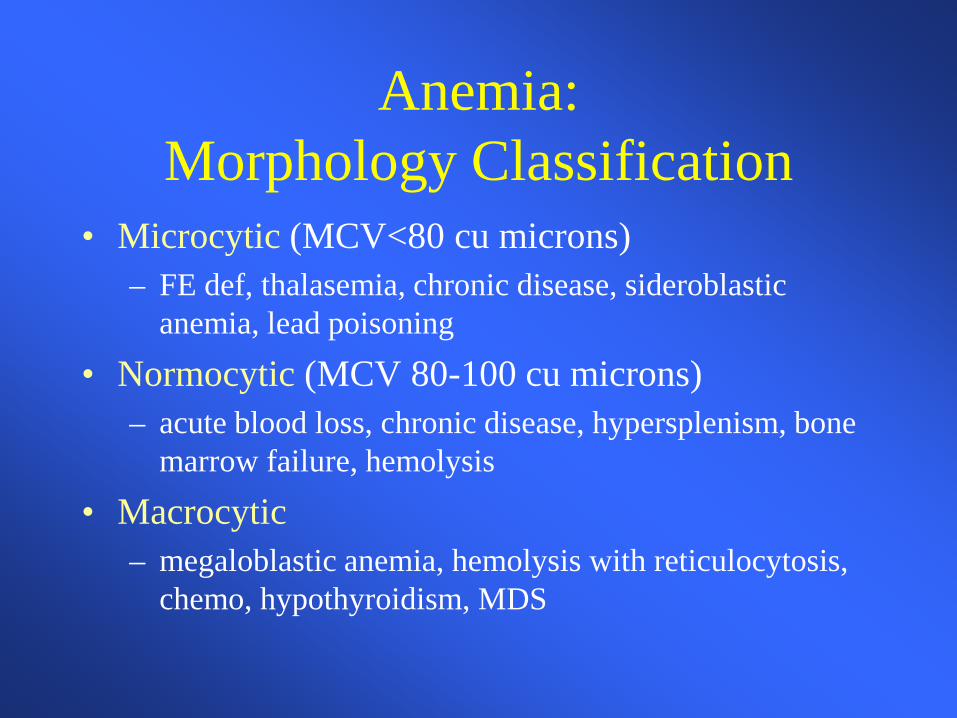
• Microcytic (MCV<80 cu microns) – FE def, thalasemia, chronic disease, sideroblastic
anemia, lead poisoning • Normocytic (MCV 80-100 cu microns)
– acute blood loss, chronic disease, hypersplenism, bone marrow failure, hemolysis
• Macrocytic – megaloblastic anemia, hemolysis with reticulocytosis,
chemo, hypothyroidism, MDS
Anemia-Case 1
• Fe deficiency – Diagnosis – Differential – Iron studies – Treatment – Length of therapy
Case 2
• B-12 deficiency – Differential diagnosis – Pathophysiology – Diagnostic entities – Associated conditions – Treatment Options
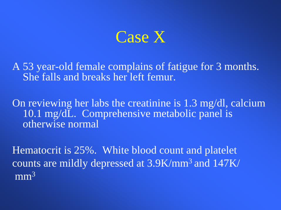
Case X A 53 year-old female complains of fatigue for 3 months.
She falls and breaks her left femur. On reviewing her labs the creatinine is 1.3 mg/dl, calcium
10.1 mg/dL. Comprehensive metabolic panel is otherwise normal
Hematocrit is 25%. White blood count and platelet counts are mildly depressed at 3.9K/mm3 and 147K/ mm3
Case X
• What is the most likely diagnosis?
• How do you want to proceed with your evaluation? – What is the next step? – How would you make the definitive diagnosis?
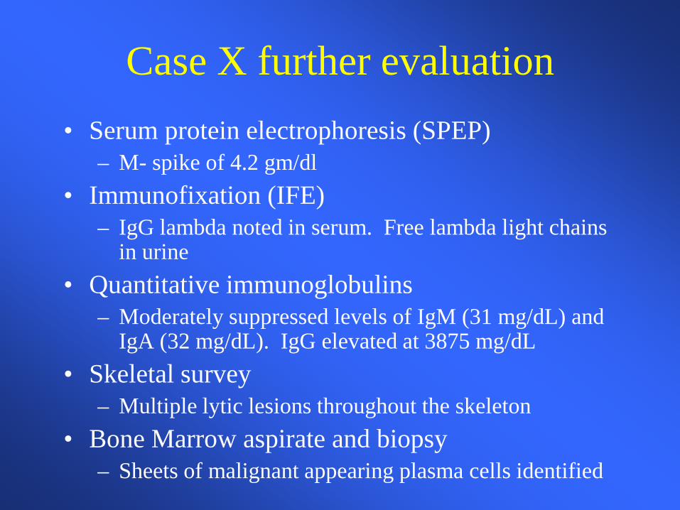
Case X further evaluation • Serum protein electrophoresis (SPEP)
– M- spike of 4.2 gm/dl • Immunofixation (IFE)
– IgG lambda noted in serum. Free lambda light chains in urine
• Quantitative immunoglobulins – Moderately suppressed levels of IgM (31 mg/dL) and
IgA (32 mg/dL). IgG elevated at 3875 mg/dL • Skeletal survey
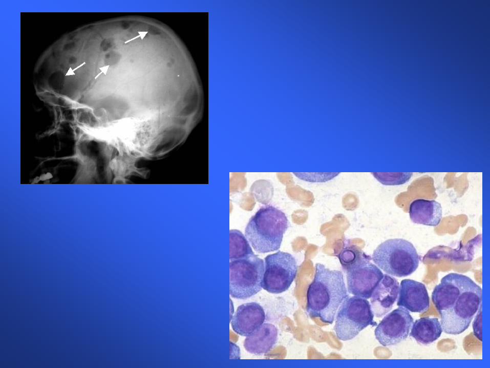
– Multiple lytic lesions throughout the skeleton • Bone Marrow aspirate and biopsy
– Sheets of malignant appearing plasma cells identified
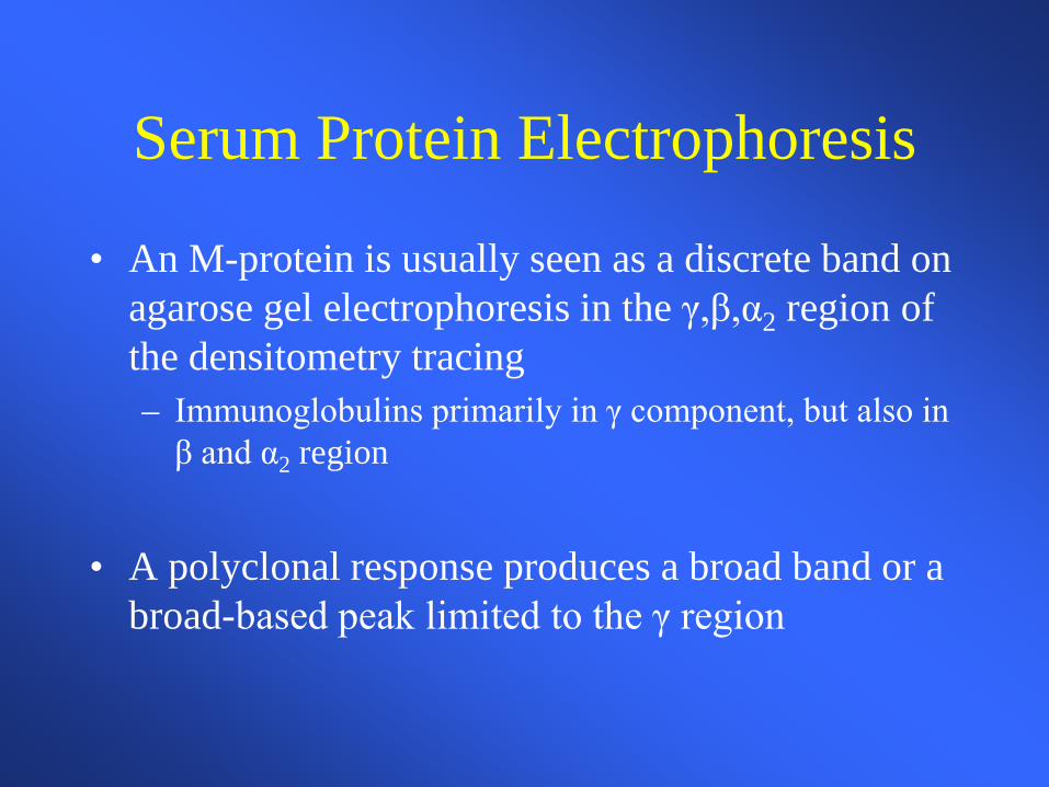
Serum Protein Electrophoresis
• An M-protein is usually seen as a discrete band on agarose gel electrophoresis in the γ,β,α2 region of the densitometry tracing – Immunoglobulins primarily in γ component, but also in
β and α2 region
• A polyclonal response produces a broad band or a broad-based peak limited to the γ region
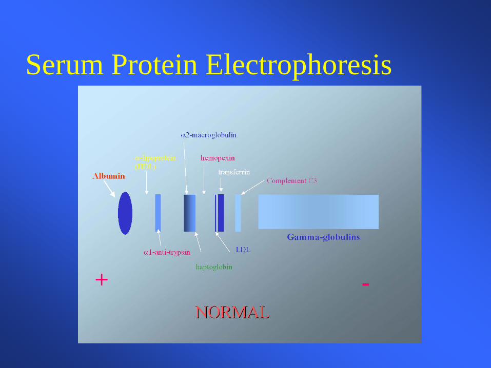
Serum Protein Electrophoresis
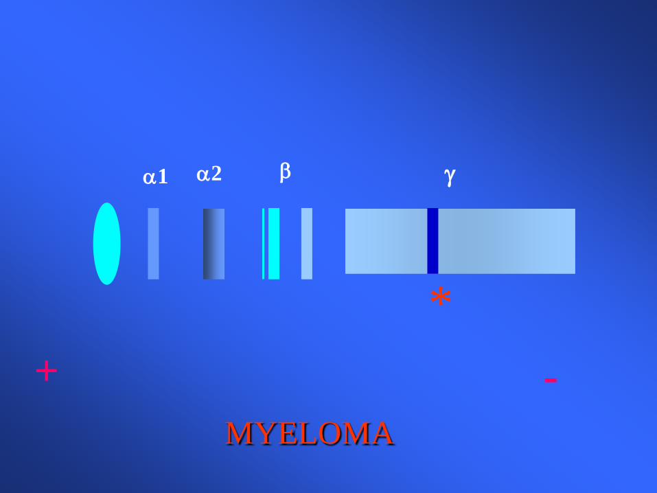
+ - MYELOMA
α1 α2 β γ
*
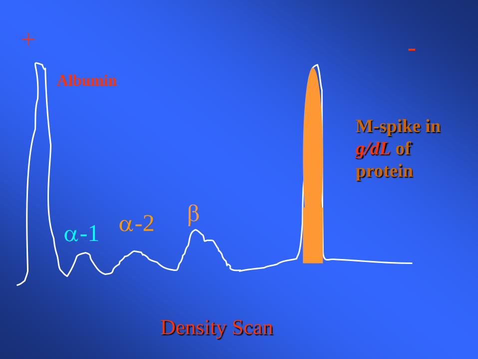
+ - Albumin
α-1 α-2
β
Density Scan
M-spike in g/dL of protein
When to order a SPEP
• When you suspect multiple myeloma, Waldenstrom’s macroglobulinemia or amyloidosis
• With unexplained – Weakness, fatigue, anemia, back pain, fractures, hypercalcemia,
renal insufficiency – Recurrent infections – Sensorimotor neuropathy, carpel tunnel syndrome, CHF, syncope
Immunofixation (IFE)
• Proteins are fractioned on electrophoretic strips – Each lane overlaid with monospecific antisera
against IgG, IgA, IgM, and light chains
– Immunoglobulins are precipitated by antisera • Wash away nonprecipitated proteins
– Precipitated proteins are stained
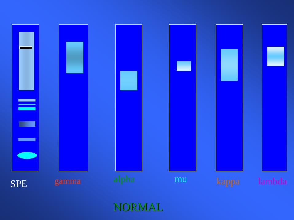
gamma alpha
mu kappa lambda SPE
NORMAL
When to order IFE • To type the paraprotein (M spike) identified on SPEP
• Further evaluate an equivocal SPEP
• To search for a low level paraprotein with a negative SPEP
– clinical suspicion of a lymphoplasmacytic disorder is high – unexplained symptoms such as neuropathy, renal failure, etc..
• When searching for Bence-Jones proteinuria
• In treated myeloma patients with a negative SPEP
Quantitative Immunoglobulins • Useful to quantitate
– the amount of monoclonal protein – suppression of uninvolved immunoglobulins in a
monoclonal disorder – identify a congenital or acquired deficiency state of an
individual immunoglobulin
• Serum light chain assays are newer and very sensitive technique for measuring serum light chains – Adjunct or replacement of urine protein electrophoresis
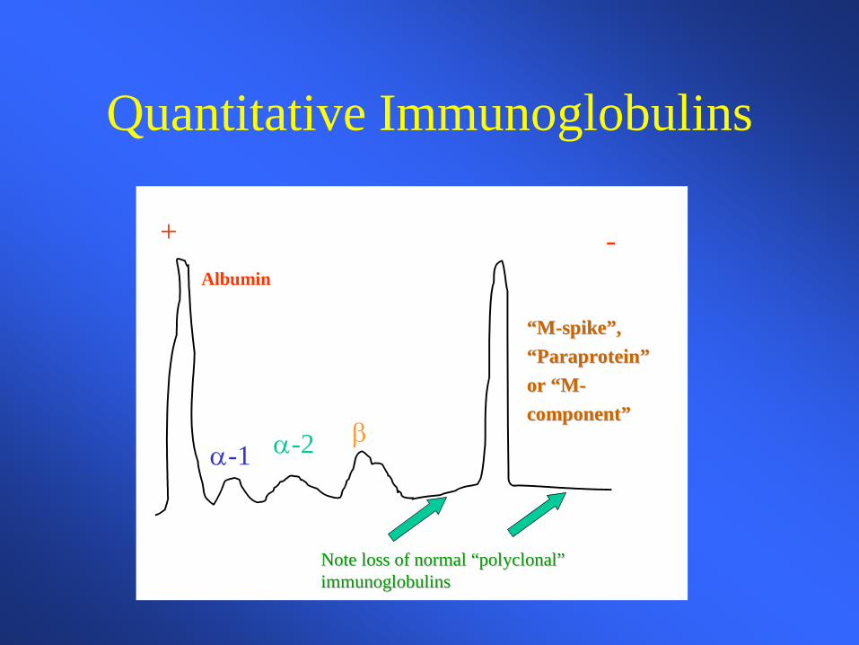
Quantitative Immunoglobulins
+ -Albumin
α-1 α-2 β
Note loss of normal “polyclonal” Note loss of normal “polyclonal” immunoglobulinsimmunoglobulins
“M“M--spike”, spike”, “Paraprotein” “Paraprotein” or “Mor “M--component”component”
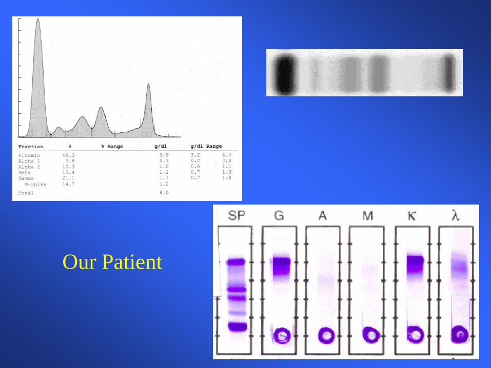
Our Patient
Stage III multiple myeloma The patient is started on systemic chemotherapy and an
autologous bone marrow transplant is planned • Multiple new and effective agents exist for MM
– Thalidomide or Revlimid and other anti-angiogenic agents – Proteosome inhibitors (Velcade) – Double autologous transplant – Mini allogeneic transplant
• Remains non-curative except possibly for allogeneic
transplant
Case 3
A woman in her mid thirties has suffered from episodes of pleurisy and has been told she may have SLE. Her prior blood cell counts have been normal. She now complains of exertional dyspnea, fatigue and yellowing of her eyes. Physical exam is normal except for mild scleral icterus, and moderate splenomegaly.
Case 3
Initial labs reveal: hgb=7.9 gm/dl hct=23.9% wbc=4000 /mm3 with a normal differential platelet=138,000 /mm3 What other initial labs / values would you like
to see?
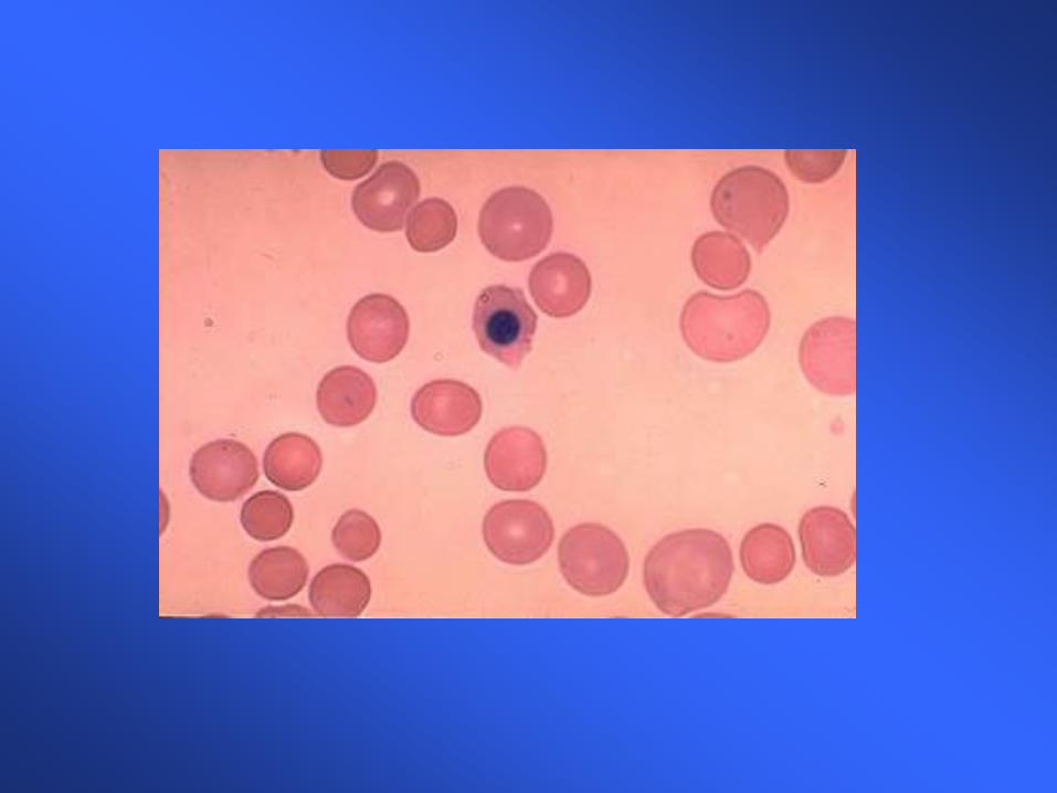
Case 3
MCV=114 cubic microns Retic count=14.2% uncorrected LDH=2343 U/L Bilirubin=4.3 mg/dl, .8 direct mg/dl The peripheral blood smear reveals
macrovalocytes, polychromasia and an occasional nucleated red blood cell.
Case 3
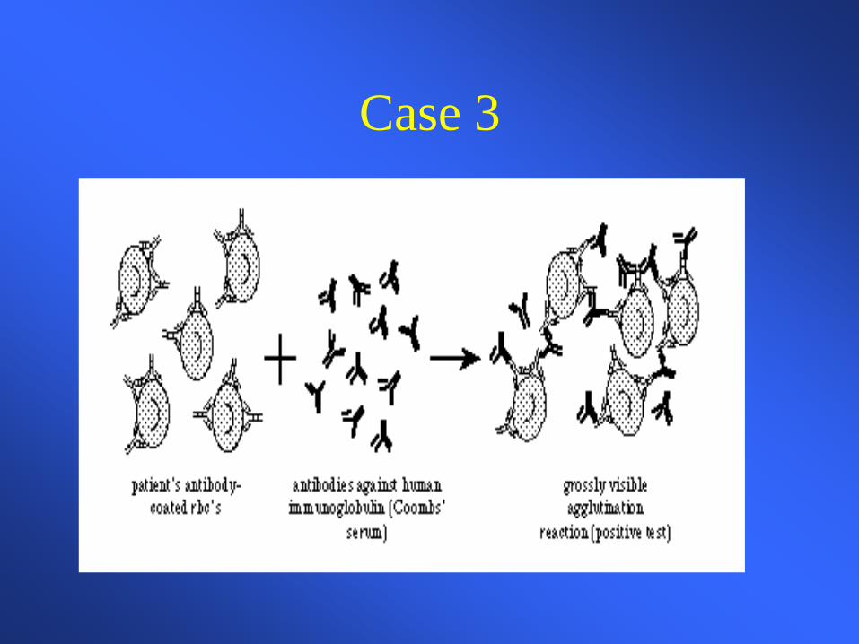
You suspect an autoimmune hemolytic anemia based on her history, PE, labs and smear. What further tests would you like to order?
Case 3
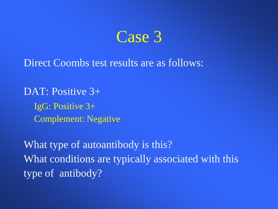
Case 3 Direct Coombs test results are as follows: DAT: Positive 3+ IgG: Positive 3+ Complement: Negative What type of autoantibody is this? What conditions are typically associated with this type of antibody?
Case 3
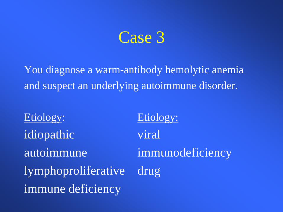
You diagnose a warm-antibody hemolytic anemia and suspect an underlying autoimmune disorder. Etiology: Etiology: idiopathic viral autoimmune immunodeficiency lymphoproliferative drug immune deficiency
Case 3
You decide to avoid blood transfusion because of: 1.) the difficulties in obtaining cross match
compatible blood AND 2.) the expected short half life of transfused blood What treatment do you recommend?
Case 3
TREATMENT • Prednisone • Splenectomy • Immunosuppressive agents
– cytoxan, immuran, cyclosporin • Danazol • Plasma exchange / immunoadsorption
Case 3
You begin her on 60mg Prednisone and she has a good response. However upon several attempts at tapering the prednisone she has a relapse with worsening of the hemolytic anemia. As you are concerned about the long term side effects of Prednisone you send her for a Procedure.
Case 3
She responds well to splenectomy, but still requires very low maintenance doses of Prednisone. Over the years she occasionally requires higher doses of prednisone when she is ill or has a lupus flare
Case 4
Mrs. P is a previously healthy 32 year-old female with no prior medical history. She reports feeling “unwell” for the past month. However in the past three days she has experienced fevers, a ‘rash” on her legs, weakness and shortness of breath. She has been to weak to get out of bed the last 24 hours and has experienced spontaneous nose bleeds. How is this case different from the previous?
Case 4
Physical exam reveals an ill appearing female: T=38.6, pulse 122, bp123/54 dried blood in the nares, pale conjunctiva, dried mucous membrane tachycardia and tachypnea pettechia over the lower extremities
Case 4
Routine labs include: WBC=1700/mm3 Hgb=5.6 gm/dl Hct=18.2% Plt=12,000/mm3 What important piece of laboratory information is missing from the WBC?
Case 4
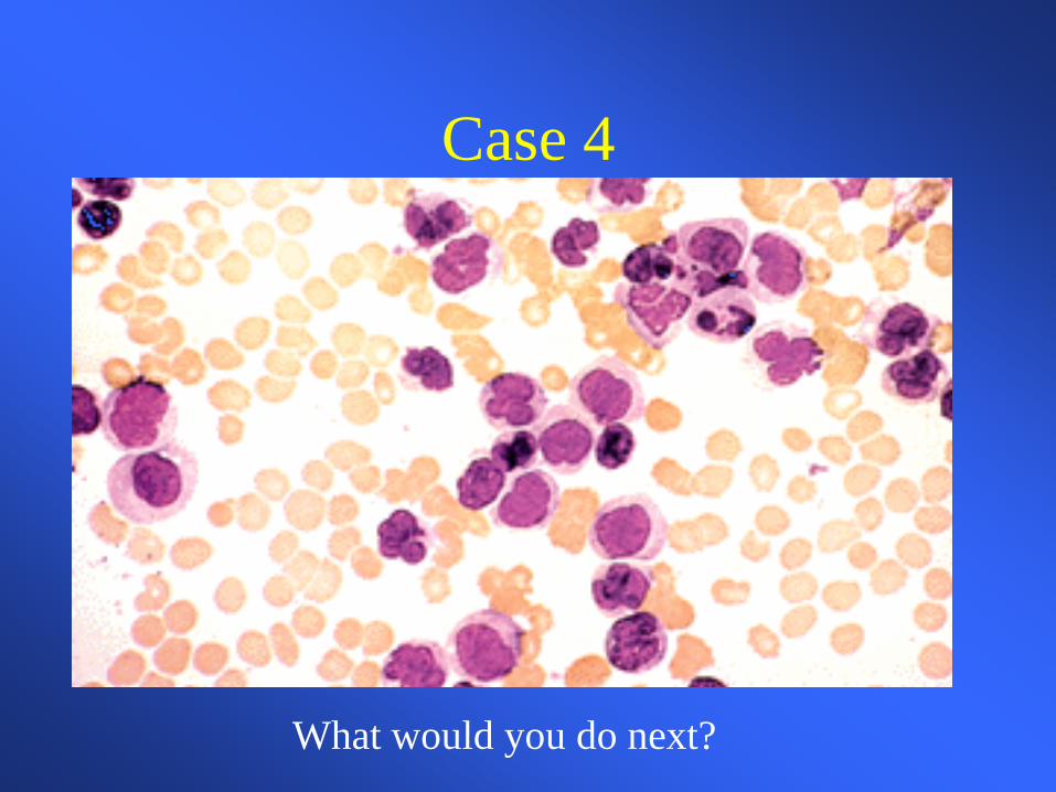
Differential of the white blood cell count reveals at least 50% of young white cells, the lab thinks are blasts reticulocyte count corrected is 0.4% electrolytes are consistent with dehydration potassium and creatinine are slightly elevated
Case 4
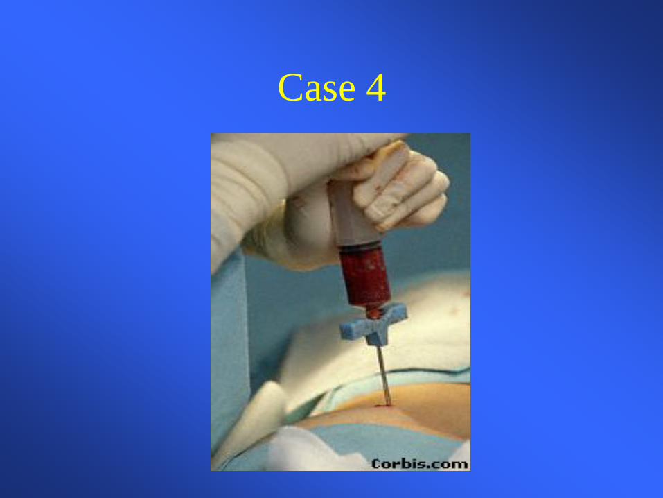
What would you do next?
Case 4
Case 4
Case 4
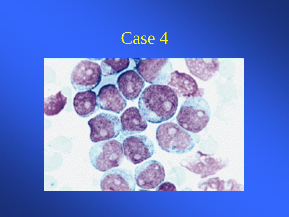
Hematopathology evaluation revealed AML subtype M1 ? Chromosomal studies did not reveal any abnormalities What should you do prior to beginning chemotherapy in this patient?
Case 4
The patient tolerated induction chemotherapy well and remains in remission 4 months after completing induction and consolidation chemotherapy. Her blood counts have all now normalized. If she was to relapse what therapy would you recommend?
Case 5 You are referred a pleasant 34 year-old African American female who has been known to have a mild microcytic anemia, which was picked up some years ago on routine blood work. She is entirely asymptomatic. She has been prescribed iron several times over the years without a response. What data would you like to review?
Case 5
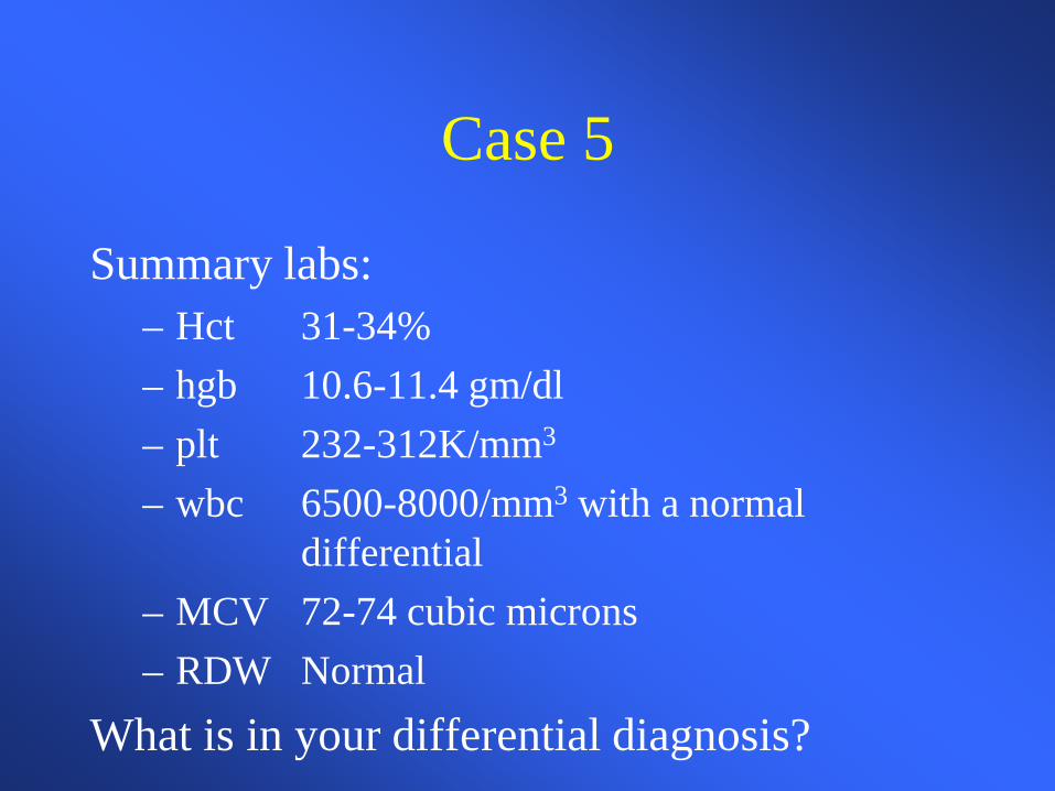
Summary labs: – Hct 31-34% – hgb 10.6-11.4 gm/dl – plt 232-312K/mm3 – wbc 6500-8000/mm3 with a normal
differential – MCV 72-74 cubic microns – RDW Normal
What is in your differential diagnosis?
Case 5 • FE deficiency anemia
– noncompliance, inadequate dosing, incorrect formulation
• Beta-thalassemia • Alpha-thalassemia • Anemia of chronic inflammation/ disease • Sideroblastic anemia • Lead What further laboratory tests would you like to order?
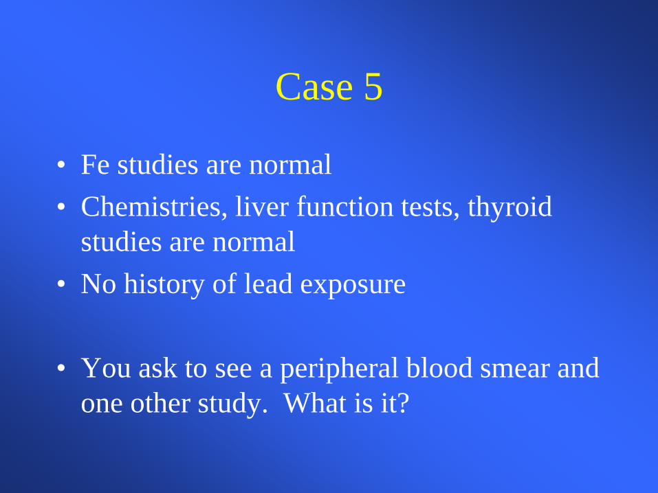
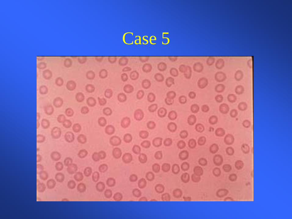
Case 5
• Fe studies are normal • Chemistries, liver function tests, thyroid
studies are normal • No history of lead exposure
• You ask to see a peripheral blood smear and
one other study. What is it?
Case 5
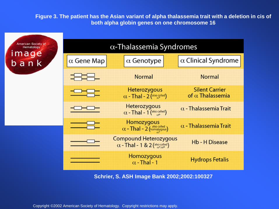
Copyright ©2002 American Society of Hematology. Copyright restrictions may apply.
Schrier, S. ASH Image Bank 2002;2002:100327
Figure 3. The patient has the Asian variant of alpha thalassemia trait with a deletion in cis of both alpha globin genes on one chromosome 16
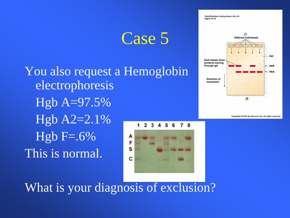
Case 5
You also request a Hemoglobin electrophoresis
Hgb A=97.5% Hgb A2=2.1% Hgb F=.6% This is normal. What is your diagnosis of exclusion?
Case 5
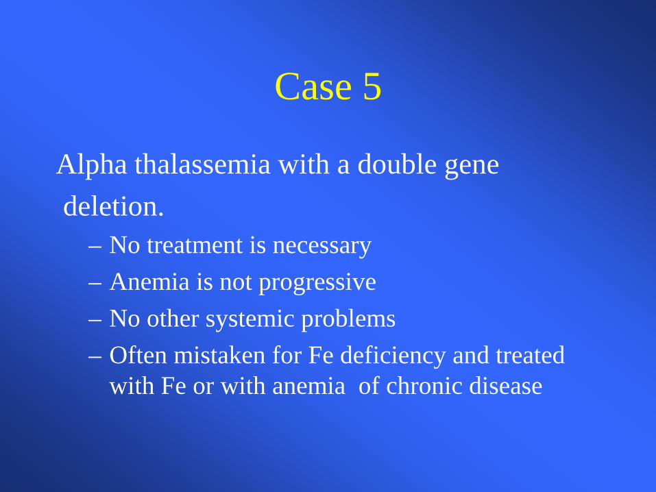
Alpha thalassemia with a double gene deletion.
– No treatment is necessary – Anemia is not progressive – No other systemic problems – Often mistaken for Fe deficiency and treated
with Fe or with anemia of chronic disease
Case 6
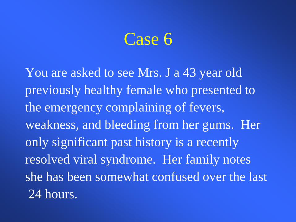
You are asked to see Mrs. J a 43 year old previously healthy female who presented to the emergency complaining of fevers, weakness, and bleeding from her gums. Her only significant past history is a recently resolved viral syndrome. Her family notes she has been somewhat confused over the last 24 hours.
Case 6
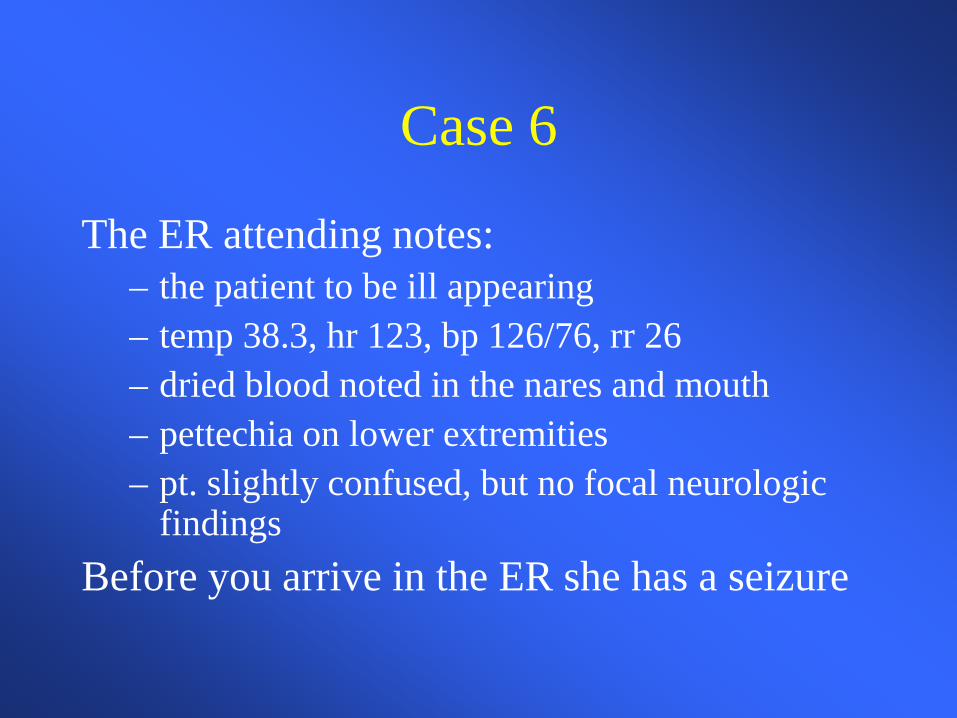
The ER attending notes: – the patient to be ill appearing – temp 38.3, hr 123, bp 126/76, rr 26 – dried blood noted in the nares and mouth – pettechia on lower extremities – pt. slightly confused, but no focal neurologic
findings Before you arrive in the ER she has a seizure
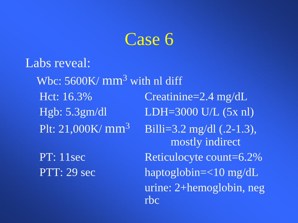
Case 6 Labs reveal: Wbc: 5600K/ mm3 with nl diff Hct: 16.3% Creatinine=2.4 mg/dL Hgb: 5.3gm/dl LDH=3000 U/L (5x nl) Plt: 21,000K/ mm3 Billi=3.2 mg/dl (.2-1.3),
mostly indirect PT: 11sec Reticulocyte count=6.2% PTT: 29 sec haptoglobin=<10 mg/dL urine: 2+hemoglobin, neg
rbc
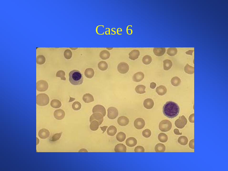
Case 6
Case 6
What is your differential diagnosis based on the history, physical, labs and blood smear What else could give you a similar blood smear?
Case 6
You diagnosis TTP based on the classic pentad and consistent blood smear:
– microangiopathic hemolytic anemia – thrombocytopenia – fever – renal failure – MS changes
What is the first line treatment of TTP?
Case 6 You begin daily plasma exchange procedures using FFP as your replacement fluid. Her mental status improves by the next day. Her platelet count normalizes and LDH decreases over the next week. Her anemia slowly improves as does her renal failure. She is weaned off plasma exchange and has a single relapse, which responds to similar therapy.
Pathophysiology
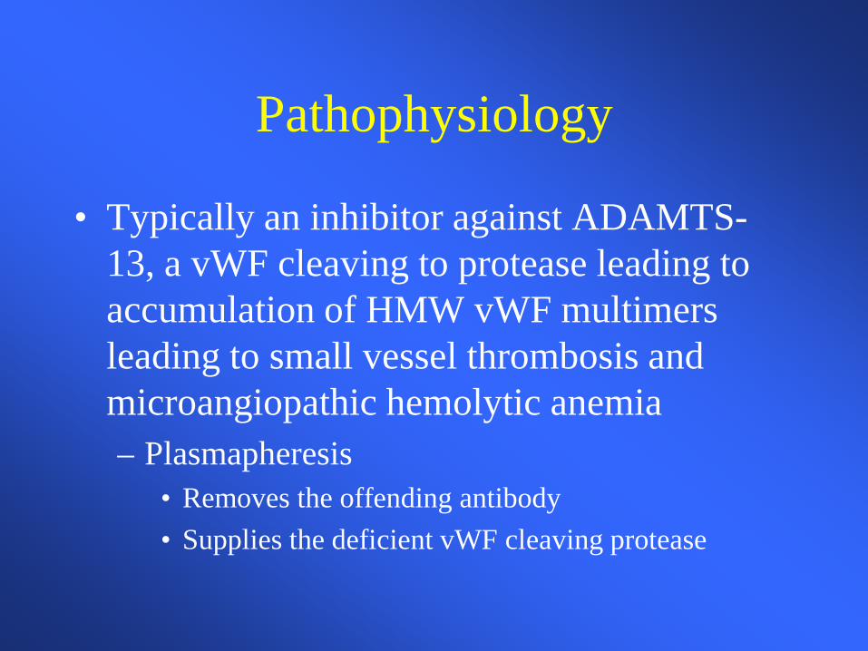
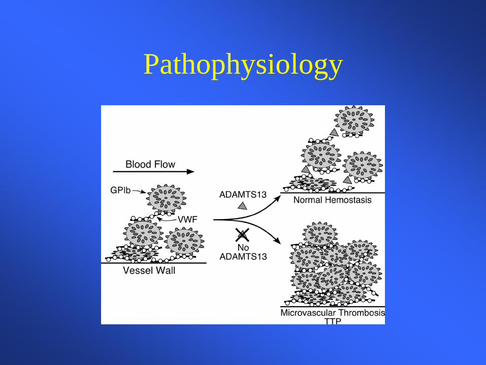
• Typically an inhibitor against ADAMTS-13, a vWF cleaving to protease leading to accumulation of HMW vWF multimers leading to small vessel thrombosis and microangiopathic hemolytic anemia – Plasmapheresis
• Removes the offending antibody • Supplies the deficient vWF cleaving protease
Pathophysiology
Case 7 A 36 year-old white male with a history of progressive renal failure over the last 4 years comes to you for work-up of weakness and progressive anemia. He has recently begun dialysis. His renal failure initially appeared after an acute febrile illness and the etiology was never determined.
Case 7
Physical exam is only significant for pale sclera and an AV shunt in his left arm CBC revealed: Wbc=6400/ mm3 MCV=78 / mm3 Hgb=7.6gm/dl Plt=242K/ mm3 Hct=23.8% What further test would you like to order?
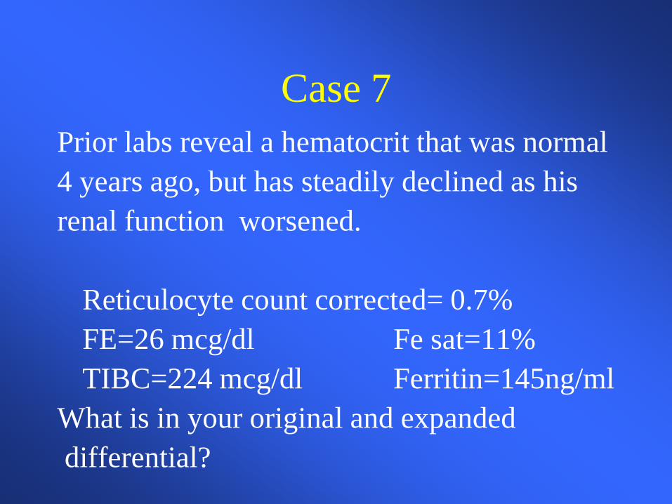
Case 7 Prior labs reveal a hematocrit that was normal 4 years ago, but has steadily declined as his renal function worsened. Reticulocyte count corrected= 0.7% FE=26 mcg/dl Fe sat=11% TIBC=224 mcg/dl Ferritin=145ng/ml What is in your original and expanded differential?
Case 7
Case 7 • Anemia of renal failure • Fe deficiency anemia • Thalassemia • Lead poisoning • Myelodysplasia • Mixed microcytic and macrocytic anemia
You request one more study to support your suspicion. What is it?
Case 7
You order an erythropoietin level which returns at 12 IU ( 4.1-19.5 ) within the normal limits. What do you make of this value? Should you proceed to a bone marrow exam?
Case 7
You place the patient on 10,000 units of EPO SQ tiw with dialysis. He comes to see you in one month feeling better. Hct=34% Retic corrected=3.2% Hgb=11.2 gm/dl MCV=83 mm3

Case 7
He continues on his EPO injections for four months. Soon after he again becomes fatigued, but otherwise doing well. Labs reveal: Wbc=6.2K/ mm3 Plt=234K/ mm3 Hct=27% MCV=76 cubic microns hgb=8.9 gm/dl Retic corr=.6%
Case 7
Case 7
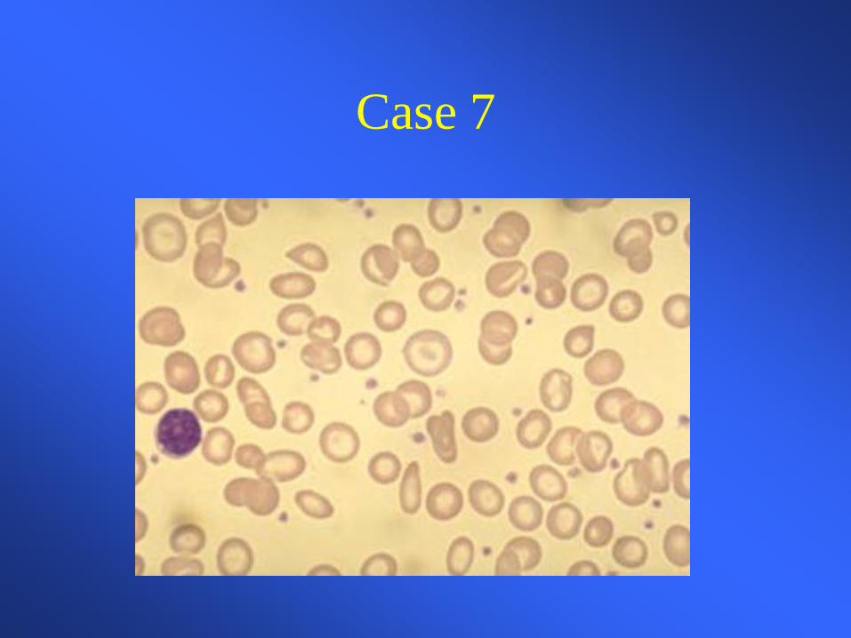
You suspect with his increased reticulocytosis and hgb/hct over the last few months he has become iron deficient. You confirm this with lab assay. You give him 1 gram of intravenous iron and ask him to come back in 2 weeks.
Case 7
He again feels much improved and is tolerating the iron fairly well. His hematocrit has increased to 33%, corrected reticulocyte count to 3.2%, and the MCV is now 80 cubic microns. He continues on Iron and EPO injections and is considering renal transplant
Anemia of Chronic Inflammation-Definition
• Mild to moderate anemia that is persistent for greater than 1-2 months in patients with infectious, inflammatory, or neoplastic diseases – Other causes excluded – Hypoproliferative-low reticulocyte count – Normocytic-MCV 80-100
• May be microcytic in later stages – Iron studies show low serum iron, low percent saturation, and
normal to elevated ferritin • Adequate reticuloendothelial iron stores
Anemia of chronic inflammation • Modest (<10%) shortening of RBC life span
– RBC life span may be decreased to 90 days – Creates increased demand for RBC production by the bone marrow
• Blunting of the expected production of EPO
– Inappropriately normal levels
• Decreased response of erythroid progenitors to EPO
• Impaired mobilization of reticuloendothelial iron stores and decreased absorption of intestinal iron
– Advantageous to the host sequester iron from bacterial pathogens with infectious etiology
– May not be adaptive and even detrimental in non-infectious etiology – Key role of hepcidin
Conclusions
The causes of anemia are very diverse, but the
workup can be successfully guided by only
a
few initial tests
Case 7
• Prior CBCP • Family history • Recent CBCP
– attention to MCV – attention to other blood counts
• Peripheral Smear • Reticulocyte count
Case #1 A 45 yo white male presents to his physician complaining of tiredness and fatigue over the last several months. He is otherwise healthy. His only other complaint is vague abdominal discomfort. The only medications he takes are daily NSAIDS for a knee injury he sustained while playing tennis. You are concerned he might be anemic. What questions should you ask in the H and P?
Case #1
• Progression of symptoms • Blood in stool • Other meds or toxins, including ETOH • Prior history of anemia, prior cbc values • Other constitutional symptoms, recent illness • family history • diet • craving ice or starch
Case #1
The patient does note he has had black tarry stools for a few weeks prior to his visit. You suspect GI blood loss induced by NSAID use. What should you key in on the physical exam?
Case #1
• Vital signs • pallor • skin color, turgor • mucous membranes, glossitis • liver, spleen size • rectal exam with guiac of stool
Case #1
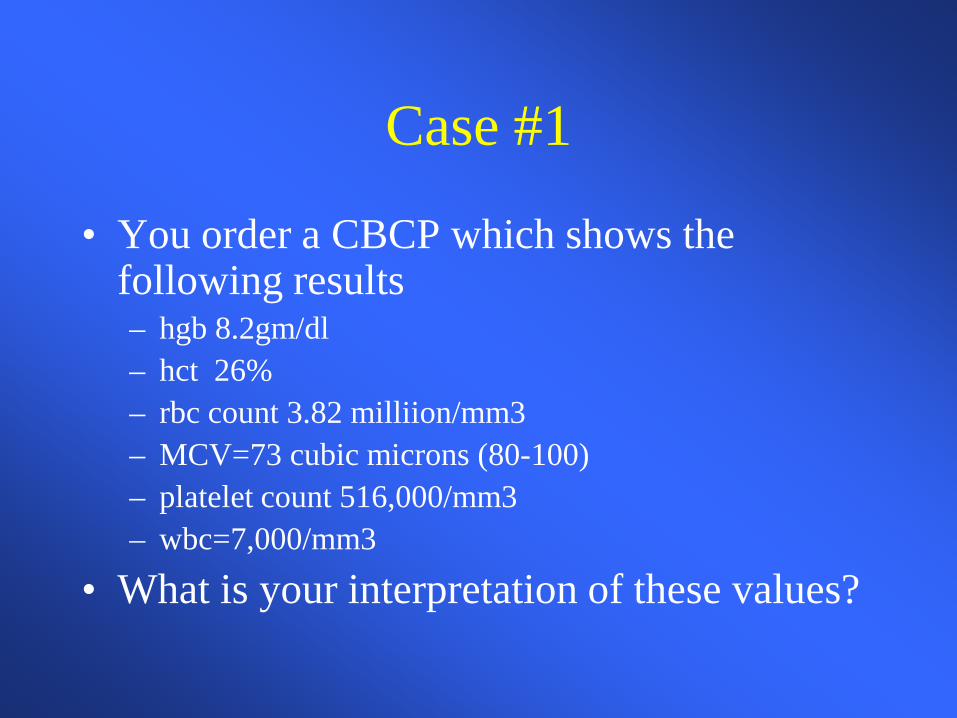
• You order a CBCP which shows the following results – hgb 8.2gm/dl – hct 26% – rbc count 3.82 milliion/mm3 – MCV=73 cubic microns (80-100) – platelet count 516,000/mm3 – wbc=7,000/mm3
• What is your interpretation of these values?
Case 1
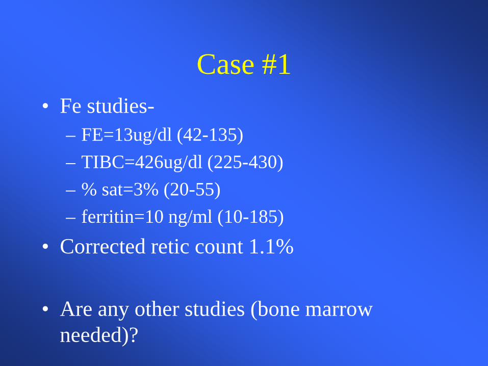
Case #1 • Fe studies-
– FE=13ug/dl (42-135) – TIBC=426ug/dl (225-430) – % sat=3% (20-55) – ferritin=10 ng/ml (10-185)
• Corrected retic count 1.1%
• Are any other studies (bone marrow needed)?
Case #1
You confirm iron deficiency likely secondary to NSAID use. An endoscopy confirms a H.pylori negative ulcer with chronic bleeding. He is started on Omeprazole and his NSAID is stopped. You prescribe oral Fe Sulfate 325 mg titrated to TID which he tolerates fairly well. He comes back to see you in 4 weeks.
Case #1
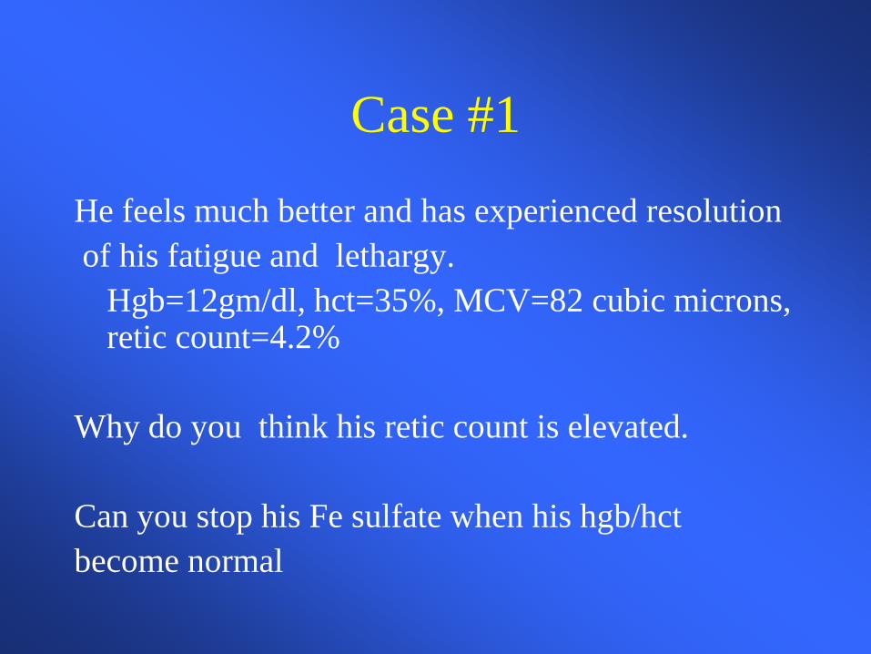
He feels much better and has experienced resolution of his fatigue and lethargy. Hgb=12gm/dl, hct=35%, MCV=82 cubic microns,
retic count=4.2% Why do you think his retic count is elevated. Can you stop his Fe sulfate when his hgb/hct become normal
Case 1
Case 2
A 48 year-old female with a history of IDDM and treated hypothyroidsm is referred to you for evaluation of anemia. Her complaints leading to this diagnosis included weakness, fatigue, weight loss, and mild numbness in her feet bilaterally. Exam was essentially normal except for mild loss of proprioception in her feet bilaterally
Case 2
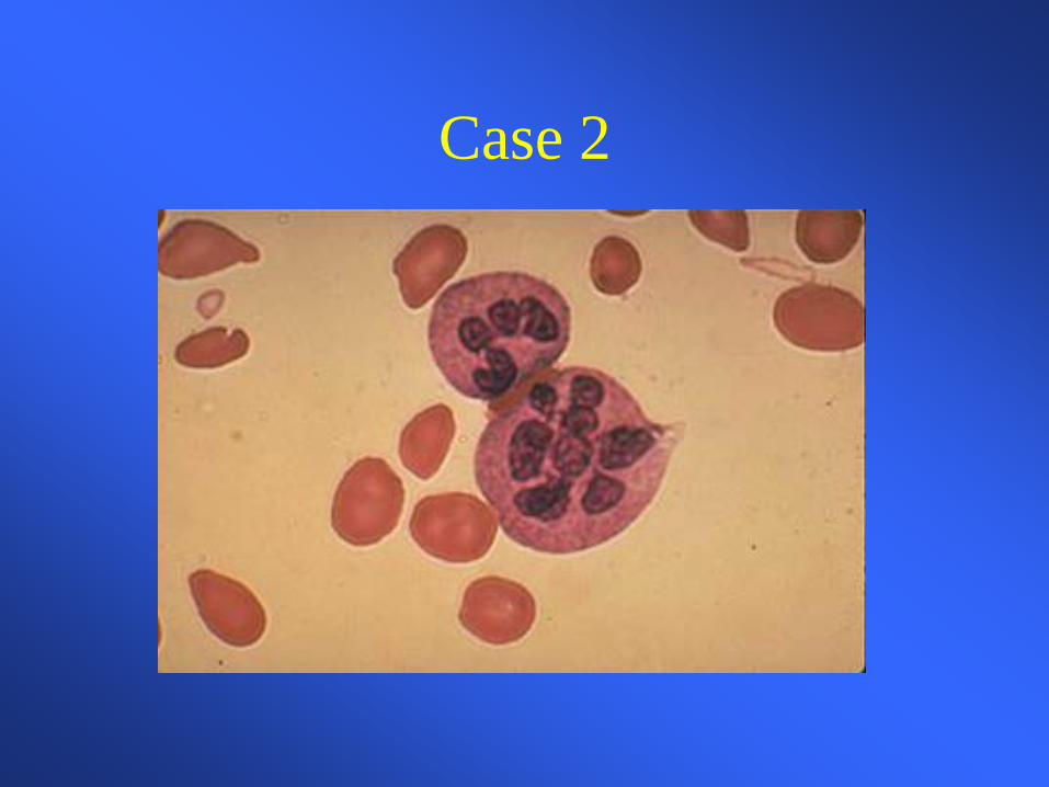
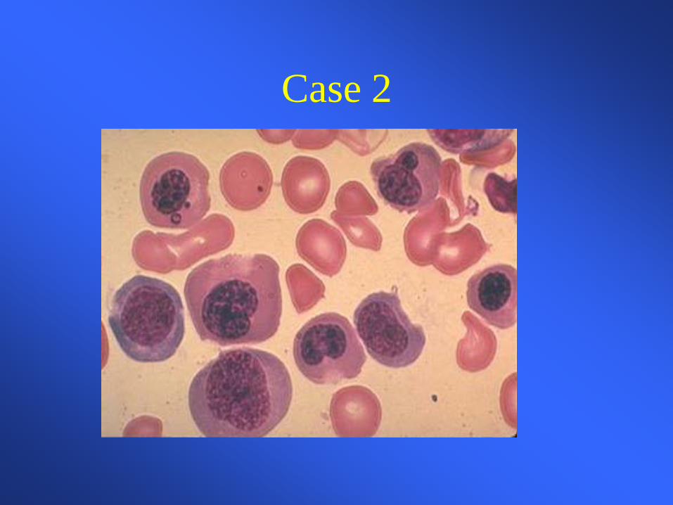
Laboratory data revealed: hgb=8.3, hct=27%, wbc=3.7, plt=152K MCV=123 retic count=.3 Two years ago labs showed: hgb 11.2, hct=33, wbc =4.8, plt=187 MCV=103 retic=1.1
Case 2
Case 2
Case 2
What tests or procedures do you want to perform to further workup this patient? Folate=8.3ng/ml (>3) Vit b-12=73pg/ml (180-914) TSH=1.5 Do you need a bone marrow exam?
Case 2
You diagnose B-12 deficiency and prescribe B-12 injections 1000ug weekly x4 than 1000ug a month indefinitely. In 1 month the patient feels remarkably better and her blood counts have all improved?
Case 2
• What are potential causes of b-12 deficiency?
• What do you suspect in this case?
• What further tests can you perform to verify
your suspicion?
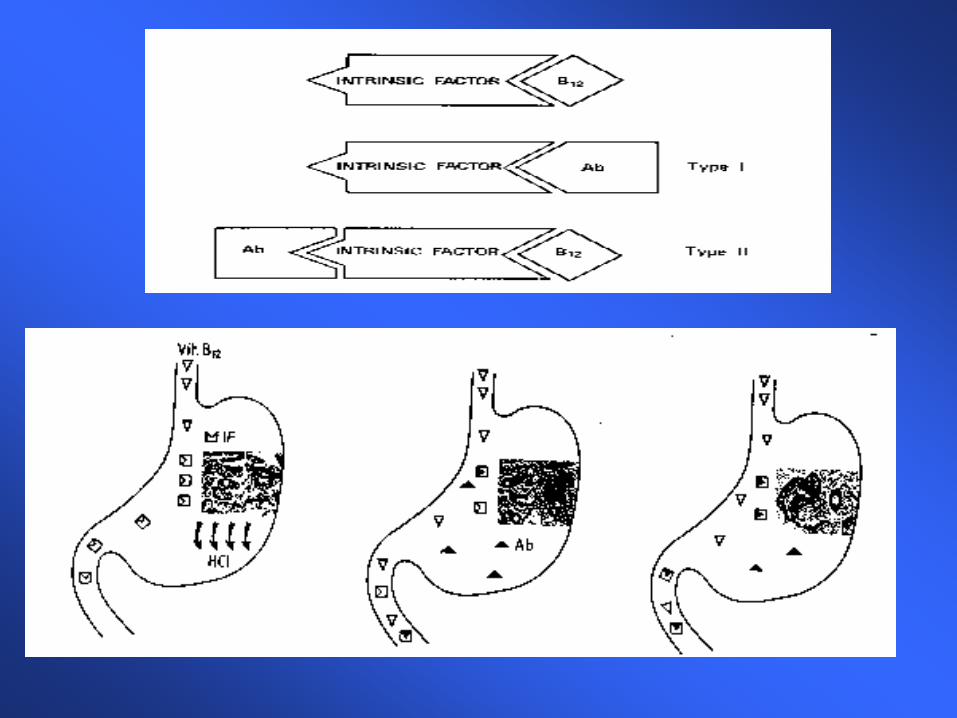
Case 2 You correctly suspect pernicious anemia which you confirm with anti-parietal cell and anti-intrinsic factor antibody tests. What is the mechanism of b-12 deficiency in this syndrome? Why does B-12 deficiency lead to blood abnormalities?
Case 2
• Vitamin B-12 deficiency leads to abnormal DNA sytnthesis leading to many problems including abnormal blood cell maturation in the bone marrow (innefective erythropoeisis)
• Responds very well and quickly to B-12 replacement. Neurologic symptoms may not be reversible