Embed Size (px)
Citation preview

0
CIRCLE OF WILLIS MORPHOLOGY AND
CEREBROVASCULAR COMPLICATIONS IN CAROTID
ATHEROSCLEROSIS
PhD thesis
Andrea Varga MD
Basic and Translational Medicine Doctoral School Semmelweis University
Supervisor: Péter Banga, MD, PhD
Official reviewers: Sándor Nardai, MD, PhD
Dávid László Tárnoki, MD PhD Head of the Complex Examination Committee:
Péter Barsi MD, PhD Members of the Complex Examination Committee:
Beáta Rosdy, MD PhD
Lajos Kozák, MD PhD
Budapest 2021

1
1. Introduction
Reduction of blood flow due to a severe internal carotid artery (ICA) stenosis or an
intraoperative cross clamping of the ICA requires compensation via other pathways to
maintain sufficient perfusion of the affected vascular territory. The circle of Willis
(CoW) is considered the primary collateral pathway which may allow blood supply
form the contralateral ICA through the contralateral precommunicating segment of the
anterior cerebral artery (A1), the anterior communicant artery (AComA) and through
the ipsilateral A1. Collateral flow from the vertebrobasilar system to the obliterated ICA
can be provided through the ipsilateral precommunicating segment of the posterior
cerebral artery (P1) with flow reversal in the posterior communicant artery (PComA).
The potential to develop these collateral pathways depends on the continuity of the CoW.
The CoW has been widely investigated showing substantial individual differences by the earliest non-selected post mortem studies. Selected autopsy studies found that the
prevalence of absent or hypoplastic segments was increased in stroke patients as
compared to normal subjects. Discontinuity of the CoW in patients with symptomatic
ICA stenosis was associated with higher risk of transient ischemic attack (TIA) and
ischemic stroke. Subjects with high-grade ICA stenosis or occlusion with nil or one
ipsilateral collateral vessel had a higher likelihood of stroke when compared to patients
with two functional ipsilateral collaterals. The majority of imaging studies showed
higher prevalence of hypoplastic or absent CoW segments (1, 2).
Carotid endarterectomy (CEA) is a frequent vascular surgical procedure with low
reported complication rates. Cross-clamping during CEA may result in cerebral
ischemia, which can be prevented by shunt usage. Those CEA patients who have collaterals supplying the operative side are less prone to perioperative stroke or
intolerance to cross clamping (3).
Although several articles addressed the CoW anatomy using different imaging
modalities, only a few reports have been published with the use of multi-detector
computerized tomography (CT) angiography (CTA), in particular focusing on patients
with ICA stenosis. The impact of multiple incompleteness of the CoW has not been
thoroughly studied either. CTA could be incorporated in an imaging-based prediction
model for prevention of unnecessary shunting, while establishing more precise
indications of shunting for non-routine shunt-user vascular surgeons.
2. Aims
1) Assessing the prevalence of anatomical variants of the CoW which may hamper
collateral supply in a cohort of 544 CEA subjects compared to 196 controls. 2) Correlating these variants with cerebral ischemia proved by cerebral CT or magnetic
resonance imaging (MRI).
3) Determining the reproducibility of CTA in CoW assessment.
4) Evaluating the impact of an incomplete CoW with an isolated MCA (iMCA) on
immediate neurological events (INE) after CEA.
Aims 1)-3) were referred as radioanatomical, whereas aim 4) as clinical approach.

2
3. Methods 3. 1. Study group
After approval from the Institutional Review Board (IRB) was obtained (216/2016), we
retrospectively analyzed the data of our registry from the Heart and Vascular Center of
Semmelweis University. We recruited all CEA patients from January 2013 to November
2015. Eligibility to CEA was stated as ICA stenosis of >70% (in exceptional cases
>50%) for symptomatic or ICA stenosis of >70% for asymptomatic subjects (Class I,
Level of Evidence A). ICA stenosis severity was established according to the North American Symptomatic Carotid Endarterectomy Trial (NASCET) method.
Symptomatic ICA stenosis was defined as history of stroke, amaurosis fugax or TIA
involving the ipsilateral ICA territory and in the period of 180 days prior to CEA.
Patients with a disabling stroke due to large infarcts were not subjected to carotid
revascularisation. Patients without adequate preoperative CTA to evaluate the CoW and
those who had shunting were excluded. One subject was removed from the
radioanatomical study because of suboptimal preoperative imaging quality, but for the
important outcome in this particular case the patient was included in the clinical study,
explaining the difference in patient numbers (544 vs 545). The patient’s CoW was
assessed on the postoperative CTA, which was performed to check the patency of the
operated ICA, since he had a major stroke after CEA.
3. 2. Eversion endarterectomy of the carotid arteries
All CEAs were performed under general anaesthesia without specific neuromonitoring.
Technical success was considered when the plaque removal and the arterial wall
reconstruction was achieved with <30% residual stenosis. Intra-arterial shunting was
rare and based on the individual decision of the operating surgeon. Shunting was
performed when the carotid lesion was too high for eversion, or in case of a relatively
large acute brain infarct, or with multiple supra-aortic occlusions.
3. 3. Brain CT and carotid CTA Examinations
All examinations were performed on a 256-detector scanner (Brilliance iCT 256, Philips
Healthcare). Brain CT was followed by CTA from the aortic arch to the vertex. Bolus tracking technique was used with 50 ml of iodinated contrast agent (Iomerone 400,
Bracco) followed by a 40 ml saline bolus, both injected at 5 ml/s. Continuous sections
were reconstructed with 0.67 mm slice thickness and 512×512 matrix using hybrid
iterative reconstruction technique (iDose, Philips Healthcare). Images were evaluated
on a dedicated workstation (IntelliSpace Portal, Philips Healthcare).
The CT and CTA assessment was carried out by two skilled radiologists (R1 with 13
years; R2, with 8 years of experience).
3
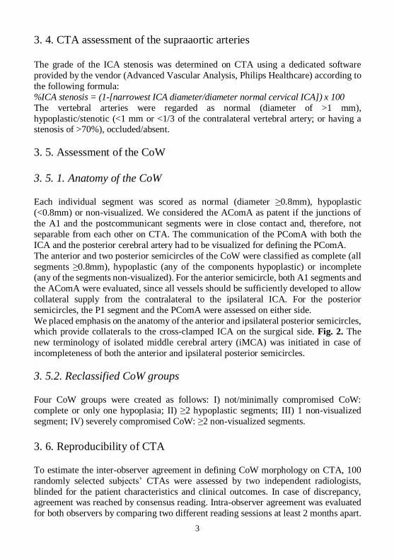
3. 4. CTA assessment of the supraaortic arteries
The grade of the ICA stenosis was determined on CTA using a dedicated software
provided by the vendor (Advanced Vascular Analysis, Philips Healthcare) according to
the following formula:
%ICA stenosis = (1-[narrowest ICA diameter/diameter normal cervical ICA]) x 100
The vertebral arteries were regarded as normal (diameter of >1 mm),
hypoplastic/stenotic (<1 mm or <1/3 of the contralateral vertebral artery; or having a stenosis of >70%), occluded/absent.
3. 5. Assessment of the CoW
3. 5. 1. Anatomy of the CoW Each individual segment was scored as normal (diameter ≥0.8mm), hypoplastic
(<0.8mm) or non-visualized. We considered the AComA as patent if the junctions of
the A1 and the postcommunicant segments were in close contact and, therefore, not
separable from each other on CTA. The communication of the PComA with both the
ICA and the posterior cerebral artery had to be visualized for defining the PComA.
The anterior and two posterior semicircles of the CoW were classified as complete (all
segments ≥0.8mm), hypoplastic (any of the components hypoplastic) or incomplete
(any of the segments non-visualized). For the anterior semicircle, both A1 segments and
the AComA were evaluated, since all vessels should be sufficiently developed to allow
collateral supply from the contralateral to the ipsilateral ICA. For the posterior
semicircles, the P1 segment and the PComA were assessed on either side.
We placed emphasis on the anatomy of the anterior and ipsilateral posterior semicircles, which provide collaterals to the cross-clamped ICA on the surgical side. Fig. 2. The
new terminology of isolated middle cerebral artery (iMCA) was initiated in case of
incompleteness of both the anterior and ipsilateral posterior semicircles.
3. 5.2. Reclassified CoW groups Four CoW groups were created as follows: I) not/minimally compromised CoW:
complete or only one hypoplasia; II) ≥2 hypoplastic segments; III) 1 non-visualized
segment; IV) severely compromised CoW: ≥2 non-visualized segments.
3. 6. Reproducibility of CTA To estimate the inter-observer agreement in defining CoW morphology on CTA, 100
randomly selected subjects’ CTAs were assessed by two independent radiologists,
blinded for the patient characteristics and clinical outcomes. In case of discrepancy, agreement was reached by consensus reading. Intra-observer agreement was evaluated
for both observers by comparing two different reading sessions at least 2 months apart.

4
Fig. 1. Definition of the circle of Willis segments: normal: ≥0.8mm; hypoplastic <0.8mm; non-visualized/absent. AComA = anterior communicating artery; A1 =
precommunicating segment of the anterior cerebral artery; PComA = posterior
communicating artery; P1 = precommunicating segment of the posterior cerebral artery.
3. 7. Radioanatomical approach
3. 7. 1. Control group Having reviewed all carotid CTAs from January 2014 to November 2017, we identified
all subjects with either negative CTA or minor/mild carotid atherosclerosis to provide a
sex-matched control group.
3. 7. 2. Brain CT assessment in the patient group
Any detectable ICA territory infarct on the side of surgery evidenced by CT
hypoattenuation was considered as a positive CT regardless of the infarct’s features (acute, subacute or chronic; territorial, lacunar or watershed). Fig. 2. The lack of infarct
in the corresponding ICA territory was classified as a negative CT result.
3. 7. 3. Brain MRI examinations
72 brain MRIs were performed in our center (IRB approval number 169/2015), between
January 2016-May 2017, one day before CEA on a 1.5T MR scanner (Achieva1.5,
Philips Healthcare). The diffusion weighted imaging (DWI), fluid-attenuated inversion
recovery (FLAIR) sequences were evaluated on a picture archiving and communication system workstation (IMPAX 6.5.2, AGFA Healthcare). An experienced
neuroradiologist (R1) reviewed the MRIs and recorded all ipsilateral recent infarcts on
DWI and old infarcts on FLAIR. All subjects had a CTA to determine CoW anatomy.
Hypoplastic
<0.8mm
ACom
A Non-
visualized A1
Normal
≥0.8 mm
P1
PComA

5
3. 8. Clinical approach
3. 8. 1. Outcome measures
The primary outcome was an INE, including any TIA or stroke immediately after CEA.
Stroke was defined as an acute neurological event with focal symptoms, lasting for ≥24
hours, consistent with focal cerebral ischemia, assessed by the modified Rankin scale
by an independent neurologist on the first postoperative day. TIA was defined as a brief
episode of neurological dysfunction caused by focal brain or retinal ischemia lasting
≤24 hours and without evidence of acute infarction. Upon INE urgent Duplex ultrasound was performed to exclude ICA occlusion. In case of a patent ICA, brain CT
with carotid CTA was done to exclude embolisation or treatable intracranial bleeding.
At 4-6 days, the brain CT was repeated to reveal any new ischemic lesions.
Early secondary outcomes were defined as any significant events during the hospital
stay. Recorded events were in-hospital stroke, myocardial infarction and death.
3. 9. Statistical analysis All statistical analysis was performed using the SPSS software (SPSS v.23; IBM Corp.,
Armonk, NY) according to the reporting standards of the Society of Vascular Surgery.
Continuous variables were expressed as mean ± standard deviation (SD), or median ± range, as appropriate. Categorical variables were expressed as counts and percentages.
Distributions were given according to the three different approaches to classify the
CoW.
3. 9. 1. Radioanatomical approach Bivariate association analysis was performed using the ANOVA for continuous
variables or χ2 test for categorical variables. All variables that were significantly
different between the study patients and controls at bivariate analysis were entered into
multivariate logistic regression analysis for ordinal data. In the χ2 test used for brain CT
and MRI analysis p≤0.05 was considered statistically significant. Yates correction was
applied in the MRI analysis. Intra-observer and inter-observer agreement was estimated
using the Cohen κ statistics. Cohen’s κ values were interpreted as: 0.81–1.00, excellent;
0.61–0.80, good.
3. 9. 2. Clinical approach Fisher exact test or Pearson χ2 test for categorical variables, Mann–Whitney U test for
ordinal variables were used, as appropriate. Two sample t-test was applied for
continuous variables. Uni- and multivariate logistic models were used to predict INE. The threshold of significance was p≤0.05.

6
Fig. 2. The circle of Willis. The anterior semicircle was defined from the contralateral to the ipsilateral internal carotid artery (ICA), the posterior semicircle from the ICA to the basilar artery.
Fig. 3. Multiplanar reformats of unenhanced brain CTs. a) Subtle left postcentral hypodensity
corresponding to a recent left middle cerebral artery (MCA) infarct (arrows). b) Marked hypodensity in the postcentral region, contributable to a chronic right MCA infarct (arrows).
4. Results
4. 1. Radioanatomical approach
4. 1. 1. Characteristics of the radioanatomical study group
From 902 consecutive CEA patients in the study period, we initially excluded 4 subjects due to shunt usage plus ICA patching. Further exclusion criteria were i) poor image
b
)
a
)
Anterior semicircle
Middle cerebral
artery
R
L Posterior semicircle
L
Contralateral
ICA
7
quality (21/898, 2%), ii) missing or incomplete CTA (302/898, 34%), iii) shunting
during CEA (3/898, 0.3%) or the combination of the previous two criteria (28/898, 3%).
Altogether 358 patients were excluded (40%) from the radioanatomical study. The
remaining 544 study subjects were analyzed (331 males, mean age 69±8 years, range
44–90 years). Of them, 205 (38%) had symptomatic ICA stenosis, including 59 patients
(11%) with previous minor stroke, 25 (5%) with amaurosis fugax and 121 (22%) with TIA. The 3 subjects with stenosis of <70% were all symptomatic. The demographics
and co-morbidities of the 544 study subjects and 196 controls are summarized in Table 1.
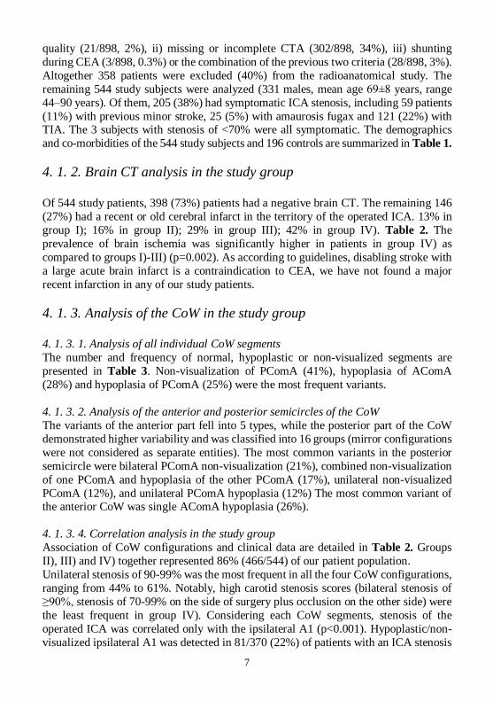
4. 1. 2. Brain CT analysis in the study group
Of 544 study patients, 398 (73%) patients had a negative brain CT. The remaining 146
(27%) had a recent or old cerebral infarct in the territory of the operated ICA. 13% in
group I); 16% in group II); 29% in group III); 42% in group IV). Table 2. The
prevalence of brain ischemia was significantly higher in patients in group IV) as
compared to groups I)-III) (p=0.002). As according to guidelines, disabling stroke with
a large acute brain infarct is a contraindication to CEA, we have not found a major
recent infarction in any of our study patients.
4. 1. 3. Analysis of the CoW in the study group
4. 1. 3. 1. Analysis of all individual CoW segments
The number and frequency of normal, hypoplastic or non-visualized segments are
presented in Table 3. Non-visualization of PComA (41%), hypoplasia of AComA
(28%) and hypoplasia of PComA (25%) were the most frequent variants.
4. 1. 3. 2. Analysis of the anterior and posterior semicircles of the CoW
The variants of the anterior part fell into 5 types, while the posterior part of the CoW
demonstrated higher variability and was classified into 16 groups (mirror configurations
were not considered as separate entities). The most common variants in the posterior
semicircle were bilateral PComA non-visualization (21%), combined non-visualization
of one PComA and hypoplasia of the other PComA (17%), unilateral non-visualized
PComA (12%), and unilateral PComA hypoplasia (12%) The most common variant of the anterior CoW was single AComA hypoplasia (26%).
4. 1. 3. 4. Correlation analysis in the study group
Association of CoW configurations and clinical data are detailed in Table 2. Groups
II), III) and IV) together represented 86% (466/544) of our patient population.
Unilateral stenosis of 90-99% was the most frequent in all the four CoW configurations,
ranging from 44% to 61%. Notably, high carotid stenosis scores (bilateral stenosis of
≥90%, stenosis of 70-99% on the side of surgery plus occlusion on the other side) were
the least frequent in group IV). Considering each CoW segments, stenosis of the
operated ICA was correlated only with the ipsilateral A1 (p<0.001). Hypoplastic/non-
visualized ipsilateral A1 was detected in 81/370 (22%) of patients with an ICA stenosis

8
of ≥90% while only in 14/174 (8%) of patients with a stenosis of <90%. The percentage
of smokers was the lowest in patients with severely compromised CoW. The other
comparisons showed no significant difference.
Considering the anterior and two posterior semicircles of the CoW (bottom part of
Table 3), the frequency of normal, hypoplastic, incomplete anterior part was 47%, 41%,
and 12%, respectively. The percentages of normal, hypoplastic, incomplete posterior semicircles were 22%, 32%, and 46%. Only 19/544 patients (3.5%) had an entirely
complete CoW with all segments ≥0.8 mm.
Examples of CoW groups I)-IV) are shown in Fig. 4.a-d).
Table 1. Demographics, co-morbidities and different configurations of the circle of
Willis in 544 study subjects and 196 controls
CoW = circle of Willis; SD = standard deviation
Study subjects
(n=544)
Controls
(n=196) p-value
Demographics
Male gender 331 (61%) 117 (60%) 0.777
Mean age ± SD (years) 69 ± 8 66±11 <0.001
Symptomatic 205 (38%) -
Cardiovascular risk factors,
N (%)
Data available
in 173
Hypertension 500 (92%) 110 (64%) <0.001
Cigarette smoking 175 (32%) 16 (9%) <0.001
Hyperlipidemia 234 (43%) 49 (28%) 0.001
Coronary artery disease 170 (31%) 30 (17%) <0.001
Chronic pulmonary disease 53 (10%) 13 (8%) 0.377
Chronic kidney disease
(Stage IIIb-V) 16 (3%) 6 (3%) 0.726
Diabetes mellitus 203 (37%) 32 (18%) <0.001
CoW groups N (%)
Group I) 78 (14%) 55 (28%)
<0.001 Group II) 97 (18%) 52 (27%)
Group III) 191 (35%) 55 (28%)
Group IV) 178 (33%) 34 (17%)

9
Table 2. Association of circle of Willis configurations and demographics,
cardiovascular risk factors and prevalence of brain ischemia
Variable
Group I)
Complete
CoW or 1 hypoplasia
(n = 78)
Group II)
≥2 hypo-
plasia
(n = 97)
Group III)
1 non-
visualized segment
(n = 191)
Group IV)
≥2 non-
visualized segments
(n = 178)
p
Demographics
Male gender, N (%) 48 (62%) 51 (53%) 121 (63%) 111 (62%) 0.32
Age ± SD (years) 68 ± 9 67 ± 8 69 ± 8 70 ± 8 0.11
Symptomatic, N (%) 21 (27%) 38 (39%) 68 (36%) 78 (44%) 0.07
Cardiovascular risk factors, N (%)
Hypertension 70 (90%) 89 (92%) 175 (92%) 166 (93%) 0.81
Cigarette smoking 26 (33%) 39 (40%) 73 (38%) 37 (21%) 0.001
Hyperlipidemia 41 (48%) 41 (42%) 77 (40%) 75 (44%) 0.32
Coronary artery
disease 34 (44%) 31 (32%) 52 (27%) 53 (30%) 0.07
Chr. pulmonary
disease 9 (9%) 14 (14%) 19 (10%) 11 (7%) 0.29
Chr. kidney disease
(IIIb-V) 2 (2%) 5 (5%) 4 (2%) 5 (3%) 0.47
Diabetes mellitus 26 (32%) 36 (37%) 69 (36%) 72 (42%) 0.71
Brain CT N (%)
Negative 398 (73%) 61 (15%) 74 (19%) 151 (38%) 112 (28%) 0.002*
Positive 146 (27%) 19 (13%) 24 (16%) 42 (29%) 61 (42%)
chr = chronic; contralat. = contralateral; ICA = internal carotid artery; SD = standard
deviation
*2 test between pooled Groups I-III) versus Group IV)
4. 1. 4. Reproducibility of the CTA
The inter-observer agreement of CTA in the assessment of AComA was good (κ=0.75)
while the intra-observer agreement was excellent (κ=0.84 for R1 and κ=0.96 for R2).
The inter-observer (κ=0.82–0.92) and intra-observer (κ=0.84–1.0) agreement for both
readers were excellent for all other segments.
We evaluated 3808 (544×7) segments altogether and encountered 212/3808 inter-
observer discrepancies (5.5%), mainly 98/212 for the PComA (46%) and 60/212 (28%)
for the AComA. These were mostly one-category discrepancies (hypoplasia versus

10
normal/non-visualization) in 196/212 (92.5%). Final agreement was reached by
consensus reading.
4. 1. 5. Characteristics of the radioanatomical control group
Data of 196 control subjects were analysed (117 males, mean age 66±11 years, range
37–93 years). The indication for CTAs was: 1) positive ultrasound scan with mild-
moderate carotid artery stenosis on CTA (30%); 2) brachiocephalic/subclavian artery stenosis or aneurysm (14%); diagnostic work-up before cardiac surgery/intervention
(14.5%); or 4) vascular intervention/surgery (7.5%); 5) neurology referral (29%); 6)
carotid artery dissection (2%); 7) neck tumour (2%); and 8) vascular malformation
(1%). Further details are reported in Table 1.
4. 1. 6. Analysis of the CoW of the radioanatomical control subjects
4. 1. 6. 1. Analysis of All Individual Segments
Hypoplasia of PComA (31%), non-visualization of PComA (28%) and hypoplasia of
AComA (20%) were the most frequent variants in controls. Table 3. Non-visualization
of the A1 segment and AComA was rare, 4/392 (1%) and 1/196 (0.5%).
4. 1. 6. 2. Analysis of the anterior and posterior semicircles of the CoW in controls The most common variants in the posterior part of the CoW were unilateral PComA
hypoplasia (22%), bilateral PComA hypoplasia (21%), bilateral PComA non-
visualization (16%), combined non-visualization of one PComA and hypoplasia of the
other PComA (15%) and unilateral PComA non-visualization (12%). The most
common variant of the anterior CoW was AComA hypoplasia (20%).
Considering the anterior and two posterior semicircles of the CoW (bottom part of
Table 3), the frequency of normal, hypoplastic, incomplete anterior parts was 73%,
24%, and 3%, respectively; 31%, 39%, and 29% for the posterior semicircle.
Of 196 control subjects, 21 (11%) had an entirely complete CoW.
4. 1. 7. Comparison of the patients and controls
Groups I), II), III) and IV accounted for 28%, 27%, 28% and 17% of control subjects;
whereas in study patients the percentages were 14%, 18%, 35% and 33%. Table 1. The difference between study patients and controls was statistically significant (p<0.001).
The bivariate analysis (Table 1.) found, that the study patients and the controls were
significantly different in terms of five cardiovascular risk factors and coronary artery
disease (p<0.001). However, by multivariate logistic regression analysis ICA stenosis
was the only independent predictor of CoW morphology (p<0.001). Except from
hypoplasia of PComA and P1, all other variants had lower percentages in controls as
compared to patients. The analysis of each CoW segment and the anterior/posterior
semicircles confirmed a significantly higher rate of hypoplasia or non-visualization
(p≤0.008) in the study group versus controls except for the P1 (Table 3).

11
Table 3. Number (frequency) of normal, hypoplastic, non-visualized/incomplete
individual segments, anterior and posterior semicircles of the circle of Willis
AComA = anterior communicating artery; A1 = precommunicating segment of the anterior
cerebral artery; PComA = posterior communicating artery; P1 = precommunicating segment of the posterior cerebral artery; post. = posterior. *Comparison of study subjects and controls,
pooling hypoplasia and non-visualization versus normal
4. 1. 8. Brain MRI study
Brain MRI has been performed in 72 cases (45 males, mean age 66±9). On CTA 46 out
of 72 patients (64%) had an incomplete CoW (with ≥1 non-visualized segments), only
26/72 (36%) had a complete CoW (with normal or hypoplastic segments).
With incomplete CoW we detected 11 subjects with recent infarcts on DWI, and 16
patients with late subacute/chronic infarcts on FLAIR (11/46, 24% and 16/46, 35%,
respectively). With complete CoW the number of subjects with acute and late
subacute/chronic infarcts was 2 and 6, respectively (2/26, 8% and 6/26, 23%).
The prevalence of brain ischemia (recent + old) was significantly higher (p=0.04 with
Yates correction) in subjects with incomplete CoW (59%) as compared to complete (normal or hypoplastic) CoW (31%). The rate of ipsilateral recent ischemic lesions
alone was three times higher in incomplete CoW (24%) relative to complete CoW (8%).
Nevertheless, this difference was not significant (p=0.09).
4. 2. Clinical approach
4. 2. 1. Characteristics of the clinical study group
We included 545 patients in the clinical study, 332 males, mean age 69±8 years, range
44–90 years). One patient’s CEA was unsuccessful (as detailed below) and he suffered
study subjects (n=544) controls (n=196) p*
Segment/
Semicircle Normal Hypoplasia
Non-visua-
lization Normal Hypoplasia
Non-visua-
lization
AComA (n) 369
(68%) 154
(28%) 21
(4%) 155
(79%) 40
(20%) 1
(<1%) 0.003
A1 (n x 2) 964
(89%)
81
(7%)
43
(4%)
380
(97%)
8
(2%)
4
(1%) <0.001
PComA
(n x 2)
366
(34%)
275
(25%)
447
(41%)
161
(41%)
121
(31%)
110
(28%) 0.008
P1 (n x 2) 948
(87%)
81
(7.5%)
59
(5.5%)
354
(90%)
33
(8%)
5
(1%) 0.098
Anterior
semicircle (n)
257
(47%)
223
(41%)
64
(12%)
143
(73%)
48
(24%)
5
(3%) <0.001
Post. semi-
circle (n x 2)
234
(22%)
351
(32%)
503
(46%)
123
(31%)
154
(39%)
115
(29%) <0.001

12
a) Complete CoW (Group I) b) Hypoplasia of the AComA,
right PComA (Group II)
c) Group III). Non-visualized d) (Group IV). Non-visualized
right PComA. The filiform communicant arteries.
contrast filled structure is the
vein of Rosenthal (red star)
Fig. 4a-d). Thick slab maximal intensity projection reformats from CT angiographies illustrating the 4 groups of the circle of Willis. The hypoplastic or non-visualized
segments are indicated with long arrows. AComA = anterior communicating artery;
CoW = circle of Willis; PComA = posterior communicating artery
a major stroke. Although his preoperative CTA was suboptimal for CoW assessment,
the CoW could be studied on the postoperative CTA performed to check ICA patency.
The excluded and included subjects, the preoperative and postoperative variables are
detailed in Table 4.
Intra-arterial shunting was rare (31 and the primarily excluded 4 cases) and based on
the individual decision of the vascular surgeon. In 8 cases long calcified ICA plaques
were found, too high for eversion. 10 patients had a recent stroke with established
ischemic infarcts, 4 had contralateral ICA occlusion. For the remaining cases the cause
of shunting was not known. 15 patients had a bilateral reconstruction (the second operation 3-82 weeks apart the first).
Demographics and cardiovascular risk factors of study subjects with and without INE
are presented in Table 5.

13
4. 2. 2. Surgical procedural details
The CEA was technically successful in 99% of cases. The average carotid clamping
time was 25±9 minutes. Plaque removal with the eversion technique was unsuccessful
in one case. Polytetrafluoroethylene interposition between the common carotid and the
endarterecomized internal carotid arteries was performed, which occluded immediately.
Several thrombectomies were attempted with no success. The patient suffered
immediate stroke, remained unconscious and later passed away. 3 further CEA subjects had successful primary reconstruction and were asymptomatic after the operation, but
later (within the first 24 hours) their ICA occluded resulting in a neurologic event.
4. 2. 3. Mortality and major adverse events Only the patient, with unsuccessful CEA discussed above, died in the early
postoperative period (0.2%). Two patients suffered myocardial infarction (0.4%), both
were treated with successful coronary intervention. Reoperation was needed in 20 cases
(3.7%), haematoma evacuation in 17 (3.1%), thrombectomy in one (0.2%), and
reocclusion followed by polytetrafluoroethylene interposition in two (0.4%). The
ischemic events are detailed further below.
Table 4. Excluded and included study subjects with preoperative variables and early
outcomes (stroke, immediate stroke, immediate transient ischemic attack and mortality)
of the clinical study
Preoperative
variables
missing
imaging
n=302
poor image
quality
n=20
n=21¥
shunting±
poor quality
n=35
excluded
total
n=357
n=358 ¥
included
n=545
n=544 ¥
p
Symptomatic 103 (34%) 2 (10%) 16 (46%) 121 (34%) 205 (38%) 0.28
Contralateral occlusion
20 (7%) 0 4 (11%) 24 (7%) 35 (6%) 0.86
Early
outcomes
All stroke 4 (1%) 0 1 (3%) 5 (1.5%) 12 (2%) 0.46
Immediate
stroke 1(<1%) 0 1 (3%) 2 (<1%) 8 (1.5%) 0.33
Immediate
TIA 6 (2%) 0 0 6 (2%) 12 (2%) 0.63
Death 0 0 0 0 1(<1%) 1.0
TIA = Transient Ischemic Attack
The differences between the clinical and radioanatomical studies are in bold italic
typeset. For details refer to text. ¥Exclusions and final study cohort of the radioanatomical study.

14
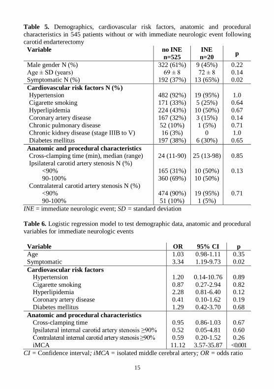
4. 2. 4. Immediate postoperative neurologic events
Of the 545 cases, eight immediate strokes and 12 TIAs were diagnosed immediately
after CEA (overall 20 INEs). We entered our data in a binary logistic regression model
including hypertension, smoking, diabetes mellitus, hyperlipidemia, carotid clamping
time, ipsilateral ICA stenosis of ≥90%, contralateral ICA stenosis of ≥90%, symptoms
180 days before surgery, and iMCA. The model revealed a significant difference
(p=0.001; -2LL=137.56; Nagelkerke R2=0.18), iMCA being an independent predictor of INE (odds ratio (OR): 11.12; 95% confidence interval (CI): 3.57-35.87; p<0.001).
Apart from iMCA, only symptomatic ICA stenosis showed a significant association
with INE (OR: 3.34; 95% CI: 1.19-9.73; p=0.02). The other parameters were non-
significant. Table 6.
4. 2. 5. Relation of CoW configuration and neurologic events
62 subjects out of 545 (12%) had a normal anterior semicircle and a normal ipsilateral
posterior semicircle, including 19 subjects with a fully normal CoW (3.5%). Among
these patients only one INE (stroke) was detected (1.5%).
268 patients had a hypoplastic or non-visualized segment either in the anterior (72) or
the posterior semicircle (196), whereas the other semicircle was complete. Out of these
268 subjects with one affected semicircle, only three suffered INE (two strokes, one TIA); the posterior semicircle was incomplete in all these cases. The statistical analysis
showed no significant difference (p=0.57) in INE between the patients with complete
ipsilateral semicircles (1/62) versus those with one affected semicircle (3/268).
The difference became significant (p<0.001) when both the anterior and the ipsilateral
posterior semicircles were affected having hypoplastic and/or non-visualized segments
at the same time (215 subjects). Among these 215 patients we encountered 16 INEs (5
strokes and 11 TIAs). Out of those 34 patients with an iMCA (incompleteness of both
semicircles) two had a stroke and six suffered TIA (8 INEs in total; 23.5%), which is a
significantly higher rate (p<0.001), when compared with the 8 INEs (3 strokes, 5 TIAs)
in the remainder of patients with two affected semicircles (8/181; 4.4%).
Out of the three patients with early postoperative stroke due to ICA reocclusion, two
had a normal anterior semicircle with a hypoplastic posterior semicircle. The third patient had hypoplasia both in the anterior and posterior semicircles.
The CoW configurations of the detailed subgroups are presented in Table 7.
4. 2. 6. Configurations of the isolated middle cerebral artery
Seven types of iMCA configurations were found. The most frequent type was combined
non-visualization of the AComA and that of the ipsilateral PComA (12/34, 35%),
followed by non-visualized ipsilateral A1 and PComA (11/34, 32%), non-visualized
contralateral A1 and ipsilateral PComA (4/34, 12%), finally non-visualized ipsilateral
A1 and P1 segments (4/34, 12%). Fig. 5 shows the graphical illustrations of these
configurations.
15
Table 5. Demographics, cardiovascular risk factors, anatomic and procedural
characteristics in 545 patients without or with immediate neurologic event following
carotid endarterectomy Variable no INE
n=525
INE
n=20 p
Male gender N (%) 322 (61%) 9 (45%) 0.22
Age ± SD (years) 69 ± 8 72 ± 8 0.14 Symptomatic N (%) 192 (37%) 13 (65%) 0.02
Cardiovascular risk factors N (%)
Hypertension 482 (92%) 19 (95%) 1.0
Cigarette smoking 171 (33%) 5 (25%) 0.64
Hyperlipidemia 224 (43%) 10 (50%) 0.67
Coronary artery disease 167 (32%) 3 (15%) 0.14
Chronic pulmonary disease 52 (10%) 1 (5%) 0.71
Chronic kidney disease (stage IIIB to V) 16 (3%) 0 1.0
Diabetes mellitus 197 (38%) 6 (30%) 0.65
Anatomic and procedural characteristics
Cross-clamping time (min), median (range) 24 (11-90) 25 (13-98) 0.85
Ipsilateral carotid artery stenosis N (%)
<90% 165 (31%) 10 (50%) 0.13
90-100% 360 (69%) 10 (50%) Contralateral carotid artery stenosis N (%)
<90% 474 (90%) 19 (95%) 0.71
90-100% 51 (10%) 1 (5%)
INE = immediate neurologic event; SD = standard deviation
Table 6. Logistic regression model to test demographic data, anatomic and procedural
variables for immediate neurologic events
Variable OR 95% CI p
Age 1.03 0.98-1.11 0.35
Symptomatic 3.34 1.19-9.73 0.02
Cardiovascular risk factors
Hypertension 1.20 0.14-10.76 0.89
Cigarette smoking 0.87 0.27-2.94 0.82 Hyperlipidemia 2.28 0.81-6.40 0.12
Coronary artery disease 0.41 0.10-1.62 0.19
Diabetes mellitus 1.29 0.42-3.70 0.68
Anatomic and procedural characteristics
Cross-clamping time 0.95 0.86-1.03 0.67
Ipsilateral internal carotid artery stenosis ≥90% 0.52 0.05-4.81 0.60
Contralateral internal carotid artery stenosis ≥90% 0.59 0.20-1.52 0.26
iMCA 11.12 3.57-35.87 <0.001
CI = Confidence interval; iMCA = isolated middle cerebral artery; OR = odds ratio

16
Table 7. Circle of Willis anatomy in patients with or without immediate neurologic
event after carotid endarterectomy
Semicircles
No INE
N=525
INE
N=20
p
Complete anterior + ipsilateral posterior 61 (12%) 1 (5%) 0.72
Entirely complete CoW (all segments normal) 18 (3%) 1 (5%) 0.51
Single affected
Hypoplasic anterior 60 (11%) 0 0.15 Incomplete anterior 12 (2%) 0 1.0
Hypoplasic ipsilateral posterior 89 (17%) 0 0.06
Incomplete ipsilateral posterior 104 (20%) 3 (10%) 0.39
Both affected
Hypoplasic anterior + ipsilateral posterior 65 (12%) 2 (10%) 1.0
Hypoplastic anterior + incomplete ipsilateral
posterior
91 (17%) 5 (25%) 0.37
Incomplete anterior + hypoplastic ipsilateral
posterior
17 (3%) 1 (5%) 0.50
Incomplete anterior +incomplete ipsilateral
posterior (iMCA)
26 (5%) 8 (40%) <0.001
Preoperatively symptomatic 14 (54%) 5 (63%) 1.0
CoW = circle of Willis; INE = immediate neurologic event, iMCA = isolated middle cerebral artery
Fig. 5. The four most frequent configurations of the isolated middle cerebral artery (iMCA) are shown (right internal carotid artery (ICA) stenosis and cross clamping was
assumed). The arrows indicate the non-visualized segments. The iMCA is considered
isolated if there is non-continuity of collaterals both from the contralateral ICA (anterior
semicircle) and the basilar artery (posterior semicircle). Percentages expressed from the
total number of patients with iMCA (n= 34).
4. 2. 7. Risk factors associated with iMCA
The statistical analysis showed fewer iMCA cases in smokers (p=0.01). Fewer iMCA
was found among patients with diabetes mellitus (p=0.02) or in coronary artery disease
L
32% 12% 12% 35%
R

17
(p=0.04). The prevalence of iMCA was higher among the preoperatively symptomatic
subjects (p=0.03).
4. 2. 8. Rate of early postoperative strokes
In the cohort of 545 CEA patients, the overall in-hospital ischemic stroke rate was 2.2%
(12/545); 3.9% (8/205) for the preoperatively symptomatic and 1.2% (4/340) for the
asymptomatic patients. Of the 12 stroke cases, eight were diagnosed immediately after
surgery. Early ICA reocclusion resulted in three strokes 1 to 3 hours following surgery.
Two of the three patients had a major stroke, one had only minor symptoms (hand
weakness). Another patient had a major stroke 6 hours after the procedure with patent
ICA and brain CT/CTA confirmed MCA territory embolisation.
One intracranial haemorrhage occurred in a preoperatively symptomatic patient secondary to hypertensive crisis resulting in minor symptoms.
The in-hospital rate of major stroke was 1.9% (4/205) among the preoperatively
symptomatic patients and 0.3% (1/340) in our asymptomatic group.
5. Discussion
To our knowledge ours is one of the most comprehensive studies on CoW morphology
using a 256-detector row CT in a large cohort of CEA subjects. Our major findings are
the high prevalence of compromised circles (86%), the significant difference in CoW
morphology between the study and control groups, as well as the significant association
of CoW configuration with brain infarcts and immediate postoperative ischemic
neurologic complications in the study group.
Some of the CoW variations may predispose to ischemic events. With preoperative CTA assessment of the CoW these patients with a high risk for cross clamping ischemia
can be identified. We found statistically higher odds for neurologic complications when
both the anterior and ipsilateral posterior semicircles were incomplete impeding
collateral recruitment towards the MCA on the side of the clamped ICA.
With contrast to MR angiography (MRA), CTA has higher spatial resolution and is not
dependent on flow velocity, thereby allowing accurate documentation of vessel
diameters. Although single phase CTA does not provide information about flow
dynamics, we hypothesized that competent component vessels are potentially capable
of supplying collateral flow.
5. 1. Radioanatomical approach
5. 1. 2. Segmental analysis of the CoW
5. 1. 2. 1. Non-visualization of the invidiual CoW segments
The non-visualization of any segment was more frequently reported in the
cerebrovascular patients as compared to healthy subjects.

18
Correlating our patients’ data with imaging studies from cerebrovascular patients we
found lower prevalence of AComA non-visualization: 4% vs 7-40%. The prevalence of
absent A1 segments in this study was 4%, at the lower limit of the reported range, and
in particular lower as compared to the other CTA studies of patients with
cerebrovascular disorders: 5.5-15%. The non-visualization of the PComA was reported
more frequent in most of the CTA studies in patients with cerebrovascular diseases as compared to ours: 47-66% vs 41%. The absence of the P1 segment was 5.5% in or study,
in line with the previously published data (3-10%).
The fact that CTA is not dependent of flow velocity as opposed to MRA and the better
spatial resolution achieved by CTA might have contributed to the lower prevalence of
non visualized AComA, A1 and PComA. However, a certain percentage of the absent
segments in this study may be hypoplastic, bellow the resolution of CTA, as in autopsy
studies absence was found rarely (0-3.5%), AComP aplasia being the most common.
5. 1. 2. 2. Hypoplasia of the individual CoW segments
Fewer data were published on hypoplasia of the CoW components, mostly from CTA
studies.
Comparing patients with control subjects lower percentage of AComA hypoplasia (4-11% vs 23%) and PComA hypoplasia (6-18% vs 38-41%) was demonstrated. In
contrast, A1 hypoplasia showed a tendency towards higher percentages in the
cerebrovascular group relative to healthy controls (8-24% vs 4-10%). In our
investigation PComA hypoplasia was also less frequent among controls relative to the
study group (31% vs 25%). Although with a little difference, the same applied to P1
hypoplasia (8.5% vs 7.5%). We might assume that in absence of significant ICA
stenosis these segments, in particular the PComA, are not recruited as collaterals and
remain small in calibre.
Comparing our study subjects with imaging studies from cerebrovascular patients, we
found higher prevalence of AComA (28% vs 4-11%) and PComA hypoplasia (25% vs
6-18%). The frequency of A1 hypoplasia was below the lower limit of the reported range (7.5% vs 8-24%). The prevalence of P1 hypoplasia was in the reported range
(7.5% vs 1-8%).
The higher detection rate of hypoplastic communicant arteries due to the better spatial
resolution of a modern MD CT equipment relative to the 16-40 row scanners or the
MRA techniques used by other investigators might partly account for this difference.
5. 1. 3. Clinical aspects of the radioanatomical approach
Discontinuity of the CoW in patients with ICA stenosis/occlusion was associated with
higher risk of TIA and ischemic stroke. This is in agreement with the higher prevalence
of brain ischemia found in our patients with severely compromised CoW (p=0.002).
Our MRI substudy also showed significantly higher percentage in the composite of old
and recent brain ischemia in subjects with incomplete CoW vs complete CoW (p=0.04). The high frequency of compromised circles suggests that our cohort of patients have
fewer functional segments, which might hinder hemodynamic adaptation. The study

19
group and controls were significantly different in terms of 5 cardiovascular risk factors
and coronary artery disease. The multivariate regression logistic analysis showed that
ICA stenosis was the only independent predictor of CoW morphology (p<0.001). The
higher frequency of ipsilateral A1 hypoplasia/non-visualization was positively
associated to ipsilateral ICA stenosis of ≥90% (p<0.001), also implying a correlation
between carotid artery disease and hindered collateral recruitment.
5. 2. Clinical approach (correlation of INE and CoW morphology)
In our study cohort entirely complete CoW (3.5 %) was surprisingly rare and only one
INE occurred in this subgroup. In subjects with at least one adequate collateral pathway,
the odds to suffer a complication were not different from those with two complete
ipsilateral semicircles. In 215 cases both collateral pathways proved to be insufficient
and we encountered 5 strokes and 11 TIAs, which is a significantly higher complication
rate as compared to those with at least one adequate collateral network.
Isolated MCA was an independent risk factor for INE, with odds three times as high as
the other significant risk factor of INE, ie. preoperatively symptomatic ICA stenosis.
Our results are concordant with other investigations, in which incomplete versus
complete CoW were shown to have a statistically significant risk to develop carotid
clamping intolerance/intraoperative TIA. Cerebral blood flow can be maintained by the placement of a shunt, but routine or
selective shunting, or routine non-shunting can have drawbacks. A meta-analysis
showed a small difference in perioperative stroke rate between routine shunt (1.4%) and
non-shunt use (2%). Regarding selective shunting, ischemia detection on test clamping
may lead to prompt declamping with subsequent reclamping, increasing embolic risk,
or to a delayed ischemia management with the test clamp left in place while the shunt
is readied. Routine shunt use may be hazardous for patients not requiring a shunt,
potentially resulting in thromboembolic complications or arterial injury. Eversion
endarterectomy may have the advantage of lower embolisation rate due to the absence
of any non-autologous material. Most surgeons in favour of the eversion technique do
not routinely use a shunt, as shunt can only be inserted when the eversion is completed. Of the 34 patients without iMCA, eight had neurologic complications. The
complication-free 26 subjects might have had sufficient secondary collaterals. Future
research of the complex collateral network may provide a better understanding of stroke
development and more personalized carotid revascularization strategies.
5. 3. Limitations
Some of the limitations of our study are the lack of comparison with digital substraction
angiography (DSA) as reference of standard, the possible bias between the study and
control groups, and the fact that the study group was limited to patients eligible for CEA
(either asymptomatic ICA stenosis or symptomatic stenosis with TIA or minor ischemic stroke). CTA does not provide information on flow dynamics, nor on secondary
collaterals, which is a further technical limitation. In some cases the non-visualization

20
of a CoW segment is in fact hypoplasia beyond the spatial resolution of CTA. The
overall small caliber of the CoW elements precludes the direct assessment of
intracranial atherosclerotic plaques either.
Further limitation is the external validity of the anatomic variances of the CoW. The
main limitation of our study is that we cannot entirely exclude embolisation as a
background cause of INEs.
6. Conclusions
CTA is a highly reproducible imaging method to evaluate CoW anatomy. It helps to
detect those variants which predispose to cerebral ischemia and thus to tailor surgical
or endovascular management of patients with extracranial atherosclerotic disease.
Distribution of CoW variants significantly differed between the study and control
groups. CoW variants were frequent in or study group and significantly associated with
cerebral ischemia proven by CT or MRI, or clinically by immediate neurologic complications following CEA.
Multiple incompleteness of the CoW on the surgical side carries an 11-fold risk of INEs
after CEA with cross-clamping and no shunt protection. If iMCA is detected on
preoperative CTA, routine shunting is recommended to prevent INEs.
References
1. Varga A, Di Leo G, Mihály Z. (2019) Association of Circle of Willis Variants
and Carotid Plaque Morphology with Cerebral Infarcts in Carotid Endarterectomy Subjects. In: ECR2019, doi:doi:10.26044/ecr2019/C-1137,
European Society of Radiology
2. Varga A, Di Leo G, Banga PV, Csobay-Novak C, Kolossvary M, Maurovich-
Horvat P, Huttl K. (2019) Multidetector CT angiography of the Circle of
Willis: association of its variants with carotid artery disease and brain
ischemia. Eur Radiol, 29: 46-56.
3. Banga PV, Varga A, Csobay-Novak C, Kolossvary M, Szanto E, Oderich GS,
Entz L, Sotonyi P. (2018) Incomplete circle of Willis is associated with a
higher incidence of neurologic events during carotid eversion endarterectomy
without shunting. J Vasc Surg, 68: 1764-1771.

21
List of publications
Publications related to the present thesis (cumulative IF 7.344)
articles
Varga A, Di Leo G, Banga PV, Csobay-Novak C, Kolossvary M, Maurovich-Horvat
P, Huttl K. (2019) Multidetector CT angiography of the Circle of Willis: association of
its variants with carotid artery disease and brain ischemia. Eur Radiol, 29: 46-56 (IF:
4.101).
Banga PV, Varga A, Csobay-Novak C, Kolossvary M, Szanto E, Oderich GS, Entz L,
Sotonyi P. (2018) Incomplete circle of Willis is associated with a higher incidence of
neurologic events during carotid eversion endarterectomy without shunting. J Vasc Surg, 68: 1764-1771 (IF: 3.243).
congress paper
Varga A, Di Leo G, Mihály Z. (2019) Association of Circle of Willis Variants and
Carotid Plaque Morphology with Cerebral Infarcts in Carotid Endarterectomy Subjects.
In 25th European Congress of Radiology (ECR 2019),
https://epos.myesr.org/poster/esr/ecr2019/C-1137, European Society of Radiology.
Publications not related to the thesis
articles (in Hungarian)
Varga A, György B, Hüttl K. (2010) Az extracranialis artériák CT és MR angiográfiás
vizsgálata. Vascularis Neurologia 2: 38-44.
Varga A, Barsi P. (2016) A szuszceptibilitás leképezése a rutin agyi MRI-protokoll
részeként? Példák a klinikai alkalmazásra. Magyar Radiológia Online 70:1-42.
Congress papers
Varga A, Suhai FI, Szilveszter B, A. B, Panajotu A, Jermendy A, Apor A, Merkely B,
Maurovich-Horvat P. (2020) Association of antiplatelet or anticoagulant therapy with
cerebral microbleeds in transfemoral aortic valve replacement (RETORIC substudy). In European Congress of Radiology (ECR 2020),
https://epos.myesr.org/poster/esr/ecr2020/C-11271, European Society of Radiology.
Varga A, Di Leo G, Nagy AI, Suhai FI, Apor A, Kolossvary M, Panajotu A, Merkely
B, Maurovich-Horvat P. (2018) Brain diffusion tensor imaging in patients with
transcatheter aortic valve implantation: early results of the RETORIC study. In
European Congress of Radiology (ECR 2018),
https://epos.myesr.org/poster/esr/ecr2018/C-1822, European Society of Radiology.