Embed Size (px)
Citation preview

Page 1/19
A Novel Animal Model of Primary Blast Lung Injuryand Its Pathological Changes in MiceXiangyan Meng
Tianjin UniversityQianying Lu
Tianjin University https://orcid.org/0000-0002-0810-4483Jianfeng Zhang
Tianjin UniversityJunfeng Li
Tianjin UniversityMingyu Shi
Tianjin UniversitySiyi Huang
Tianjin UniversitySifan Yu
Tianjin UniversityYanmei Zhao ( [email protected] )
Tianjin University https://orcid.org/0000-0002-8572-6470Haojun Fan
Tianjin University
Research
Keywords: Lung, Blast injury, Animal Model, Shock wave, In�ammation
Posted Date: October 20th, 2021
DOI: https://doi.org/10.21203/rs.3.rs-970772/v1
License: This work is licensed under a Creative Commons Attribution 4.0 International License. Read Full License

Page 2/19
AbstractBackground Primary blast lung injury (PBLI) is a major cause of death in military con�ict and terroristattacks on civilian populations. However, the mechanisms of PBLI are not well understood, and astandardized animal model is urgently needed. This study aimed to establish an animal model of PBLIfor laboratory study and observe the pathophysiological changes in mice lung caused by shock wave.
Methods The animal model of PBLI was established using a self-made mini shock tube simulationdevice. In brief, mice were randomly divided into two groups: the control group and the model group, themodel group were suffered 0.5 bar shock pressures. Mice were sacri�ced at 2 h, 4 h, 6 h, 12 h and 24 hafter injury. Lung tissue gross observation, hematoxylin and eosin (H&E) staining and lung pathologyscoring were performed to evaluated lung tissue damage. Evans blue dye (EBD) leakage andbronchoalveolar lavage �uid (BALF) examination were performed to evaluated pulmonary edema. Therelative expression levels of in�ammation factors were measured by real-time qPCR and Western blottinganalysis. The release of neutrophil extracellular traps (NETs) was observed by immuno�uorescence stain.
Results In the model group, the gross observation and H&E staining assay showed the in�ammatory cellin�ltration, intra-alveolar hemorrhage, and damaged lung tissue structure. The EBD and BALFexamination revealed that the lung tissue permeability and edema was signi�cantly increased after injury.Real-time qPCR and Western blotting assays showed that IL-1β, IL-6, TNF-α were up-regulated in themodel group. Immuno�uorescence assay showed that the level of NETs in the lung tissue increasedsigni�cantly in the model group.
Conclusions The self-made mini shock tube simulation device can be used to establish the animal modelof PBLI successfully. Pathological changes of PBLI mice were characterized by mechanical damage andin�ammatory response in lung tissue.
IntroductionAccidental explosion are common in various �elds such as military, industry and daily life. Blast injuriesare the most common fatal injuries sustained during military actions, terrorist attacks, and peacetimeaccidents [1]. Research from WHO staff suggests that blast injuries account for 79% of combat-relatedinjuries [2]. In a review by Iain M. J. Mackenzie, among the 517 blast injured patients admitted to intensivecare at the University Hospital of Birmingham between 1 July 2008 and 15 January 2010, 95 (18.4%) diedbefore receiving treatment and 17 died during subsequent treatment [3]. In the battle�eld and terroristattacks, among the blast injuries caused by explosive weapons, brain, ear, limbs, chest, blood vessels aremainly damaged, among which chest trauma accounts for a large proportion with the highest morbidityand mortality [4]. Chest trauma mainly includes penetrating injury, blunt trauma and blast injury. The lungin the chest is the most important target organ in blast injury due to its air-containing, which is calledblast lung injury (BLI).

Page 3/19
The pattern of blast injury could be divided into �ve grades: primary, secondary, tertiary, quaternary andquinary blast injuries. Damage caused only by the blast wave was de�ned as primary blast injury [5].Primary blast lung injury (PBLI) is also de�ned as "radiological and clinical evidence of acute lung injuryoccurring within 12 h of exposure and not due to secondary or tertiary injury". In the war of Afghanistan,PBLI was identi�ed in 6-11% of military casualties surviving to reach a �eld hospital [6]. The incidence ofPBLI can reach almost 80% in blast exposed non-survivors [7]. About 94% of severe casualties in theMadrid train bombings suffered PBLI [8]. Therefore, it is very important to study the pathogenesis andtreatment of PBLI and reduce casualties in the war. Though a vast amount of blast injury animalresearches have been undertaken since the Second World War, the laboratory animal model that couldfully simulate PBLI remains absent.
Shock wave is the important cause of blast injury and can induce complex damage to multiple systemsand organs owing to instantaneous overpressure. In this study, we aim to design a mini shock tubesimulation device for the establishment of PBLI animal model suitable for laboratory study, and explorethe complex pathophysiological changes caused by blast injury to the lungs in mice.
Materials And Methods
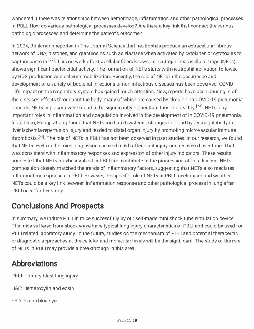
The self-made mini shock tube simulation deviceIn this study, the mouse PBLI model was induced by a self-made mini shock tube simulation device. Thedevice consists of �ve parts: compression chamber, release chamber, pricking device, pressure sensorand computer control system (Figure 1). This device weighs about 80 kg. Both compress chamber andrelease chamber are vertical steel tubes with a diameter of 400 mm and a height of 200mm and 500mm,respectively. The two chamber are separated by multilevel tin foils in the middle and secured by liftingring bolts, where a damaging shock wave is generated with peak pressures ranging from 0 bar to 5 bar.The device has two pressure sensors: one is located in the cylinder wall of the compress chamber and isused to measure the pre-explosion pressure in the compress chamber; another is located in the shockrelease port of the release chamber and measures the peak pressure of the shock wave actually appliedto the experimental animals. The two sensors are both connected to a computer control system to recordand display real-time pressure in both two chambers at a frequency of 200 Hz/s. A special rubber platewith holes (its diameter is 200mm) is �xed on the upper part of the release chamber, which is also seenas a platform for animal placement. The pricking device is installed in the middle of the release chamber.The computer control system can set the data of burst pressure and display the real-time change of airpressure in the two chambers, store and transmit the relevant data. The compression chamber isconnected with an air compressor. The compressed air is injected into the compression chamber throughthe air compressor when the control system is turned on. When the pressure-setting value is reached, thepricking device pierces the aluminum �lm and produces instantaneous shock wave. Scatter plots of thepeak pressure were drawn using origin software version 9.0 and �tted by a linear line to determine theshock-wave loading trend.

Page 4/19
Animals and statement of ethicsMale C57BL/6 mice, aged 6-8 weeks and weighing 18-24 g were purchased from Beijing Vital RiverLaboratory Animal Technology Co., Ltd. (Beijiing, China). All mice were housed in a temperature-controlled and speci�c-pathogen-free environment with a 12 h light/dark cycle, and free access to foodand water. Animal welfare and experimental design were approved by the Institutional Animal Care andUse Committee of Yi Shengyuan Gene Technology (Tianjin) (YSY-DWLL-2021013).
Establishment of mouse PBLI model40 mice were randomly divided into 4 groups. After anesthesia, mice were placed in the prone position onthe rubber plate with the chest directly above the hole. The mice were suffering from 0.5 bar, 1 bar, 1.5 baror 2 bar shock pressures, respectively. Physiological status and survival were observed every 2 h for the�rst day after injury. From the second day after the injury, mice survival was observed once a day.
According to the results of survival study above, 0.5 bar was �nally selected as the injury pressure.Another 48 mice were randomly divided into the control group and the model group. The model groupwere suffered 0.5 bar shock pressures, and the control group was treated similarly but without exposureto shock wave. The lungs of the mice in each group were harvest and weighted at 2 h, 4 h, 6 h, 12 h and24 h after injury (n=8).
Hematoxylin and eosin (H&E) staining and lung pathologyscoringThe right lung of mice was harvest and placed in 4% paraformaldehyde �x solution. Para�n embeddingand slices were used for H&E staining. The lung pathological injury was scored according to the H&Eresults of lung tissue sections and lung pathology scoring standard [9]. Pathological injury was scoredaccording to the following variables: alveolar and interstitial in�ammation, alveolar and interstitialhemorrhage, edema, alveolar fusion, alveolar septal thickening. The severity of each of the sevenindicators is graded as follows: no damage was graded as 0 points, damage with 25% was 1 points,damage with 50% was 2 points, damage with 75% was 3 points, and diffuse damage was 4 points. Thehighest score is 28 points and the lowest is 0 points.
Lung vascular permeability assayLung vascular permeability was evaluated by measuring Evans blue dye (EBD) leakage. In brief, 4%Evans blue (5 mL/kg) was injected intravenously at each time point after blast injury. The mice weresacri�ced 2 h after injection. A small incision was made in the left atrium with ophthalmic scissors, andPBS containing heparin sodium perfusion through the right ventricle was performed followed by a 20 mLrinse. Then the lungs were removed and placed into 1 mL Dimethylformamide. After incubated at 60℃for 24 h, the tissue were centrifuged at 4,000 r/min for 10 min. The supernatant was collected and theabsorbance was detected at 620 nm. According to the standard curve, the content of EBD in lung tissuewas calculated.
Page 5/19
Bronchoalveolar lavage �uid (BALF) examinationAfter sacri�ced, the trachea of the mice was exposed. A small incision was made below the thyroidcartilage and an 18 G endotracheal intubation was inserted, ligated and �xed. Mice trachea was lavagedwith 0.4 ml PBS each time, repeated for 3 times, and collected BALF solution. Then the BALF wascentrifuged at 6,000 r/min for 15 min, the supernatant was collected and the protein concentration wasdetected. After washing twice with PBS, the precipitation was mixed with 2% glacial acetic acid, the totalnumber of leukocyte was counted under a microscope. Leukocyte cell smears were prepared, and thetotal leukocyte were counted under a microscope after staining with Giemsa solution.
Real-time qPCRThe total RNA was extracted from the lung tissues using the TRIzol reagent (Invitrogen, USA), theconcentration and purity of RNA were detected using NanoDrop One Microvolume UV-VisSpectrophotometer. RNA reverse transcription was performed using PrimeScript RT reagent Kit (TaKaRa,Japan) and Mastercycler pro PCR instrument (Eppendorf, GER). Real-time qPCR was performed usingHieffTM qPCR SYBR® Green Master Mix (Yeasen, China) and LightCycle 96 instrument (Roche, Swiss).Gene expression was analyzed by LightCycle 96 software. The expression level was normalized to that ofβ-actin, and was calculated using the 2−△△Ct method. The primer sequences and running program in real-time qPCR are shown in Table 1.
Table 1Sequence of Primers and real-time qPCR running program.
Gene Primer Sequence of primers real-time qPCR
running program
IL-1β Forward TGCCACCTTTTGACAGTGATG 1. Preincubation
¬ 95℃ for 5 min
2. Amplication (50 cycles)
¬ 95℃ for 10 s
¬ 56℃ for 20 s
¬ 72℃ for 20 s
3. Melting
¬ 95℃ for 10 s
¬ 65℃ for 60 s
¬ 97℃ for 1 s
4. Cooling
37℃ for 30 s
Reverse AAGGTCCACGGGAAAGACAC
IL-6 Forward TAGTCCTTCCTACCCCAATTTCC
Reverse TTGGTCCTTAGCCACTCCTTC
TNF-α Forward CATCTTCTAAAATTCGAGTGACAA
Reverse TGGGAGTAGACAAGGTACAACCC
β-actin Forward AGTGTGACGTTGACATCCGT
Reverse GCAGCTCAGTAACAGTCCGC

Page 6/19
Western blotThe lung tissues were lysed by lysis buffer. After incubating on ice, the supernatant was obtained bycentrifugation at 12,000 rpm for 15 min. The protein concentration was measured by BCA assay. Theprotein was added SDS-PAGE sample loading buffer, boiled and denatured for 10 min. After SDS-PAGEgel electrophoresis, the protein sample was transferred to the PVDF membrane and sealed with 5%skimmed milk for 2 h. Then the appropriate primary antibody IL-1β (1:1000; cat. no. ab205924; Abcam,Cambridge, UK), IL-6 (1:1000; cat. no. #12912; Cell Signaling Technolgy, Boston, USA), TNF-α (1:1000; cat.no. ab215188; Abcam, Cambridge, UK), MPO (1:500; cat. no. sc-390109; Santa Cruz Biotechnology, Inc.,Dallas, TX, USA), β-actin (1:5000; cat. no. bs-0061R; Bioss, Beijing, China) were added and incubatedovernight at 4 ℃. Then, the membrane was washed three times with TBST, and a horseradish peroxidase-labeled anti-mouse secondary antibody (1:2000; cat. no. CW0102; Cowin Biosciences, Jiangsu, China),anti-rabbit secondary antibody (1:5000; cat. no. EF0002; Sparkjade, Shandong, China) were incubated for1 h at room temperature. The membrane was washed three times with TBST. Proteins were visualizedusing an ECL hypersensitive chemiluminescence kit (Sparkjade, cat. no. ED0016-B; Shandong, China) anda Tanon 5200 Full automatic chemiluminescence image analysis system (Tanon Science andTechnology Co., Ltd, Shanghai, China).
Immuno�uorescence stainingThe lung tissues section was depara�nized with xylene and dehydrated with ethanol gradient, and theantigens were repaired with 0.01 M citrate buffer (pH 6.0). After 30 min incubation with 0.1% Triton X-100,the tissues were sealed with 10% sheep serum for 30 min. Then the primary antibody, MPO (1:50; cat. no.sc-390109; Santa Cruz Biotechnology, Inc., Dallas, TX, USA), citH3 (1:50; cat. no. ab5103; Abcam,Cambridge, UK) were incubated at 4℃ overnight. After washing with PBS for 3 times, the �uorescentsecondary antibody was added. Finally, the nuclei were stained by 4,6-diamino-2-phenyl indole (DAPI). Allsections were analyzed under confocal microscopy.
Statistical analysisStatistical analysis was performed using the Excel software V2019. All data were presented as mean ±SE; The homogeneity variance and one way ANOVA were applied to the whole sample study. TheKaplan–Meier method was used to evaluate the survival rates of the experimental animals, and alogarithmic survival curve was plotted. The χ2 test was used to compare the survival rates among eachgroup. All the statistical tests were two-tailed tests, with statistical signi�cance de�ned as p<0.05.
Result
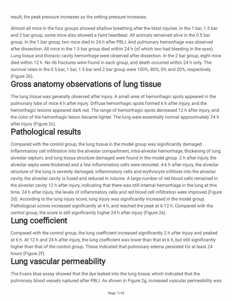
Parameters of shock wave and survival rate of animalsThe self-made mini shock tube simulation device (Figure 1) was used to establish the PBLI animal model.The peak pressure with a setting pressure of 0.5 bar, 1 bar, 1.5 bar and 2 bar are shown in Figure 2a. As a
Page 7/19
result, the peak pressure increases as the setting pressure increases.
Almost all mice in the four groups showed shallow breathing after the blast injuries. In the 1 bar, 1.5 barand 2 bar group, some mice also showed a faint heartbeat. All animals remained alive in the 0.5 bargroup. In the 1 bar group, two mice died in 24 h after PBLI. And pulmonary hemorrhage was observedafter dissection. All mice in the 1.5 bar group died within 24 h (of which two had bleeding in the eyes).Lung tissue and thoracic cavity hemorrhage were observed after dissection. In the 2 bar group, eight micedied within 12 h. No rib fractures were found in each group, and death occurred within 24 h only. Thesurvival rates in the 0.5 bar, 1 bar, 1.5 bar and 2 bar group were 100%, 80%, 0% and 20%, respectively(Figure 2b).
Gross anatomy observations of lung tissueThe lung tissue was generally observed after injury. A small area of hemorrhagic spots appeared in thepulmonary lobe of mice 4 h after injury. Diffuse hemorrhagic spots formed 6 h after injury, and thehemorrhagic lesions appeared dark red. The range of hemorrhagic spots decreased 12 h after injury, andthe color of the hemorrhagic lesion became lighter. The lung were essentially normal approximately 24 hafter injury (Figure 2c).
Pathological resultsCompared with the control group, the lung tissue in the model group was signi�cantly damaged.In�ammatory cell in�ltration into the alveolar compartment, intra-alveolar hemorrhage, thickening of lungalveolar septum, and lung tissue structure damaged were found in the model group. 2 h after injury, thealveolar septa were thickened and a few in�ammatory cells were recruited. 4-6 h after injury, the alveolarstructure of the lung is severely damaged, in�ammatory cells and erythrocyte in�ltrate into the alveolarcavity, the alveolar cavity is fused and reduced in volume. A large number of red blood cells remained inthe alveolar cavity 12 h after injury, indicating that there was still internal hemorrhage in the lung at thistime. 24 h after injury, the levels of in�ammatory cells and red blood cell in�ltration were improved (Figure2d). According to the lung injury score, lung injury was signi�cantly increased in the model group.Pathological scores increased signi�cantly at 4 h, and reached the peak at 6-12 h. Compared with thecontrol group, the score is still signi�cantly higher 24 h after injury (Figure 2e).
Lung coe�cientCompared with the control group, the lung coe�cient increased signi�cantly 2 h after injury and peakedat 6 h. At 12 h and 24 h after injury, the lung coe�cient was lower than that at 6 h, but still signi�cantlyhigher than that of the control group. These indicated that pulmonary edema persisted for at least 24hours (Figure 2f).
Lung vascular permeabilityThe Evans blue assay showed that the dye leaked into the lung tissue, which indicated that thepulmonary blood vessels ruptured after PBLI. As shown in Figure 2g, increased vascular permeability was
Page 8/19
observed at model groups. And the level of EB in lung tissue reached the peak at 6 h, which indicated thatthe diffuse vascular leakage in the lung tissue of the mice was highest at 6 h after injury.
Protein content and leukocyte number in BALFThe total protein and cell content in BALF show the changes of permeability of pulmonary capillaries andalveolar epithelial cells. Compared with the control group, the total protein content in BALF increasedsigni�cantly at model group (P < 0.05) after injury (Figure 2h). As for the total cells number of leukocytein BALF, it is increased signi�cantly after injury and reached a peak at 2 h (Figure 2i).
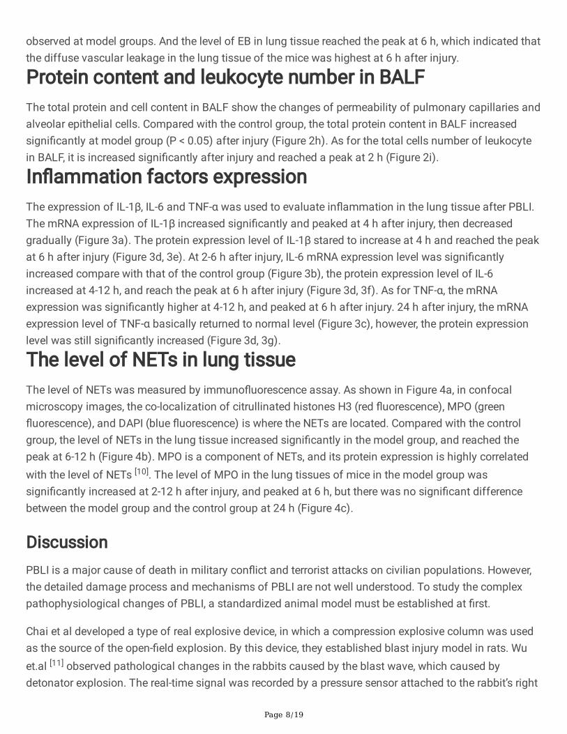
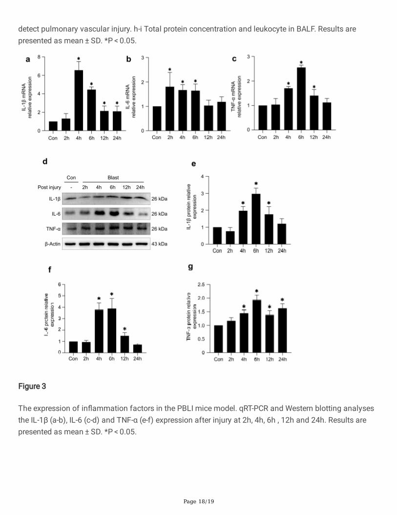
In�ammation factors expressionThe expression of IL-1β, IL-6 and TNF-α was used to evaluate in�ammation in the lung tissue after PBLI.The mRNA expression of IL-1β increased signi�cantly and peaked at 4 h after injury, then decreasedgradually (Figure 3a). The protein expression level of IL-1β stared to increase at 4 h and reached the peakat 6 h after injury (Figure 3d, 3e). At 2-6 h after injury, IL-6 mRNA expression level was signi�cantlyincreased compare with that of the control group (Figure 3b), the protein expression level of IL-6increased at 4-12 h, and reach the peak at 6 h after injury (Figure 3d, 3f). As for TNF-α, the mRNAexpression was signi�cantly higher at 4-12 h, and peaked at 6 h after injury. 24 h after injury, the mRNAexpression level of TNF-α basically returned to normal level (Figure 3c), however, the protein expressionlevel was still signi�cantly increased (Figure 3d, 3g).
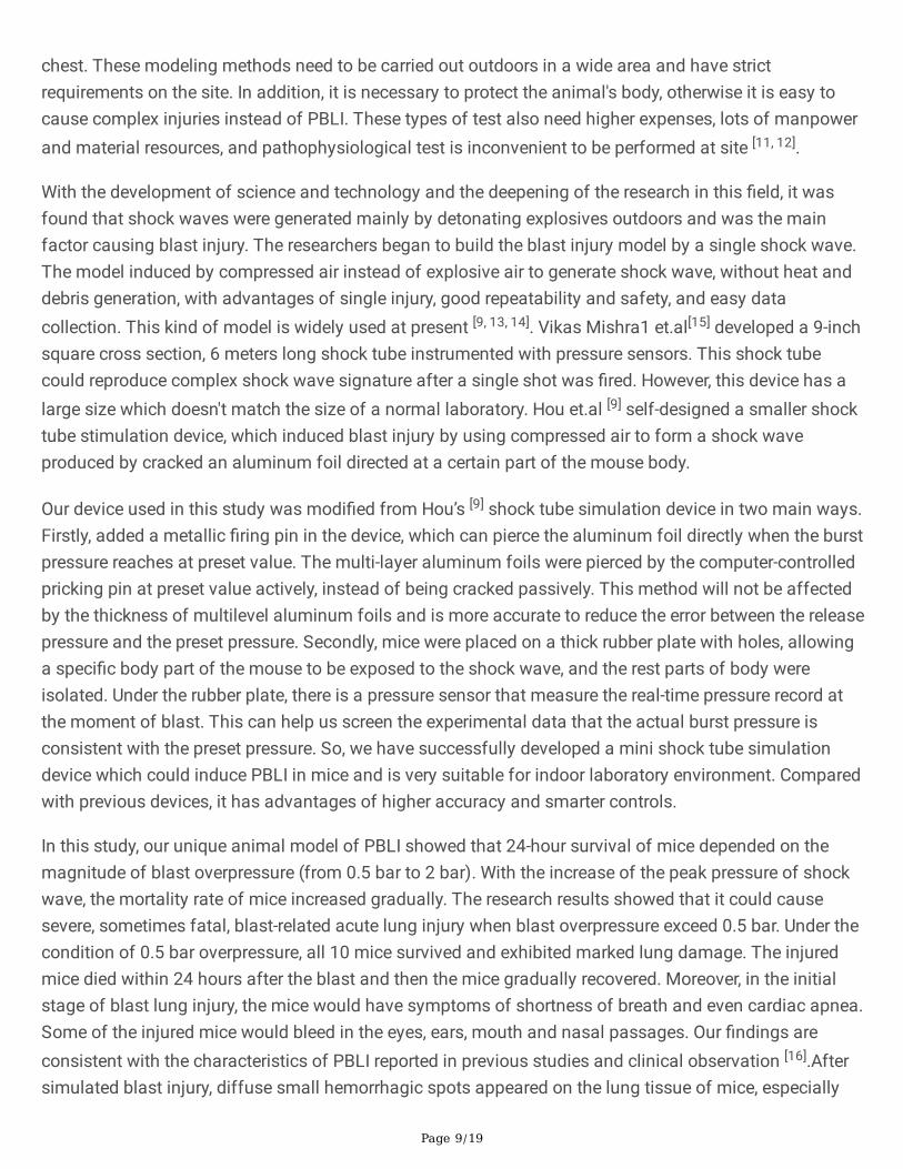
The level of NETs in lung tissueThe level of NETs was measured by immuno�uorescence assay. As shown in Figure 4a, in confocalmicroscopy images, the co-localization of citrullinated histones H3 (red �uorescence), MPO (green�uorescence), and DAPI (blue �uorescence) is where the NETs are located. Compared with the controlgroup, the level of NETs in the lung tissue increased signi�cantly in the model group, and reached thepeak at 6-12 h (Figure 4b). MPO is a component of NETs, and its protein expression is highly correlatedwith the level of NETs [10]. The level of MPO in the lung tissues of mice in the model group wassigni�cantly increased at 2-12 h after injury, and peaked at 6 h, but there was no signi�cant differencebetween the model group and the control group at 24 h (Figure 4c).
DiscussionPBLI is a major cause of death in military con�ict and terrorist attacks on civilian populations. However,the detailed damage process and mechanisms of PBLI are not well understood. To study the complexpathophysiological changes of PBLI, a standardized animal model must be established at �rst.
Chai et al developed a type of real explosive device, in which a compression explosive column was usedas the source of the open-�eld explosion. By this device, they established blast injury model in rats. Wuet.al [11] observed pathological changes in the rabbits caused by the blast wave, which caused bydetonator explosion. The real-time signal was recorded by a pressure sensor attached to the rabbit’s right
Page 9/19
chest. These modeling methods need to be carried out outdoors in a wide area and have strictrequirements on the site. In addition, it is necessary to protect the animal's body, otherwise it is easy tocause complex injuries instead of PBLI. These types of test also need higher expenses, lots of manpowerand material resources, and pathophysiological test is inconvenient to be performed at site [11, 12].
With the development of science and technology and the deepening of the research in this �eld, it wasfound that shock waves were generated mainly by detonating explosives outdoors and was the mainfactor causing blast injury. The researchers began to build the blast injury model by a single shock wave.The model induced by compressed air instead of explosive air to generate shock wave, without heat anddebris generation, with advantages of single injury, good repeatability and safety, and easy datacollection. This kind of model is widely used at present [9, 13, 14]. Vikas Mishra1 et.al[15] developed a 9-inchsquare cross section, 6 meters long shock tube instrumented with pressure sensors. This shock tubecould reproduce complex shock wave signature after a single shot was �red. However, this device has alarge size which doesn't match the size of a normal laboratory. Hou et.al [9] self-designed a smaller shocktube stimulation device, which induced blast injury by using compressed air to form a shock waveproduced by cracked an aluminum foil directed at a certain part of the mouse body.
Our device used in this study was modi�ed from Hou’s [9] shock tube simulation device in two main ways.Firstly, added a metallic �ring pin in the device, which can pierce the aluminum foil directly when the burstpressure reaches at preset value. The multi-layer aluminum foils were pierced by the computer-controlledpricking pin at preset value actively, instead of being cracked passively. This method will not be affectedby the thickness of multilevel aluminum foils and is more accurate to reduce the error between the releasepressure and the preset pressure. Secondly, mice were placed on a thick rubber plate with holes, allowinga speci�c body part of the mouse to be exposed to the shock wave, and the rest parts of body wereisolated. Under the rubber plate, there is a pressure sensor that measure the real-time pressure record atthe moment of blast. This can help us screen the experimental data that the actual burst pressure isconsistent with the preset pressure. So, we have successfully developed a mini shock tube simulationdevice which could induce PBLI in mice and is very suitable for indoor laboratory environment. Comparedwith previous devices, it has advantages of higher accuracy and smarter controls.
In this study, our unique animal model of PBLI showed that 24-hour survival of mice depended on themagnitude of blast overpressure (from 0.5 bar to 2 bar). With the increase of the peak pressure of shockwave, the mortality rate of mice increased gradually. The research results showed that it could causesevere, sometimes fatal, blast-related acute lung injury when blast overpressure exceed 0.5 bar. Under thecondition of 0.5 bar overpressure, all 10 mice survived and exhibited marked lung damage. The injuredmice died within 24 hours after the blast and then the mice gradually recovered. Moreover, in the initialstage of blast lung injury, the mice would have symptoms of shortness of breath and even cardiac apnea.Some of the injured mice would bleed in the eyes, ears, mouth and nasal passages. Our �ndings areconsistent with the characteristics of PBLI reported in previous studies and clinical observation [16].Aftersimulated blast injury, diffuse small hemorrhagic spots appeared on the lung tissue of mice, especially

Page 10/19
obviously at 6 h after injury. Obvious pulmonary edema occurred in the model group, and the highestdegree of the edema was found at 6 h as showed by increased lung coe�cient and vascular permeability.Pathological examination of lung tissue showed the ruptured alveolar septal and the in�ltration ofin�ammatory cells in the lung tissue of the injured mice. The recruitment of in�ammatory cells in lungtissue indicates the occurrence of in�ammatory response in lung tissue from the perspective ofpathological injury. The degree of pathological injury of lung tissue reached the peak at 6 h, and thengradually decreased after 12 h. Blood cells also appear in the interstitial lung signi�cantly 6 h after injury,suggesting the occurrence of intrapulmonary hemorrhage. All these results suggest symptoms ofpneumothorax and internal bleeding at this time, which are closely consistent with those seen in clinicalpatients with PBLI and in laboratory animals in other studies. The reasons for these changes are that thevolume of the mouse chest immediately decreased after suffering the blast shock wave and instantlyrebounded when the external force was eliminated. During this process, the intrathoracic pressure risesinitially and then falls sharply, resulting in the rupture of the alveoli and the pulmonary blood vessels. Thepermeability of alveoli-capillary membrane increased signi�cantly, and a large number of proteins andcells exude through the blood-gas barrier [11, 16–18]. As seen in this study, shock waves induced by ourself-made device could cause mice lung damage, which may lead to a variety of pathophysiologicalchanges, including hemorrhage, edema, and in�ammatory cells in�ltration in the lung.
In this study, the expression of IL-1β, IL-6 and TNF-α in the mice lung tissues was signi�cantly increasedin the model group, and peaked within 2 and 6 h, respectively. The expression levels of in�ammatoryfactors in the mice lung tissues were signi�cantly changed only in the earlier acute phase of PBLI, andreturned to normal at 24 h after injury. Combined with pathological observation and molecular levelexploration, it was not di�cult to �nd that the lung tissue, as the target organ of the shock wave, occurredobvious in�ammatory response. The increase of in�ammatory factors in this in�ammatory response ispositively correlated with the degree of recruitment of in�ammatory cells. In�ammatory cells number andrecruitment degree are closely related to the degree of lung tissue damage. Previous researchessuggested that when lung tissue were damaged by shock waves, the damaged cells released a largenumber of cytokines, inducing various in�ammatory factors [9, 19]. Previous studies have reported thatPBLI involves an immediate autonomic response, followed by intrapulmonary hemorrhage andhistopathological injury, and �nally the production of cytokines associated with in�ammation [18]. Our�ndings are basically consistent with the previous results, except that the in�ammatory response appearsearlier, which is more consistent with the clinical manifestations of critical illness. It indicated that ourPBLI animal model could be used in the diagnosis and treatment of PBLI and other related studies in thepre-hospital phase.
Clinical studies have shown that the initial signs of PBLI are cough, hemoptysis, and other pulmonarysymptoms [20, 21]. Some of them have hemothorax and/or combined pneumothorax. The victim at thescene of the explosion often have various degrees of visceral injury without signi�cant surface damage, aminority of patients developed rapidly progressive pneumonia leading to acute respiratory distresssyndrome (ARDS). The main cause of death is shock, respiratory failure, septicemia, even ARDS. We
Page 11/19
wondered if there was relationships between hemorrhage, in�ammation and other pathological processesin PBLI. How do various pathological processes develop? Are there a key link that connect the variouspathologic processes and determine the patient's outcome?
In 2004, Brinkmann reported in The Journal Science that neutrophils produce an extracellular �brousnetwork of DNA, histones, and granulocins such as elastase when activated by cytokines or cytotoxins tocapture bacteria [22]. This network of extracellular �bers known as neutrophil extracellular traps (NETs),shows signi�cant bactericidal activity. The formation of NETs starts with neutrophil activation followedby ROS production and calcium mobilization. Recently, the role of NETs in the occurrence anddevelopment of a variety of bacterial infections or non-infectious diseases has been observed. COVID-19’s impact on the respiratory system has gained much attention. Now, reports have been pouring in ofthe disease’s effects throughout the body, many of which are caused by clots [23]. In COVID-19 pneumoniapatients, NETs in plasma were found to be signi�cantly higher than those in healthy [24]. NETs playimportant roles in in�ammation and coagulation involved in the development of in COVID-19 pneumonia.In addition, Hongji Zhang found that NETs mediated systemic changes in blood hypercoagulability inliver ischemia-reperfusion injury and leaded to distal organ injury by promoting microvascular immunethrombosis [24]. The role of NETs in PBLI has not been observed in past studies. In our research, we foundthat NETs levels in the mice lung tissues peaked at 6 h after blast injury and recovered over time. Thatwas consistent with in�ammatory responses and expression of other injury indicators. These resultssuggested that NETs maybe involved in PBLI and contribute to the progression of this disease. NETscomposition closely matched the trends of in�ammatory factors, suggesting that NETs also mediatesin�ammatory responses in PBLI. However, the speci�c role of NETs in PBLI mechanism and weatherNETs could be a key link between in�ammation response and other pathological process in lung afterPBLI need further study.
Conclusions And ProspectsIn summary, we induce PBLI in mice successfully by our self-made mini shock tube simulation device.The mice suffered from shock wave have typical lung injury characteristics of PBLI and could be used forPBLI related laboratory study. In the future, studies on the mechanism of PBLI and potential therapeuticor diagnostic approaches at the cellular and molecular levels will be the signi�cant. The study of the roleof NETs in PBLI may provide a breakthrough in this area.
AbbreviationsPBLI: Primary blast lung injury
H&E: Hematoxylin and eosin
EBD: Evans blue dye

Page 12/19
BALF: Bronchoalveolar lavage �uid
NETs: Neutrophil extracellular traps
BLI: Blast lung injury
DAPI: 4,6-diamino-2-phenyl indole
ARDS: Acute respiratory distress syndrome
DeclarationsAvailability of data and materials
The data and materials used in the current study are all available from the corresponding author uponreasonable request.
Funding
This study was supported by the Open Scienti�c Research Program of Military Logistics by Hao-Jun Fan(BLB19J006) and Yan-Mei Zhao (BLB20J009).
Author information
Xiang-Yan Meng, Qian-Ying Lu and Jian-Feng Zhang contributed equally to this work.
A�liations
Institute of Disaster and Emergency Medicine, Tianjin University, Tianjin, 300072, China
Xiang-Yan Meng, Qian-Ying Lu, Jian-Feng Zhang, Jun-Feng Li, Ming-Yu Shi, Si-Yu Huang, Si-Fan Yu, Yan-Mei Zhao and Hao-Jun Fan
Tianjin Key Laboratory of Disaster Medicine Technology, Tianjin, 300072, China
Xiang-Yan Meng, Qian-Ying Lu, Jian-Feng Zhang, Jun-Feng Li, Ming-Yu Shi, Si-Yu Huang, Si-Fan Yu, Yan-Mei Zhao and Hao-Jun Fan
Authors’ contributions
YMZ, HJF, XYM and QYL conceived and designed the experiments; XYM, QYL and JFZ performed theexperiments; JFL, MYS and SFY contributed reagents/ materials; JFZ and SYH contributed data analyses;JFZ wrote the manuscript and XYM, QYL revised it. All authors read and approved the �nal manuscript
Corresponding authors
Page 13/19
Correspondence to Yan-Mei Zhao and Hao-Jun Fan.
Acknowledgements
Not applicable.
Ethics declarations
Ethics approval and consent to participate
The experiments involving animals were approved by the animal care and use ethical committee of YiShengyuan Gene Technology (Tianjin) (YSY-DWLL-2021013) and complied with the Guide for the Careand Use of Laboratory Animals approved by the National Institutes of Health.
Consent for publication
Not applicable.
Competing interests
The authors declare that there are no competing interests.
References1. Chang Y, Zhang DH, Liu LY, Yu YH, Wu Y, Zang LW, et al. Simulation of blast lung injury induced by
shock waves of �ve distances based on �nite element modeling of a three-dimensional rat. Sci Rep.2019;9(1):3440.
2. Krug EG, Sharma GK, Lozano R. The global burden of injuries. Am J Public Health. 2000;90(4):523–6.
3. Mackenzie IM, Tunnicliffe B. Blast injuries to the lung: epidemiology and management. Philos TransR Soc Lond B Biol Sci. 2011;366(1562):295–9.
4. Lichtenberger JP, Kim AM, Fisher D, TatumPS, Neubauer B, Peterson PG, et al. Imaging of Combat-Related Thoracic Trauma - Blunt Trauma and Blast Lung Injury. Mil Med. 2018;183(3-4):e89–96.
5. Wolf SJ, Bebarta VS, Bonnett CJ, Pons PT, Cantrill SV. Blast injuries. Lancet. 2009;374(9687):405–15.
�. Smith JE. The epidemiology of blast lung injury during recent military con�icts: a retrospectivedatabase review of cases presenting to deployed military hospitals, 2003-2009. Philos Trans R SocLond B Biol Sci. 2011;366(1562):291–4.
7. Cooper GJ, Townend DJ, Cater SR, Pearce BP. The role of stress waves in thoracic visceral injury fromblast loading: modi�cation of stress transmission by foams and high-density materials. J Biomech.1991;24(5):273–85.

Page 14/19
�. Cooper GJ, Pearce BP, Sedman AJ, Bush IS, Oakley CW. Experimental evaluation of a rig to simulatethe response of the thorax to blast loading. J Trauma. 1996;40(Suppl 3):38-11.
9. Tong CC, Liu YN, Zhang YB, Cong PF, Shi XY, Liu Y, et al. Shock waves increase pulmonary vascularleakage, in�ammation, oxidative stress, and apoptosis in a mouse model. Exp Biol Med (Maywood).2018;243(11):934–14.
10. von Meijenfeldt FA, Stravitz RT, Zhang JW, Adelmeijer J, Zen Y, Durkalski V, et al. Generation ofneutrophil extracellular traps in patients with acute liver failure is associated with poor outcome.Hepatology. 2021. doi:10.1002/hep.32174.
11. Wu SY, Han GF, Kang JY, Zhang LC, Wang AM, Wang JM. Pulmonary microvascular dysfunction andpathological changes induced by blast injury in a rabbit model. Ulus Travma Acil Cerrahi Derg.2016;22(5):405–11.
12. Zhao Y, Zhou YG. The past and present of blast injury research in China. Chin J Traumatol.2015;18(4):194–10.
13. Yang ZS, Aderemi OA, Zhao QW, Edsall PR, Simovic MO, Lund BJ, et al. Early Complement andFibrinolytic Activation in a Rat Model of Blast-Induced Multi-Organ Damage. Mil Med.2019;184(Suppl 1):282–10.
14. Li YS, Yang ZS, Chavko M, Liu B, Aderemi OA, Simovic MO, et al. Complement inhibition amelioratesblast-induced acute lung injury in rats: Potential role of complement in intracellular HMGB1-mediatedin�ammation. PLoS One. 2018. doi:10.1371/journal.pone.0202594.
15. Mishra V, Skotak M, Schuetz H, Heller A, Haorah J, Chandra N. Primary blast causes mild, moderate,severe and lethal TBI with increasing blast overpressures: Experimental rat injury model. Sci Rep.2016. doi:10.1038/srep26992.
1�. Scott T, Hulse E, Haque M, Kirkman E, Hardman J, Mahoney P. Modelling primary blast lung injury:current capability and future direction. J R Army Med Corps. 2017;163(2):84–8.
17. de Candole CA. Blast injury. Can Med Assoc J. 1967;96(4):207–14.
1�. Scott TE, Kirkman E, Haque M, Gibb IE, Mahoney P, Hardman JG. Primary blast lung injury - a review.Br J Anaesth. 2017;118(3):311–6.
19. Barnett-Vanes A, Sharrock A, Eftaxiopoulou T, Arora H, Macdonald W, Bull AMJ, et al. CD43Loclassical monocytes participate in the cellular immune response to isolated primary blast lung injury.J Trauma Acute Care Surg. 2016;81(3):500–11.
20. Zhang Y, Meng WZ, Wang MJ, Ren WJ, Wang GZ, Zhang GB, et al. The epidemiological features ofblast injury of lungs caused by gas explosion. Zhonghua Lao Dong Wei Sheng Zhi Ye Bing Za Zhi.2012;30(8):582–3.
21. Lee K, Yoon J, Min K, Lee J, Kang S, Hong S, et al. An objective index to estimate the survival rate ofprimary blast lung injury. Annu Int Conf IEEE Eng Med Biol Soc. 2014;2014:1206–9.
22. Brinkmann V, Reichard U, Goosmann C, Fauler B, Uhlemann Y, Weiss DS, et al. Neutrophil extracellulartraps kill bacteria. Science. 2004;303(5663):1532–5.
Page 15/19
23. Willyard C. Coronavirus blood-clot mystery intensi�es. Nature. 2020;581(7808):250.
24. Skendros P, Mitsios A, Chrysanthopoulou A, Mastellos DC, Metallidis S, Rafailidis P, et al.Complement and tissue factor-enriched neutrophil extracellular traps are key drivers in COVID-19immunothrombosis. J Clin Invest. 2020;130(11):6151–7.
Figures
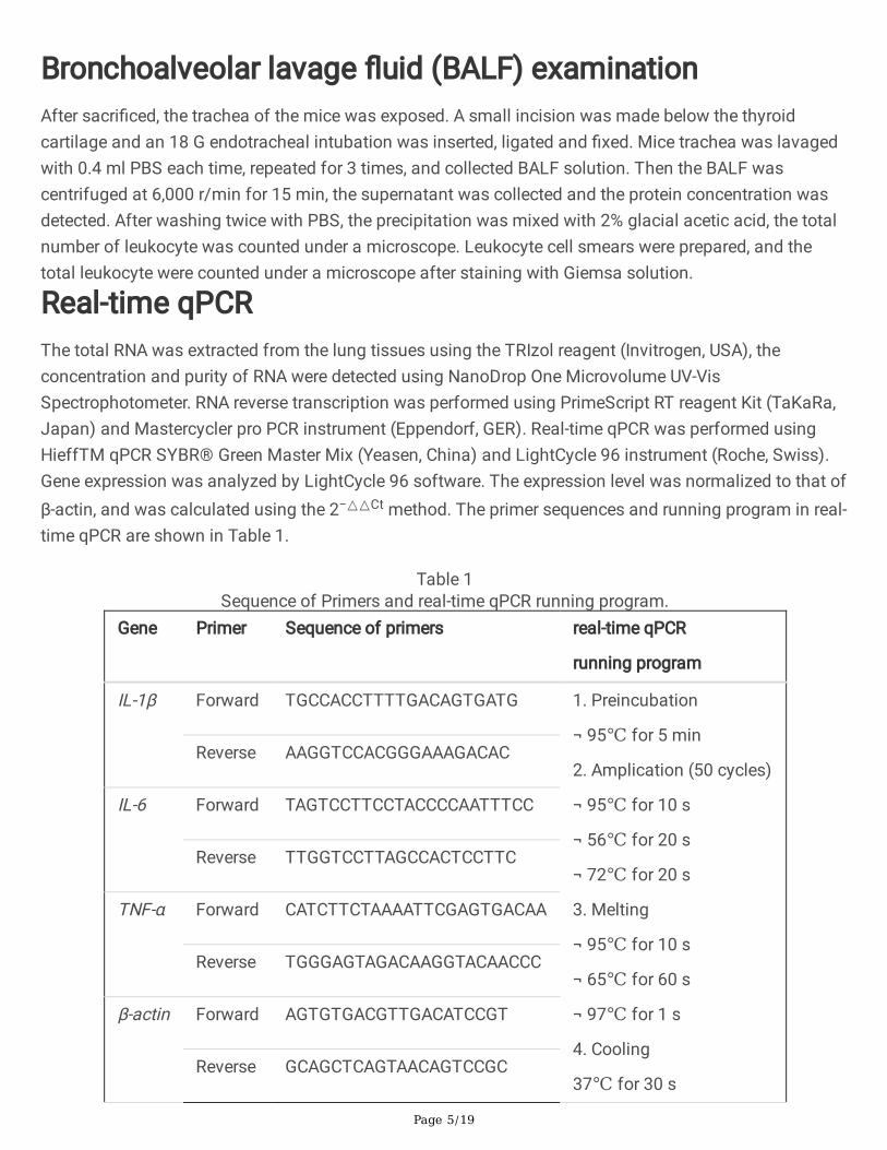
Figure 1
Page 16/19
The self-made mini shock tube simulation device. a Model diagram of the device. The device consists of�ve parts: compression chamber, release chamber, pricking device, pressure transducer sensor andcomputer control system. Both the compress chamber and the release chamber are respectively verticalsteel tubes and are separated by multilevel tin foils in the middle. The compression chamber is connectedwith an air compressor. The compressed air is injected into the compression chamber through the aircompressor when the control system is turned on. When the pressure-setting value is reached, thepricking device pierces the aluminum �lm and produces instantaneous shock wave. The shock wavegenerated by the device ranges from 0-2bar. b-f Photos of the device. b Shock tube main body. c Releasechamber. d Compression chamber. e Pricking device. f Pressure sensor

Page 17/19
Figure 2
Lung tissue damage in mice after explosion. a Diagram of the peak pressure of shock wave generated bythe device changing with time (0.5- 2.0 bar). b Survival analysis of the animals with blast injuries atdifferent shock wave pressure (0.5- 2.0 bar). c Gross observation of lung tissue. d-e H&E staining andpathological scores of lung tissues. f The lung coe�cient in each group. g Evans blue dye leakage test to
Page 18/19
detect pulmonary vascular injury. h-i Total protein concentration and leukocyte in BALF. Results arepresented as mean ± SD. *P < 0.05.
Figure 3
The expression of in�ammation factors in the PBLI mice model. qRT-PCR and Western blotting analysesthe IL-1β (a-b), IL-6 (c-d) and TNF-α (e-f) expression after injury at 2h, 4h, 6h , 12h and 24h. Results arepresented as mean ± SD. *P < 0.05.

Page 19/19
Figure 4
The level of NETs in lung tissue. a Immuno�uorescence assay shows the level of NETs in the lung tissueafter injury. Citrullinated histone H3 is stained red, Myeloperoxidase is stained green and DNA is stainedblue. b Western blotting analyses the expression of MPO.