Embed Size (px)
Citation preview

! www.clutchprep.com
!
ANATOMY & PHYSIOLOGY - CLUTCH
CH. 5 - MEMBRANE DYNAMICS

CONCEPT: DIFFUSION
Diffusion, Qualitatively:
● Diffusion is the simple movement of solute from an area of higher concentration to an area of lower concentration.
□ Entirely passive—no _______________________ expenditure is required.
□ Happens from higher concentration→lower concentration.
- Net movement until concentrations are equal everywhere.
□ ↑Diffusion rate for ↑Temperature and ↓Molecular Size/Weight.
● Diffusion into and out of cells must happen across cell membranes, which are ___________________________.
□ Lipophilic (i.e. nonpolar) solutes are the major solutes that enter cells via simple diffusion.
□ Hydrophilic (i.e. polar) solutes need to be transported across the membrane (more later).
EXAMPLE: Nonpolar solutes cross the cell membrane into cells via diffusion, moving from an area of higher concentration (the extracellular space) to an area of lower concentration (the intracellular space).
ANATOMY & PHYSIOLOGY - CLUTCH
CH. 5 - MEMBRANE DYNAMICS
Page 2
Diffusion, Quantitatively—Fick’s Law:
● Mathematically, the rate of diffusion through membranes can be described by Fick’s Law: J=PA(ΔC), where:
□ J is the flux (the amount of solute moving per unit time) across the membrane.
□ P is the permeability of the membrane to the solute; if the solute can cross more easily, it crosses more quickly.
- A higher value of P means that the solute crosses more _______________________.
- Nonpolar molecules or molecules that can be transported typically have higher values of P.
□ A is the surface area of the membrane. More surface area means more “opportunity” for solute to cross.
□ ΔC is concentration gradient (the difference in solute concentration between the two side).
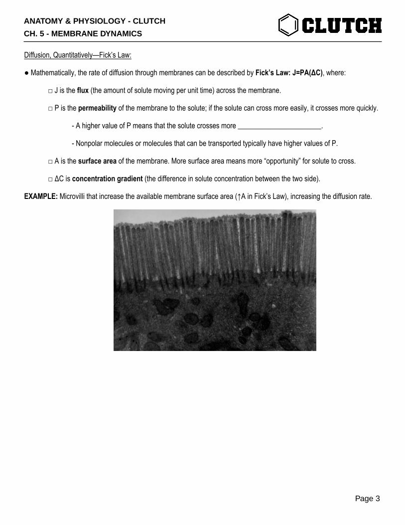
EXAMPLE: Microvilli that increase the available membrane surface area (↑A in Fick’s Law), increasing the diffusion rate.
ANATOMY & PHYSIOLOGY - CLUTCH
CH. 5 - MEMBRANE DYNAMICS
Page 3
PRACTICE 1: The walls of capillaries—where all of the exchange between the blood and body takes place—are made of endothelial cells. Tight junctions are proteins that hold adjacent endothelial cells together and prevent too much from crossing. After injury, damaged cells release molecules that loosen tight junctions. Which variable in Fick’s Law is affected by injury?
a) Permeability (P). b) Surface Area (A). c) Concentration Gradient (ΔC)
PRACTICE 2: The walls of capillaries—where all of the exchange between the blood and body takes place—are made of endothelial cells. Tight junctions are proteins that hold adjacent endothelial cells together and prevent too much from crossing. After injury, damaged cells release molecules that loosen tight junctions. How will this affect flux (J)?
a) Increase J (more flux). b) Decrease J (less flux). c) No change.
ANATOMY & PHYSIOLOGY - CLUTCH
CH. 5 - MEMBRANE DYNAMICS
Page 4
CONCEPT: OSMOSIS: OSMOLARITY, OSMOTIC PRESSURE, AND TONICITY
Osmolarity:
● The concentration of a specific solute is calculated by taking the amount of that solute and dividing by the volume it’s in.
□ Concentrations are expressed in units like molar (M, mols/liter) or millimolar (mM, millimoles/liter).
● Osmolarity (adjective: osmolar, OsM) is the concentration of any and every solute in a ___________________.
□ OsM=Total Solutes/Total Volume.
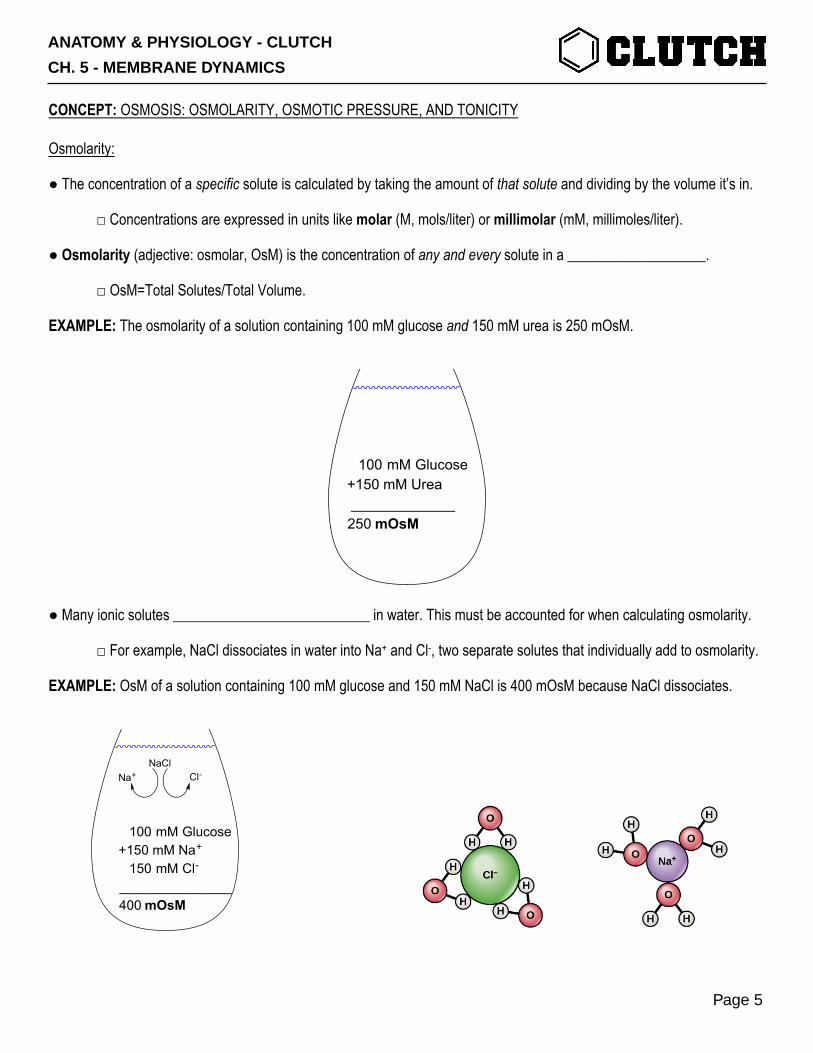
EXAMPLE: The osmolarity of a solution containing 100 mM glucose and 150 mM urea is 250 mOsM.
● Many ionic solutes ___________________________ in water. This must be accounted for when calculating osmolarity.
□ For example, NaCl dissociates in water into Na+ and Cl-, two separate solutes that individually add to osmolarity.
EXAMPLE: OsM of a solution containing 100 mM glucose and 150 mM NaCl is 400 mOsM because NaCl dissociates.
100 mM Glucose+150 mM Urea _____________250 mOsM
100 mM Glucose+150 mM Na+
150 mM Cl-
_______________400 mOsM
NaClNa+ Cl-
ANATOMY & PHYSIOLOGY - CLUTCH
CH. 5 - MEMBRANE DYNAMICS
Page 5

Hyper- vs. Hypo- vs. Isoosmotic:
● It’s often useful to compare the osmolarities of 2+ solutions separated from each other by a ______________________.
□ Hyperosmotic means the OsM of one solution is greater than the OsM of the other.
□ Hypoosmotic means the OsM of one solution is less than the OsM of the other.
□ Isoosmotic means that both solutions have the same OsM.
EXAMPLE: Two solutions—one with 100 mM NaCl, the other with 100 mM Glucose are separated by a membrane. The NaCl solution is 200 mOsM, and is therefore hyperosmotic to the 100 mOsM solution.
100 mM NaCl 100 mM Glucose
200 mOsM 100 mOsM
HYPEROSMOTIC HYPOOSMOTIC
ANATOMY & PHYSIOLOGY - CLUTCH
CH. 5 - MEMBRANE DYNAMICS
Page 6
Osmosis:
● Osmosis is the process by which water moves across a ________________________ from one solution to another.
□ This depends heavily on the permeability of the membrane—what can cross the membrane and what can’t.
● If the membrane separating two solutions is permeable to both the solute and water, the two solutions will equilibrate so that there are equal volumes, concentrations of individual solutes, and osmolarities on both sides.
● If the membrane is permeable to water but impermeable to solute, water will move to the more concentrated solution.
□ This serves to dilute the more concentrated solution with increased volume, thus equilibrating osmolarities.
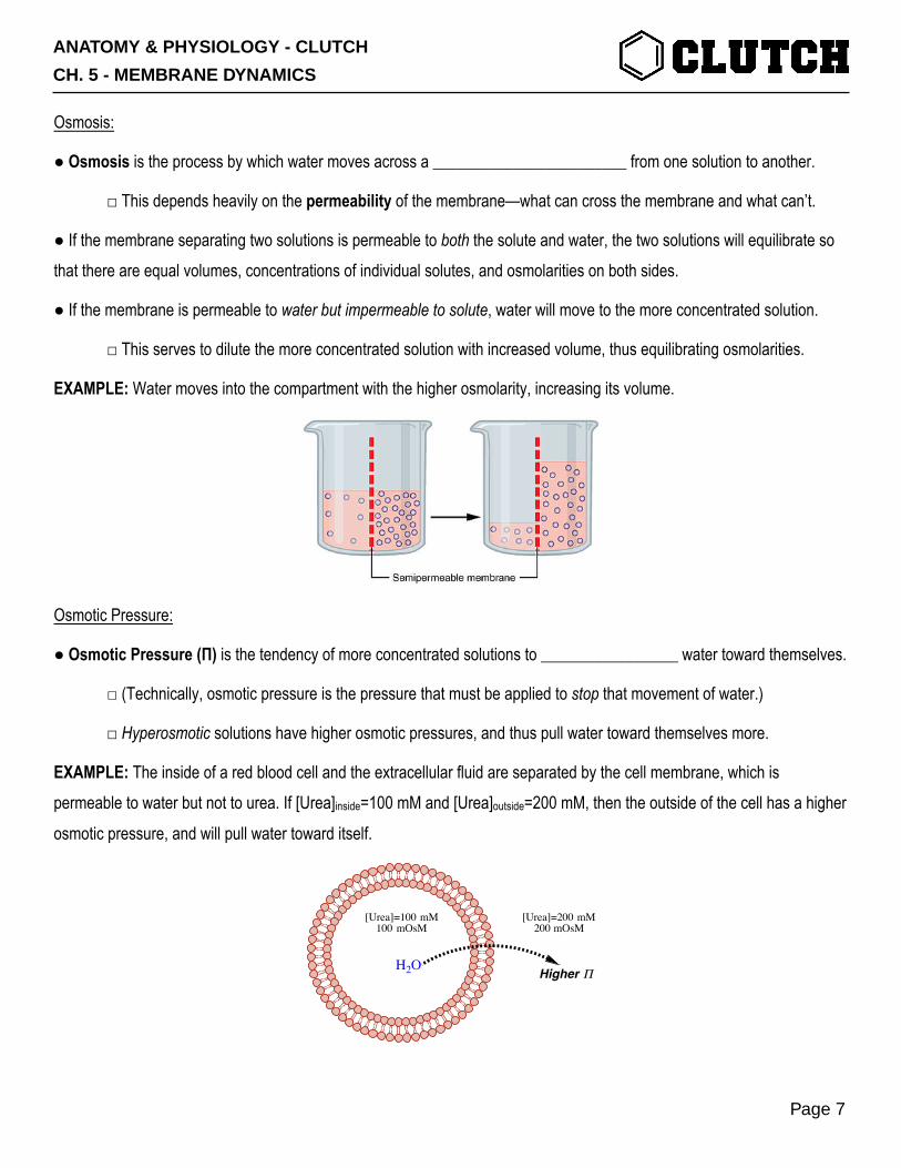
EXAMPLE: Water moves into the compartment with the higher osmolarity, increasing its volume.
Osmotic Pressure:
● Osmotic Pressure (Π) is the tendency of more concentrated solutions to _________________ water toward themselves.
□ (Technically, osmotic pressure is the pressure that must be applied to stop that movement of water.)
□ Hyperosmotic solutions have higher osmotic pressures, and thus pull water toward themselves more.
EXAMPLE: The inside of a red blood cell and the extracellular fluid are separated by the cell membrane, which is permeable to water but not to urea. If [Urea]inside=100 mM and [Urea]outside=200 mM, then the outside of the cell has a higher osmotic pressure, and will pull water toward itself.
[Urea]=100 mM100 mOsM
[Urea]=200 mM200 mOsM
H2O Higher Π
ANATOMY & PHYSIOLOGY - CLUTCH
CH. 5 - MEMBRANE DYNAMICS
Page 7

Tonicity:
● Osmotic pressure determines the direction that water actually travels across a membrane.
● The tonicity of a solution describes whether or not water actually moves into it.
□ The hypertonic side of a membrane pulls water more strongly, and thus gains volume.
□ The hypotonic side of a membrane loses water, and thus loses volume.
□ In an isotonic state, neither side’s volume changes.
● Hyperosmotic Solution→Higher Osmotic Pressure→Hypertonic Solution.
EXAMPLE: Cells suspended in hypertonic solutions shrink, while cells in hypotonic solutions swell.
ANATOMY & PHYSIOLOGY - CLUTCH
CH. 5 - MEMBRANE DYNAMICS
Page 8
Isoosmotic But Not Isotonic—A More Complicated Example:
● There are cases where each side starts out isosmotic. But, because of the permeability of the membrane, individual solutes may follow their concentration gradients and cross the membrane, bringing water with them.
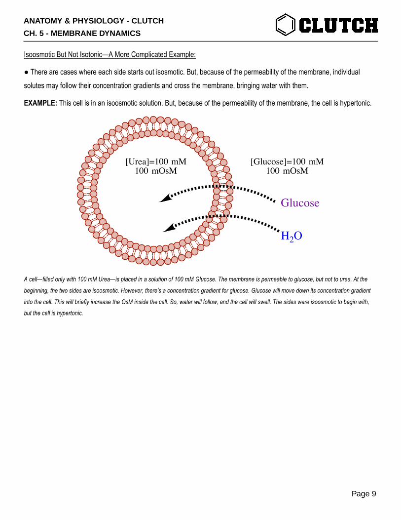
EXAMPLE: This cell is in an isoosmotic solution. But, because of the permeability of the membrane, the cell is hypertonic.
A cell—filled only with 100 mM Urea—is placed in a solution of 100 mM Glucose. The membrane is permeable to glucose, but not to urea. At the
beginning, the two sides are isoosmotic. However, there’s a concentration gradient for glucose. Glucose will move down its concentration gradient
into the cell. This will briefly increase the OsM inside the cell. So, water will follow, and the cell will swell. The sides were isoosmotic to begin with,
but the cell is hypertonic.
[Urea]=100 mM100 mOsM
[Glucose]=100 mM100 mOsM
H2O
Glucose
ANATOMY & PHYSIOLOGY - CLUTCH
CH. 5 - MEMBRANE DYNAMICS
Page 9
PRACTICE 1: An undergraduate playing around in lab combines 1 L of pure water, 150 mmols of glucose, and 150 mmols of KCl. Assuming complete dissociation, which of the following is the osmolarity of the resulting solution?
a) 150 mOsM. b) 300 mOsM. c) 450 mOsM. d) 600 mOsM.
PRACTICE 2: Filtrate inside of the nephron (part of the kidney) has an osmolarity of 500 mOsM. Fluid outside of the nephron has an osmolarity of 750 mOsM. Which of the following describes the fluid inside of the nephron relative to the fluid
outside of the nephron.
a) Hyperosmotic. b) Hypoosmotic. c) Isoosmotic.
PRACTICE 3: Filtrate inside of the nephron (part of the kidney) has an osmolarity of 500 mOsM. Fluid outside of the nephron has an osmolarity of 750 mOsM. Assume that water can move between the compartments, but solute cannot. Which compartment is hypertonic, and what is going to happen to the volume of that compartment?
a) Inside the nephron; increase in volume. b) Inside the nephron; decrease in volume. c) Outside the nephron; increase in volume. d) Outside the nephron; decrease in volume.
ANATOMY & PHYSIOLOGY - CLUTCH
CH. 5 - MEMBRANE DYNAMICS
Page 10

PRACTICE 4: The cytosol of red blood cells is approximately 300 mOsM, mostly from NaCl. You place the RBC in a extracellular solution of 600 mM sucrose. RBC membranes are not permeable to NaCl or sucrose. Circle the answer.
a) Which solution is hyperosmotic? ( Cytosol / Extracellular Solution )
b) Which solution has a higher osmotic pressure? ( Cytosol / Extracellular Solution )
c) Which direction will water move? ( Toward Cytosol / Toward Extracellular Solution )
d) Which solution is hypertonic? ( Cytosol / Extracellular Solution )
e) What will happen to the volume of the RBC? ( Increase / Decrease )
ANATOMY & PHYSIOLOGY - CLUTCH
CH. 5 - MEMBRANE DYNAMICS
Page 11
CONCEPT: PROTEIN-MEDIATED TRANSPORT
Passive Transport:
● Passive Transport is the movement of molecules across cell membranes down their concentration ________________.
□ No energy/ATP required.
□ Accomplished by transmembrane channel proteins and carrier proteins.
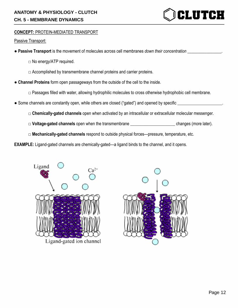
● Channel Proteins form open passageways from the outside of the cell to the inside.
□ Passages filled with water, allowing hydrophilic molecules to cross otherwise hydrophobic cell membrane.
● Some channels are constantly open, while others are closed (“gated”) and opened by specific _____________________.
□ Chemically-gated channels open when activated by an intracellular or extracellular molecular messenger.
□ Voltage-gated channels open when the transmembrane _____________________ changes (more later).
□ Mechanically-gated channels respond to outside physical forces—pressure, temperature, etc.
EXAMPLE: Ligand-gated channels are chemically-gated—a ligand binds to the channel, and it opens.
ANATOMY & PHYSIOLOGY - CLUTCH
CH. 5 - MEMBRANE DYNAMICS
Page 12
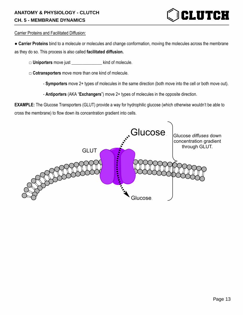
Carrier Proteins and Facilitated Diffusion:
● Carrier Proteins bind to a molecule or molecules and change conformation, moving the molecules across the membrane as they do so. This process is also called facilitated diffusion.
□ Uniporters move just ______________ kind of molecule.
□ Cotransporters move more than one kind of molecule.
- Symporters move 2+ types of molecules in the same direction (both move into the cell or both move out).
- Antiporters (AKA “Exchangers”) move 2+ types of molecules in the opposite direction.
EXAMPLE: The Glucose Transporters (GLUT) provide a way for hydrophilic glucose (which otherwise wouldn’t be able to cross the membrane) to flow down its concentration gradient into cells.
GlucoseNa+
GlucoseGlucose Na + Glucose diffuses down concentration gradient
through GLUT.GLUT
ANATOMY & PHYSIOLOGY - CLUTCH
CH. 5 - MEMBRANE DYNAMICS
Page 13
Active Transport:
● Active Transport moves molecules across cell membranes against their concentration gradients.
□ This process requires some source of ______________________.
- Primary Active Transport uses ATP as the energy source.
- Secondary Active Transport uses the concentration gradients of one molecule to power the movement of another molecule.
● Primary Active Transport uses ATP as the energy source, so the proteins doing this are called ATPases.
□ Cells can use primary active transport to establish concentration gradients across their membranes.
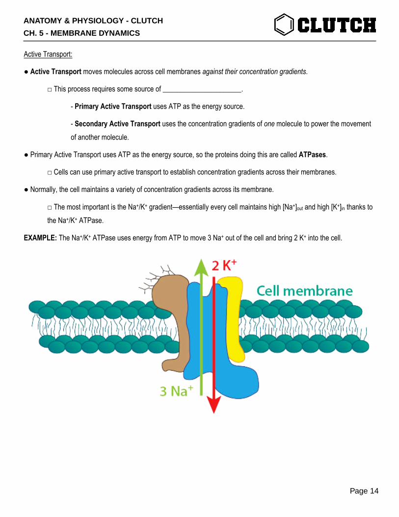
● Normally, the cell maintains a variety of concentration gradients across its membrane.
□ The most important is the Na+/K+ gradient—essentially every cell maintains high [Na+]out and high [K+]in thanks to the Na+/K+ ATPase.
EXAMPLE: The Na+/K+ ATPase uses energy from ATP to move 3 Na+ out of the cell and bring 2 K+ into the cell.
ANATOMY & PHYSIOLOGY - CLUTCH
CH. 5 - MEMBRANE DYNAMICS
Page 14
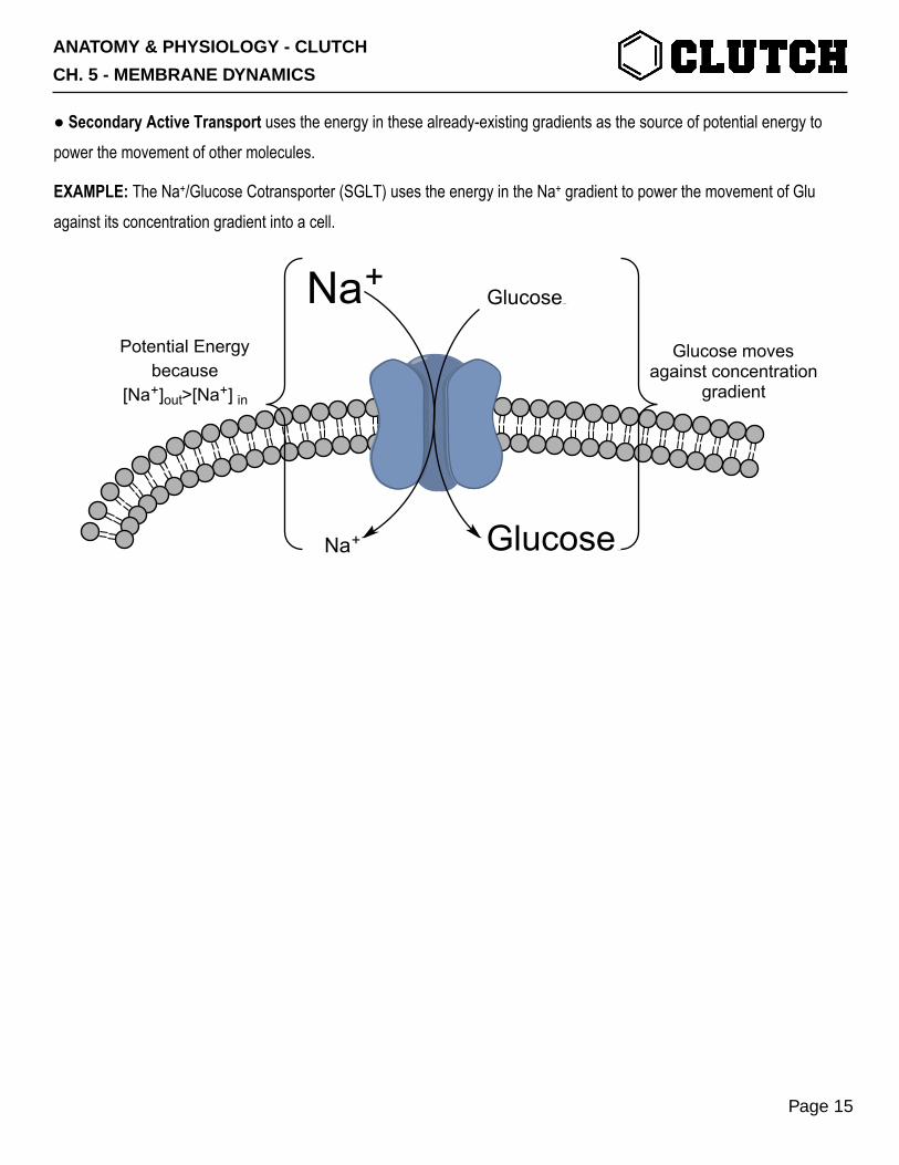
● Secondary Active Transport uses the energy in these already-existing gradients as the source of potential energy to power the movement of other molecules.
EXAMPLE: The Na+/Glucose Cotransporter (SGLT) uses the energy in the Na+ gradient to power the movement of Glu against its concentration gradient into a cell.
Na+GlucoseNa+
GlucoseGlucose Na +Na+
Potential Energybecause
[Na+]out>[Na+] in
Glucose moves against concentration
gradient
ANATOMY & PHYSIOLOGY - CLUTCH
CH. 5 - MEMBRANE DYNAMICS
Page 15
PRACTICE 1: A transporter moves Na+, K+, and Cl- into the cell. What type of protein-mediated transport does this channel perform? (Hint: [Na+] is higher outside the cell, [K+] and [Cl-] are higher inside the cell).
a) Passive transport through a chemically-gated channel. b) Facilitated Diffusion. c) Primary Active Transport. d) Secondary Active Transport.
PRACTICE 2: Nicotinic acetylcholine receptors (nAchRs) are inserted in the membranes of skeletal muscle fibers. When they bind to acetylcholine, a channel within them opens and allows Na+ and K+ to cross the skeletal muscle cell membrane. Which of the following describes the category into which nAchRs fit?
a) Mechanically-gated ion channels. b) Voltage-gated ion channels. c) Ligand-gated ion channels.
PRACTICE 3: Parietal cells of the stomach express H+/K+ ATPase proteins. These proteins use energy from the hydrolysis of ATP to pump one H+ out of the cell into the stomach while, at the same time, bringing one K+ from the stomach into the cell. Which of the following accurately describes this type of transport?
a) Facilitated diffusion. b) Primary active transport. c) Secondary active transport.
ANATOMY & PHYSIOLOGY - CLUTCH
CH. 5 - MEMBRANE DYNAMICS
Page 16
CONCEPT: VESICLE-BASED TRANSPORT—ENDOCYTOSIS AND EXOCYTOSIS
Bulk Transport:
● Bulk Transport is the movement of larger solutes—carbohydrates and proteins, mostly—across cell membranes.
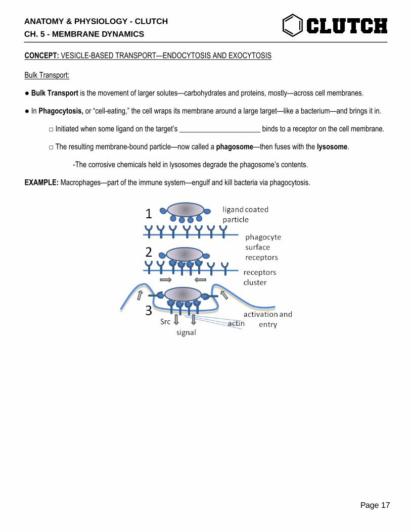
● In Phagocytosis, or “cell-eating,” the cell wraps its membrane around a large target—like a bacterium—and brings it in.
□ Initiated when some ligand on the target’s ______________________ binds to a receptor on the cell membrane.
□ The resulting membrane-bound particle—now called a phagosome—then fuses with the lysosome.
-The corrosive chemicals held in lysosomes degrade the phagosome’s contents.
EXAMPLE: Macrophages—part of the immune system—engulf and kill bacteria via phagocytosis.
ANATOMY & PHYSIOLOGY - CLUTCH
CH. 5 - MEMBRANE DYNAMICS
Page 17
Endocytosis:
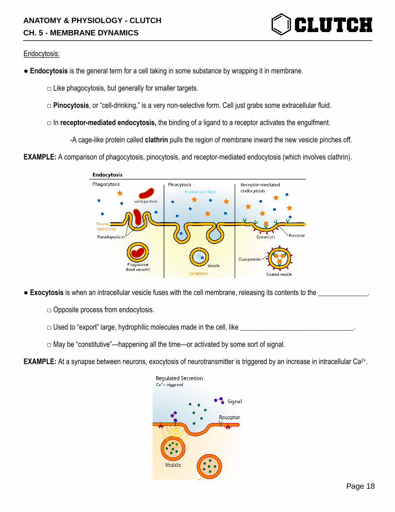
● Endocytosis is the general term for a cell taking in some substance by wrapping it in membrane.
□ Like phagocytosis, but generally for smaller targets.
□ Pinocytosis, or “cell-drinking,” is a very non-selective form. Cell just grabs some extracellular fluid.
□ In receptor-mediated endocytosis, the binding of a ligand to a receptor activates the engulfment.
-A cage-like protein called clathrin pulls the region of membrane inward the new vesicle pinches off.
EXAMPLE: A comparison of phagocytosis, pinocytosis, and receptor-mediated endocytosis (which involves clathrin).
● Exocytosis is when an intracellular vesicle fuses with the cell membrane, releasing its contents to the ______________.
□ Opposite process from endocytosis.
□ Used to “export” large, hydrophilic molecules made in the cell, like ________________________________.
□ May be “constitutive”—happening all the time—or activated by some sort of signal.
EXAMPLE: At a synapse between neurons, exocytosis of neurotransmitter is triggered by an increase in intracellular Ca2+.
ANATOMY & PHYSIOLOGY - CLUTCH
CH. 5 - MEMBRANE DYNAMICS
Page 18
PRACTICE 1: One of the functions of antibodies—proteins that are part of the immune system—is to bind to and coat bacteria. This makes the bacteria more likely to be engulfed and destroyed by macrophages. Which of the following processes is likely facilitated by antibodies?
a) Endocytosis. b) Exocytosis. c) Phagocytosis.
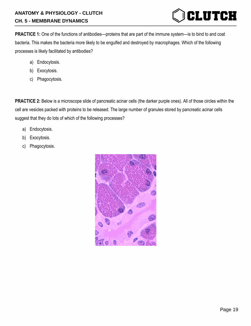
PRACTICE 2: Below is a microscope slide of pancreatic acinar cells (the darker purple ones). All of those circles within the cell are vesicles packed with proteins to be released. The large number of granules stored by pancreatic acinar cells suggest that they do lots of which of the following processes?
a) Endocytosis. b) Exocytosis. c) Phagocytosis.
ANATOMY & PHYSIOLOGY - CLUTCH
CH. 5 - MEMBRANE DYNAMICS
Page 19
CONCEPT: CROSSING AN EPITHELIAL LAYER: THE TRANS- AND PARACELLULAR PATHWAYS
Epithelial Cell Layers:
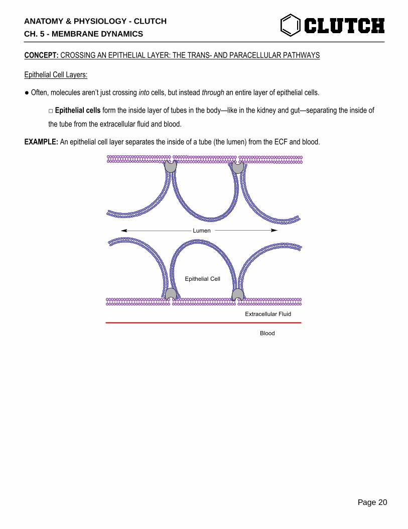
● Often, molecules aren’t just crossing into cells, but instead through an entire layer of epithelial cells.
□ Epithelial cells form the inside layer of tubes in the body—like in the kidney and gut—separating the inside of the tube from the extracellular fluid and blood.
EXAMPLE: An epithelial cell layer separates the inside of a tube (the lumen) from the ECF and blood.
Blood
Extracellular Fluid
Epithelial Cell
Lumen
ANATOMY & PHYSIOLOGY - CLUTCH
CH. 5 - MEMBRANE DYNAMICS
Page 20

The Transcellular and Paracellular Pathways:
● For a layer of epithelial cells lined-up one next to another:
□ The lumen is the inside of a tube lined with epithelial cells (e.g. the inside of the small intestine).
□ The apical membrane is the surface of cell membrane facing the ___________________.
□ The basolateral membrane (aka serosal membrane) is/are the side(s) that aren’t apical, usually facing ECF.
□ Tight junctions are connections that hold _________________________ epithelial cells together.
● There are two possible pathways a molecule can take from the lumen to the ECF.
□ In the transcellular pathway, the substance crosses both the apical and basolateral membranes of one cell.
□ In the paracellular pathway, the substance goes __________________ the cells and through the tight junctions.
EXAMPLE: A schematic of the above vocabulary and the transcellular and paracellular pathways.
Lumen
Blood
Extracellular FluidBasolateral Membrane
TightJunction
Apical Membrane
TRANSCELLULARPATHWAY
PARACELLULARPATHWAY
ANATOMY & PHYSIOLOGY - CLUTCH
CH. 5 - MEMBRANE DYNAMICS
Page 21
Transport via the Transcellular Pathway:
● Using the transcellular pathway means the solute must cross _______________ membranes—the apical and basolateral.
● Usually, the transcellular pathway is accomplished by two transporters—one per membrane.
□ Typically, one of these transport processes is active transport, and the other is passive (or facilitated diffusion).
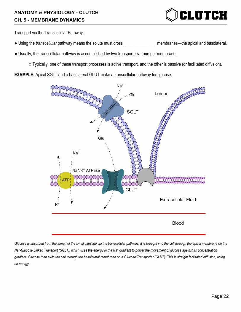
EXAMPLE: Apical SGLT and a basolateral GLUT make a transcellular pathway for glucose.
Glucose is absorbed from the lumen of the small intestine via the transcellular pathway. It is brought into the cell through the apical membrane on the
Na+-Glucose Linked Transport (SGLT), which uses the energy in the Na+ gradient to power the movement of glucose against its concentration
gradient. Glucose then exits the cell through the basolateral membrane on a Glucose Transporter (GLUT). This is straight facilitated diffusion, using
no energy.
Lumen
Extracellular Fluid
Blood
SGLT
Na+/K+ ATPase
Na+
K+
Na+
Glu
Glu
GLUT
ATP
ANATOMY & PHYSIOLOGY - CLUTCH
CH. 5 - MEMBRANE DYNAMICS
Page 22
PRACTICE 1: Blood vessels in the brain are lined by epithelial cells with extremely close, repellent tight junctions—essentially nothing can pass through these tight junctions. For a substance to cross from the blood into the brain interstitial fluid, which pathway must it use?
a) Transcellular pathway. b) Paracellular pathway. c) Pathway to true enlightenment.
ANATOMY & PHYSIOLOGY - CLUTCH
CH. 5 - MEMBRANE DYNAMICS
Page 23
CONCEPT: THE RESTING MEMBRANE POTENTIAL I: FORCES AND OHM’S LAW
Concentration Gradients as Forces:
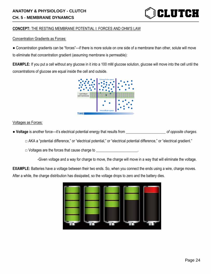
● Concentration gradients can be “forces”—if there is more solute on one side of a membrane than other, solute will move to eliminate that concentration gradient (assuming membrane is permeable):
EXAMPLE: If you put a cell without any glucose in it into a 100 mM glucose solution, glucose will move into the cell until the concentrations of glucose are equal inside the cell and outside.
Voltages as Forces:
● Voltage is another force—it’s electrical potential energy that results from _____________________ of opposite charges.
□ AKA a “potential difference,” or “electrical potential,” or “electrical potential difference,” or “electrical gradient.”
□ Voltages are the forces that cause charge to ______________________.
-Given voltage and a way for charge to move, the charge will move in a way that will eliminate the voltage.
EXAMPLE: Batteries have a voltage between their two ends. So, when you connect the ends using a wire, charge moves. After a while, the charge distribution has dissipated, so the voltage drops to zero and the battery dies.
ANATOMY & PHYSIOLOGY - CLUTCH
CH. 5 - MEMBRANE DYNAMICS
Page 24
RESISTANCE
CURRENT
VOLTAGE
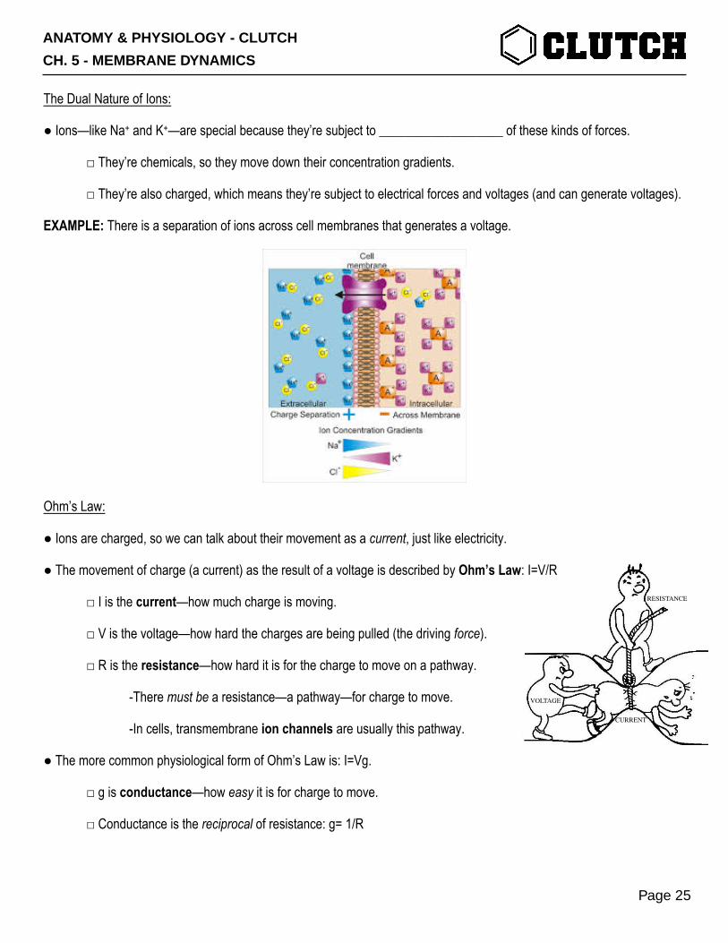
The Dual Nature of Ions:
● Ions—like Na+ and K+—are special because they’re subject to ___________________ of these kinds of forces.
□ They’re chemicals, so they move down their concentration gradients.
□ They’re also charged, which means they’re subject to electrical forces and voltages (and can generate voltages).
EXAMPLE: There is a separation of ions across cell membranes that generates a voltage.
Ohm’s Law:
● Ions are charged, so we can talk about their movement as a current, just like electricity.
● The movement of charge (a current) as the result of a voltage is described by Ohm’s Law: I=V/R
□ I is the current—how much charge is moving.
□ V is the voltage—how hard the charges are being pulled (the driving force).
□ R is the resistance—how hard it is for the charge to move on a pathway.
-There must be a resistance—a pathway—for charge to move.
-In cells, transmembrane ion channels are usually this pathway.
● The more common physiological form of Ohm’s Law is: I=Vg.
□ g is conductance—how easy it is for charge to move.
□ Conductance is the reciprocal of resistance: g= 1/R
ANATOMY & PHYSIOLOGY - CLUTCH
CH. 5 - MEMBRANE DYNAMICS
Page 25
PRACTICE 1: Normally, Na+ ions can’t cross cell membranes very easily, because they’re hydrophilic cations and the membrane is lipophilic. If a Na+ channel in the membrane opens and increases the permeability of the membrane to Na+, which variable in Ohm’s Law (I=Vg) is directly changed by the opening of the channel?
a) Current (I). b) Voltage (V). c) Conductance (g).
PRACTICE 2: Normally, Na+ ions can’t cross cell membranes very easily, because they’re hydrophilic cations and the membrane is lipophilic. If a Na+ channel in the membrane opens and increases the permeability of the membrane to Na+, how will that change affect the transmembrane current of Na+?
a) Increase INa+. b) Decrease INa+. c) No change in INa+.
PRACTICE 3: A cell is sitting in a KCl solution. Outside the cell, [K+]=10 mM. Inside of the cell, [K+]=100 mM. As a result of this separation of K+, there is a transmembrane voltage V= -61 mV (i.e. the inside of the cell is more negative than the outside). Which of the following accurately describes the type(s) and direction of force(s) acting on the K+ inside of the cell? (Choose all that apply.)
a) Concentration gradient; out of the cell. b) Concentration gradient; in to the cell. c) Electrical force; out of the cell. d) Electrical force; in to the cell.
ANATOMY & PHYSIOLOGY - CLUTCH
CH. 5 - MEMBRANE DYNAMICS
Page 26
CONCEPT: THE RESTING MEMBRANE POTENTIAL II: DEVELOPMENT OF A MEMBRANE POTENTIAL
Development of Charge Separation Across a Membrane:
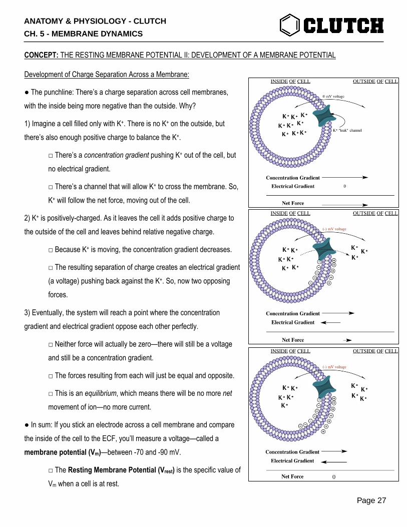
● The punchline: There’s a charge separation across cell membranes, with the inside being more negative than the outside. Why?
1) Imagine a cell filled only with K+. There is no K+ on the outside, but there’s also enough positive charge to balance the K+.
□ There’s a concentration gradient pushing K+ out of the cell, but no electrical gradient.
□ There’s a channel that will allow K+ to cross the membrane. So, K+ will follow the net force, moving out of the cell.
2) K+ is positively-charged. As it leaves the cell it adds positive charge to the outside of the cell and leaves behind relative negative charge.
□ Because K+ is moving, the concentration gradient decreases.
□ The resulting separation of charge creates an electrical gradient (a voltage) pushing back against the K+. So, now two opposing forces.
3) Eventually, the system will reach a point where the concentration gradient and electrical gradient oppose each other perfectly.
□ Neither force will actually be zero—there will still be a voltage and still be a concentration gradient.
□ The forces resulting from each will just be equal and opposite.
□ This is an equilibrium, which means there will be no more net movement of ion—no more current.
● In sum: If you stick an electrode across a cell membrane and compare the inside of the cell to the ECF, you’ll measure a voltage—called a membrane potential (Vm)—between -70 and -90 mV.
□ The Resting Membrane Potential (Vrest) is the specific value of Vm when a cell is at rest.
ANATOMY & PHYSIOLOGY - CLUTCH
CH. 5 - MEMBRANE DYNAMICS
Page 27
PRACTICE 1: A neuron is sitting at rest in a dish. Its membrane potential is at Vrest= -70 mV; [K+]inside=100 mM and [K+]outside=10 mM. Which of the following force(s) acting on the K+ is equal to zero? (Choose all that apply.)
a) Concentration gradient force. b) Electrical gradient force. c) Concentration Gradient Force+Electrical Gradient Force (i.e. the net force). d) Force of sexual attraction.
ANATOMY & PHYSIOLOGY - CLUTCH
CH. 5 - MEMBRANE DYNAMICS
Page 28
CONCEPT: THE RESTING MEMBRANE POTENTIAL III: THE NERNST AND GOLDMAN-HODGKIN-KATZ EQUATIONS
The Nernst Equation and The Equilibrium Potential:
● The Nernst Equation lets us calculate the Vm at which the concentration gradient force and electrical gradient force are ___________________ and opposite to each other for a particular ion. This value is called the Equilibrium Potential (Eion).
□ General form: E"#$ =&'(ℱln( "#$ ./0
"#$ 12)
-R= universal gas constant, T=temp (in Kelvin), z=ionic charge (z=+1 for Na+ and K+), F=Faraday Constant.
□ Simplified: 𝐄𝐢𝐨𝐧 =𝟔𝟏𝐳𝐥𝐨𝐠( 𝐢𝐨𝐧 𝐨𝐮𝐭
𝐢𝐨𝐧 𝐢𝐧).
-Assumes body temperature and a base-10 logarithm.
● The Nernst Equation—the Eion—tells you the value of Vm at which there is no net _________________ on a particular ion.
□ If an ion can cross the membrane, it will move in a way that will work to bring Vm toward Eion.
● Nernst is used when you have an ionic concentration gradient and an ionic current and want to know how that ionic
current will affect Vm.
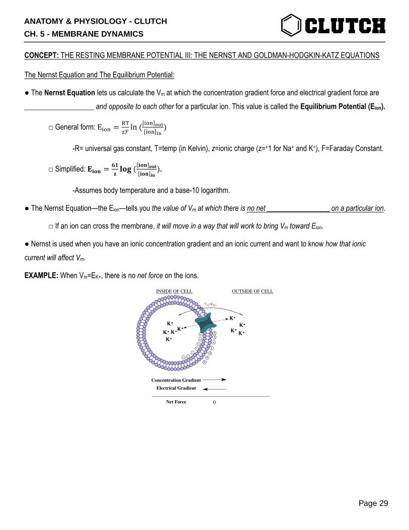
EXAMPLE: When Vm=EK+, there is no net force on the ions.
INSIDE OF CELL OUTSIDE OF CELL
Vm=EK+
Concentration GradientElectrical Gradient
Net Force
K+K+K+ K+
K+
K+K+K+
K+
0
ANATOMY & PHYSIOLOGY - CLUTCH
CH. 5 - MEMBRANE DYNAMICS
Page 29

The Goldman-Hodgkin-Katz Equation—Estimating Vm:
● The Nernst Equation tells us the value that Vm will approach because of a particular ionic current.
□ But, it doesn’t give any information about the instantaneous value of Vm.
● The Goldman-Hodgkin-Katz Equation provides an estimate of Vm for a cell by calculating a weighted ______________ of equilibrium potentials for all the ions capable of crossing the membrane. For Na+, K+, and Cl- :
□ 𝑉@ = 61𝑙𝑜𝑔(FGHI[KL
I]NOPQFRI[SI]NOPQFTUV[WXV]YZ)
(FGHI[KLI]YZQFRI[SI]YZQFTUV[WXV]NOP), where Pion is the permeability of the membrane to that ion.
□ Ions that can cross the membrane more easily—can make a larger current—have a larger effect on Vm.
Why Does Vrest ≅ -70 mV?
● Essentially every cell in the body expresses Na+/K+ ATPase pumps in their cell membranes.
□ These pumps continuously pump Na+ out of the cell and K+ into the cell→a concentration gradient for these ions:
- Typically: [Na+]out≅150 mM and [Na+]in≅15 mM ⇒ ENa+≅+61 mV.
- Typically: [K+]out≅5 mM and [K+]in≅150 mM ⇒ EK+≅-90 mV.
● Essentially every cell in the body also expresses K+ leak channels in their cell membranes.
□ These channels provide a ________________ for K+ to cross the membrane and pull Vm toward EK+ even at rest.
□ This is why the resting membrane potential of most cells is so close to EK+ at about -70 to -90 mV.
EXAMPLE: The combination of the concentration gradients for Na+ and K+ established by the Na+/K+ ATPase and the presence of K+ leak channels causes the membrane potential of resting cells to be close to EK+.
Na+/K+ ATPaseNa+
ATP
K+K+K+K+K+
K+
ANATOMY & PHYSIOLOGY - CLUTCH
CH. 5 - MEMBRANE DYNAMICS
Page 30
PRACTICE 1: Assume that, for a given cell: [Ca2+]extracellular fluid=120 mM and [Ca2+]cytosol=1.2 mM. Which of the following is true for this cell?
a) ECa2+= +61 mV b) ECa2+= -61 mV c) ECa2+= +122 mV d) ECa2+= -122 mV
PRACTICE 2: A typical neuron is sitting at Vrest=- 70 mV. Many Na+ channels in the neuron’s membrane open and stay open. [Na+]outside=150 mM and [Na+]inside=15 mM. After a new equilibrium is reached, you measure the new membrane potential. Which of the following is likely to be the value of that new membrane potential?
a) Vm= -70 mV. b) Vm= -10 mV. c) Vm= 0 mV. d) Vm= +61 mV.
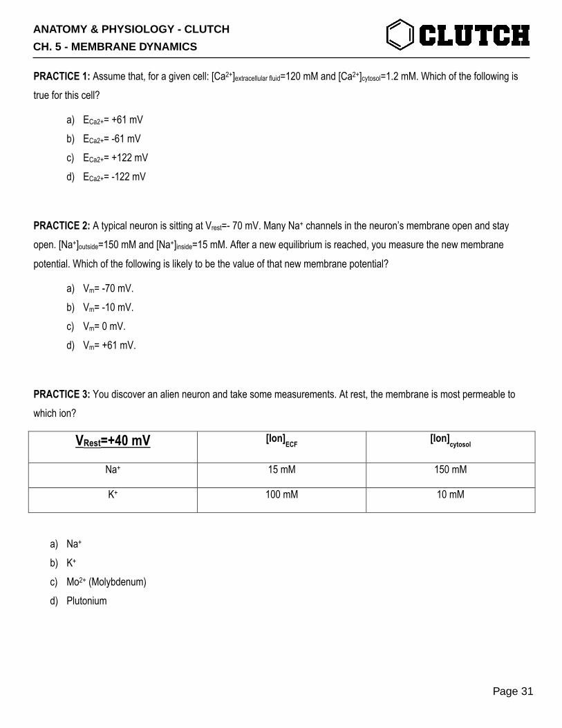
PRACTICE 3: You discover an alien neuron and take some measurements. At rest, the membrane is most permeable to which ion?
VRest=+40 mV [Ion]ECF [Ion]cytosol
Na+ 15 mM 150 mM
K+ 100 mM 10 mM
a) Na+ b) K+ c) Mo2+ (Molybdenum) d) Plutonium
ANATOMY & PHYSIOLOGY - CLUTCH
CH. 5 - MEMBRANE DYNAMICS
Page 31
PRACTICE 4: A cell is initially at Vm=0 mV. The concentrations of K+ inside and outside the cell are: [K+]out=5 mM, [K+]in=150 mM. If a channel for K+ opens, will K+ flow into or out of the cell?
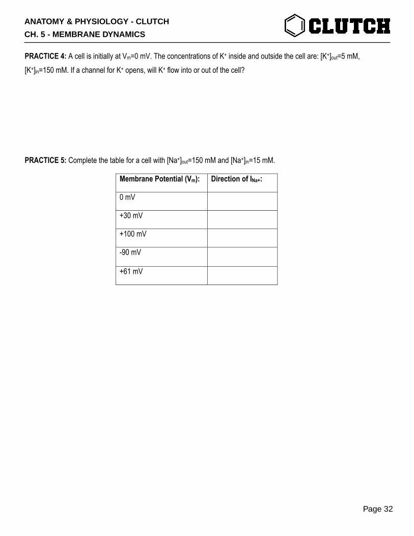
PRACTICE 5: Complete the table for a cell with [Na+]out=150 mM and [Na+]in=15 mM.
Membrane Potential (Vm): Direction of INa+:
0 mV
+30 mV
+100 mV
-90 mV
+61 mV
ANATOMY & PHYSIOLOGY - CLUTCH
CH. 5 - MEMBRANE DYNAMICS
Page 32