Embed Size (px)
Citation preview
Anatomy and Three-DimensionalReconstructions of the Brain of the
White Whale (Delphinapterus leucas)From Magnetic Resonance Images
LORI MARINO,1* TIMOTHY L. MURPHY,2 AMY L. DEWEERD,2
JOHN A. MORRIS,3 ARCHIBALD J. FOBBS,4 NATHALIE HUMBLOT,4
SAM H. RIDGWAY,5 AND JOHN I. JOHNSON2,3
1Neuroscience and Behavioral Biology Program, Psychology Building,Emory University, Atlanta, Georgia
2Radiology Department, Michigan State University, East Lansing, Michigan3Neuroscience Program, Michigan State University, East Lansing, Michigan
4National Museum of Health and Medicine, Armed Forces Institute of Pathology,Washington, DC
5Navy Marine Mammal Program, San Diego, California
ABSTRACTMagnetic resonance imaging offers a means of observing the internal
structure of the brain where traditional procedures of embedding, sectioning,staining, mounting, and microscopic examination of thousands of sections arenot practical. Furthermore, internal structures can be analyzed in their precisequantitative spatial interrelationships, which is difficult to accomplish afterthe spatial distortions often accompanying histological processing. For thesereasons, magnetic resonance imaging makes specimens that were traditionallydifficult to analyze, more accessible. In the present study, images of the brainof a white whale (Beluga) Delphinapterus leucas were scanned in the coronalplane at 119 antero-posterior levels. From these scans, a computer-generatedthree-dimensional model was constructed using the programs VoxelView andVoxelMath (Vital Images, Inc.). This model, wherein details of internal andexternal morphology are represented in three-dimensional space, was thenresectioned in orthogonal planes to produce corresponding series of “virtual”sections in the horizontal and sagittal planes. Sections in all three planesdisplay the sizes and positions of such structures as the corpus callosum,internal capsule, cerebral peduncles, cerebral ventricles, certain thalamic nu-clear groups, caudate nucleus, ventral striatum, pontine nuclei, cerebellarcortex and white matter, and all cerebral cortical sulci and gyri. Anat Rec 262:429–439, 2001. © 2001 Wiley-Liss, Inc.
Key words: brain; neuroanatomy; cetacean; odontocete; whitewhale; Beluga; MRI
Odontocetes (toothed whales, dolphins, and porpoises)have undergone a number of evolutionary modificationsfrom their terrestrial ancestral state. Among thesechanges was a major increase in relative brain size. Sev-eral modern odontocete species possess encephalizationlevels second only to modern humans when brain-bodyallometry is taken into account (Ridgway and Brownson,1984; Marino, 1998). An arguably equally dramatic trans-formation of odontocetes occurred in the anatomical struc-ture and organization of their brains. Compared withmany other mammalian brains, odontocete brain mor-
phology is unusual in many respects. Researchers havestated that “…the lobular formations in the dolphin brain
Grant sponsor: Division of Integrative Biology and Neuro-science, National Science Foundation; Grant numbers: 9812712,9814911, and 9814912.
*Correspondence to: Lori Marino, PhD, Psychology Building,Emory University, Atlanta, GA 30322. E-mail: [email protected]
Received 8 May 2000; Accepted 19 November 2000Published online 28 February 2001
THE ANATOMICAL RECORD 262:429–439 (2001)
© 2001 WILEY-LISS, INC.
are organized in a pattern fundamentally different fromthat seen in the brains of primates or carnivores” (Mor-gane et al., 1980). Because of the fifty-five to sixty millionyear divergence between cetaceans and other mammals,odontocete brains represent a blend of early mammalianfeatures along with unique derived characteristics (Ridg-way, 1986, 1990; Glezer et al., 1988; Manger et al., 1998).The differences between odontocete and other mammalianbrains of similar size are present at the level of corticalcytoarchitecture and immunohistochemistry (Garey et al.,1985; Garey and Leuba, 1986; Glezer and Morgane, 1990;Hof et al., 1992, 1995; Glezer et al., 1990, 1992a,b, 1993,1998), cortical surface morphology (Jacobs et al., 1979;Morgane et al., 1980; Haug, 1987), noncortical structuresand features (Tarpley and Ridgway, 1994; Glezer et al.,
1995a,b), and ontogenesis (Oelschlager and Buhl, 1985;Buhl and Oelschlager, 1988; Oelschlager and Kemp,1998).
Although there are a number of published descriptionsof cetacean neuroanatomy (see Morgane et al., 1986; Ridg-way, 1990; for reviews of this literature) there are only ahandful of studies in which morphometric analyses wereconducted in a systematic way permitting quantitativecomparative analysis with other mammals (Jacobs et al.,1984; Johnson et al., 1984; Schwerdtfeger et al., 1984;Garey and Leuba, 1986; Johnson et al., 1994; Tarpley andRidgway, 1994; Manger et al., 1998; Marino, 1998). Fur-thermore, with the exception of Morgane et al. (1980),Ridgway and Brownson (1984), Haug (1987), and Tarpleyand Ridgway (1994) there are no systematic anatomical
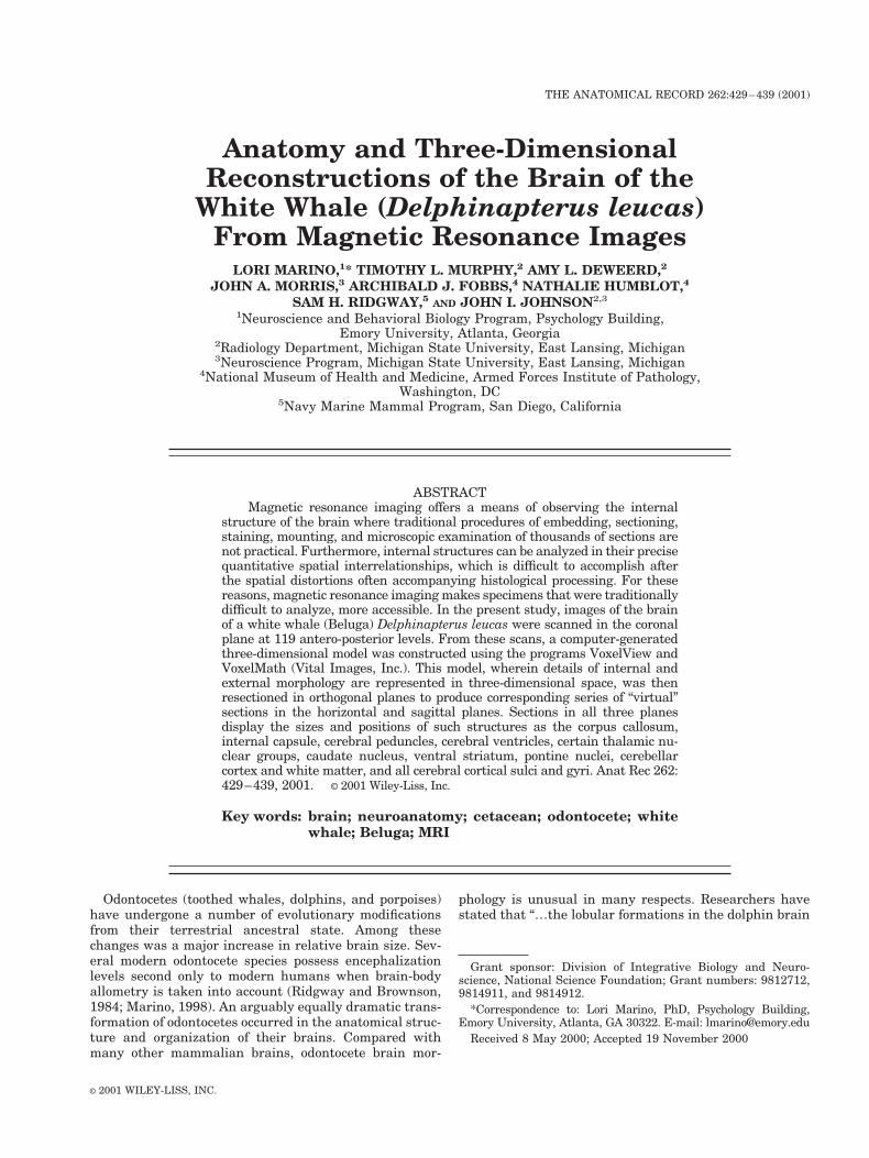
Fig. 1. Ventral surface of a three-dimensional digital reconstruction of the whole brain and labeled schematic illustration of the same image.
430 MARINO ET AL.
descriptions of whole cetacean brains and substructuresat the qualitative level. There currently exists no compre-hensive cetacean neuroanatomical atlas either in paper orelectronic format on which to base studies of cetaceanbrain organization and function. This situation is mainlydue to the time and practicality associated with the prep-aration of such large brain specimens. Magnetic resonanceimaging (MRI) offers a means of observing the internalstructure of the brain where traditional procedures ofembedding, sectioning, staining, mounting, and micro-scopic examination of thousands of sections are not prac-tical. Furthermore internal structures can be analyzed intheir precise spatial interrelationships, which is difficult
to accomplish after the spatial distortions often accompa-nying histological processing. This study presents an an-atomically-labeled three-dimensional atlas, created fromMRI images, of the brain of one of the most behaviorallystudied odontocetes, the white whale (Delphinapterus leu-cas).
MATERIALS AND METHODSSpecimen
The specimen is the postmortem brain, fixed in 10%buffered formalin, of an adult female white whale (Delphi-napterus leucas) who died of natural causes. The whalehad been involved in several behavioral studies including
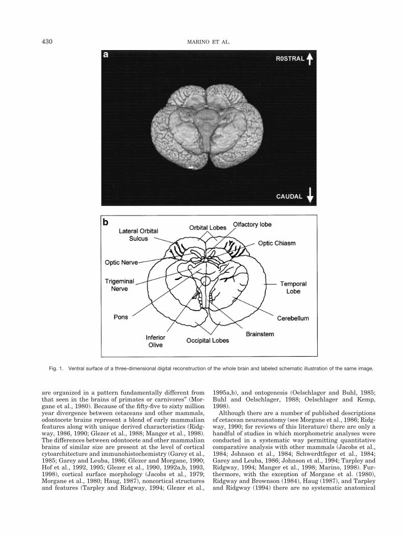
Fig. 2. Three-dimensional digital reconstructions of the whole brain and resectioning to produce “virtual” horizontal sections.
Fig. 3. Three-dimensional digital reconstructions of the whole brain and resectioning to produce “virtual” sagittal sections.
431ANATOMY OF THE WHITE WHALE BRAIN
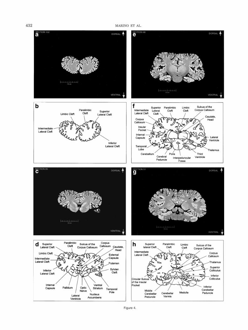
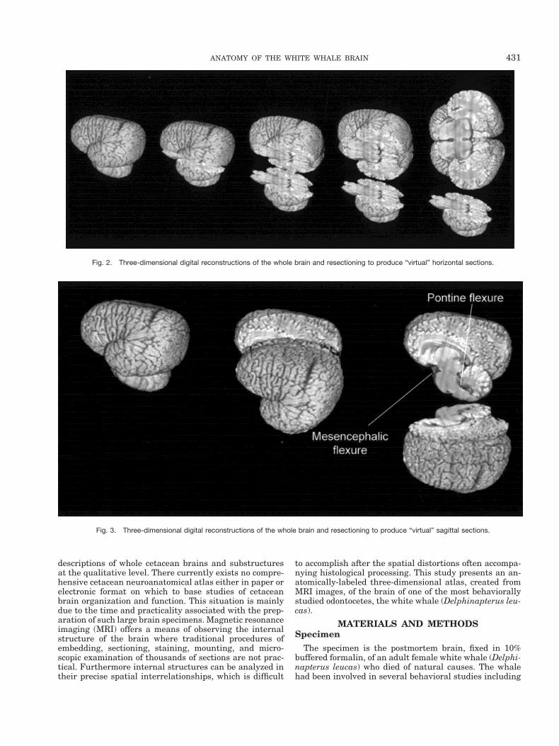
Figure 4.
432 MARINO ET AL.
studies of its hearing (Awbrey et al., 1988). At death, thebrain was extracted from the skull, weighed, and placed inneutral buffered formalin for 4 years before scanning.Fresh brain weight was 1,871 g. Fixed brain weight was1,755 g at the time of scanning.
Magnetic Resonance ImagingThe brain was removed from the fluid and placed in a
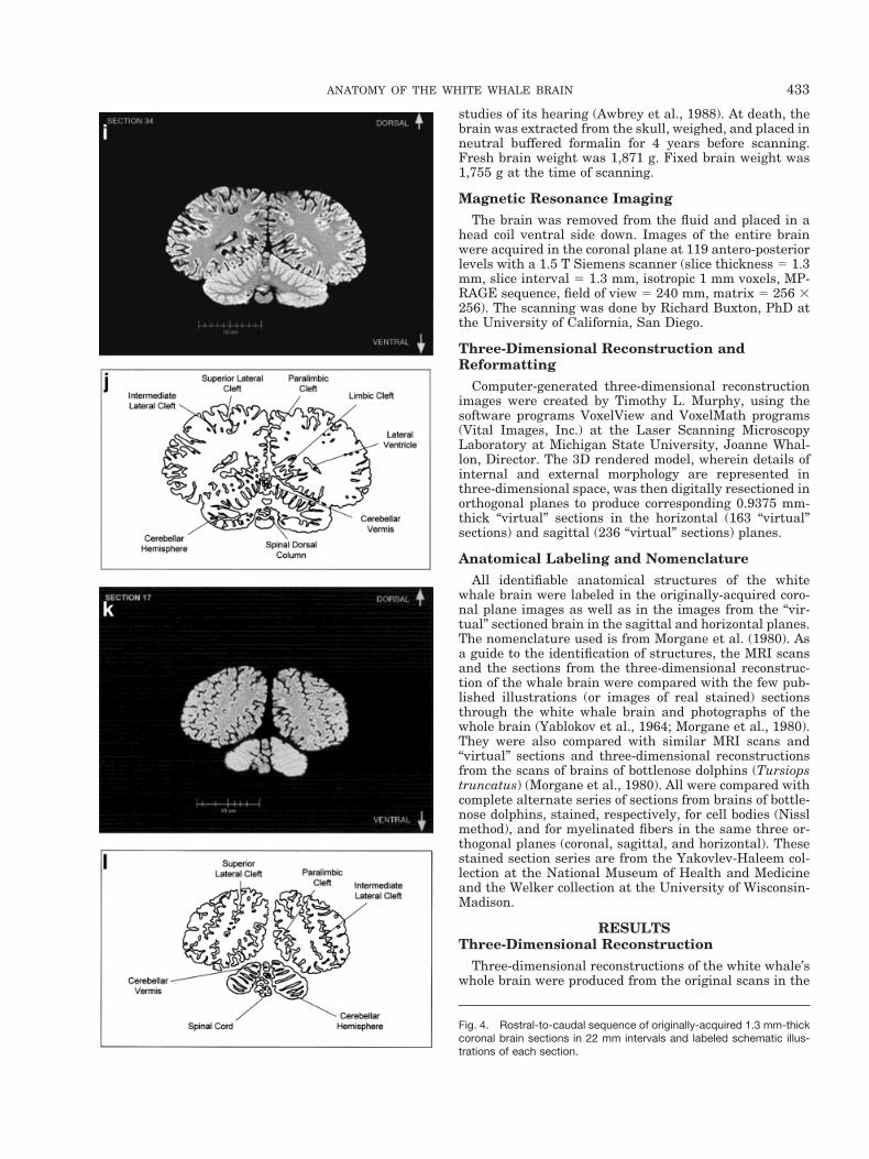
head coil ventral side down. Images of the entire brainwere acquired in the coronal plane at 119 antero-posteriorlevels with a 1.5 T Siemens scanner (slice thickness 5 1.3mm, slice interval 5 1.3 mm, isotropic 1 mm voxels, MP-RAGE sequence, field of view 5 240 mm, matrix 5 256 3256). The scanning was done by Richard Buxton, PhD atthe University of California, San Diego.
Three-Dimensional Reconstruction andReformatting
Computer-generated three-dimensional reconstructionimages were created by Timothy L. Murphy, using thesoftware programs VoxelView and VoxelMath programs(Vital Images, Inc.) at the Laser Scanning MicroscopyLaboratory at Michigan State University, Joanne Whal-lon, Director. The 3D rendered model, wherein details ofinternal and external morphology are represented inthree-dimensional space, was then digitally resectioned inorthogonal planes to produce corresponding 0.9375 mm-thick “virtual” sections in the horizontal (163 “virtual”sections) and sagittal (236 “virtual” sections) planes.
Anatomical Labeling and NomenclatureAll identifiable anatomical structures of the white
whale brain were labeled in the originally-acquired coro-nal plane images as well as in the images from the “vir-tual” sectioned brain in the sagittal and horizontal planes.The nomenclature used is from Morgane et al. (1980). Asa guide to the identification of structures, the MRI scansand the sections from the three-dimensional reconstruc-tion of the whale brain were compared with the few pub-lished illustrations (or images of real stained) sectionsthrough the white whale brain and photographs of thewhole brain (Yablokov et al., 1964; Morgane et al., 1980).They were also compared with similar MRI scans and“virtual” sections and three-dimensional reconstructionsfrom the scans of brains of bottlenose dolphins (Tursiopstruncatus) (Morgane et al., 1980). All were compared withcomplete alternate series of sections from brains of bottle-nose dolphins, stained, respectively, for cell bodies (Nisslmethod), and for myelinated fibers in the same three or-thogonal planes (coronal, sagittal, and horizontal). Thesestained section series are from the Yakovlev-Haleem col-lection at the National Museum of Health and Medicineand the Welker collection at the University of Wisconsin-Madison.
RESULTSThree-Dimensional Reconstruction
Three-dimensional reconstructions of the white whale’swhole brain were produced from the original scans in the
Fig. 4. Rostral-to-caudal sequence of originally-acquired 1.3 mm-thickcoronal brain sections in 22 mm intervals and labeled schematic illus-trations of each section.
433ANATOMY OF THE WHITE WHALE BRAIN
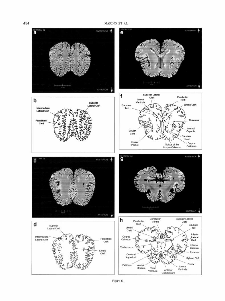
Figure 5.
434 MARINO ET AL.
coronal plane. Figure 1 displays an image (or view) of theposterior-ventral surface of a computer-generated three-dimensional reconstruction of the whole brain and a la-beled illustration of the image. Figures 2 and 3 also dis-play three-dimensional reconstructions of the whole brainand the brain digitally recut in the horizontal and sagittalplanes, respectively. These three-dimensional reconstruc-tions clearly display many noted characteristics of thecetacean brain that diverge from most other terrestrialmammalian brains (Morgane et al., 1980). The foreshort-ened orbital lobes are evident in Figure 3 as is the pro-nounced bitemporal width of the brain in Figures 1 andthe last “virtual” cut in Figure 2. The labeled “virtual” cutin Figure 3 shows the mesencephalic and pontine flexuresreminiscent of brainstem flexure patterns in the embry-onic state of most terrestrial mammals. These flexuresremain present in adult cetacean brains.
Anatomically-Labeled Two-Dimensional MRISections
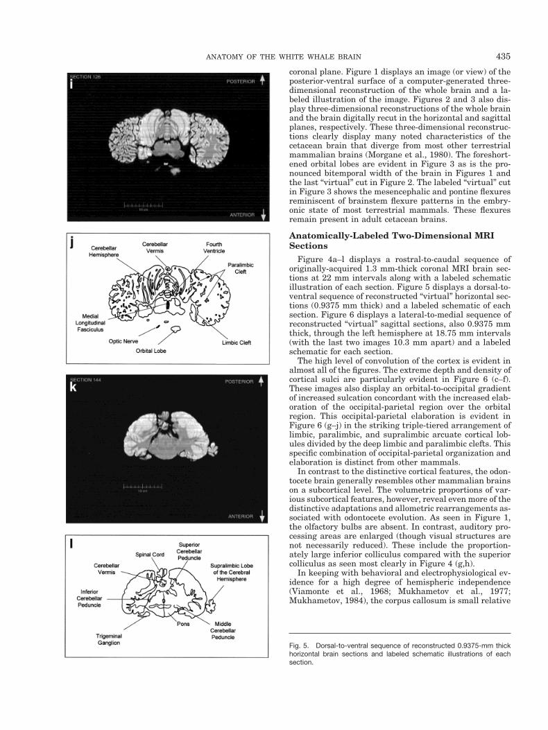
Figure 4a–l displays a rostral-to-caudal sequence oforiginally-acquired 1.3 mm-thick coronal MRI brain sec-tions at 22 mm intervals along with a labeled schematicillustration of each section. Figure 5 displays a dorsal-to-ventral sequence of reconstructed “virtual” horizontal sec-tions (0.9375 mm thick) and a labeled schematic of eachsection. Figure 6 displays a lateral-to-medial sequence ofreconstructed “virtual” sagittal sections, also 0.9375 mmthick, through the left hemisphere at 18.75 mm intervals(with the last two images 10.3 mm apart) and a labeledschematic for each section.
The high level of convolution of the cortex is evident inalmost all of the figures. The extreme depth and density ofcortical sulci are particularly evident in Figure 6 (c–f).These images also display an orbital-to-occipital gradientof increased sulcation concordant with the increased elab-oration of the occipital-parietal region over the orbitalregion. This occipital-parietal elaboration is evident inFigure 6 (g–j) in the striking triple-tiered arrangement oflimbic, paralimbic, and supralimbic arcuate cortical lob-ules divided by the deep limbic and paralimbic clefts. Thisspecific combination of occipital-parietal organization andelaboration is distinct from other mammals.
In contrast to the distinctive cortical features, the odon-tocete brain generally resembles other mammalian brainson a subcortical level. The volumetric proportions of var-ious subcortical features, however, reveal even more of thedistinctive adaptations and allometric rearrangements as-sociated with odontocete evolution. As seen in Figure 1,the olfactory bulbs are absent. In contrast, auditory pro-cessing areas are enlarged (though visual structures arenot necessarily reduced). These include the proportion-ately large inferior colliculus compared with the superiorcolliculus as seen most clearly in Figure 4 (g,h).
In keeping with behavioral and electrophysiological ev-idence for a high degree of hemispheric independence(Viamonte et al., 1968; Mukhametov et al., 1977;Mukhametov, 1984), the corpus callosum is small relative
Fig. 5. Dorsal-to-ventral sequence of reconstructed 0.9375-mm thickhorizontal brain sections and labeled schematic illustrations of eachsection.
435ANATOMY OF THE WHITE WHALE BRAIN
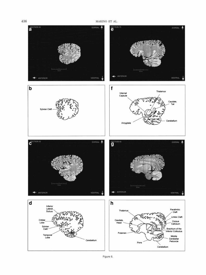
Figure 6.
436 MARINO ET AL.
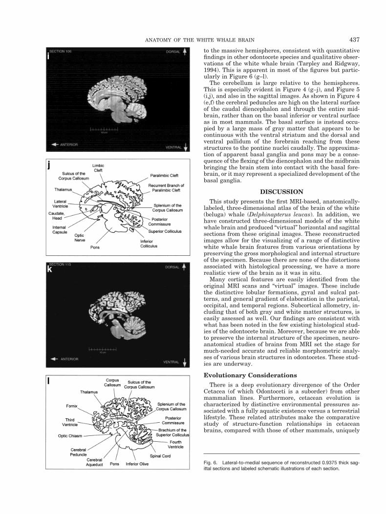
to the massive hemispheres, consistent with quantitativefindings in other odontocete species and qualitative obser-vations of the white whale brain (Tarpley and Ridgway,1994). This is apparent in most of the figures but partic-ularly in Figure 6 (g–l).
The cerebellum is large relative to the hemispheres.This is especially evident in Figure 4 (g–j), and Figure 5(i,j), and also in the sagittal images. As shown in Figure 4(e,f) the cerebral peduncles are high on the lateral surfaceof the caudal diencephalon and through the entire mid-brain, rather than on the basal inferior or ventral surfaceas in most mammals. The basal surface is instead occu-pied by a large mass of gray matter that appears to becontinuous with the ventral striatum and the dorsal andventral pallidum of the forebrain reaching from thesestructures to the pontine nuclei caudally. The approxima-tion of apparent basal ganglia and pons may be a conse-quence of the flexing of the diencephalon and the midbrainbringing the brain stem into contact with the basal fore-brain, or it may represent a specialized development of thebasal ganglia.
DISCUSSIONThis study presents the first MRI-based, anatomically-
labeled, three-dimensional atlas of the brain of the white(beluga) whale (Delphinapterus leucas). In addition, wehave constructed three-dimensional models of the whitewhale brain and produced “virtual” horizontal and sagittalsections from these original images. These reconstructedimages allow for the visualizing of a range of distinctivewhite whale brain features from various orientations bypreserving the gross morphological and internal structureof the specimen. Because there are none of the distortionsassociated with histological processing, we have a morerealistic view of the brain as it was in situ.
Many cortical features are easily identified from theoriginal MRI scans and “virtual” images. These includethe distinctive lobular formations, gyral and sulcal pat-terns, and general gradient of elaboration in the parietal,occipital, and temporal regions. Subcortical allometry, in-cluding that of both gray and white matter structures, iseasily assessed as well. Our findings are consistent withwhat has been noted in the few existing histological stud-ies of the odontocete brain. Moreover, because we are ableto preserve the internal structure of the specimen, neuro-anatomical studies of brains from MRI set the stage formuch-needed accurate and reliable morphometric analy-ses of various brain structures in odontocetes. These stud-ies are underway.
Evolutionary ConsiderationsThere is a deep evolutionary divergence of the Order
Cetacea (of which Odontoceti is a suborder) from othermammalian lines. Furthermore, cetacean evolution ischaracterized by distinctive environmental pressures as-sociated with a fully aquatic existence versus a terrestriallifestyle. These related attributes make the comparativestudy of structure-function relationships in cetaceanbrains, compared with those of other mammals, uniquely
Fig. 6. Lateral-to-medial sequence of reconstructed 0.9375 thick sag-ittal sections and labeled schematic illustrations of each section.
437ANATOMY OF THE WHITE WHALE BRAIN
valuable for improving our understanding of the parame-ters of mammalian brain evolution.
The brain of the white whale as revealed in this study ischaracterized by similar morphological trends as thosefound in the bottlenose dolphin and other cetaceans (Mor-gane et al., 1980). Although there are differences amongcetacean brains, these differences are relatively minorcompared with the striking dissimilarities to brains ofother mammals. The most obvious difference betweencetacean brains and those of other mammals is in thegross morphological configuration of the whole structureand the lobules of the cerebral hemispheres. These arewell-visualized in MRI scans. Evolution of overall brainshape in cetaceans may have been partly due to migrationof the blowhole and telescoping of the skull, i.e., antorbitalelongation and postorbital compression. This in turn mayaccount for the distinctive construction of the midbrain,i.e., the corticopontine, corticobulbar and corticospinal fi-bers travel high on the lateral surface whereas the ventralsurface is occupied by a large continuous mass of graymatter extending from the diencephalon rostrally to thepontine nuclei caudally. There may be distinctive organi-zational features of the basal ganglia that also contributeto this uniquely cetacean architecture.
There is also adequate evidence that many of the ana-tomical changes in the cetacean brain represent changesin function, e.g., loss of olfactory structures and enlarge-ment of acoustic structures. Similar, convergent changesin function, along with their neuroanatomical correlates,are observed in several brains of unrelated clades, such asmany bats and primates (Johnson et al., 1984, 1994). Ingeneral, the cetacean brain possesses some common mam-malian features in combination with specialized andhighly unusual features, the function of which we havebarely begun to understand.
CONCLUSIONSIf we are to eventually understand the functional sig-
nificance of this mosaic of typical mammalian anduniquely cetacean features, the structural organization ofthe cetacean brain must be further elucidated. This can berapidly and effectively accomplished by MRI-based stud-ies of neuroanatomy. In comparison, already-existing datafrom the more traditional methods of sectioning and stain-ing are very time-intensive, expensive, and vulnerable tospatial distortion compared with the data acquired byMRI. Studies like the present one are crucial for estab-lishing the structural basis of and templates for futurefunctional studies using non-invasive neuroimaging tech-niques to investigate the neurobiological basis of cetaceancognition and behavior.
ACKNOWLEDGMENTSWe wish to thank R.C. Switzer III for discussions and
identifications of the basal ganglia in the cetacean speci-mens. We also thank Joanne Whallon for use of the VoxelView programs and Silicon Graphics, Inc. workstations atthe Laser Scanning Microscopy Laboratory at MichiganState University, Cheryl Short for technical assistance,Rick Buxton for conducting the MRI scanning at the Uni-versity of California, San Diego, and Patsy Bryan forexcellent artwork. This study was supported by an EmoryUniversity University Research Committee Award.
LITERATURE CITEDAwbrey FT, Thomas JA, Kastelein RA.1988. Low-frequency underwa-
ter hearing sensitivity in belugas, Delphinapterus leucas. J AcoustSoc Am 84:2273–2275
Buhl EH, Oelschlager HA. 1988. Morphogenesis of the brain in theharbor porpoise. J Comp Neurol 277:109–125.
Garey LJ, Leuba G. 1986. A quantitative study of neuronal and glialnumerical density in the visual cortex of the bottlenose dolphin:evidence for a specialized subarea and changes with age. J CompNeurol 247:491–496.
Garey LJ, Winkelman E, Brauer K. 1985. Golgi and Nissl studies ofthe visual cortex of the bottlenose dolphin. J Comp Neurol 240:305–321.
Glezer II, Hof PR, Leranth C, Morgane PJ. 1992a. Morphological andhistological features of odontocete visual neocortex: immunocyto-chemical analysis of pyramidal and nonpyramidal populations ofneurons. In: Thomas JA, Kastelein RA, Supin AY, editors. Marinemammal sensory systems. New York: Plenum Press. p 1–38.
Glezer II, Hof PR, Morgane PJ. 1992b. Calretinin-immunoreactiveneurons in the primary visual cortex of dolphin and human brains.Brain Res 595:181–188.
Glezer II, Hof PR, Istomin VV, Morgane PJ. 1995a. Comparativeimmunocytochemistry of calcium-binding protein-positive neuronsin visual and auditory systems of cetacean and primate brains. In:Kastelein RA, Thomas JA, Nachtigall PE, editors. Sensory systemsof aquatic mammals. The Netherlands: De Spil Publishers. p 477–513.
Glezer II, Hof PR, Leranth C, Morgane PJ. 1993. Calcium-bindingprotein-containing neuronal populations in mammalian visualcortex: a comparative study in whales, insectivores, bats, rodents,and primates. Cereb Cortex 3:249–272.
Glezer II, Hof PR, Morgane PJ. 1995b. Cytoarchitectonics and immu-nocytochemistry of the inferior colliculus of midbrains in cetaceans.FASEB J 9:A247.
Glezer II, Hof PR, Morgane PJ. 1998. Comparative analysis of calci-um-binding protein-immunoreactive neuronal populations in theauditory and visual systems of the bottlenose dolphin (Tursiopstruncatus) and the macaque monkey (Macaca fascicularis). J ChemNeuro15:203–237.
Glezer II, Jacobs M, Morgane P. 1988. Implications of the ‘initialbrain’ concept for brain evolution in Cetacea. Behav Brain Sci11:75–116.
Glezer II, Morgane, PJ. 1990. Ultrastructure of synapses and Golgianalysis of neurons in neocortex of the lateral gyrus (visual cortex)of the dolphin and pilot whale. Brain Res Bull 24:401–427.
Glezer II, Morgane PJ, Leranth C. 1990. Immunohistochemistry ofneurotransmitters in visual cortex of several toothed whales: lightand electron microscopic study. In: Thomas JA, Kastelein RA, edi-tors. Sensory abilities of Cetaceans: laboratory and field evidence.New York: Plenum Press. p 39–60.
Haug H. 1987. Brain sizes, surfaces and neuronal sizes of the cortexcerebri. A stereological investigation of man and his variability anda comparison with some mammals (primates, whales, marsupialia,insectivores and one elephant). Am J Anat 180:126–142.
Hof PR, Glezer II, Archin N, Janssen WG, Morgane PJ, Morrison JH.1992. The primary auditory cortex in cetacean and human brain: acomparative analysis of neurofilament protein-containing pyrami-dal neurons. Neurosci Lett 146:91–95.
Hof PR, Glezer II, Revishchin AV, Bouras C, Charnay Y, Morgane PJ.1995. Distribution of dopaminergic fibers and neurons in visual andauditory cortices of the harbor porpoise and pilot whale. Brain ResBull 36:275–284.
Jacobs MS, Galaburda AM, McFarland WL, Morgane PJ. 1984. Theinsular formations of the dolphin brain: quantitative cytoarchitec-tonic studies of the insular component of the limbic lobe. J CompNeurol 225:396–432.
Jacobs MS, McFarland WL, Morgane PJ. 1979. The anatomy of thebrain of the bottlenose dolphin (Tursiops truncatus). Rhinic lobe(rhinencephalon): the archicortex. Brain Res Bull 4 (suppl.):1–108.
438 MARINO ET AL.
Johnson JI, Kirsch JA, Switzer III RC. 1984. Brain traits throughphylogeny: the evolution of neural characters. Brain Behav Evol20:97–117.
Johnson JI, Kirsch JA, Reep RL, Switzer III RC. 1994. Phylogenythrough brain traits: more characters for the analysis of mamma-lian evolution. Brain Behav Evol 43:319–347.
Manger P, Sum M, Szymanski M, Ridgway S, Krubitzer L. 1998.Modular subdivisions of dolphin insular cortex: does evolutionaryhistory repeat itself? J Cognitive Neurosci 10: 153–166.
Marino L. 1998. A comparison of encephalization levels between adultAnthropoid Primates and Odontocetes (toothed whales). Brain Be-hav Evol 51:230–238.
Morgane P, Jacobs M, Galaburda A. 1986. Evolutionary morphologyof the dolphin brain. In: Schusterman RJ, Thomas JA, Wood FG,editors. Dolphin cognition and behavior: a comparative approach.Hillsdale, NJ: Lawrence Erlbaum Associates. p 5–30.
Morgane PJ, Jacobs MS, MacFarland WL. 1980. The anatomy of thebrain of the bottlenose dolphin (Tursiops truncatus). Surface con-figurations of the telencephalon of the bottlenose dolphin with com-parative anatomical observations in four other Cetacean species.Brain Res Bull 5 (suppl.):1–107.
Mukhametov LM. 1984. Sleep in marine mammals. Exp Brain Res8:227–238.
Mukhametov LM, Supin AY, Polyakova IG. 1977. Interhemisphericasymmetry of the electroencephalographic sleep patterns in dol-phins. Brain Res 134:581–584.
Oelschlager HA, Buhl, EH. 1985. Occurrence of an olfactory bulb inthe early development of the harbor porpoise (Phocoena phocoena
L.). In: Duncker HR, Fleischer G, editors. Functional morphology invertebrates. New York: Fischer. p 695–698.
Oelschlager HA, Kemp B. 1998. Ontogenesis of the sperm whalebrain. J Comp Neurol 399:210–228.
Ridgway SH. 1986. The central nervous system of the bottlenosedolphin. In: Schusterman RJ, Thomas JA, Wood FG, editors. Dol-phin cognition and behavior: a comparative approach. Hillsdale,NJ: Lawrence Erlbaum Associates. p 31–60.
Ridgway SH. 1990. The central nervous system of the bottlenosedolphin. In: Leatherwood S, Reeves R, editors. The bottlenose dol-phin. San Diego: Academic Press. p 69–97.
Ridgway SH, Brownson RH. 1984. Relative brain sizes andcortical surface areas in odontocetes. Acta Zool Fennica 172:149 –152.
Schwerdtfeger WK, Oelschlager HA, Stephan H. 1984. Quantitativeneuroanatomy of the brain of the La Plata dolphin, Pontoporiablainvillei. Anat Embryol 170:11–19.
Tarpley RL, Ridgway, SH. 1994. Corpus callosum size in delphinidcetaceans. Brain Behav Evol 44:156–165.
Viamonte M, Morgane PJ, Galliano RE, Nagel EL, McFarland WL.1968. Angiography in the living dolphin and observations on bloodsupply to the brain. Am J Physiol 214:1225–1249.
Yablokov, AV, Bel’kovich BM, Tarasevich MN. 1964. The centralnervous system and sense organs. In: Kleinenberg SE, YablokovAV, Bel’kovich BM, editors. Beluga (Delphinapterus leucas) inves-tigation of the species. USSR: Academy of Sciences of the USSR. p169–183.
439ANATOMY OF THE WHITE WHALE BRAIN