Embed Size (px)
Citation preview

© 2012 Pearson Education, Inc.
Dr. McGehee’s
Anatomy and Physiology I
Fall 2013 (BIOL 2401)
• Sign-in with your full name and hccs.edu
email address
• Grab a blank piece of scratch paper

© 2012 Pearson Education, Inc.
© 2012 Pearson Education, Inc.
Tell me about yourself!
Name?
Have you taken Biology 1406 (General Biology I)?
Have you taken any other science courses?
Why are you taking A&P I?
Have you taken this class before?
What are your career goals?
TEAM-BASED LEARNING
© 2012 Pearson Education, Inc.
Syllabus
• Go to the HCC homepage
• Click on Course Syllabi/ Faculty CV
• Type in my name—mcgehee
• Anatomy and Physiology I Fall 2013 (BIOL
2401)
• You will find the syllabus and upcoming lectures,
notes, and other class materials posted.

© 2012 Pearson Education, Inc.
• Study Strategies
• Attend all lectures, labs, and exam reviews
• Read your lecture and laboratory assignments before going to class or lab
• Devote a block of time each day to you’re A&P I course
• Set up a study schedule and stick to it
• Do not procrastinate
• Approach the information in different ways
• Develop your own method of studying-do what works for you!
• Notecards
• Quizlet app
• Chapter Summaries/Outline
• Form Study Groups
• As soon as you experience difficulty with the course, seek assistance

© 2012 Pearson Education, Inc.
Memorizing Learning

© 2012 Pearson Education, Inc.
Study Strategies
• Key Concepts
• Illustrations, Tables,
and Photos
• Pronunciation Guides
• Concept Check
Questions
• Making Connections
with previously learned
material
• Tips & Tricks
• Chapter Review and
Summary Figures
• www.masteringaandp.com
• Tutorials
• eText
• Practice Tests
• BioFlix
• Important Features of the Textbook—use what is available to you!

© 2012 Pearson Education, Inc.
PowerPoint® Lecture Presentations prepared by
Jason LaPres
Lone Star College—North Harris
1 An Introduction to
Anatomy and
Physiology

© 2012 Pearson Education, Inc.
An Introduction to Studying the Human Body
• You must be able to define anatomy and
physiology, describe the origins of anatomical and
physiological terms
• Explain the relationship between anatomy and
physiology, and describe various specialties of
each discipline.
• Identify the major levels of organization in
organisms, from the simplest to the most
complex, and identify major components of each
organ system.
• Explain the concept of homeostasis.

© 2012 Pearson Education, Inc.
An Introduction to Studying the Human Body
• Learning Outcomes
• Describe how negative feedback and positive
feedback are involved in homeostatic regulation,
and explain the significance of homeostasis.
• Use anatomical terms to describe body
sections, body regions, and relative positions.
• Identify the major body cavities and their
subdivisions, and describe the functions of each.

© 2012 Pearson Education, Inc.
An Introduction to Studying the Human Body
• Classification of Living Things
• Humans and many other animals are vertebrates
• Characterized by a segmented vertebral column
• Homeostasis
• The goal of physiological regulation and the key to
survival in a changing environment

© 2012 Pearson Education, Inc.
1-1 Anatomy and Physiology Directly Affect
Your Life
• Anatomy
• Is the oldest medical science
• Physiology
• Is the study of function

© 2012 Pearson Education, Inc.
1-3 Anatomy and Physiology
• Anatomy
• Describes the structures of the body
• What they are made of
• Where they are located
• Associated structures
• Physiology
• Is the study of:
• Functions of anatomical structures
• Individual and cooperative functions
Can you give examples
of how structure
relates to function in
the body?
Think, pair, share…

© 2012 Pearson Education, Inc.
1-4 Relationships between Anatomy and
Physiology
• Anatomy (macroscopic vs microscopic)
• Gross anatomy, or macroscopic anatomy, examines
large, visible structures
• Surface anatomy: exterior features—superficial
markings
• Regional anatomy: body areas-neck, head
• Systemic anatomy: organ systems
• Developmental anatomy: from conception to death
• Clinical anatomy: medical specialties (anatomical
features change when you are in a diseased state)
© 2012 Pearson Education, Inc.
1-4 Relationships between Anatomy and
Physiology
• Anatomy
• Microscopic anatomy examines cells and molecules
• Cytology: study of cells and their structures
• cyt- = cell
• Histology: study of tissues and their structures

© 2012 Pearson Education, Inc.
1-4 Relationships between Anatomy and
Physiology
• Physiology
• Cell physiology: processes within and between cells
• Organ physiology: functions of specific organs
• Systemic physiology: functions of an organ system
• Pathological physiology: effects of diseases

© 2012 Pearson Education, Inc.
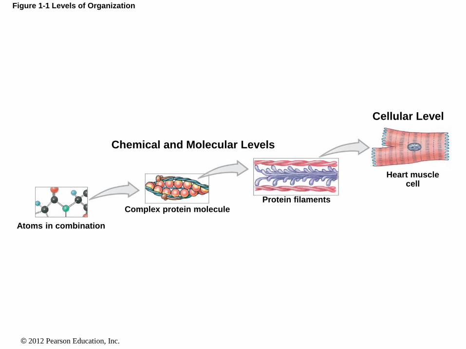
1-5 Levels of Organization
• The Chemical (or Molecular) Level
• Atoms are the smallest chemical units
• Molecules are a group of atoms working together
• The Cellular Level
• Cells are a group of atoms, molecules, and organelles working
together
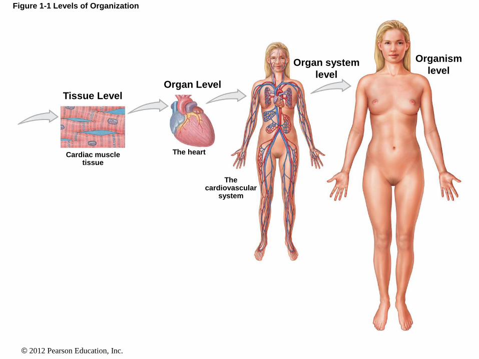
• The Tissue Level
• A tissue is a group of similar cells working together
• The Organ Level
• An organ is a group of different tissues working together
© 2012 Pearson Education, Inc.
1-5 Levels of Organization
• The Organ System Level
• An organ system is a group of organs working together
• Humans have 11 organ systems
• The Organism Level
• A human is an organism
Molecule < cell < tissue < organ < system < organism
© 2012 Pearson Education, Inc.
Figure 1-1 Levels of Organization
Chemical and Molecular Levels
Cellular Level
Atoms in combination
Complex protein molecule Protein filaments
Heart muscle cell
© 2012 Pearson Education, Inc.
Figure 1-1 Levels of Organization
Tissue Level Organ Level
Cardiac muscle tissue
The heart
The cardiovascular
system
Organ system
level
Organism
level
© 2012 Pearson Education, Inc.
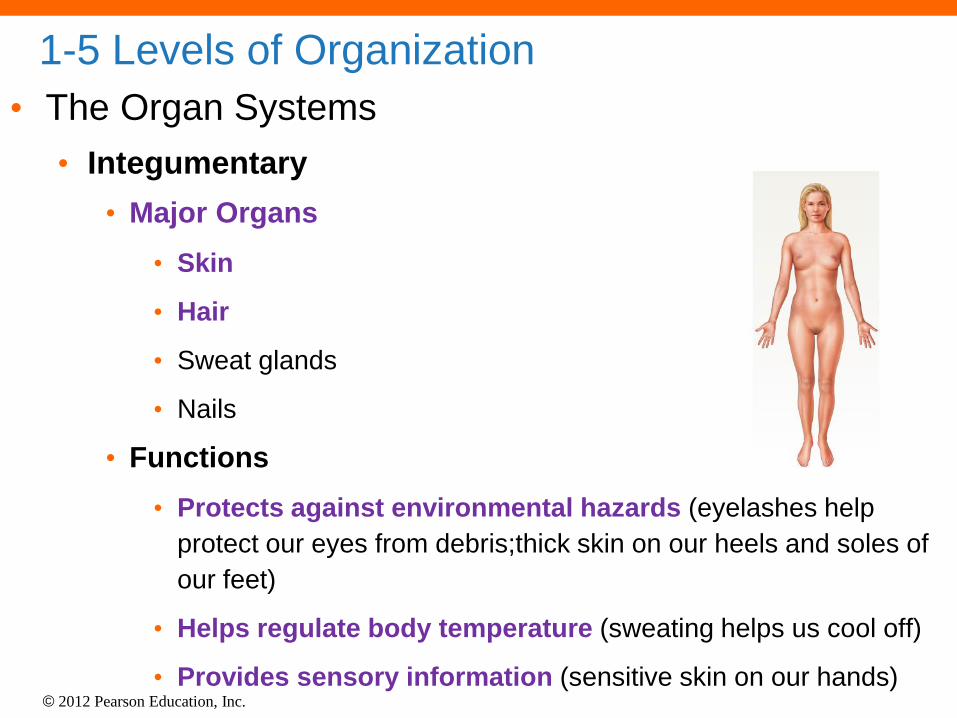
1-5 Levels of Organization
• The Organ Systems
• Integumentary
• Major Organs
• Skin
• Hair
• Sweat glands
• Nails
• Functions
• Protects against environmental hazards (eyelashes help
protect our eyes from debris;thick skin on our heels and soles of
our feet)
• Helps regulate body temperature (sweating helps us cool off)
• Provides sensory information (sensitive skin on our hands)
© 2012 Pearson Education, Inc.
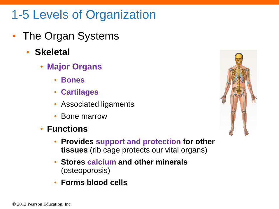
1-5 Levels of Organization
• The Organ Systems
• Skeletal
• Major Organs
• Bones
• Cartilages
• Associated ligaments
• Bone marrow
• Functions
• Provides support and protection for other tissues (rib cage protects our vital organs)
• Stores calcium and other minerals (osteoporosis)
• Forms blood cells

© 2012 Pearson Education, Inc.
1-5 Levels of Organization
• The Organ Systems
• Muscular
• Major Organs
• Skeletal muscles and associated tendons
• Functions
• Provides movement
• Provides protection and support for other
tissues
• Generates heat that maintains body
temperature
© 2012 Pearson Education, Inc.
1-5 Levels of Organization • The Organ Systems
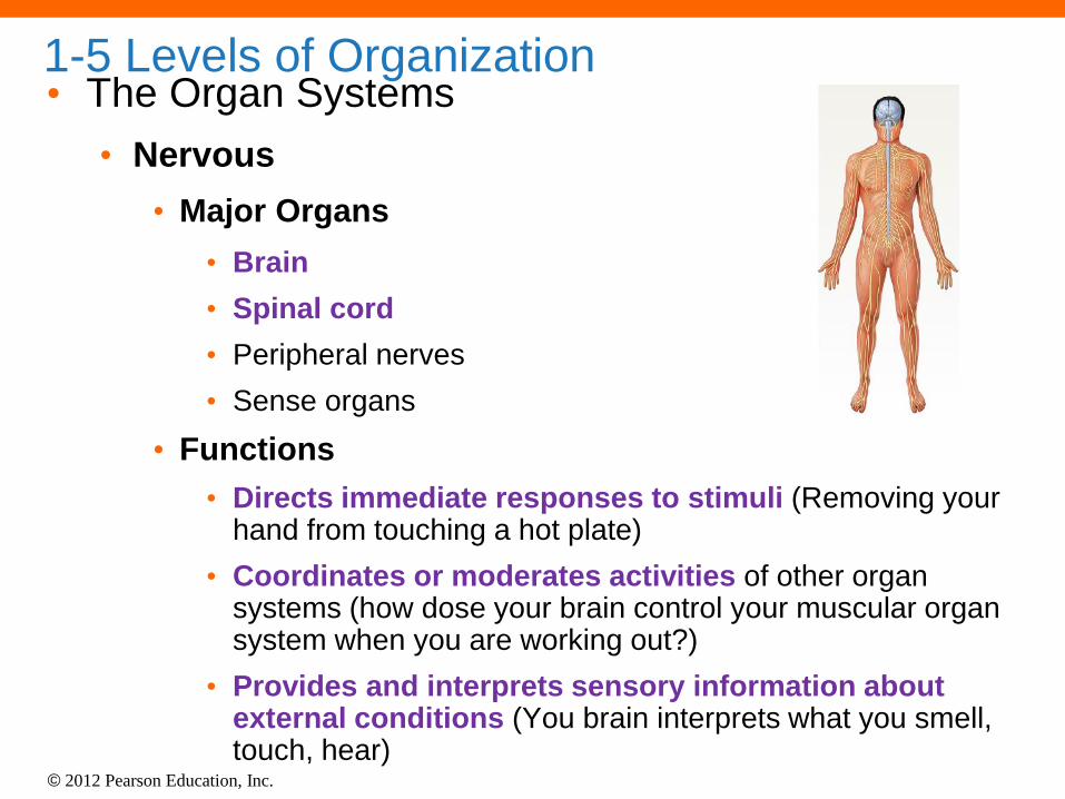
• Nervous
• Major Organs
• Brain
• Spinal cord
• Peripheral nerves
• Sense organs
• Functions
• Directs immediate responses to stimuli (Removing your hand from touching a hot plate)
• Coordinates or moderates activities of other organ systems (how dose your brain control your muscular organ system when you are working out?)
• Provides and interprets sensory information about external conditions (You brain interprets what you smell, touch, hear)

© 2012 Pearson Education, Inc.
• The Organ Systems
• Endocrine
• Major Organs
• Pituitary gland
• Pancreas
• Gonads
• Endocrine tissues in other systems
• Functions
• Directs long-term changes in the activities of
other organ systems
• Adjusts metabolic activity and energy use by the body
• Controls many structural and functional changes during
development (think about how your body changes during
puberty/growth spurt!)
• Thyroid gland
• Adrenal glands
1-5 Levels of Organization

© 2012 Pearson Education, Inc.
1-5 Levels of Organization
• The Organ Systems
• Cardiovascular
• Major Organs
• Heart
• Blood
• Blood vessels
• Functions
• Distributes blood cells, water and dissolved
materials including nutrients, waste
products, oxygen, and carbon dioxide
• Distributes heat and assists in control of
body temperature

© 2012 Pearson Education, Inc.
1-5 Levels of Organization • The Organ Systems
• Lymphatic
• Major Organs
• Spleen
• Thymus
• Lymphatic vessels
• Lymph nodes
• Tonsils
• Functions
• Defends against infection and disease (think about
why the doctor feels for swollen lymph nodes in your
neck when you are sick)
• Returns tissue fluids to the bloodstream

© 2012 Pearson Education, Inc.
1-5 Levels of Organization
• The Organ Systems
• Respiratory
• Major Organs
• Nasal cavities
• Sinuses
• Larynx
• Trachea
• Bronchi
• Lungs
• Alveoli

© 2012 Pearson Education, Inc.
1-5 Levels of Organization
• The Organ Systems
• Respiratory
• Functions
• Delivers air to alveoli (sites in lungs where
gas exchange occurs)
• Provides oxygen to bloodstream
• Removes carbon dioxide from
bloodstream
• Produces sounds for communication
(talking)

© 2012 Pearson Education, Inc.
1-5 Levels of Organization
• The Organ Systems
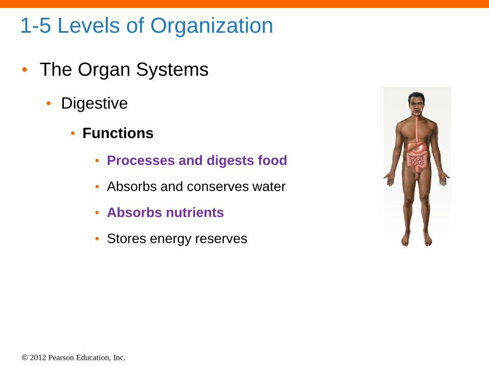
• Digestive
• Major Organs
• Teeth
• Tongue
• Pharynx
• Esophagus
• Stomach
• Small intestine
• Large intestine
• Liver
• Gallbladder
• Pancreas
© 2012 Pearson Education, Inc.
1-5 Levels of Organization
• The Organ Systems
• Digestive
• Functions
• Processes and digests food
• Absorbs and conserves water
• Absorbs nutrients
• Stores energy reserves

© 2012 Pearson Education, Inc.
1-5 Levels of Organization
• The Organ Systems
• Urinary
• Major Organs
• Kidneys
• Ureters
• Urinary bladder
• Urethra
• Functions
• Excretes waste products from the blood
• Controls water balance by regulating volume of
urine produced (hydration versus dehydration)
• Stores urine prior to voluntary elimination
• Regulates blood ion concentrations and pH

© 2012 Pearson Education, Inc.
1-5 Levels of Organization
• The Organ Systems
• Male Reproductive
• Major Organs
• Testes
• Epididymides
• Ductus deferentia
• Seminal vesicles
• Prostate gland
• Penis
• Scrotum

© 2012 Pearson Education, Inc.
1-5 Levels of Organization
• The Organ Systems
• Male Reproductive
• Functions
• Produces male sex cells (sperm),
suspending fluids, and hormones
• Sexual intercourse
© 2012 Pearson Education, Inc.
1-5 Levels of Organization
• The Organ Systems
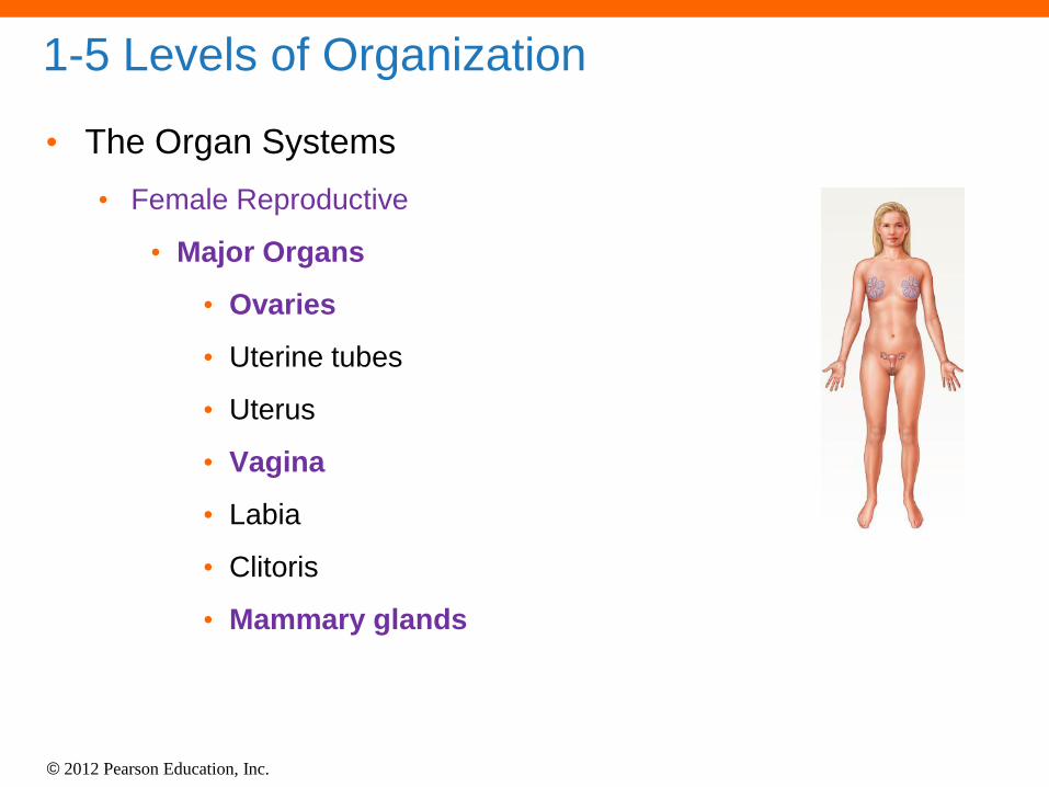
• Female Reproductive
• Major Organs
• Ovaries
• Uterine tubes
• Uterus
• Vagina
• Labia
• Clitoris
• Mammary glands

© 2012 Pearson Education, Inc.
1-5 Levels of Organization
• The Organ Systems
• Female Reproductive
• Functions
• Produces female sex cells (oocytes)
and hormones
• Supports developing embryo from
conception to delivery
• Provides milk to nourish newborn infant
• Sexual intercourse
© 2012 Pearson Education, Inc.
1-6 Homeostasis
• Homeostasis
• All body systems working together to maintain a
stable internal environment (remember by
thinking you are comfortable at home)
• Systems respond to external and internal changes
to function within a normal range (body
temperature, fluid balance)

© 2012 Pearson Education, Inc.
1-6 Homeostasis • Mechanisms of Regulation
• Autoregulation (intrinsic)
• Automatic response in a cell, tissue, or organ to some environmental
change
• Ex: When cells within a tissue need more oxygen, they release chemicals
that cause an increase in blood flow to the area, providing more oxygen to
the region.
• Extrinsic regulation
• Responses controlled by nervous and endocrine systems
• Ex: When you are exercising, your nervous system issues commands that
increase the heart rate so that blood will circulate faster.
• The nervous system reduces blood flow to organs, such as the digestive
tract, that are relatively inactive (oxygen in circulating blood is thus saved for
the active muscles.)

© 2012 Pearson Education, Inc.
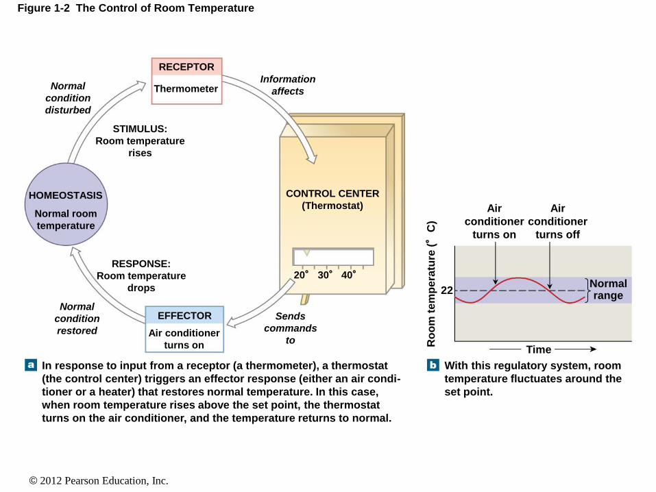
1-6 Homeostasis
• Receptor
• Receives the stimulus
• Control center
• Processes the signal and sends instructions
• Effector
• Carries out instructions
© 2012 Pearson Education, Inc.
Figure 1-2 The Control of Room Temperature
Normal
condition
disturbed
Information
affects
RECEPTOR
Thermometer
HOMEOSTASIS
STIMULUS:
Room temperature
rises
Normal room
temperature
RESPONSE:
Room temperature
drops
CONTROL CENTER
(Thermostat)
Normal
condition
restored
EFFECTOR
Air conditioner
turns on
Sends
commands
to
20° 30° 40°
In response to input from a receptor (a thermometer), a thermostat
(the control center) triggers an effector response (either an air condi-
tioner or a heater) that restores normal temperature. In this case,
when room temperature rises above the set point, the thermostat
turns on the air conditioner, and the temperature returns to normal.
With this regulatory system, room
temperature fluctuates around the
set point.
Air
conditioner
turns on
Air
conditioner
turns off
Time
Ro
om
tem
pe
ratu
re (°
C)
22 Normal range
© 2012 Pearson Education, Inc.
1-7 Negative and Positive Feedback
• The Role of Negative Feedback
• The response of the effector negates the stimulus
• Body is brought back into homeostasis
• Normal range is achieved

© 2012 Pearson Education, Inc.
Figure 1-3 Negative Feedback in the Control of Body Temperature
Normal
temperature
disturbed
Information
affects
RECEPTORS
Temperature sensors in skin
and hypothalamus
HOMEOSTASIS
STIMULUS:
Body temperature
rises
Normal body
temperature RESPONSE:
Increased heat loss,
body temperature
drops
CONTROL
CENTER
Normal
temperature
restored
EFFECTORS
• Sweat glands in skin increase secretion • Blood vessels in skin dilate
Sends
commands
to
Events in the regulation of body temperature, which are
comparable to those shown in Figure 12. A control center
in the brain (the hypothalamus) functions as a thermostat
with a set point of 37°C. If body temperature exceeds
37.2°C, heat loss is increased through enhanced blood flow
to the skin and increased sweating.
The thermoregulatory center keeps
body temperature fluctuating
within an acceptable range, usually
between 36.7 and 37.2°C.
Vessels
dilate,
sweating
increases
Vessels
constrict,
sweating
decreases
Time Bo
dy t
em
pera
ture
(°
C)
37.2 Normal range 37
36.7
Thermoregulatory
center in brain

© 2012 Pearson Education, Inc.
1-7 Negative and Positive Feedback
• The Role of Positive Feedback
• The response of the effector increases change of the
stimulus
• Body is moved away from homeostasis
• Normal range is lost
• Used to speed up processes

© 2012 Pearson Education, Inc.
Figure 1-4 Positive Feedback: Blood Clotting
Clotting
accelerates
Positive
feedback
loop
Blood clot Chemicals
This escalating process
is a positive feedback
loop that ends with the
formation of a blood clot,
which patches the vessel
wall and stops the bleeding.
As clotting continues,
each step releases
chemicals that further
accelerate the process.
The chemicals start chain
reactions in which cells,
cell fragments, and
soluble proteins in the
blood begin to form a clot.
Damage to cells in the
blood vessel wall releases
chemicals that begin the
process of blood clotting.
Chemicals

© 2012 Pearson Education, Inc.
1-7 Negative and Positive Feedback
• Systems Integration
• Systems work together to maintain homeostasis
• Homeostasis is a state of equilibrium (equal)
• Opposing forces are in balance
• Dynamic equilibrium — continual adaptation
(always changing to reach a balance)
• Physiological systems work to restore balance
• Failure results in disease or death

© 2012 Pearson Education, Inc.
Table 1-1 The Roles of Organ Systems in Homeostatic Regulation
Know some examples from each column. (1)
Name a system involve a system involved in
regulating body temperature. (2) What is the
systems function? (1) Integumentary (2) heat
loss by sweating
© 2012 Pearson Education, Inc.
1-8 Anatomical Terminology
• Superficial Anatomy
• Locating structures on or near the body surface
• Anatomical Landmarks
• Anatomical position: hands at sides, palms forward
• Supine: lying down, face up
• Prone: lying down, face down
© 2012 Pearson Education, Inc.
1-8 Anatomical Terminology
• Superficial Anatomy
• Anatomical Landmarks
• References to tangible structures
• Anatomical Regions
• Body regions
• Abdominopelvic quadrants
• Abdominopelvic regions
• Anatomical Directions
• Reference terms based on subject
Why do health
care professionals
use these specific
terms when
referring to the
human body?

© 2012 Pearson Education, Inc.
Figure 1-5a Anatomical Landmarks
Cephalic or head
Frontal or
forehead
Cranial or skull
Facial or face
Oral or mouth
Mental or chin
Axillary or armpit
Brachial or arm
Antecubital or front of
elbow
Umbilical or navel
Trunk Abdominal (abdomen)
Mammary or breast
Thoracic or thorax, chest
Cervical or neck
Buccal or cheek
Otic or ear
Nasal or nose
Ocular, orbital or eye
Anterior view
© 2012 Pearson Education, Inc.
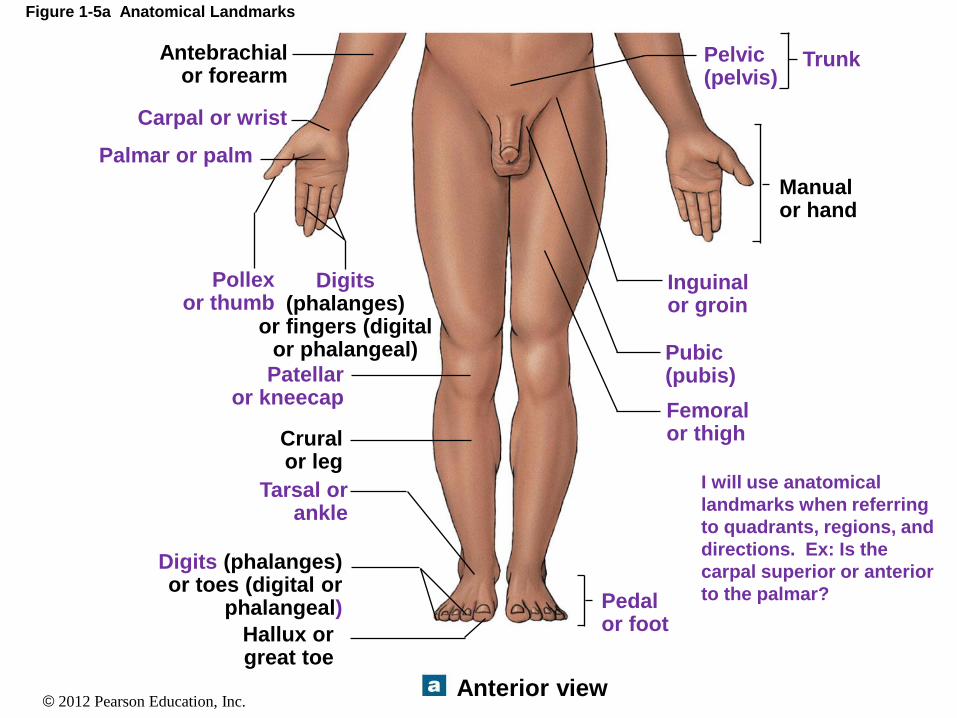
Figure 1-5a Anatomical Landmarks
Antebrachial or forearm
Carpal or wrist
Palmar or palm
Pollex or thumb
Digits (phalanges)
or fingers (digital or phalangeal)
Patellar or kneecap
Crural or leg
Digits (phalanges) or toes (digital or
phalangeal)
Tarsal or ankle
Anterior view
Hallux or great toe
Pedal or foot
Femoral or thigh
Pubic (pubis)
Inguinal or groin
Manual or hand
Pelvic (pelvis)
Trunk
I will use anatomical
landmarks when referring
to quadrants, regions, and
directions. Ex: Is the
carpal superior or anterior
to the palmar?
© 2012 Pearson Education, Inc.
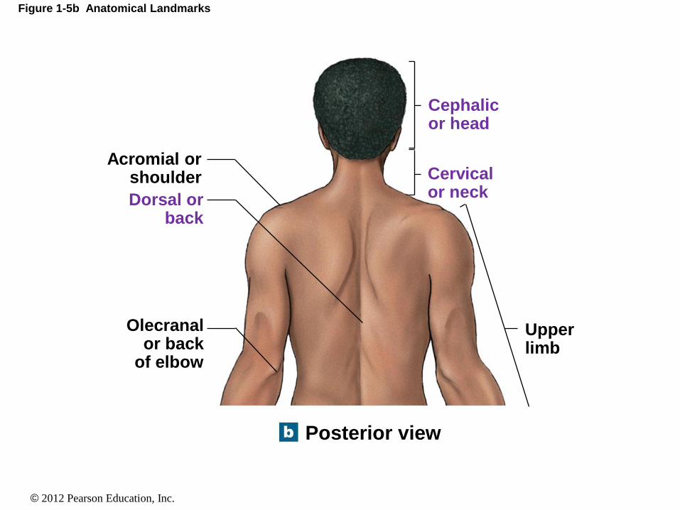
Figure 1-5b Anatomical Landmarks
Acromial or shoulder
Olecranal or back
of elbow
Dorsal or back
Upper limb
Cervical or neck
Cephalic or head
Posterior view

© 2012 Pearson Education, Inc.
Figure 1-5b Anatomical Landmarks
Posterior view
Lumbar or loin
Gluteal or buttock
Popliteal or back of knee
Sural or calf
Calcaneal or heel of foot
Plantar or sole of foot
Lower limb
Upper limb
© 2012 Pearson Education, Inc.
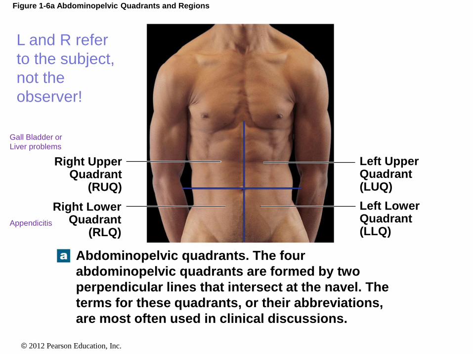
Figure 1-6a Abdominopelvic Quadrants and Regions
Abdominopelvic quadrants. The four
abdominopelvic quadrants are formed by two
perpendicular lines that intersect at the navel. The
terms for these quadrants, or their abbreviations,
are most often used in clinical discussions.
Right Upper Quadrant
(RUQ)
Right Lower Quadrant
(RLQ)
Left Upper Quadrant (LUQ)
Left Lower Quadrant (LLQ)
L and R refer
to the subject,
not the
observer!
Appendicitis
Gall Bladder or
Liver problems

© 2012 Pearson Education, Inc.
Figure 1-6b Abdominopelvic Quadrants and Regions
Right hypochondriac
region
Right lumbar region
Right inguinal
region
Abdominopelvic regions. The nine abdominopelvic
regions provide more precise regional descriptions.
Left hypochondriac region
Left lumbar region
Left inguinal region
Epigastric region
Umbilical region
Hypogastric (pubic) region

© 2012 Pearson Education, Inc.
Figure 1-6c Abdominopelvic Quadrants and Regions
Stomach
Spleen
Urinary bladder
Liver
Gallbladder
Large intestine
Small intestine
Appendix
Anatomical relationships. The relationship between
the abdominopelvic quadrants and regions and the
locations of the internal organs are shown here.
© 2012 Pearson Education, Inc.
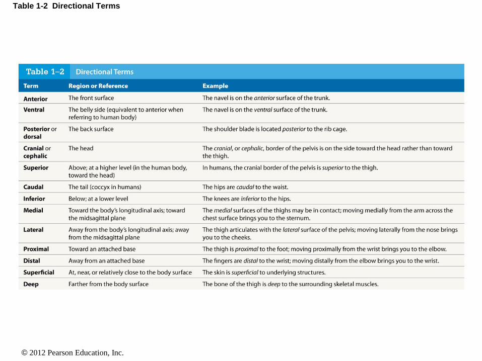
Table 1-2 Directional Terms
© 2012 Pearson Education, Inc.
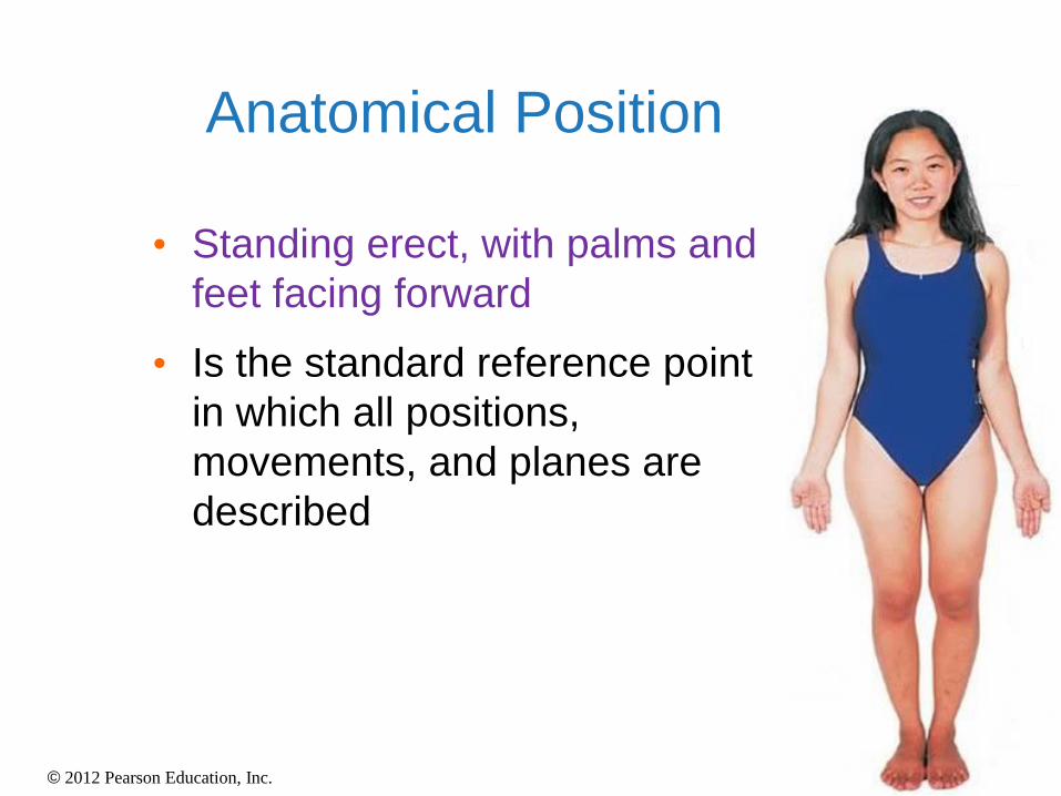
Anatomical Position
• Standing erect, with palms and
feet facing forward
• Is the standard reference point
in which all positions,
movements, and planes are
described

© 2012 Pearson Education, Inc.
Positions and Directions
Superior
• Refers to a structure being closer to the head or higher than another structure in the body
Inferior
• Refers to a structure being closer to the feet or lower than another structure in the body

© 2012 Pearson Education, Inc.
Positions and Directions
Anterior
• Refers to a structure being
more in front than another
structure in the body
Posterior
• Refers to a structure being
more in back than another
structure in the body
© 2012 Pearson Education, Inc.
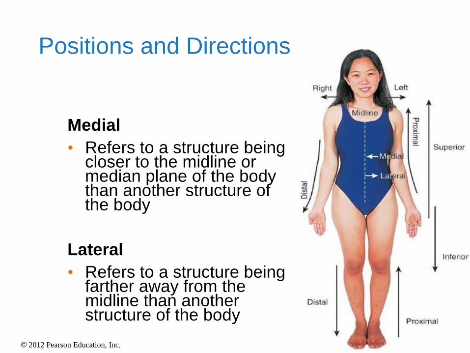
Positions and Directions
Medial
• Refers to a structure being closer to the midline or median plane of the body than another structure of the body
Lateral
• Refers to a structure being farther away from the midline than another structure of the body

© 2012 Pearson Education, Inc.
Positions and Directions
• Ipsilateral
• Structures are on the same side of the boy
• Contralateral
• Structures are on the opposite sides of the body

© 2012 Pearson Education, Inc.
Positions and Directions
Distal (Reference to the extremities only)
• Refers to a structure being further away from the root of the limb than another structure in the limb
Proximal (Reference to the extremities only)
• Refers to a structure being closer to the root of the limb than another structure in that limb
© 2012 Pearson Education, Inc.
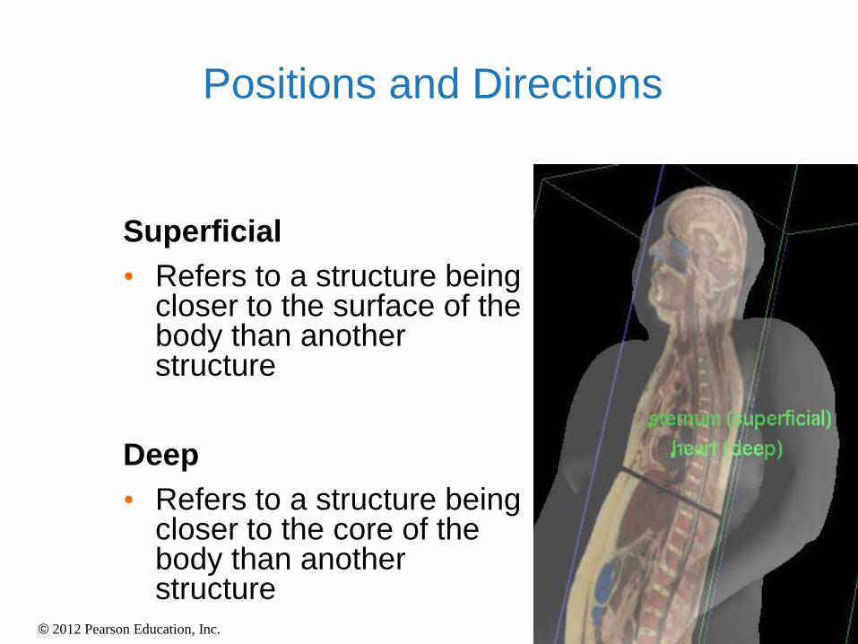
Positions and Directions
Superficial
• Refers to a structure being closer to the surface of the body than another structure
Deep
• Refers to a structure being closer to the core of the body than another structure

© 2012 Pearson Education, Inc.
Positions and Directions
Ventral
• Towards the front or
belly
Dorsal
• Towards the back

© 2012 Pearson Education, Inc.
Positions and Directions
Prone
• Lying face down
Supine
• Lying face up
Unilateral
• Pertaining to one side of the body
Bilateral
• Pertaining to both sides of the body

© 2012 Pearson Education, Inc.
1-8 Anatomical Terminology
• Sectional Anatomy
• Planes and sections
• Plane: a three-dimensional axis
• Section: a slice parallel to a plane
• Used to visualize internal organization and
structure
• Important in radiological techniques
• MRI
• PET
• CT

© 2012 Pearson Education, Inc.
Table 1-3 Terms That Indicate Sectional Planes

© 2012 Pearson Education, Inc.
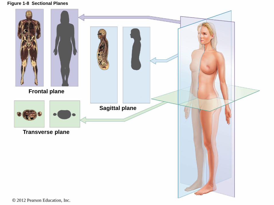
Anatomical Planes
Sagittal plane
• The plane dividing the
body into right and left
portions
• Midsagittal or median
are names for the plane
dividing the body into
equal right and left
halves
• Parasagittal divides the
structure into unequal
left and right planes.
© 2012 Pearson Education, Inc.
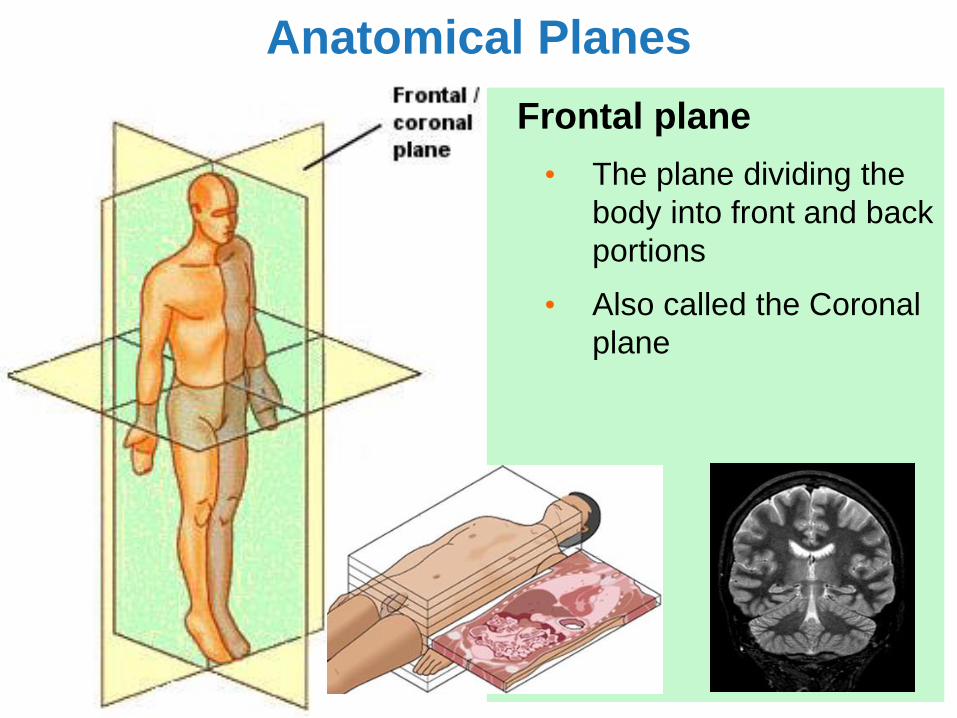
Anatomical Planes
Frontal plane
• The plane dividing the
body into front and back
portions
• Also called the Coronal
plane
© 2012 Pearson Education, Inc.
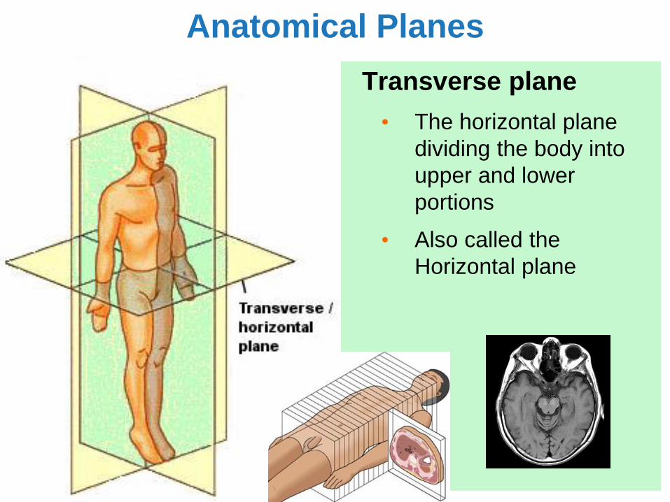
Anatomical Planes
Transverse plane
• The horizontal plane
dividing the body into
upper and lower
portions
• Also called the
Horizontal plane
© 2012 Pearson Education, Inc.
Figure 1-8 Sectional Planes
Frontal plane
Transverse plane
Sagittal plane

© 2012 Pearson Education, Inc.
Word Association Activity
From your list of Anatomical Terminology, choose 3 terms that
may be difficult for you to understand or remember. In this
activity, you will associate these terms and their definition with
a creative picture or drawing.
Dorsal -Towards the back
-Like the dorsal fin of a dolphin
Think, Pair,
Share!
© 2012 Pearson Education, Inc.
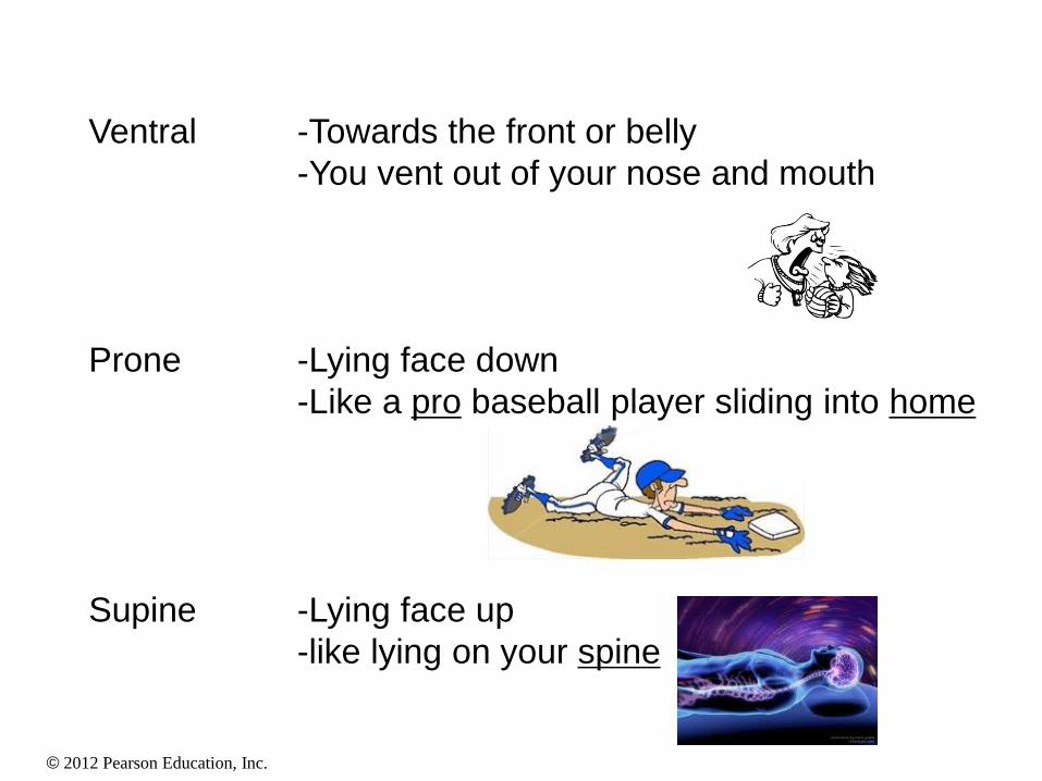
Prone -Lying face down
-Like a pro baseball player sliding into home
Supine -Lying face up
-like lying on your spine
Ventral -Towards the front or belly
-You vent out of your nose and mouth
© 2012 Pearson Education, Inc.
1-9 Body Cavities
• Essential Functions of Body Cavities
1. Protect organs from accidental shocks
2. Permit changes in size and shape of internal organs
• Ventral body cavity (coelom)
• Divided by the diaphragm
• Thoracic cavity
• Abdominopelvic cavity
© 2012 Pearson Education, Inc.
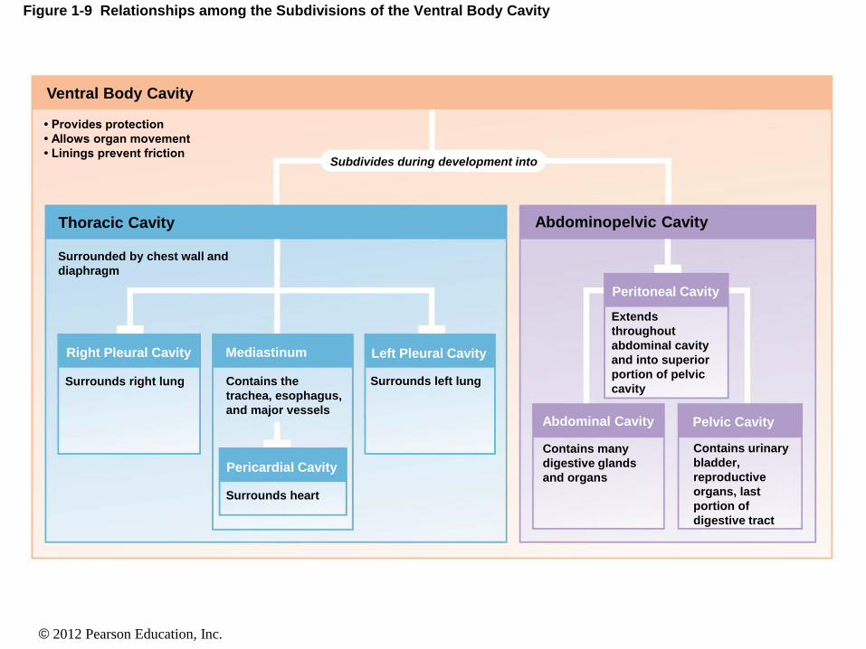
Figure 1-9 Relationships among the Subdivisions of the Ventral Body Cavity
• Provides protection
• Allows organ movement
• Linings prevent friction
Ventral Body Cavity
Thoracic Cavity
Surrounded by chest wall and
diaphragm
Surrounds right lung Contains the
trachea, esophagus,
and major vessels
Mediastinum Right Pleural Cavity
Peritoneal Cavity
Surrounds left lung
Subdivides during development into
Surrounds heart
Pericardial Cavity
Contains many
digestive glands
and organs
Abdominal Cavity
Abdominopelvic Cavity
Extends
throughout
abdominal cavity
and into superior
portion of pelvic
cavity
Pelvic Cavity
Contains urinary
bladder,
reproductive
organs, last
portion of
digestive tract
Left Pleural Cavity

© 2012 Pearson Education, Inc.
Figure 1-10a The Ventral Body Cavity and Its Subdivisions
POSTERIOR ANTERIOR
Pleural
cavity
Pericardial
cavity
Thoracic
cavity
Peritoneal
cavity
Abdominal
cavity
Pelvic
cavity
Diaphragm
Abdominopelvic
cavity
© 2012 Pearson Education, Inc.
1-9 Body Cavities
• Serous Membranes
• Line body cavities and cover organs
• Consist of parietal layer and visceral layer
• Parietal layer — lines cavity
• Visceral layer — covers organ
© 2012 Pearson Education, Inc.
1-9 Body Cavities
• The Thoracic Cavity
• Right and left pleural cavities
• Contain right and left lungs
• Mediastinum
• Upper portion filled with blood vessels, trachea,
esophagus, and thymus
• Lower portion contains pericardial cavity
• The heart is located within the pericardial
cavity

© 2012 Pearson Education, Inc.
Figure 1-10a The Ventral Body Cavity and Its Subdivisions
POSTERIOR ANTERIOR
Pleural
cavity
Pericardial
cavity
Thoracic
cavity
Peritoneal
cavity
Abdominal
cavity
Pelvic
cavity
Diaphragm
Abdominopelvic
cavity

© 2012 Pearson Education, Inc.
Figure 1-10b The Ventral Body Cavity and Its Subdivisions
Visceral
pericardium
Pericardial
cavity
Parietal
pericardium
Heart Air space
Balloon
Parietal layer — lines cavity
Visceral layer — covers organ

© 2012 Pearson Education, Inc.
Figure 1-10c The Ventral Body Cavity and Its Subdivisions
Spinal cord
Mediastinum
Parietal pleura
Pleural cavity
Pericardial cavity
Right lung
POSTERIOR
Left lung
ANTERIOR
Mediastinum
Upper portion filled with
blood vessels, trachea,
esophagus, and thymus

© 2012 Pearson Education, Inc.
Figure 1-9 Relationships among the Subdivisions of the Ventral Body Cavity
• Provides protection
• Allows organ movement
• Linings prevent friction
Ventral Body Cavity
Thoracic Cavity
Surrounded by chest wall and
diaphragm
Surrounds right lung Contains the
trachea, esophagus,
and major vessels
Mediastinum Right Pleural Cavity
Peritoneal Cavity
Surrounds left lung
Subdivides during development into
Surrounds heart
Pericardial Cavity
Contains many
digestive glands
and organs
Abdominal Cavity
Abdominopelvic Cavity
Extends
throughout
abdominal cavity
and into superior
portion of pelvic
cavity
Pelvic Cavity
Contains urinary
bladder,
reproductive
organs, last
portion of
digestive tract
Left Pleural Cavity
© 2012 Pearson Education, Inc.
1-9 Body Cavities
• The Abdominopelvic Cavity
• Peritoneal cavity: chamber within abdominopelvic
cavity
• Parietal peritoneum: lines the internal body wall
• Visceral peritoneum: covers the organs
© 2012 Pearson Education, Inc.
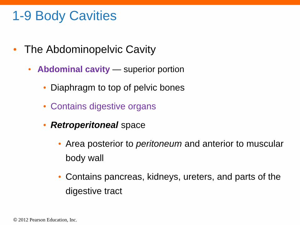
1-9 Body Cavities
• The Abdominopelvic Cavity
• Abdominal cavity — superior portion
• Diaphragm to top of pelvic bones
• Contains digestive organs
• Retroperitoneal space
• Area posterior to peritoneum and anterior to muscular
body wall
• Contains pancreas, kidneys, ureters, and parts of the
digestive tract

© 2012 Pearson Education, Inc.
1-9 Body Cavities
• The Abdominopelvic Cavity
• Pelvic cavity — inferior portion
• Within pelvic bones
• Contains reproductive organs, rectum, and bladder

© 2012 Pearson Education, Inc.
BREAK!
• 20 minute break, and then return to room 224
promptly
© 2012 Pearson Education, Inc.
Lab Safety-General
• No eating or drinking
• Only registered students allowed in the class
• Long hair must be tied back
• Closed-toed shoes must be worn at all times
• Familiarize yourself with the emergency stations
(eyewash, fire extinguisher, exits and first aid kits)
• Do not mark on the models!
• Inform me of any medical conditions that will
interfere with participation in the lab
© 2012 Pearson Education, Inc.
Lab Safety-Cleaning
• Clean up your workstation!
• Return all materials to the storage sites
• You MUST return slides to the proper labeled rack
• Clean glassware and wipe down countertops
• Follow directions for disposing used slides,
solutions, or other biohazard waste.
• Wash your hands
• I must approve your bench table cleanliness
before your team is allowed to leave.

© 2012 Pearson Education, Inc.
Lab Safety-The Microscope
• Carry the microscope with 2 hands
• Clean oculars and objectives with lens paper
before and after use
• Do no drag/slide the microscope, fully lift it to
move it
• We will not be using immersion oil
• Turn off the lamp before unplugging the
microscope
• You must show me your microscope before
you put it up the cabinet (cleanliness, lamp
switch, and wrapped cord).

© 2012 Pearson Education, Inc.
Dissection
• We will be performing online virtual
dissections.