Embed Size (px)
DESCRIPTION
Anatomy First YearOradea
Citation preview

Embryology
- generalities, history, evolution -
Prof. Dr. Teodor T. Maghiar
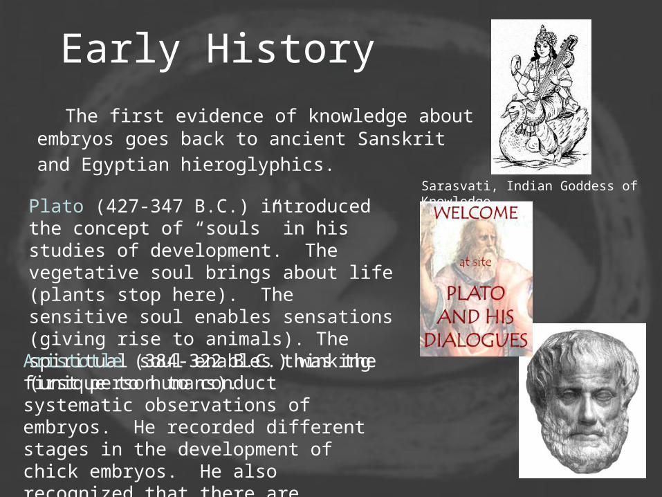
Early History
The first evidence of knowledge about embryos goes back to ancient Sanskrit and Egyptian hieroglyphics.
Sarasvati, Indian Goddess of Knowledge
Plato (427-347 B.C.) introduced the concept of “souls” in his studies of development. The vegetative soul brings about life (plants stop here). The sensitive soul enables sensations (giving rise to animals). The spiritual soul enables thinking (unique to humans).Aristotle (384-322 B.C.) was the first person to conduct systematic observations of embryos. He recorded different stages in the development of chick embryos. He also recognized that there are multiple ways that organisms reproduce.

Renaissance History Like most areas of Western civilization little
progress was made beyond the Greeks until the fifteenth century
Volcher Coiter (1514-1576) published a detailed study of chick embryology. Largely unheard of, he is now rightfully recognized as the “father of embryology.”
William Harvey (1578-1657) is best known for his work on blood, but he also contributed significantly to embryology by expanding and correcting Aristotle’s work. He coined the term epigenesis. Perhaps his greatest contribution was his idea “omne vivum ex ovo” promoting the existence of ova, even in humans

Renaissance History
Antoni von Leeuwenhoek (1632-1723) discovered “animalcules” in semen. He argued that a tiny preformed human was already present in these animalcules. Leeuwenhoek’s “discovery” led to the Preformationist period. A long argument ensued over which sex produced the “homunculus.” Malpighi and Swamerdam were the chief ovists.The debate decayed into a philosophical
dead-end (the homunculi should have even smaller homunculi inside them who had even smaller homunculi and so on ad infinitum).
Lazzaro Spallanzani (1729-1799) finally put an end to this idea when he successfully performed the first artifical insemination (using frog eggs).

Epigenesis and VitalismCaspar Friedrich Wolff (1738-1794) was the first person to demonstrate morphogenesis. He saw the development of structure out of structureless yolk material.
Friedrich Blumenbach (1742-1840) introduced the concept of “vis vitalis” an immaterial virtue or life force. Vitalism supposedly explained how structure could develop from an amorphous state. This concept brought about serious attempts to discover the nature of this force. Carl Ernst von Baer (1792-1876) made significant strides in descriptive embryology searching for the vital force. He was the first person to note the many similarities between the embryos of vertebrates particularly amniotes.

Ontogeny recapitulates PhylogenyErnst Haeckel (1834--1919) followed in von Baer’s footsteps as the leading authority in embryology during the late 1800’s. He developed the concept of Ontogeny recapitulates Phylogeny – Individual development progresses through the adult stages of the organism’s ancestors.
Haeckel’s ideas on race and human development and evolution were later incorporated into the pseudo-scientific basis for Nazism

Synthesis of Embryology with Genetics
August Weismann (1834-1914) developed germ plasm theory. In 1892 he published the idea of self reproducing determinants as the guiding agent in morphogenesis. He also proposed that these determinants were located on some newly discovered structures called chromosomes. He believed that cell differentiation was the result of cells acquiring different chromosomes during replication.
Oscar Hertwig (1849-1922) and his brother Richard Hertwig (1850-1937) are credited with providing the first demonstration of fertilization (sea urchins). They also discovered the existence of polar bodies, leading to significant advances in the understanding of meiosis.

Experimental EmbryologyHans Spemann (1869-1941) conducted the first rigorous experimental procedures on living embryos. He discovered the process of induction.
Spemann won the Nobel Prize for Medicine in 1935 for his discovery of the organizer effect (by studying the role of the dorsal lip of the blastopore) in embryology. This work was carried out jointly with his graduate student Hilde Proescholdt Mangold. Unfortunately she died the year of the publication (Nobel Prizes are not awarded posthumously).

Model Organisms
Wilhelm Roux
(1850-1924)
Hans Driesch
(1867-1941)
Thomas Hunt Morgan
(1866-1945)
Prior to the 1900’s there were no model organisms targeted by large numbers of scientists. Isolated advances were the work of individual genius. With the development of standard experimental subjects numerous workers could contribute to our knowledge of Embryology and Genetics.

Mitosis and Meiosis
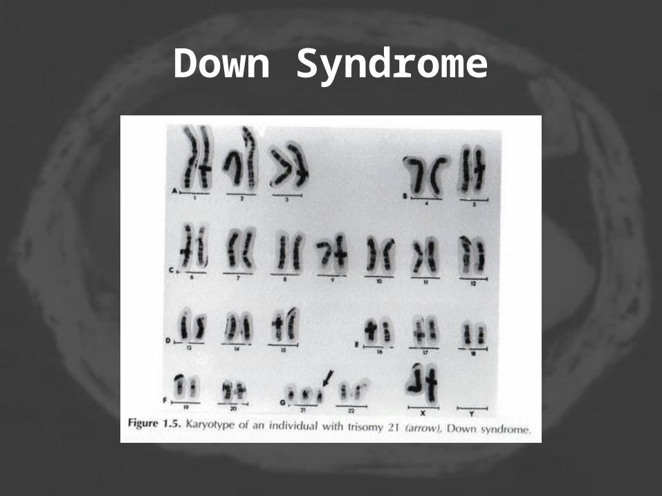
Down Syndrome


Embryology
-chromosomes, meiosis, cromosome X and Y-

SEXUALLY REPRODUCING
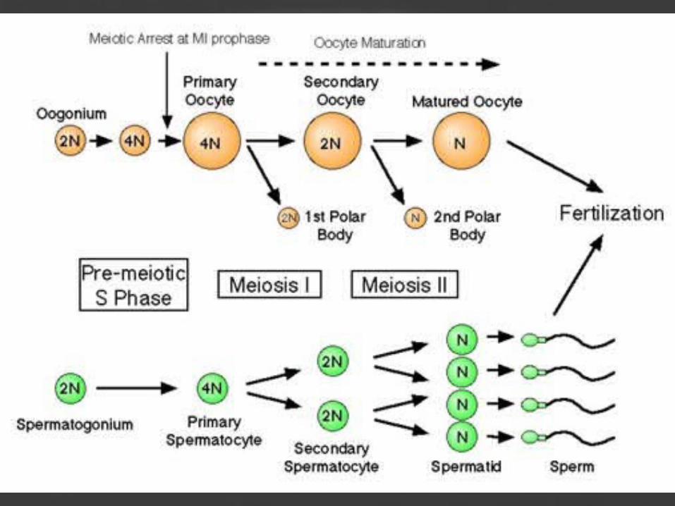
• Sexually reproducing species have somatic cells (body cells), which are diploid [2n] having two sets of chromosomes, one from the mother and one from the father. Gametes, reproductive cells, are haploid [n]: they have one set of chromosomes. Gametes are produced by meiosis of a diploid germ line cell. During meiosis, the matching chromosomes of father and mother can exchange small parts of themselves (crossover), and thus create new chromosomes that are not inherited solely from either parent. When a male and a female gamete merge (fertilization), a new diploid organism is formed.

Sexual reproduction • Formation of new individual by a combination of
two haploid sex cells (gametes). • Fertilization- combination of genetic information
from two separate cells that have one half the original genetic information
• Gametes for fertilization usually come from separate parents – Female- produces an egg – Male produces sperm
• Both gametes are haploid, with a single set of chromosomes
• The new individual is called a zygote, with two sets of chromosomes (diploid).
• Meiosis is a process to convert a diploid cell to a haploid gamete, and cause a change in the genetic information to increase diversity in the offspring.

A = adenine G = guanine C = cytosine T = thymine U = uracil

Chromosomes were first observed in plant cells by a Swiss botanist named
Karl Wilhelm von Nägeli in 1842, and independently in Ascaris worms by Belgian scientist Edouard Van Beneden (1846-1910).
The use of basophilic aniline dyes was a fundamentally new technique for effectively
staining the chromatin material in the nucleus. Their behavior in animal (
salamander) cells was later described in detail by German cytologist and professor of anatomy Walther Flemming, the discoverer of mitosis, in 1882. The name was invented later
by another German anatomist, Heinrich von Waldeyer in 1888.

A scheme of a condensed (metaphase) chromosome. (1) Chromatid - one of the two identical parts of the chromosome after S phase.
(2) Centromere - the point where the two chromatids touch, and where the microtubules attach.
(3) Short arm. (4) Long arm.
A chromosome is a single long molecule of DNA, and is the organized form of DNA found in cells. Chromosomes contain one very long, continuous piece of DNA (a single DNA molecule), which contains many genes, regulatory elements and other intervening nucleotide sequences. A broader definition of "chromosome" also includes the DNA-bound proteins which serve to package and manage the DNA. In eukaryotes nuclear chromosomes are packaged by proteins into a condensed structure called chromatin. This allows the massively-long DNA molecules to fit into the cell nucleus. The structure of chromatin varies through the cell cycle, and is responsible for the organisation of chromosomes into the classic four-arm structure during mitosis and meiosis
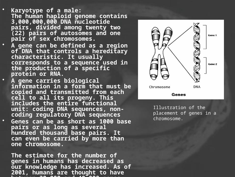
• Karyotype of a male: The human haploid genome contains 3,000,000,000 DNA nucleotide pairs, divided among twenty two (22) pairs of autosomes and one pair of sex chromosomes.
• A gene can be defined as a region of DNA that controls a hereditary characteristic. It usually corresponds to a sequence used in the production of a specific protein or RNA.
• A gene carries biological information in a form that must be copied and transmitted from each cell to all its progeny. This includes the entire functional unit: coding DNA sequences, non-coding regulatory DNA sequences
• Genes can be as short as 1000 base pairs or as long as several hundred thousand base pairs. It can even be carried by more than one chromosome.
The estimate for the number of genes in humans has decreased as our knowledge has increased. As of 2001, humans are thought to have between 30,000 and 40,000 genes.
Illustration of the placement of genes in a chromosome.
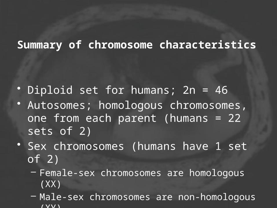
Summary of chromosome characteristics
• Diploid set for humans; 2n = 46 • Autosomes; homologous chromosomes, one
from each parent (humans = 22 sets of 2) • Sex chromosomes (humans have 1 set of 2)
– Female-sex chromosomes are homologous (XX) – Male-sex chromosomes are non-homologous (XY)

A = adenine G = guanine C = cytosine T = thymine U = uracil


Human Karyotype• Karyotyping is a technique used to determine
the (diploid) number of nuclear chromosomes of a eukaryotic organism, and may be used for determining sex and spotting chromosomal abnormalities. Cells can be locked part way through division (in metaphase) in vitro (in a reaction vial) with colchicine. These cells are then stained, photographed and arranged into a karyotype (an ordered set of chromosomes), also called karyogram.
Like many sexually reproducing species, humans have special gonosomes (sex chromosomes, in contrast to autosomes). These are XX in females and XY in males, and can be seen in the karyotype


Chromosomal mutations produce changes in whole chromosomes (more than one gene) or in the number of chromosomes present.
• Deletion - loss of part of a chromosome • Duplication - extra copies of a part of a
chromosome • Inversion - reverse the direction of a part
of a chromosome • Translocation - part of a chromosome
breaks off and attaches to another chromosome

The gain or loss of chromosome material can lead to a variety of genetic disorders. Human examples include:
• Down's Syndrome - trisomy of chromosome 21 • Patau Syndrome - trisomy of chromosome 13 • Edward Syndrome - trisomy of chromosome 18 • Klinefelter Syndrome - extra X chromosomes in
males - ie XXY, XXXY, XXXXY • Turner Syndrome - atypical X chromosome dosage
in females - ie XO, XXX, XXXX • XYY Syndrome - an extra Y chromosome in males

Chromosome numbers
• Common fruit fly - 8• Guinea Pig - 16
• Dove - 16• Earthworm - 36• Tibetan fox – 36
• Domestic cat – 38• Domestic pig – 38• Lab mouse - 40
• Lab rat – 42• Rabbit – 44• Human – 46
• Gorillas, Chimpanzees – 48• Elephants – 56
• Cow – 60• Donkey – 62• Horse – 64• Dog – 78
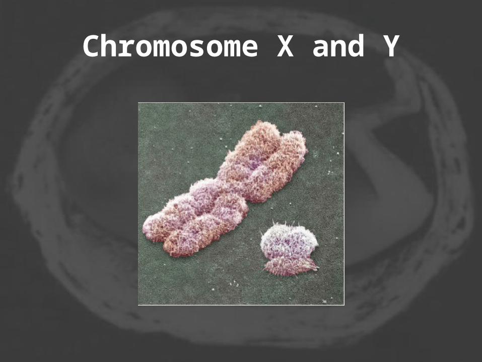
Chromosome X and Y

The X Chromosome• The X chromosome carries hundreds of genes but few, if
any, of these have anything to do directly with sex. However, the inheritance of these genes follows special rules. These arise because:
• males have only a single X chromosome • almost all the genes on the X have no counterpart on the
Y; thus • any gene on the X, even if recessive in females, will be
expressed in males. • Genes inherited in this fashion are described as sex-
linked or, more precisely, X-linked. • The sex chromosomes are one of the 23 homologous
pairs of human chromosomes. The X chromosome spans more than 153 million base pairs (the building material of DNA) and represents about 5% of the total DNA in women's cells, 2.5% in men's.
• Each person normally has one pair of sex chromosomes in each cell. Females have two X chromosomes, while males have one X and one Y chromosome.

The Y Chromosome
• In making sperm by meiosis, the X and Y chromosomes must separate in anaphase just as homologous autosomes do. This occurs without a problem because, like homologous autosomes, the X and Y chromosome synapse during prophase of meiosis I.
• Crossing over occurs in two regions of pairing, called the pseudoautosomal regions. These are located at opposite ends of the chromosome.


• The Pseudoautosomal Regions
• The pseudoautosomal regions get their name because any genes located within them (so far only 9 have been found) are inherited just like any autosomal genes. Males have two copies of these genes: one in the pseudoautosomal region of their Y, the other in the corresponding portion of their X chromosome. So males can inherit an allele originally present on the X chromosome of their father and females can inherit an allele originally present on the Y chromosome of their father.

• Meiosis was discovered and described for the first time in sea urchin eggs in 1876, by noted German biologist Oscar Hertwig (1849-1922). It was described again in 1883, at the level of chromosomes, by Belgian zoologist Edouard Van Beneden (1846-1910), in Ascaris worms' eggs. The significance of meiosis for reproduction and inheritance, however, was described only in 1890 by German biologist August Weismann (1834-1914), who noted that two cell divisions were necessary to transform one diploid cell into four haploid cells if the number of chromosomes had to be maintained. In 1911 the American geneticist Thomas Hunt Morgan (1866-1945) observed crossover in Drosophila melanogaster meiosis and provided the first true genetic interpretation of meiosis.

• meiosis is the process by which one diploid eukaryotic cell divides to generate four haploid cells often called gametes. The word "meiosis" comes from the Greek meioun, meaning "to make smaller," since it results in a reduction in chromosome number in the gamete cell.
• Meiosis is essential for sexual reproduction and therefore occurs in all eukaryotes (including single-celled organisms) that reproduce sexually.