Embed Size (px)
Citation preview
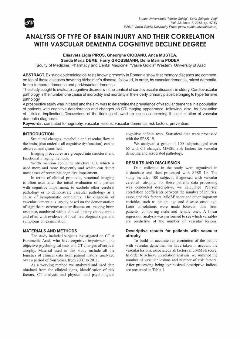
AnALYSIS oF TYPe oF BRAIn InJURY AnD TheIR coRReLATIon wITh vAScULAR DemenTIA coGnITIve DecLIne DeGRee
Elisaveta Ligia PIroS, Gheorghe cIoBAnu, Anca MuStEA, Sanda Maria dEME, harry GroSSMAnn, delia Marina PodEA
Faculty of Medicine, Pharmacy and Dental Medicine, “Vasile Goldis” Western University of Arad
ABStrAct. Existing epidemiological tests known presently in Romania show that memory diseases are common, on top of those diseases hovering Alzheimer’s disease, followed, in order, by vascular dementia, mixed dementia, fronto-temporal dementia and parkinsonian dementia.The study sought to evaluate cognitive disorders in the context of cardiovascular diseases in eldery. Cardiovascular pathology is the number one cause of morbidity and mortality in the elderly, primary place belonging to hypertensive pathology.A prospective study was initiated and the aim was to determine the prevalence of vascular dementia in a population of patients with cognitive deterioration and changes on CT-imaging appearance, following, also, by evaluation of clinical implications.Discussions of the findings showed up issues concerning the delimitation of vascular dementia diagnosis.Keywords: computed tomography, vascular lesions, vascular dementia, risk factors, prevention.
IntroductIonStructural changes, metabolic and vascular flow in
the brain, (that underlie all cognitive dysfunction), can be observed and quantified.
Imaging procedures are grouped into structural and functional imaging methods.
Worth mention about the structural CT, which is used more and more frequently and which can detect most cases of reversible cognitive impairment.
In terms of clinical protocols, structural imaging is often used after the initial evaluation of a patient with cognitive impairment, to exclude other cerebral pathology or to demonstrate vascular pathology as a cause of symptomatic complaints. The diagnosis of vascular dementia is largely based on the demonstration of significant cerebrovascular disease on imaging brain response, combined with a clinical history characteristic and often with evidence of focal neurological signs and symptoms on examination.
MAtErIALS And MEthodSThe study included subjects investigated on CT at
Euromedic Arad, who have cognitive impairment, the objective psychological tests and CT changes of cortical atrophy. Material used in this study include all the logistics of clinical data from patient history, analyzed over a period of four years, from 2007 to 2011.
As a working method we analyzed and used data obtained from the clinical signs, identification of risk factors, CT analysis and physical and psychological
cognitive deficits tests. Statistical data were processed with the SPSS 19.
We analyzed a group of 100 subjects aged over 65 with CT changes, MMSE, risk factors for vascular dementia and associated pathology.
rESuLtS And dIScuSSIonData collected in the study were organized in
a database and then processed with SPSS 19. The study includes 100 subjects, diagnosed with vascular cerebral atrophy. For these patients data processing was conducted descriptive, we calculated Pearson correlation coefficients between the number of injuries, associated risk factors, MMSE score and other important variables such as patient age and disease onset age. Later correlations were made between data from patients, comparing male and female ones. A linear regression analysis was performed to see which variables are predictive of the number of vascular lesions.
descriptive results for patients with vascular atrophy
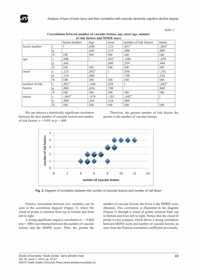
To build an accurate representation of the people with vascular dementia, we have taken in account the vascular lesions, associated risk factors and MMSE score. In order to achieve correlation analysis, we summed the number of vascular lesions and number of risk factors. After processing being synthesized descriptive indices are presented in Table 1.
Studia Universitatis “Vasile Goldiş”, Seria Ştiinţele VieţiiVol. 22, issue 1, 2012, pp. 47-51
©2012 Vasile Goldis University Press (www.studiauniversitatis.ro)
48 Studia Universitatis “Vasile Goldiş”, Seria Ştiinţele VieţiiVol. 22, issue 1, 2012, pp. 47-51
©2012 Vasile Goldis University Press (www.studiauniversitatis.ro)
Table 1Descriptive indices for the number of vascular lesions, number of risk factors and MMSE score
Number of lesions Number of risk factors MMSEAverage 7,1837 2,3061 25,9694Median 7,0000 2,0000 27,0000Standard deviation 1,85754 ,91277 2,34830Minimum 4,00 1,00 20,00Maximum 12,00 4,00 29,00
Notice that subjects enrolled in this stage have at least four vascular lesions, vascular lesions maximum number for a patient being 12 and average vascular lesions 7.18 (sd = 1.85). If we investigate the presence of risk factors, at least one risk factor for each patient and a maximum of 4 risk factors (m = 2.30, SD = 0.91) were found. MMSE scores of patients range between 20 and 29, with an average m = 25.96 and standard deviation sd = 2.34.
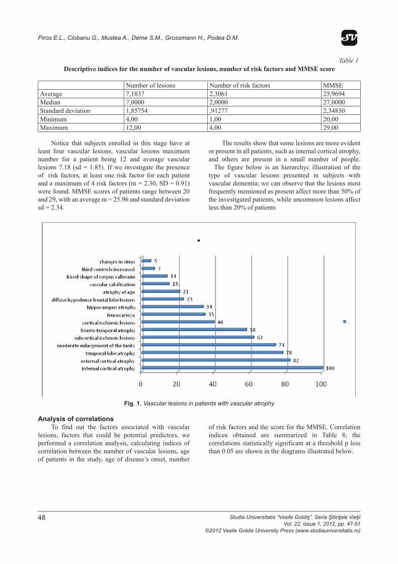
The results show that some lesions are more evident or present in all patients, such as internal cortical atrophy, and others are present in a small number of people. The figure below is an hierarchyc illustration of the type of vascular lesions presented in subjects with vascular dementia; we can observe that the lesions most frequently mentioned as present affect more than 50% of the investigated patients, while uncommon lesions affect less than 20% of patients
Piros E. Ligia., Ciobanu Gh., Mustea Anca, Deme Sanda Maria, Grossmann H., Podea Delia Marina
Studia Universitatis “Vasile Goldiş”, Seria Ştiinţele Vieţii
Table 1 Descriptive indices for the number of vascular lesions, number of risk factors and MMSE score
Number of lesions Number of risk factors MMSE Average 7,1837 2,3061 25,9694 Median 7,0000 2,0000 27,0000 Standard deviation 1,85754 ,91277 2,34830 Minimum 4,00 1,00 20,00Maximum 12,00 4,00 29,00
Notice that subjects enrolled in this stage have at least four vascular lesions, vascular lesions maximum number for a patient being 12 and average vascular lesions 7.18 (sd = 1.85). If we investigate the presence of risk factors, at least one risk factor for each patient and a maximum of 4 risk factors (m = 2.30, SD = 0.91) were found. MMSE scores of patients range between 20 and 29, with an average m = 25.96 and standard deviation sd = 2.34.
The results show that some lesions are more evident or present in all patients, such as internal cortical
atrophy, and others are present in a small number of people. The figure below is an hierarchyc illustration of the type of vascular lesions presented in subjects with vascular dementia; we can observe that the lesions most frequently mentioned as present affect more than 50% of the investigated patients, while uncommon lesions affect less than 20% of patients
Fig. 1. Vascular lesions in patients with vascular atrophy
Analysis of correlations To find out the factors associated with vascular
lesions, factors that could be potential predictors, we performed a correlation analysis, calculating indices of correlation between the number of vascular lesions, age
of patients in the study, age of disease’s onset, number of risk factors and the score for the MMSE. Correlation indices obtained are summarized in Table 8; the correlations statistically significant at a threshold p less than 0.05 are shown in the diagrams illustrated below.
Vol. 21, issue 2, 2011, pp. 157-162 © 2011 Vasile Goldiş University Press (www.studiauniversitatis.ro)
158
Fig. 1. Vascular lesions in patients with vascular atrophy
Analysis of correlationsTo find out the factors associated with vascular
lesions, factors that could be potential predictors, we performed a correlation analysis, calculating indices of correlation between the number of vascular lesions, age of patients in the study, age of disease’s onset, number
of risk factors and the score for the MMSE. Correlation indices obtained are summarized in Table 8; the correlations statistically significant at a threshold p less than 0.05 are shown in the diagrams illustrated below.
Piros E.L., Ciobanu G., Mustea A., Deme S.M., Grossmann H., Podea D.M.
Studia Universitatis “Vasile Goldiş”, Seria Ştiinţele VieţiiVol. 22, issue 1, 2012, pp. 47-51©2012 Vasile Goldis University Press (www.studiauniversitatis.ro)
49
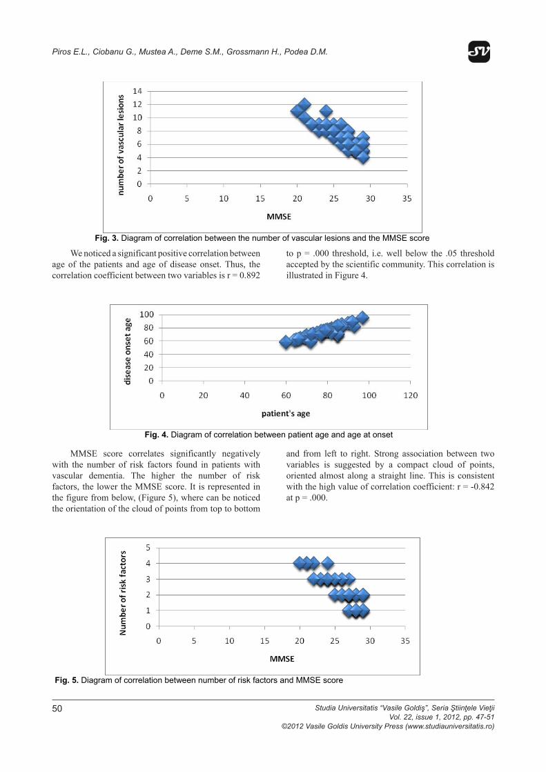
Table 2Correlations between number of vascular lesions, age, onset age, number
of risk factors and MMSE scorelesion number Age onset number of risk factors mmse
lesion number r 1 ,048 ,125 ,891** -,864**
p ,642 ,219 ,000 ,000N 100 100 100 100 100
age r ,048 1 ,892** -,046 -,078p ,642 ,000 ,654 ,444N 100 100 100 100 100
onset r ,125 ,892** 1 ,038 -,101p ,219 ,000 ,708 ,324N 100 100 100 100 100
number of risk factors
r ,891** -,046 ,038 1 -,842**
p ,000 ,654 ,708 ,000N 100 100 100 100 100
mmse r -,864** -,078 -,101 -,842** 1p ,000 ,444 ,324 ,000N 100 100 100 100 100
We can observe a statistically significant correlation between the first number of vascular lesions and number of risk factors: r = 0.891 to p = .000.
Therefore, the greater number of risk factors the greater is the number of vascular lesions.
Analysis of type of brain injury and their correlation with vascular dementia cognitive decline degree
Studia Universitatis “Vasile Goldiş”, Seria Ştiinţele Vieţii
Table 2 Correlations between number of vascular lesions, age, onset age, number
of risk factors and MMSE score lesion number Age onset number of risk factors mmse
r 1 ,048 ,125 ,891** -,864**
p ,642 ,219 ,000 ,000lesion number
N 100 100 100 100 100 r ,048 1 ,892** -,046 -,078p ,642 ,000 ,654 ,444
age
N 100 100 100 100 100 r ,125 ,892** 1 ,038 -,101p ,219 ,000 ,708 ,324
onset
N 100 100 100 100 100 r ,891** -,046 ,038 1 -,842**
p ,000 ,654 ,708 ,000number of risk factors
N 100 100 100 100 100 r -,864** -,078 -,101 -,842** 1p ,000 ,444 ,324 ,000
mmse
N 100 100 100 100 100
We can observe a statistically significant correlation between the first number of vascular lesions and number of risk factors: r = 0.891 to p = .000.
Therefore, the greater number of risk factors the greater is the number of vascular lesions.
Fig. 2. Diagram of correlation between the number of vascular lesions and number of risk factor
Positive association between two variables can be seen in the correlation diagram (Figure 2), where the cloud of points is oriented from top to bottom and from left to right.
A strong significant negative correlation (r = -0.864 at p = .000) was obtained between the number of vascular lesions and the MMSE score. Thus, the greater the number of vascular lesions, the lower is the
MMSE score obtained. This correlation is illustrated in the diagram (Figure 3) through a cloud of points oriented from top to bottom and from left to right. Notice that the cloud of points is very compact, which shows a strong correlation between MMSE score and number of vascular lesions, as seen from the Pearson correlation coefficient previously.
Vol. 21, issue 2, 2011, pp. 157-162 © 2011 Vasile Goldiş University Press (www.studiauniversitatis.ro)
159
Fig. 2. Diagram of correlation between the number of vascular lesions and number of risk factor
Positive association between two variables can be seen in the correlation diagram (Figure 2), where the cloud of points is oriented from top to bottom and from left to right.
A strong significant negative correlation (r = -0.864 at p = .000) was obtained between the number of vascular lesions and the MMSE score. Thus, the greater the
number of vascular lesions, the lower is the MMSE score obtained. This correlation is illustrated in the diagram (Figure 3) through a cloud of points oriented from top to bottom and from left to right. Notice that the cloud of points is very compact, which shows a strong correlation between MMSE score and number of vascular lesions, as seen from the Pearson correlation coefficient previously.
Analysis of type of brain injury and their correlation with vascular dementia cognitive decline degree
50 Studia Universitatis “Vasile Goldiş”, Seria Ştiinţele VieţiiVol. 22, issue 1, 2012, pp. 47-51
©2012 Vasile Goldis University Press (www.studiauniversitatis.ro)
Piros E. Ligia., Ciobanu Gh., Mustea Anca, Deme Sanda Maria, Grossmann H., Podea Delia Marina
Studia Universitatis “Vasile Goldiş”, Seria Ştiinţele Vieţii
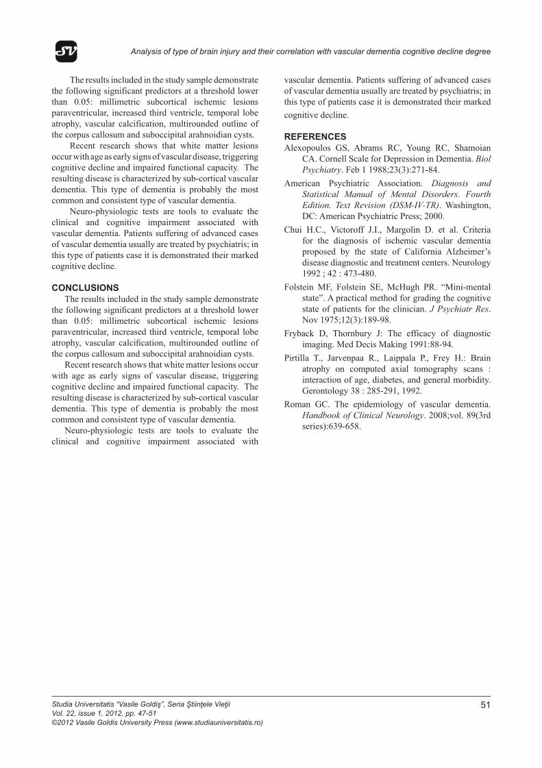
Fig. 3. Diagram of correlation between the number of vascular lesions and the MMSE score
We noticed a significant positive correlation between age of the patients and age of disease onset. Thus, the correlation coefficient between two variables is r =
0.892 to p = .000 threshold, i.e. well below the .05 threshold accepted by the scientific community. This correlation is illustrated in Figure 4.
Fig. 4. Diagram of correlation between patient age and age at onset
MMSE score correlates significantly negatively with the number of risk factors found in patients with vascular dementia. The higher the number of risk factors, the lower the MMSE score. It is represented in the figure from below, (Figure 5), where can be noticed the orientation of the cloud of points from top to
bottom and from left to right. Strong association between two variables is suggested by a compact cloud of points, oriented almost along a straight line. This is consistent with the high value of correlation coefficient: r = -0.842 at p = .000.
Fig. 5. Diagram of correlation between number of risk factors and MMSE score
Vol. 21, issue 2, 2011, pp. 157-162 © 2011 Vasile Goldiş University Press (www.studiauniversitatis.ro)
160
We noticed a significant positive correlation between age of the patients and age of disease onset. Thus, the correlation coefficient between two variables is r = 0.892
to p = .000 threshold, i.e. well below the .05 threshold accepted by the scientific community. This correlation is illustrated in Figure 4.
Piros E. Ligia., Ciobanu Gh., Mustea Anca, Deme Sanda Maria, Grossmann H., Podea Delia Marina
Studia Universitatis “Vasile Goldiş”, Seria Ştiinţele Vieţii
Fig. 3. Diagram of correlation between the number of vascular lesions and the MMSE score
We noticed a significant positive correlation between age of the patients and age of disease onset. Thus, the correlation coefficient between two variables is r =
0.892 to p = .000 threshold, i.e. well below the .05 threshold accepted by the scientific community. This correlation is illustrated in Figure 4.
Fig. 4. Diagram of correlation between patient age and age at onset
MMSE score correlates significantly negatively with the number of risk factors found in patients with vascular dementia. The higher the number of risk factors, the lower the MMSE score. It is represented in the figure from below, (Figure 5), where can be noticed the orientation of the cloud of points from top to
bottom and from left to right. Strong association between two variables is suggested by a compact cloud of points, oriented almost along a straight line. This is consistent with the high value of correlation coefficient: r = -0.842 at p = .000.
Fig. 5. Diagram of correlation between number of risk factors and MMSE score
Vol. 21, issue 2, 2011, pp. 157-162 © 2011 Vasile Goldiş University Press (www.studiauniversitatis.ro)
160
MMSE score correlates significantly negatively with the number of risk factors found in patients with vascular dementia. The higher the number of risk factors, the lower the MMSE score. It is represented in the figure from below, (Figure 5), where can be noticed the orientation of the cloud of points from top to bottom
and from left to right. Strong association between two variables is suggested by a compact cloud of points, oriented almost along a straight line. This is consistent with the high value of correlation coefficient: r = -0.842 at p = .000.
Piros E. Ligia., Ciobanu Gh., Mustea Anca, Deme Sanda Maria, Grossmann H., Podea Delia Marina
Studia Universitatis “Vasile Goldiş”, Seria Ştiinţele Vieţii
Fig. 3. Diagram of correlation between the number of vascular lesions and the MMSE score
We noticed a significant positive correlation between age of the patients and age of disease onset. Thus, the correlation coefficient between two variables is r =
0.892 to p = .000 threshold, i.e. well below the .05 threshold accepted by the scientific community. This correlation is illustrated in Figure 4.
Fig. 4. Diagram of correlation between patient age and age at onset
MMSE score correlates significantly negatively with the number of risk factors found in patients with vascular dementia. The higher the number of risk factors, the lower the MMSE score. It is represented in the figure from below, (Figure 5), where can be noticed the orientation of the cloud of points from top to
bottom and from left to right. Strong association between two variables is suggested by a compact cloud of points, oriented almost along a straight line. This is consistent with the high value of correlation coefficient: r = -0.842 at p = .000.
Fig. 5. Diagram of correlation between number of risk factors and MMSE score
Vol. 21, issue 2, 2011, pp. 157-162 © 2011 Vasile Goldiş University Press (www.studiauniversitatis.ro)
160
Piros E.L., Ciobanu G., Mustea A., Deme S.M., Grossmann H., Podea D.M.
Studia Universitatis “Vasile Goldiş”, Seria Ştiinţele VieţiiVol. 22, issue 1, 2012, pp. 47-51©2012 Vasile Goldis University Press (www.studiauniversitatis.ro)
51
The results included in the study sample demonstrate the following significant predictors at a threshold lower than 0.05: millimetric subcortical ischemic lesions paraventricular, increased third ventricle, temporal lobe atrophy, vascular calcification, multirounded outline of the corpus callosum and suboccipital arahnoidian cysts.
Recent research shows that white matter lesions occur with age as early signs of vascular disease, triggering cognitive decline and impaired functional capacity. The resulting disease is characterized by sub-cortical vascular dementia. This type of dementia is probably the most common and consistent type of vascular dementia.
Neuro-physiologic tests are tools to evaluate the clinical and cognitive impairment associated with vascular dementia. Patients suffering of advanced cases of vascular dementia usually are treated by psychiatris; in this type of patients case it is demonstrated their marked cognitive decline.
concLuSIonS The results included in the study sample demonstrate
the following significant predictors at a threshold lower than 0.05: millimetric subcortical ischemic lesions paraventricular, increased third ventricle, temporal lobe atrophy, vascular calcification, multirounded outline of the corpus callosum and suboccipital arahnoidian cysts.
Recent research shows that white matter lesions occur with age as early signs of vascular disease, triggering cognitive decline and impaired functional capacity. The resulting disease is characterized by sub-cortical vascular dementia. This type of dementia is probably the most common and consistent type of vascular dementia.
Neuro-physiologic tests are tools to evaluate the clinical and cognitive impairment associated with
vascular dementia. Patients suffering of advanced cases of vascular dementia usually are treated by psychiatris; in this type of patients case it is demonstrated their marked cognitive decline.
rEFErEncES Alexopoulos GS, Abrams RC, Young RC, Shamoian
CA. Cornell Scale for Depression in Dementia. Biol Psychiatry. Feb 1 1988;23(3):271-84.
American Psychiatric Association. Diagnosis and Statistical Manual of Mental Disorders. Fourth Edition. Text Revision (DSM-IV-TR). Washington, DC: American Psychiatric Press; 2000.
Chui H.C., Victoroff J.I., Margolin D. et al. Criteria for the diagnosis of ischemic vascular dementia proposed by the state of California Alzheimer’s disease diagnostic and treatment centers. Neurology 1992 ; 42 : 473-480.
Folstein MF, Folstein SE, McHugh PR. “Mini-mental state”. A practical method for grading the cognitive state of patients for the clinician. J Psychiatr Res. Nov 1975;12(3):189-98.
Fryback D, Thornbury J: The efficacy of diagnostic imaging. Med Decis Making 1991:88-94.
Pirtilla T., Jarvenpaa R., Laippala P., Frey H.: Brain atrophy on computed axial tomography scans : interaction of age, diabetes, and general morbidity. Gerontology 38 : 285-291, 1992.
Roman GC. The epidemiology of vascular dementia. Handbook of Clinical Neurology. 2008;vol. 89(3rd series):639-658.
Analysis of type of brain injury and their correlation with vascular dementia cognitive decline degree