Embed Size (px)
Citation preview

REVIEW ARTICLE
Analysis of the Load on the Knee Joint and Vertebral Columnwith Changes in Squatting Depth and Weight Load
Hagen Hartmann • Klaus Wirth • Markus Klusemann
Published online: 3 July 2013
� Springer International Publishing Switzerland 2013
Abstract It has been suggested that deep squats could
cause an increased injury risk of the lumbar spine and the
knee joints. Avoiding deep flexion has been recommended
to minimize the magnitude of knee-joint forces. Unfortu-
nately this suggestion has not taken the influence of the
wrapping effect, functional adaptations and soft tissue
contact between the back of thigh and calf into account.
The aim of this literature review is to assess whether squats
with less knee flexion (half/quarter squats) are safer on the
musculoskeletal system than deep squats. A search of rel-
evant scientific publications was conducted between March
2011 and January 2013 using PubMed. Over 164 articles
were included in the review. There are no realistic esti-
mations of knee-joint forces for knee-flexion angles beyond
50� in the deep squat. Based on biomechanical calculations
and measurements of cadaver knee joints, the highest ret-
ropatellar compressive forces and stresses can be seen at
90�. With increasing flexion, the wrapping effect contrib-
utes to an enhanced load distribution and enhanced force
transfer with lower retropatellar compressive forces.
Additionally, with further flexion of the knee joint a cranial
displacement of facet contact areas with continuous
enlargement of the retropatellar articulating surface occurs.
Both lead to lower retropatellar compressive stresses.
Menisci and cartilage, ligaments and bones are susceptible
to anabolic metabolic processes and functional structural
adaptations in response to increased activity and mechan-
ical influences. Concerns about degenerative changes of the
tendofemoral complex and the apparent higher risk for
chondromalacia, osteoarthritis, and osteochondritis in deep
squats are unfounded. With the same load configuration as
in the deep squat, half and quarter squat training with
comparatively supra-maximal loads will favour degenera-
tive changes in the knee joints and spinal joints in the long
term. Provided that technique is learned accurately under
expert supervision and with progressive training loads, the
deep squat presents an effective training exercise for pro-
tection against injuries and strengthening of the lower
extremity. Contrary to commonly voiced concern, deep
squats do not contribute increased risk of injury to passive
tissues.
1 Introduction
The primary objective of performance-oriented strength
training is the gain of muscle cross-sectional area (hyper-
trophy strength training) or the enhancement of inter- and
intramuscular coordination (strength/power training), for
which high training intensities are necessary [1]. For this
purpose, the squat is one of the most effective exercises in
athletic training. The Olympic barbell squat can be clas-
sified into three fundamental variations: the front squat [2],
the high-bar back squat, and the low-bar back squat [2, 3]
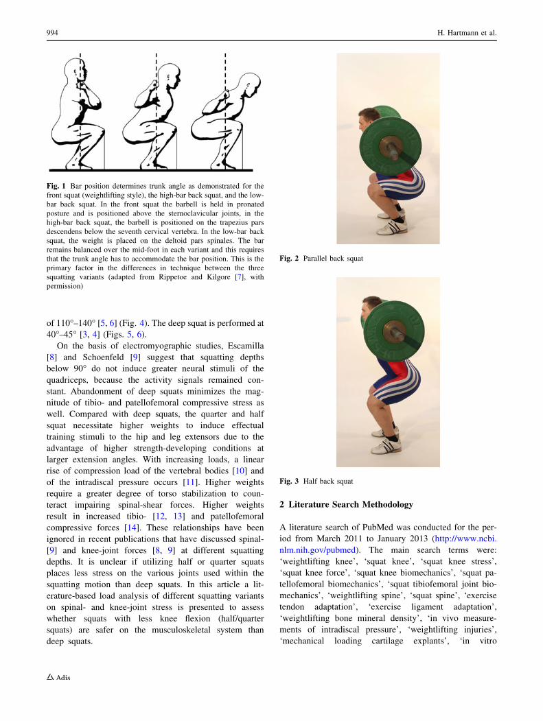
(Fig. 1). In the parallel squat, the knees are flexed until the
inguinal fold is in a straight horizontal line with the top of
the knee musculature [2] (Fig. 2). Depending on the squat
variant, the knee angles vary between 60� and 70� in this
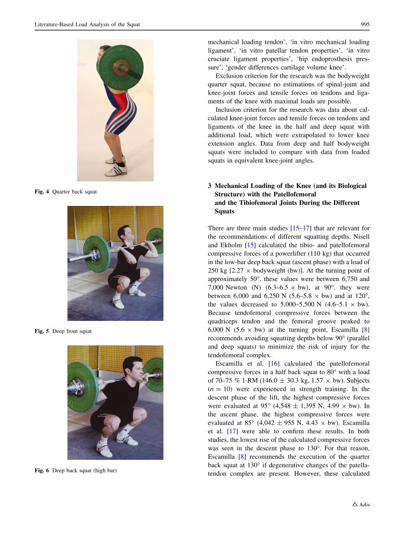
position [2, 3]. The half squat is performed at 80�–100� [4,
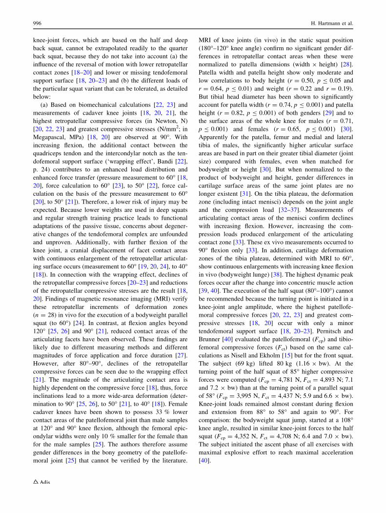
5] (Fig. 3), and the quarter squat is executed at a knee angle
H. Hartmann (&) � K. Wirth
Department of Human Movement Science and Athletic Training,
Institute of Sports Sciences, Goethe-University, Ginnheimer
Landstr. 39, 60487 Frankfurt am Main, Germany
e-mail: [email protected]
M. Klusemann
Physiology Department, Australian Institute of Sport (AIS),
Charles Sturt University, Leverrier Crescent, Bruce,
ACT 2617, Australia
Sports Med (2013) 43:993–1008
DOI 10.1007/s40279-013-0073-6
of 110�–140� [5, 6] (Fig. 4). The deep squat is performed at
40�–45� [3, 4] (Figs. 5, 6).
On the basis of electromyographic studies, Escamilla
[8] and Schoenfeld [9] suggest that squatting depths
below 90� do not induce greater neural stimuli of the
quadriceps, because the activity signals remained con-
stant. Abandonment of deep squats minimizes the mag-
nitude of tibio- and patellofemoral compressive stress as
well. Compared with deep squats, the quarter and half
squat necessitate higher weights to induce effectual
training stimuli to the hip and leg extensors due to the
advantage of higher strength-developing conditions at
larger extension angles. With increasing loads, a linear
rise of compression load of the vertebral bodies [10] and
of the intradiscal pressure occurs [11]. Higher weights
require a greater degree of torso stabilization to coun-
teract impairing spinal-shear forces. Higher weights
result in increased tibio- [12, 13] and patellofemoral
compressive forces [14]. These relationships have been
ignored in recent publications that have discussed spinal-
[9] and knee-joint forces [8, 9] at different squatting
depths. It is unclear if utilizing half or quarter squats
places less stress on the various joints used within the
squatting motion than deep squats. In this article a lit-
erature-based load analysis of different squatting variants
on spinal- and knee-joint stress is presented to assess
whether squats with less knee flexion (half/quarter
squats) are safer on the musculoskeletal system than
deep squats.
2 Literature Search Methodology
A literature search of PubMed was conducted for the per-
iod from March 2011 to January 2013 (http://www.ncbi.
nlm.nih.gov/pubmed). The main search terms were:
‘weightlifting knee’, ‘squat knee’, ‘squat knee stress’,
‘squat knee force’, ‘squat knee biomechanics’, ‘squat pa-
tellofemoral biomechanics’, ‘squat tibiofemoral joint bio-
mechanics’, ‘weightlifting spine’, ‘squat spine’, ‘exercise
tendon adaptation’, ‘exercise ligament adaptation’,
‘weightlifting bone mineral density’, ‘in vivo measure-
ments of intradiscal pressure’, ‘weightlifting injuries’,
‘mechanical loading cartilage explants’, ‘in vitro
Fig. 1 Bar position determines trunk angle as demonstrated for the
front squat (weightlifting style), the high-bar back squat, and the low-
bar back squat. In the front squat the barbell is held in pronated
posture and is positioned above the sternoclavicular joints, in the
high-bar back squat, the barbell is positioned on the trapezius pars
descendens below the seventh cervical vertebra. In the low-bar back
squat, the weight is placed on the deltoid pars spinales. The bar
remains balanced over the mid-foot in each variant and this requires
that the trunk angle has to accommodate the bar position. This is the
primary factor in the differences in technique between the three
squatting variants (adapted from Rippetoe and Kilgore [7], with
permission)
Fig. 2 Parallel back squat
Fig. 3 Half back squat
994 H. Hartmann et al.
mechanical loading tendon’, ‘in vitro mechanical loading
ligament’, ‘in vitro patellar tendon properties’, ‘in vitro
cruciate ligament properties’, ‘hip endoprosthesis pres-
sure’, ‘gender differences cartilage volume knee’.
Exclusion criterion for the research was the bodyweight
quarter squat, because no estimations of spinal-joint and
knee-joint forces and tensile forces on tendons and liga-
ments of the knee with maximal loads are possible.
Inclusion criterion for the research was data about cal-
culated knee-joint forces and tensile forces on tendons and
ligaments of the knee in the half and deep squat with
additional load, which were extrapolated to lower knee
extension angles. Data from deep and half bodyweight
squats were included to compare with data from loaded
squats in equivalent knee-joint angles.
3 Mechanical Loading of the Knee (and its Biological
Structure) with the Patellofemoral
and the Tibiofemoral Joints During the Different
Squats
There are three main studies [15–17] that are relevant for
the recommendations of different squatting depths. Nisell
and Ekholm [15] calculated the tibio- and patellofemoral
compressive forces of a powerlifter (110 kg) that occurred
in the low-bar deep back squat (ascent phase) with a load of
250 kg [2.27 9 bodyweight (bw)]. At the turning point of
approximately 50�, these values were between 6,750 and
7,000 Newton (N) (6.3–6.5 9 bw), at 90�, they were
between 6,000 and 6,250 N (5.6–5.8 9 bw) and at 120�,
the values decreased to 5,000–5,500 N (4.6–5.1 9 bw).
Because tendofemoral compressive forces between the
quadriceps tendon and the femoral groove peaked to
6,000 N (5.6 9 bw) at the turning point, Escamilla [8]
recommends avoiding squatting depths below 90� (parallel
and deep squats) to minimize the risk of injury for the
tendofemoral complex.
Escamilla et al. [16] calculated the patellofemoral
compressive forces in a half back squat to 80� with a load
of 70–75 % 1-RM (146.0 ± 30.3 kg, 1.57 9 bw). Subjects
(n = 10) were experienced in strength training. In the
descent phase of the lift, the highest compressive forces
were evaluated at 95� (4,548 ± 1,395 N, 4.99 9 bw). In
the ascent phase, the highest compressive forces were
evaluated at 85� (4,042 ± 955 N, 4.43 9 bw). Escamilla
et al. [17] were able to confirm these results. In both
studies, the lowest rise of the calculated compressive forces
was seen in the descent phase to 130�. For that reason,
Escamilla [8] recommends the execution of the quarter
back squat at 130� if degenerative changes of the patella-
tendon complex are present. However, these calculated
Fig. 4 Quarter back squat
Fig. 5 Deep front squat
Fig. 6 Deep back squat (high bar)
Literature-Based Load Analysis of the Squat 995
knee-joint forces, which are based on the half and deep
back squat, cannot be extrapolated readily to the quarter
back squat, because they do not take into account (a) the
influence of the reversal of motion with lower retropatellar
contact zones [18–20] and lower or missing tendofemoral
support surface [18, 20–23] and (b) the different loads of
the particular squat variant that can be tolerated, as detailed
below:
(a) Based on biomechanical calculations [22, 23] and
measurements of cadaver knee joints [18, 20, 21], the
highest retropatellar compressive forces (in Newton, N)
[20, 22, 23] and greatest compressive stresses (N/mm2; in
Megapascal, MPa) [18, 20] are observed at 90�. With
increasing flexion, the additional contact between the
quadriceps tendon and the intercondylar notch as the ten-
dofemoral support surface (‘wrapping effect’, Bandi [22],
p. 24) contributes to an enhanced load distribution and
enhanced force transfer (pressure measurement to 60� [18,
20], force calculation to 60� [23], to 50� [22], force cal-
culation on the basis of the pressure measurement to 60�[20], to 50� [21]). Therefore, a lower risk of injury may be
expected. Because lower weights are used in deep squats
and regular strength training practice leads to functional
adaptations of the passive tissue, concerns about degener-
ative changes of the tendofemoral complex are unfounded
and unproven. Additionally, with further flexion of the
knee joint, a cranial displacement of facet contact areas
with continuous enlargement of the retropatellar articulat-
ing surface occurs (measurement to 60� [19, 20, 24], to 40�[18]). In connection with the wrapping effect, declines of
the retropatellar compressive forces [20–23] and reductions
of the retropatellar compressive stresses are the result [18,
20]. Findings of magnetic resonance imaging (MRI) verify
these retropatellar increments of deformation zones
(n = 28) in vivo for the execution of a bodyweight parallel
squat (to 60�) [24]. In contrast, at flexion angles beyond
120� [25, 26] and 90� [21], reduced contact areas of the
articulating facets have been observed. These findings are
likely due to different measuring methods and different
magnitudes of force application and force duration [27].
However, after 80�–90�, declines of the retropatellar
compressive forces can be seen due to the wrapping effect
[21]. The magnitude of the articulating contact area is
highly dependent on the compressive force [18], thus, force
inclinations lead to a more wide-area deformation (deter-
mination to 90� [25, 26], to 50� [21], to 40� [18]). Female
cadaver knees have been shown to possess 33 % lower
contact areas of the patellofemoral joint than male samples
at 120� and 90� knee flexion, although the femoral epic-
ondylar widths were only 10 % smaller for the female than
for the male samples [25]. The authors therefore assume
gender differences in the bony geometry of the patellofe-
moral joint [25] that cannot be verified by the literature.
MRI of knee joints (in vivo) in the static squat position
(180�–120� knee angle) confirm no significant gender dif-
ferences in retropatellar contact areas when these were
normalized to patella dimensions (width 9 height) [28].
Patella width and patella height show only moderate and
low correlations to body height (r = 0.50, p B 0.05 and
r = 0.64, p B 0.01) and weight (r = 0.22 and r = 0.19).
But tibial head diameter has been shown to significantly
account for patella width (r = 0.74, p B 0.001) and patella
height (r = 0.82, p B 0.001) of both genders [29] and to
the surface areas of the whole knee for males (r = 0.71,
p B 0.001) and females (r = 0.65, p B 0.001) [30].
Apparently for the patella, femur and medial and lateral
tibia of males, the significantly higher articular surface
areas are based in part on their greater tibial diameter (joint
size) compared with females, even when matched for
bodyweight or height [30]. But when normalized to the
product of bodyweight and height, gender differences in
cartilage surface areas of the same joint plates are no
longer existent [31]. On the tibia plateau, the deformation
zone (including intact menisci) depends on the joint angle
and the compression load [32–37]. Measurements of
articulating contact areas of the menisci confirm declines
with increasing flexion. However, increasing the com-
pression loads produced enlargement of the articulating
contact zone [33]. These ex vivo measurements occurred to
90� flexion only [33]. In addition, cartilage deformation
zones of the tibia plateau, determined with MRI to 60�,
show continuous enlargements with increasing knee flexion
in vivo (bodyweight lunge) [38]. The highest dynamic peak
forces occur after the change into concentric muscle action
[39, 40]. The execution of the half squat (80�–100�) cannot
be recommended because the turning point is initiated in a
knee-joint angle amplitude, where the highest patellofe-
moral compressive forces [20, 22, 23] and greatest com-
pressive stresses [18, 20] occur with only a minor
tendofemoral support surface [18, 20–23]. Pernitsch and
Brunner [40] evaluated the patellofemoral (Fcp) and tibio-
femoral compressive forces (Fct) based on the same cal-
culations as Nisell and Ekholm [15] but for the front squat.
The subject (69 kg) lifted 80 kg (1.16 9 bw). At the
turning point of the half squat of 85� higher compressive
forces were computed (Fcp = 4,781 N, Fct = 4,893 N; 7.1
and 7.2 9 bw) than at the turning point of a parallel squat
of 58� (Fcp = 3,995 N, Fct = 4,437 N; 5.9 and 6.6 9 bw).
Knee-joint loads remained almost constant during flexion
and extension from 88� to 58� and again to 90�. For
comparison: the bodyweight squat jump, started at a 108�knee angle, resulted in similar knee-joint forces to the half
squat (Fcp = 4,352 N, Fct = 4,708 N; 6.4 and 7.0 9 bw).
The subject initiated the ascent phase of all exercises with
maximal explosive effort to reach maximal acceleration
[40].
996 H. Hartmann et al.

(b) With decreased knee flexion in the squat, the weight
loads that can be lifted increase. The influence of
increasing weight loads have not been considered in joint-
force estimations of different squatting depths. For the
quarter back squat (120� knee angle) physical education
students were shown to be capable of lifting 3.89-fold
(±0.33) their bodyweight [41]. For professional soccer
players, Hoff and Helgerud [42] recommend values of
2.75-fold of bodyweight in the half back squat. Higher
weights result in increased tibio- [12, 13] and patellofe-
moral compressive forces [14]. These data indicate that the
force values for half and deep back squats calculated by
Escamilla et al. [16] and Nisell and Ekholm [15], which
were intended to be extrapolated to lower extension angles
up to 120�–130�, are too low. For the quarter back squat,
physical education students were shown to be capable of
moving weights of 4.02-fold (±1.59) the weight of a deep
back squat and 4.38-fold (±1.02) the weight of a deep front
squat, although the majority of the subjects had minimal
strength-training experience [41]. Well trained athletes are
capable of lifting much greater weights. Based on the
weight of the deep back squat (250 kg, 2.27 9 bw) that
was moved by the powerlifter described in Nisell and
Ekholm [15], this weight would correspond to an astro-
nomical 1005 kg. This weight is out of question for train-
ing practice, because such a high barbell load is unlikely to
be stabilized by the back musculature. The back, not the
legs, would be performance-limiting, thus providing no
training effect for the legs.
McKean et al. [43] determined the movement pattern
with regards to the timing when maximum angles of the hip
and knee occurred while performing the parallel back
squat. Sixteen men and twelve women executed the squat
with no additional weight or with 0.5-fold bodyweight.
Independent of gender, phase (ascent or descent) and load,
the subjects demonstrated their maximum hip and knee
angles within 2 % of the deepest position whereby subjects
had to move their knees forward of the toes by 6.4–6.5 cm
in men and 9.3–9.7 cm in women. The difference in the
forward movement of the knees between genders could be
explained by the greater body height of the men (167 vs.
179 cm). The subject’s height and tibial length has been
shown to account for 69 % of the explained variances in
male subjects being capable of keeping their heels on the
ground while performing the parallel back squat [44].
These anthropometric factors have an influence on the
torso inclination and hence the forward movement of the
knees [43]. In training and practice of strength training it is
sometimes suggested that the tibia should move anteriorly
only to the point where the knee joints and the toes form a
vertical line to minimize the tibiofemoral shear forces. This
recommendation is based on video analyses of parallel
back squats (mean weight 201.85 kg, 2.23 9 bw) of 12
male weightlifters. Data of only three subjects are pre-
sented. The subject with the greatest forward movement of
the knees while performing the squat had the greatest tib-
iofemoral shear forces [45]. The restriction of the forward
knee displacement will result in changes to the knee-hip
coordination [43] with greater forward leaning [46] and
ventral flexion of the thoracic and lumbar spine [47]. This
evasive movement elicits greater anterior shear forces on
intervertebral discs [48] and causes tensile forces on
intervertebral ligaments [48, 49]. For that reason, instruc-
tions about a restriction of the forward knee displacement
have to be strictly avoided. This recommendation is based
on a misinterpretation of existing data and should be
removed in future practical literature. It is not clear from
the data of Ariel [45] whether the calculated shear forces
acted in an anterior or posterior direction. The subject with
the greatest anterior knee movement, who showed the
highest shear forces, squatted to a knee angle of 90� only.
The remaining subjects squatted to 61� and 69� knee angles
and possessed lower shear forces. The literature provides
evidence for these calculations: When initiating the turning
point at 80�–90�, higher (relative) posterior shear forces are
the result [16, 17, 50] compared with the deep squat at 50�[15] (chapter 5). Additionally, the calculations described
by Ariel [45] do not provide any information about the
squatting technique of the subjects. The high shear forces
could result from a very upright posture, which can be
demonstrated by the following calculations of the com-
pressive forces in the patellofemoral joint. For the parallel
back squat, Wretenberg et al. [3] evaluated higher mean
patellofemoral compressive forces for eight weightlifters
than for six powerlifters (4,700 ± 290 vs. 3,300 ± 1,700
N, 5.84 9 bw vs. 3.87 9 bw), despite the weightlifters
lifting lower weights than the powerlifters (101.9 ± 27.7
vs. 154.2 ± 21.1 kg, 1.24 9 bw vs. 1.85 9 bw). Consid-
ering the biomechanical calculations of Nisell and Ekholm
[23], this result was due to the higher torque values in the
knee joint, which were developed via a more upright
posture (high- vs. low-bar position of the barbell) [3], see
Fig. 1.
There are no calculations for knee flexion angles beyond
50� in the deep squat that permit an accurate estimation of
knee-joint forces because these studies did not take the
influence of the wrapping effect [51] or the influence of
soft tissue contact between the back of the thigh and the
calf into account [51–53]. Evaluations of Reilly and Mar-
tens [51] and Dahlkvist et al. [52] showed patellofemoral
compressive forces (7.6 9 bw and 7.62 9 bw) in the
turning point of the bodyweight deep squat (40�) that
exceeded those of Nisell and Ekholm [15] (6.3 9 bw),
Pernitsch and Brunner [40] (5.9 9 bw) and Wretenberg
et al. [3] (weightlifter 5.84 9 bw, powerlifter 3.87 9 bw).
It is surprising that despite only using bodyweight loading,
Literature-Based Load Analysis of the Squat 997
Reilly and Martens [51] and Dahlkvist et al. [52] measured
higher patellofemoral compressive forces than in the
studies of Nisell and Ekholm [15] and Wretenberg et al. [3]
using up to 70 % 1-RM loads in the deep (50�) and parallel
squat (64�–69�). The subjects described by Dahlkvist et al.
[52] experienced difficulties in maintaining their balance
while performing the squat. However, an uncontrolled
execution leads to distinctly higher knee-joint forces [45].
The same contradictions between the studies of Dahlkvist
et al. [52] and Nisell and Ekholm [15] exist for the cal-
culated posterior shear forces.
The influence of soft tissue contact between the back of
the thigh and the calf plays a prominent role in reducing the
knee-joint forces beyond 40� of knee extension [54–56].
The calculation of knee-joint forces, which did not take
into account the soft tissue contact in deep flexion angles
(25�) [53], must be examined critically. This soft tissue
contact depends upon the cross-sectional area of the ham-
string and the calf muscles and can begin at higher degrees
of extension from approximately 60� [57], thereby reduc-
ing tibiofemoral [54–56] and patellofemoral joint forces
[54, 55]. The structures of the knee (such as meniscal and
cartilage tissue and ligaments) that benefit from soft-tissue
contact remain unclear. The apparent higher risk for
degenerative changes, such as osteochondrosis dissecans of
the odd facet in deep squats [8], seems unfounded and is
unproven. Recommendations for half or quarter squats to
avoid degenerative changes in the knee joint may be
counterproductive. If the cartilage tissue of the odd facet is
inadequately stressed then it receives insufficient nourish-
ment that leads to consequent degeneration and atrophy
[58]. This result is confirmed by animal studies in which
cartilage tissue was exposed to hypopression by unweigh-
ting the extremities [59, 60].
3.1 Adaptation Effects and Damage of Meniscal
and Cartilage Tissue of the Knee Joint
The training of weightlifters confronts the athlete with deep
front/back squats and exercises in deep knee bends with
acceleration and deceleration of high barbell loads. With
ten training sessions per week at the international level
[61], it could be suggested that these athletes are predis-
posed to a high prevalence of acute and chronic knee
injuries with long training interruptions. However, from 27
weightlifters from different US Olympic training centres,
Calhoon et al. [62] determined 3.3 injuries per 1000
training hours over a 6-year period. Of missed training time
due to knee problems, 95.3 % lasted 1 day or less, and in
the remaining cases, lasted less than 1 week. These training
interruptions were primarily related to tendinitis due to
chronic overuse and in very few cases to muscle strains due
to acute injuries [62]. A 4-year retrospective study of 1,109
weightlifters (age 12–20 years), who participated in
national or international competitions, did not show any
injuries (i.e. at the epiphyseal joints) that required surgery
or hospitalization [63]. An examination of questionnaires
completed by weightlifters aged 13–16 years (n = 1,634)
and based on 168,551 hours of training, indicated that the
injury rate of American weightlifters is 0.0017 per
100 hours of training. This prevalence is much lower than
the injury rate in US basketball players (0.03), US track-
and-field athletes (0.57), American football players (0.10)
and US gymnasts (0.044) [64]. According to a question-
naire conducted by 80 weightlifters, knee problems were
not common in the deep squat. There have been no me-
niscectomies among the lifters. No knee clicks or pops
were reported. Acute knee injuries like sprains usually
occurred in the catch phase in the deep-squat position of
the clean and jerk with high loads and not in the deep-squat
exercise [65].
According to the findings of Pernitsch and Brunner [40],
for knee joint forces in the range of 60�–110� the accel-
eration achieved determines the compressive forces more
than the weight load. The higher the lowering speed in the
descent phase, the higher the developing deceleration phase
to avoid a ‘dipping movement’ and hence a rising increase
of tibiofemoral shear and compressive forces in the turning
point of the squat [45]. For that reason, care should be
taken to complete a slow and controlled execution [45]
comprising a descent phase of 3 and 4 seconds in the deep
squat corresponding to an average angular velocity in the
knee joint of 46.66 and 35�/sec, respectively. For com-
parison, international-level weightlifters produced a
10-fold and 13-fold higher average angular velocity in the
knee joint of 465.67�/sec in the snatch (1-RM) in the
descent movement under the barbell (descent phase
238 msec) [66]. At the turning point (knee angle 17.17�)
[66], this will cause increases of the knee-joint forces
accordingly. The following calculations of the clean and
jerk with a weight of 120 kg are indicative of this point.
For an elite weightlifter (71 kg) Collins [57] calculated the
tibiofemoral compressive forces at the turning point (knee
angle 35�–40�), which accounted for maximal values of
24-fold the bodyweight of the athlete. However, cross-
sectional findings, determined using MRI, verify that knee
joints of professional weightlifters (n = 7) possess signif-
icantly (p \ 0.01) higher cartilage thickness for the same
patellar contact area of 14 %, in average, compared with
controls (n = 14) who are not strength-trained [67]. When
loaded, an increased retropatellar cartilage thickness shows
an increased stiffness that corresponds to an increased
mechanical stress tolerance [68]. Although neither contact
area nor cartilage thickness of the tibia plateau and the
femur condyles demonstrated significant differences [67],
in vivo evidence from cross-sectional [69] and longitudinal
998 H. Hartmann et al.
studies [70, 71] with humans show that increased activities
can lead to anabolic biochemical and structural adaptations
of the cartilage tissue, causing increased mechanical stress
tolerance and hence protective effects against degenerative
changes [70, 72, 73]. There were no tibiofemoral or pa-
tellofemoral cartilage defects in the weightlifters [67].
These athletes had begun their weightlifting training at
between 7 and 13 years and continued until the point of
data evaluation [67]. For active weightlifters at a national
and international level and a mean training experience of
17 years (n = 13, mean age 35.3 years), the prevalence of
degenerative cartilage changes of the patellofemoral and
tibiofemoral joints (grade 2–4) is not higher than in men of
the general population and of similar age (n = 162) [74].
Laboratory studies with animal [75, 76] and human
preparations [77], and animal experiments (in vivo) [78],
confirm that the menisci are responsive to anabolic meta-
bolic processes and functional structural adaptations that
are induced by dynamic loading [75–77] and increased
activity [78]. Evaluations of former professional weight-
lifters (n = 29, mean age 59.3 years) provide evidence for
this adaptation potential in humans: when compared with
former professional soccer players (n = 31, mean age
56.5 years), patellofemoral arthrosis dominated for the
weightlifters. For the soccer players, tibiofemoral arthrosis
was dominant. The prevalence of gonarthrosis in the
weightlifters (31 %) did not differ from the soccer players
(29 %), but both cohorts demonstrated a higher percentage
than former professional long-distance runners (14 %)
(n = 28, mean age 59.7 years) [79]. However, the misdi-
agnosis of functional and degenerative cartilage changes
must also be noted [80].
Based on the facts presented, it can be assumed that
many years of strength training in the full range of motion
and using the correct movement pattern results in functional
adaptations of the articulating cartilage and meniscal tissue.
4 In Vitro Pressure Thresholds of Cartilage
and Meniscal Tissue and Their Application
to In Vivo Conditions
It has been suggested that the deep squat with high loads
may exceed the compression threshold of articular cartilage
of the knee joint [81]. Pernitsch and Brunner [40] calcu-
lated at a knee-joint angle of 66� in the bodyweight squat
jump (starting angle 60�) tibiofemoral compressive forces
of 5.1-fold bodyweight (3,449 N). In cadaver knees, the
same relative force value at 60� knee angle resulted in a
measured tibiofemoral compression value of 26.6 MPa
[81]. The methodological procedure has to be critically
examined. Ex vivo conditions cannot guarantee the natural
joint movement, which is subject to muscular support
in vivo. This can lead to unnatural local peak forces.
Indenter [82] and impactor compression [83] elicited
chondrocyte death in the cell matrix at 15–20 MPa of
bovine cartilage explants [82] and at 20–30 MPa of human
bone cartilage explants [83]. Repetitive indenter compres-
sion of bovine bone cartilage explants already caused ini-
tial chondrocyte death at over 6 MPa due to summed
microscopic trauma [84]. Joint loading of hip endopros-
thesis of living persons has been shown to result in
8.9 MPa when rising from a chair [85]. This means that
(repetitive) rising from a chair or jumping from the parallel
squat position would already exceed the compression
threshold of hip and tibiofemoral articular cartilage. The
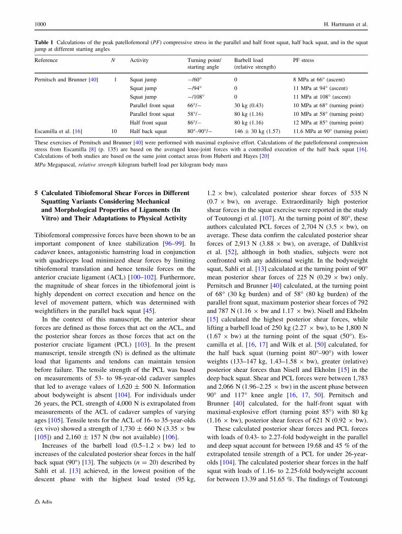
influence of the acceleration on the patellofemoral stress
can be seen when comparing the calculations of the com-
pressive stress in the bodyweight squat jump (94�) and in
the loaded front squat with 80 kg (86�), eliciting compa-
rable compressive stress (Table 1). However, the
mechanical properties and composition vary between car-
tilage tissue of articular joint areas (i.e. femoral-trochlear
vs. patellar) [86]. Furthermore, the composition, structure,
relative thickness [87] and mechanical properties [88, 89]
of cartilage tissues show species-specific differences [87–
89]. Whether the experimentally based threshold values of
in vitro studies, which were determined with indenter [82,
84] or impactor devices [83], can be extrapolated to the
in vivo (human) condition must therefore be critically
examined. In articular cartilage of intact joints, the load is
transmitted by hydrostatic compression and not by elastic
deformation [90]. When two opposing articulating cartilage
explants of bovine knee joints were compressed in a
compression cylinder, there was no cellular trauma even at
a hydrostatic pressure of 50 MPa compared with the pure
impactor compression that already caused cell death at
10 MPa [91]. These factors clarify the issue of making
general statements on compression threshold values of
articular cartilage.
To the authors’ knowledge, there are only two studies
that provide information on compression test-induced cel-
lular traumata of meniscal explants. Findings of animal
experiments point to comparable mechanical tolerance
thresholds of bovine medial [92, 93] and lateral [93]
meniscal explants compared with cartilage tissue (in vitro).
In addition to cartilage tissue, the mechanical properties of
meniscal tissue show species-specific differences [94, 95].
Joshi et al. [94] and Sweigart et al. [95] did not confirm any
statistically significant differences in the compression
behaviour of bovine and human medial meniscus explants.
However, Sweigart et al. [95] revealed significant differ-
ences in the permeability between both species. The extent
to which the findings of Nishimuta and Levenston [93] and
Kisiday et al. [92] can be extrapolated to humans must be
investigated.
Literature-Based Load Analysis of the Squat 999
5 Calculated Tibiofemoral Shear Forces in Different
Squatting Variants Considering Mechanical
and Morphological Properties of Ligaments (In
Vitro) and Their Adaptations to Physical Activity
Tibiofemoral compressive forces have been shown to be an
important component of knee stabilization [96–99]. In
cadaver knees, antagonistic hamstring load in conjunction
with quadriceps load minimized shear forces by limiting
tibiofemoral translation and hence tensile forces on the
anterior cruciate ligament (ACL) [100–102]. Furthermore,
the magnitude of shear forces in the tibiofemoral joint is
highly dependent on correct execution and hence on the
level of movement pattern, which was determined with
weightlifters in the parallel back squat [45].
In the context of this manuscript, the anterior shear
forces are defined as those forces that act on the ACL, and
the posterior shear forces as those forces that act on the
posterior cruciate ligament (PCL) [103]. In the present
manuscript, tensile strength (N) is defined as the ultimate
load that ligaments and tendons can maintain tension
before failure. The tensile strength of the PCL was based
on measurements of 53- to 98-year-old cadaver samples
that led to average values of 1,620 ± 500 N. Information
about bodyweight is absent [104]. For individuals under
26 years, the PCL strength of 4,000 N is extrapolated from
measurements of the ACL of cadaver samples of varying
ages [105]. Tensile tests for the ACL of 16- to 35-year-olds
(ex vivo) showed a strength of 1,730 ± 660 N (3.35 9 bw
[105]) and 2,160 ± 157 N (bw not available) [106].
Increases of the barbell load (0.5–1.2 9 bw) led to
increases of the calculated posterior shear forces in the half
back squat (90�) [13]. The subjects (n = 20) described by
Sahli et al. [13] achieved, in the lowest position of the
descent phase with the highest load tested (95 kg,
1.2 9 bw), calculated posterior shear forces of 535 N
(0.7 9 bw), on average. Extraordinarily high posterior
shear forces in the squat exercise were reported in the study
of Toutoungi et al. [107]. At the turning point of 80�, these
authors calculated PCL forces of 2,704 N (3.5 9 bw), on
average. These data confirm the calculated posterior shear
forces of 2,913 N (3.88 9 bw), on average, of Dahlkvist
et al. [52], although in both studies, subjects were not
confronted with any additional weight. In the bodyweight
squat, Sahli et al. [13] calculated at the turning point of 90�mean posterior shear forces of 225 N (0.29 9 bw) only.
Pernitsch and Brunner [40] calculated, at the turning point
of 68� (30 kg burden) and of 58� (80 kg burden) of the
parallel front squat, maximum posterior shear forces of 792
and 787 N (1.16 9 bw and 1.17 9 bw). Nisell and Ekholm
[15] calculated the highest posterior shear forces, while
lifting a barbell load of 250 kg (2.27 9 bw), to be 1,800 N
(1.67 9 bw) at the turning point of the squat (50�). Es-
camilla et al. [16, 17] and Wilk et al. [50] calculated, for
the half back squat (turning point 80�–90�) with lower
weights (133–147 kg, 1.43–1.58 9 bw), greater (relative)
posterior shear forces than Nisell and Ekholm [15] in the
deep back squat. Shear and PCL forces were between 1,783
and 2,066 N (1.96–2.25 9 bw) in the ascent phase between
90� and 117� knee angle [16, 17, 50]. Pernitsch and
Brunner [40] calculated, for the half-front squat with
maximal-explosive effort (turning point 85�) with 80 kg
(1.16 9 bw), posterior shear forces of 621 N (0.92 9 bw).
These calculated posterior shear forces and PCL forces
with loads of 0.43- to 2.27-fold bodyweight in the parallel
and deep squat account for between 19.68 and 45 % of the
extrapolated tensile strength of a PCL for under 26-year-
olds [104]. The calculated posterior shear forces in the half
squat with loads of 1.16- to 2.25-fold bodyweight account
for between 13.39 and 51.65 %. The findings of Toutoungi
Table 1 Calculations of the peak patellofemoral (PF) compressive stress in the parallel and half front squat, half back squat, and in the squat
jump at different starting angles
Reference N Activity Turning point/
starting angle
Barbell load
(relative strength)
PF stress
Pernitsch and Brunner [40] 1 Squat jump -/60� 0 8 MPa at 66� (ascent)
Squat jump -/94� 0 11 MPa at 94� (ascent)
Squat jump -/108� 0 11 MPa at 108� (ascent)
Parallel front squat 66�/- 30 kg (0.43) 10 MPa at 68� (turning point)
Parallel front squat 58�/- 80 kg (1.16) 10 MPa at 58� (turning point)
Half front squat 86�/- 80 kg (1.16) 12 MPa at 85� (turning point)
Escamilla et al. [16] 10 Half back squat 80�–90�/- 146 ± 30 kg (1.57) 11.6 MPa at 90� (turning point)
These exercises of Pernitsch and Brunner [40] were performed with maximal explosive effort. Calculations of the patellofemoral compression
stress from Escamilla [8] (p. 135) are based on the averaged knee-joint forces with a controlled execution of the half back squat [16].
Calculations of both studies are based on the same joint contact areas from Huberti and Hayes [20]
MPa Megapascal, relative strength kilogram barbell load per kilogram body mass
1000 H. Hartmann et al.
et al. [107] and Dahlkvist et al. [52] reach estimated values
of 67.6 and 72.83 %. These values have to be critically
examined. The calculated posterior shear forces of the
bodyweight squat (90�) of Sahli et al. [13] account for
5.63 % of the extrapolated tensile strength of the PCL only.
While Escamilla et al. [16, 17] and Wilk et al. [50] did
not calculate any anterior shear forces for the half back
squat, Toutoungi et al. [107] calculated a very low ACL
force of 28 N (0.03 9 bw), on average, in the ascent phase
of the bodyweight squat. In the deep squat (250 kg burden),
Nisell and Ekholm [15] calculated the highest anterior
shear forces in the final 30� of knee extension of approx-
imately 500 N (0.46 9 bw). Pernitsch and Brunner [40]
calculated in the parallel front squat (80 kg), in the ascent
phase of the lift, the highest anterior shear forces of 251 N
(0.37 9 bw) at 158� (maximal explosive effort). In the half
front squat, in which a higher acceleration occurred, the
same load resulted in 2.86-times higher anterior shear
forces of 719 N (1.06 9 bw) at 138� knee angle.
The calculated anterior shear forces and ACL forces
with loads of 1.16- to 2.27-fold bodyweight in the parallel
and deep squat account for between 11.62 and 28.9 % of
the tensile strength of an ACL of 16- to 35-year-olds [105,
106]. The calculated anterior shear forces in the half squat
with a load of 1.16-fold bodyweight accounts for between
33.29 and 41.56 %.
Based on these calculations, in deep squats, neither
posterior nor anterior shear forces may reach magnitudes
that could harm an intact PCL and ACL.
Training interventions of 8- to 21-weeks duration con-
firm that parallel [108] and deep back squats [109, 110] do
not cause any negative effects on knee ligament stability.
Measurements of knee stability followed immediately by
the execution of parallel back squats with 1.6-fold body-
weight show no significant changes (powerlifters, n = 24)
when compared with 19 % (p B 0.01) after a 6.2-mile road
race (distance runners, n = 12) or 19 % (p B 0.01) after
basketball training (basketball players, n = 10) [111]. In
contrast, cross-sectional studies with 28 weightlifters and
27 powerlifters confirm significantly (p B 0.005) higher
knee stability compared with 30 controls with little or no
strength training experience [109].
The adaptation potential of ligaments is known from
training studies with animals [112], i.e. the increased ten-
sile strength and enhanced stiffness and enlarged elasticity
modulus of the ACL [113]. MRI of knee joints of active
professional weightlifters (n = 9, mean age 26.1 years)
show an impressive significantly larger cross-sectional area
(CSA) (p B 0.05) of the ACL (71.7 vs. 40.56 mm2) and
PCL (64.48 vs. 44.98 mm2) compared with age-matched
untrained controls (n = 19), with no significant group
differences in body height and weight. These athletes had
already begun weightlifting at an age of between 9 and
12 years [114]. The CSA of the ACL and PCL of the
weightlifters were 61.49 and 50 % larger than those of the
cadaver studies [104, 105]. Higher tensile strength, and
hence lower risk of injury of these structures, may thus be
expected. Possible improvements of the material properties
that are unrelated to CSA, as is known for the elasticity
modulus from animal training studies [113], remain yet
unconsidered.
6 Mechanical and Morphological Properties
of Tendons (In Vitro and In Vivo) and Their
Adaptations to Physical Activity
Performing deep squats has been suggested to increase the
risk of suffering a distal tendinopathy of the patella tendon.
Based on MRI of 24 knee joints from individuals with
distal tendinopathy, Johnson et al. [115] suggest that an
impingement of the patella tendon at the apex inferior
could be responsible for the injury localization. The authors
attribute the pathogenesis to a general impingement in deep
flexion and not to chronic overuse because there were no
significant anatomical differences when compared with
asymptomatic knee joints of a control group. The mea-
surements were performed to a knee angle of 120� only.
Schmid et al. [116] were unable to verify this assumption
with MRI of 19 symptomatic and 32 asymptomatic knee
joints in deeper joint positions to 80�. As with the proximal
tendinopathy [117], the pathogenesis of the distal tendin-
opathy was ascribed to chronic overuse [116].
Strength training [118, 119] and cross-sectional studies
of badminton, fencing, volleyball, long-distance running
and weightlifting athletes [120–122], show that the
Achilles and patella tendons are responsive to increases in
CSA which is consistent with an enhanced stiffness [118–
120]. In ex vivo comparative tests with 10-mm-wide
preparations (central third) (n = 16, mean age 24.9 years),
Staubli et al. [123] determined a tensile strength of
2,173 ± 618 N for the quadriceps tendon and
1,953 ± 325 N for the patella tendon. For the tensile
strength of the patella tendon, Cooper et al. [124] revealed
both a linear relationship to the width and to the CSA of the
preparations. For the central third of the patella tendon
(width 15 mm) of young persons (mean age 28 years),
Cooper et al. [124] measured a tensile strength of 4,389 N,
on average. For male cadavers, the width of the quadriceps
tendon and patella tendon is 50 and 35.8 mm, on average
[23], thus the tensile strength of these tendons is much
greater. Adams et al. [125] determined that intact prepa-
rations from 52-year-old cadavers showed a 90 % higher
tensile strength of the quadriceps tendon (3,660 ± 830 N,
p B 0.05) compared with the patella tendon
(1,920 ± 330 N), which is based on the fact that the
Literature-Based Load Analysis of the Squat 1001

quadriceps tendon is 25–30 % wider and thicker than the
patella tendon [23]. These clearly different values of these
age groups [123–125] may be explained by differences in
testing methods, bodyweight and activities during the
lifetime of the cadavers [123].
A bony detachment of the patella tendon was calculated
at a tensile force of 14,500 N (18 9 bw of an 82.5 kg
heavy male athlete) during weightlifting in the jerk phase
[126]. A bony avulsion of the quadriceps tendon was
observed for a powerlifter at the turning point of the par-
allel back squat (382.5 kg burden) [15]. Nisell and Ekholm
[15] extrapolated a tensile strength of this tendon from
10,900 to 18,300 N (11–19 9 bw). The extrapolated ten-
sile strength of the patella tendon was 8,000–13,100 N
(8–16 9 bw). These high extrapolated values of Nisell and
Ekholm [15] and Zernicke et al. [126] support the con-
clusion that increases in tensile strength is caused by reg-
ular, long-term strength training, which is based on
increases in the CSA, but not only in its insertion sites
[118–120]. In high-performance weightlifters (n = 9,
mean age 26.1 years), Grzelak et al. [122] verified 37.1 %
(p \ 0.05) larger CSA of the patella tendon in its mid-
substance compared with age-matched untrained controls
(n = 19), with no significant group differences in body
height and weight. Because an increased tendon CSA leads
to enhanced tensile strength [124, 127, 128], it enhances
the stiffness [118–120, 129], which reduces the risk of
injury of the tendon in the long term. However, increases in
tendon stiffness are also possible via training-induced
enhancements of the elasticity modulus [118, 119, 130],
which is unrelated to the CSA. On the basis of animal
studies, this adaptation phenomenon is explained as
increments of collagen content and of collagen cross-links
[129]. These structural changes were also noted in MRI of
individuals with proximal tendinopathy after strength
training and were attributed to a remodelling process [131].
Studies confirm that in addition to tensile stimuli [132,
133], compression stimuli [134–136] stimulate or influence
the synthesis of extracellular proteins in tendons and ten-
don cells. Human cadaver preparations demonstrate that
tendons and ligaments, which wrap around bony structures,
develop compression areas with cartilaginous composition
[137]. Therefore, it is obvious that relative increases in the
thickness of the quadriceps tendon occur when the
increasing wrapping effect in deep squats causes higher
compression of this tendon.
7 Adaptation Effects and Damage of Passive Tissues
in the Spinal Joints
To the best of the authors’ knowledge, there are no calcu-
lations about the compression loads of the lumbar spine
between different squatting depths with maximal loads.
These estimations are therefore extrapolated from existing
literature. Cappozzo et al. [10] calculated the compression
loads on the L3–L4 segment on four subjects, who per-
formed half and quarter back squats. Weights of between
0.8- to 1.6-fold bodyweight resulted in compression loads
of 6- to 10-fold bodyweight at the turning point of the squat
(3,100–7,324 N). With increasing loads, these authors cal-
culated elevations in compression loads acting on the spine
[10]. Physical education students lifted 1.26- (±0.23) and
1.41- (±0.3) fold their bodyweight in deep back and deep
front squats [41]. Based on the calculations of Cappozzo
et al. [10], the compression loads of the lumbar spine were
within their previously calculated scores. Relative strength
values in the quarter back squat were, on average, 3.89-
(±0.33) fold bodyweight [41]. Based on the calculations of
Cappozzo et al. [10], compression loads acting on the L3–
L4 segment may have exceeded 20 times the bodyweight.
While in the load combination of high-axial compressive
and shear forces in ventral flexion intervertebral discs pro-
lapse [138], in axial compression, the vertebral body is the
weakest link and is the initial structure to show compression
failure in the fracture of the endplate [139, 140]. However,
high axial compression loads that act on the spine during
long-term weightlifting training result in functional adap-
tations of the vertebral bodies including enhanced com-
pression tolerance through increased bone mineral density
(BMD) [141]. In the present manuscript, the compressive
strength (N) is defined as the ultimate load a vertebral body
or segment can tolerate under axial compression before
failure. In ex vivo measurements, a positive and linear
correlation of r = 0.82 (p \ 0.00001) was found between
BMD and compressive strength of the vertebral bodies (L3,
n = 101) [142]. Hansson et al. [143] determined a com-
pressive strength of a vertebral body of 11,000 N.
The compressive strength (ex vivo) of an L4/L5 verte-
bral segment for a 22-year-old man was 8,800 N, that of an
L3/L4 vertebral segment of a 22-year-old woman was
6,200 N and of a 39-year-old man was 8,200 N [140].
Based on the calculations of Cappozzo et al. [10], the
compression loads in the quarter back squat would have
exceeded the compression tolerance limit [41]. Therefore,
these extrapolations must be examined critically. However,
these comparisons lead to the serious question of how
performance-oriented strength training with comparatively
supra-maximal loads in the quarter squat may increase the
risk of injury of the spinal column, in particular for female
athletes. Females possess significant lower compressive
strength of vertebral bodies (L3) [144] because of their
significantly lower end-plate CSA than their male coun-
terparts [145–147]. This means the female lumbar vertebral
body is exposed to higher axial compressive stress than a
male spine when subjected to an equivalent load [145].
1002 H. Hartmann et al.

Twenty-five elite weightlifters (mean age 17.4 years)
with an average training experience of 2.5 years possessed
133 % (p B 0.05) higher BMD of the L2–L4 vertebrae
than age-matched controls with no significant group dif-
ferences in body height or weight. In addition, these values
significantly exceeded the reference values of 400 adult
men of between 20 and 39 years by 113 % (p B 0.05)
[141]. One year later, an additional measurement demon-
strated a further increase in the BMD of these weightlifters
[1]. Furthermore, Neumann et al. [148] determined a
positive and linear correlation of the BMD of lumbar
vertebrae (in vitro) to both the tensile strength (r = 0.84,
p B 0.05) and to the stiffness of the anterior longitudinal
ligament (r = 0.78, p B 0.05). Increases in tensile strength
and stiffness could cause a greater passive stability of the
vertebral segments in vivo. Combined with an increased
BMD and a well developed muscle corset, regularly
practiced strength training can be attributed a protective
effect.
For a man with a bodyweight of 70 kg, lifting a beer
crate (20 kg) close to the body from a squat position
resulted in measured intradiscal (L4/L5) compression val-
ues of 1.7 MPa [149]. At 2.5 MPa, human discus explants
demonstrated increases in proteoglycan synthesis but
showed a reduction at 7.5 MPa. Intermediate values were
not evaluated [150]. In vivo experiments with rats underpin
an adaptation potential of the caudal nucleus and annulus
pulposus when subjected to 2 weeks of dynamic com-
pression at 1 MPa, which amounts to 3-fold the body-
weight of the animal [151]. Because there are differences
between humans and rats in the cell content of the inter-
vertebral disc, extrapolations to humans must be made with
a certain degree of reserve [152]. On an international level,
weightlifting includes ten training sessions per week,
involving 400 repetitions and much more in phases of high
training volume with 70–90 tons lifted [61]. However, for
weightlifters (n = 25, mean age 31.5 years) the extent of
radiographically determined degenerative changes of the
spine was not higher when compared with track-and-field
athletes (n = 25, mean age 27.0 years) [153]. MRI of the
spine (T6–T7, L5–S1) of elite athletes (mean age 26 years)
showed that weightlifters (n = 21) and ice-hockey players
possessed the highest prevalence of degenerative disc ab-
normities when compared with wrestlers (n = 13) and
orienteers (n = 18). These comparisons led to no statisti-
cally significant group differences, which was not even
significant to untrained control persons (n = 21, mean age
28 years). Fifteen years later, an additional measurement
demonstrated further deteriorations of the previously
diagnosed degenerative diseases in 88 % of the elite ath-
letes, the most of which were seen in the ice-hockey
players [154]. The investigators did not find any statistical
correlation between back pain and number of affected discs
or any type of abnormality on MRI [154]. Epidemiological
studies over 4- to 6-year periods did not report any serious
injuries of the spine in competitive weightlifters [62–64]. A
questionnaire-based survey of former weightlifters
(n = 13, age 40–61 years) emphasized a lower prevalence
of low-back pain (23 vs. 31 %) compared with a general
population (n = 716, age 40–47 years) [155]. MRI of
lumbar vertebrae of former high-performance athletes
confirm that weightlifters (n = 29, mean age 59.4 years)
possess no statistically significant group differences in disc
height (L1–L2, L5–S1) compared with long-distance run-
ners (n = 27, mean age 59.6 years), shooters (n = 28,
mean age 61.1 years) or soccer players (n = 30, mean age
56.6 years). In addition, there were no statistically signif-
icant group differences in the lumbar mobility [156]. Based
on MRI, the prevalence of disc abnormities such as reduced
disc height (T6–T7, L5–S1) is not higher in elite weight-
lifters (n = 10, mean age 42 years) compared with
untrained control persons (n = 10, mean age 43 years)
[154].
Therefore, it is obvious that human intervertebral discs
are responsive to training-induced increases of their com-
pression tolerance in the long term.
Walsh et al. [157] performed a three-dimensional
motion analysis of the lumbar spine during the half back
squat. Subjects (n = 48) had strength training experience
and lifted three submaximal-intensity loads (40–80 %
1-RM). With increasing load, subjects hyperextended their
lumbar spines significantly. These authors were concerned
about the increased pressure in the posterior annulus that
was analyzed by Adams et al. [158] in the combination of
axial compression and extension. These findings are based
on ex-vivo measurements of the L4–L5 segment. During
axial compression, 2� of extension led to significantly
increased compression stress within the posterior annulus
compared with neutral loading [158]. The expressed fears
of Walsh et al. [157] during the execution of the squat
exercise are unfounded because the hip-joint angle chan-
ges. On the contrary, the deep squat involves the risk of
dissolving the lordotic curvature in the turning point [159].
McKean et al. [159] determined a lower range of move-
ment at the sacrum (p B 0.01) with a larger range of
lumbar flexion (p B 0.01) in 18 males compared with 12
females while performing the descent phase in the parallel
back squat with a narrow stance (pelvic width). In contrast,
females demonstrated less range of lumbar flexion and
more anterior tilt of the sacrum compared with males.
Women have a lower stiffness and greater range of motion
between motion segments of the lumbar spine than men
[160]. McKean et al. [159] therefore assume that the
females were capable of developing more muscular sta-
bilization of the lumbar spine that may explain their greater
sacrum movement due to higher flexibility of the lumbar
Literature-Based Load Analysis of the Squat 1003
sacral region. Another contributing factor could be the
greater hamstring flexibility that females have been shown
to possess compared with males [161]. Subjects squatted
with a barbell load of 50 % bw only [159]. Larger weights
may also predispose females to a higher risk of ventral
flexion of the lumbar spine while squatting. Lander et al.
[162] determined five male subjects with strength training
experience in performing parallel back squats (75–80 %
1-RM) with an eight repetition maximum load. Completion
near muscular failure caused greater forward leaning,
induced by exhaustion. Quadriceps fatigue has been shown
to affect lifting technique with regard to performing a back
lift rather than a squat lift [163]. Forward leaning during
the execution of the squat increases the risk of ventral
flexion of the lumbar spine [47], in particular in fatiguing
lifting condition. The load combination of high-axial
compressive and shear forces in ventral flexion increases
the risk of a spinal disc herniation [138]. To minimize the
magnitude of this ventral flexion, it is necessary to induce a
lumbar extension manoeuvre before motion reversal. This
movement leads to increased activity of the erector spinae
and joint closing of the apophyseal joints, causing reduced
shear forces on the intervertebral discs [48]. Weightlifters
are trained to maintain the lordotic posture during move-
ment onset in the clean and jerk, also. Calhoon et al. [62]
calculated the injury prevalence of 27 Olympic weight-
lifters to 3.3 events per 1000 hours of training over an
observation period of 6 years. Missed training time caused
by lower back injuries lasted for 1 day or less for 87.3 % of
cases and less than 1 week in the remaining cases.
8 Conclusion
For elite athletes, the perennial training structure in deep
front and back squats obtains target values between 1.5- to
2-fold bodyweight [164]. It is unclear why higher risk of
injury of passive tissues in deep squats is hypothesized [5,
81], although considerably lower weights are accomplished
in this variant. When compared with half and quarter
squats, in the deep squat, lower knee joint and spinal joint
stress can be expected. Provided that the technique is
learned accurately under expert supervision and with pro-
gressive training loads, the deep squat presents an effective
training exercise for protection against injuries and
strengthening of the lower extremity.
Acknowledgements There was no funding source for this
manuscript.
Conflicts of interest The authors declare that there are no conflicts
of interest.
References
1. Fleck SJ, Kraemer WJ. Designing resistance training programs.
3rd ed. Champaign: Human Kinetics; 2004.
2. Fry AC, Aro TA, Bauer JA, et al. A comparison of methods for
determining kinematic properties of three barbell squat exer-
cises. J Hum Mov Stud. 1993;24:83–95.
3. Wretenberg P, Feng Y, Arborelius UP. High- and low-bar
squatting techniques during weight-training. Med Sci Sports
Exerc. 1996;28:218–24.
4. Caterisano A, Moss RF, Pellinger TK, et al. The effect of back
squat depth on the EMG activity of 4 superficial hip and thigh
muscles. J Strength Cond Res. 2002;16:428–32.
5. Wilson GJ. Strength and power in sport. In: Bloomfield J,
Ackland TR, Elliott BC, editors. Applied anatomy and biome-
chanics in sport. 3rd ed. Berlin: Blackwell Wissenschafts-Verlag
GmbH; 1998. p. 110–208.
6. Raastad T, Karlsen S, Madsgaard S, et al. Effects of heavy
strength training with deep or shallow squats on muscle cross
sectional area and muscle function. In: 13th Annual Congress of
the European College of Sport Science, 2008, Estoril-Portugal,
p. 515.
7. Rippetoe M, Kilgore L. Starting strength: Basic Barbell Train-
ing. 2nd ed. TX: The Aasgaard Company; 2007.
8. Escamilla RF. Knee biomechanics of the dynamic squat exer-
cise. Med Sci Sports Exerc. 2001;33:127–41.
9. Schoenfeld BJ. Squatting kinematics and kinetics and their
application to exercise performance. J Strength Cond Res.
2010;24:3497–506.
10. Cappozzo A, Felici F, Figura F, et al. Lumbar spine loading
during half-squat exercises. Med Sci Sports Exerc. 1985;17:
613–20.
11. Kuo C-S, Hu H-T, Huang K-Y, et al. Biomechanical analysis of
the lumbar spine on facet joint and intradiscal pressure—a finite
element study. BMC Musculoskelet Disord. 2010;11:1–13.
12. Andrews JG, Hay JG, Vaughan CL. Knee shear forces during a
squat exercise using a barbell and a weight machine. In: Matsui
H, Kobayashi K, editors. Biomechanics VIII-B. Champaign:
Human Kinetics; 1983. p. 923–7.
13. Sahli S, Rebai H, Elleuch MH, et al. Tibiofemoral joint kinetics
during squatting with increasing external loads. J Sport Rehabil.
2008;17:300–15.
14. Wallace DA, Salem GJ, Salinas R, et al. Patellofemoral joint
kinetics while squatting with and without an external load.
J Orthop Sports Phys Ther. 2002;32:141–8.
15. Nisell R, Ekholm J. Joint load during the parallel squat in
powerlifting and force analysis of in vivo bilateral quadriceps
tendon rupture. Scand J Sports Sci. 1986;8:63–70.
16. Escamilla RF, Fleisig GS, Zheng N, et al. Biomechanics of the
knee during closed kinetic chain and open kinetic chain exer-
cises. Med Sci Sports Exer. 1998;30:556–69.
17. Escamilla RF, Fleisig GS, Zheng N, et al. Effects of technique
variations on knee biomechanics during the squat and leg press.
Med Sci Sports Exerc. 2001;33:1552–66.
18. Hehne HJ. Biomechanics of the patellofemoral joint and its
clinical relevance. Clin Orthop Relat Res. 1990;258:73–85.
19. Hille E, Schulitz KP. Kontaktflachenbestimmung des femorop-
atellaren Gelenkes unter Berucksichtigung der Chon-
dromalazielokalisation [Determination of contact surfaces of the
femoropatellar joint with reference to the localization of chon-
dromalacia]. Z fur Orthop Ihre Grenzgeb. 1984;122:40–7
20. Huberti HH, Hayes WC. Patellofemoral contact pressures. The
influence of Q-angle and tendofemoral contact. J Bone Joint
Surg Am. 1984;66-A:715–24.
1004 H. Hartmann et al.

21. Ahmed AM, Burke DL, Yu A. In-vitro measurement of static
pressure distribution in synovial joints: part II. retropatellar
surface. J Biomech Eng. 1983;105:226–36.
22. Bandi W. Die retropatellaren Kniegelenkschaden. Pathomecha-
nik und pathologische Anatomie, Klinik und Therapie [The
retropatellar knee joint diseases. Pathomechanics and patho-
logic anatomy, clinic and therapy]. Bern: Verlag Hans Huber;
1977
23. Nisell R, Ekholm J. Patellar forces during knee extension. Scand
J Rehabil Med. 1985;17:63–74.
24. Eckstein F, Lemberger B, Gratzke C, et al. In vivo cartilage
deformation after different types of activity and its dependence
on physical training status. Ann Rheum Dis. 2005;64:291–5.
25. Csintalan RP, Schulz MM, Woo J, et al. Gender differences in
patellofemoral joint biomechanics. Clin Orthop Rel Res.
2002;402:260–9.
26. Matthews LS, Sonstegard DA, Henke JA. Load bearing char-
acteristics of the patello-femoral joint. Acta Orthop Scand.
1977;48:511–6.
27. Grelsamer RP, Weinstein CH. Applied biomechanics of the
patella. Clin Orthop Relat Res. 2001;389:9–14.
28. Besier TF, Draper CE, Gold GE, et al. Patellofemoral joint
contact area increases with knee flexion and weight-bearing.
J Orthop Res. 2005;23:345–50.
29. Eckstein F, Winzheimer M, Westhoff J, et al. Quantitative
relationships of normal cartilage volumes of the human knee
joint—assessment by magnetic resonance imaging. Anat
Embryol. 1998;197:383–90.
30. Eckstein F, Reiser M, Englmeier K-H, et al. In vivo mor-
phometry and functional analysis of human articular cartilage
with quantitative magnetic resonance imaging—from image to
data, from data to theory. Anat Embryol. 2001;203:147–73.
31. Faber SC, Eckstein F, Lukasz S, et al. Gender differences in
knee joint cartilage thickness, volume and articular surface
areas: assessment with quantitative three-dimensional MR
imaging. Skelet Radiol. 2001;30:144–50.
32. Agneskirchner JD, Hurschler Ch, Wrann CD, et al. The effects
of valgus medial opening wedge high tibial osteotomy on
articular cartilage pressure of the knee: a biomechanical study.
Arthroscopy. 2007;23:852–61.
33. Ahmed AM, Burke DL. In-vitro measurement of static pressure
distribution in synovial joints—part I: tibial surface of the knee.
J Biomech Eng. 1983;105:216–25.
34. Fukubayashi T, Kurosawa H. The contact area and pressure
distribution pattern of the knee. Acta Orthop Scand. 1980;51:
871–9.
35. Kettelkamp DB, Jacobs AW. Tibiofemoral contact area-determi-
nation and implications. J Bone Joint Surg. 1972;54-A:349–56.
36. Maquet PG, Van de Berg AJ, Simonet JC. Femorotibial weight-
bearing areas. J Bone Joint Surg. 1975;57-A:766–71.
37. Yao J, Lancianese SL, Hovinga KR, et al. Magnetic resonance
image analysis of meniscal translation and tibio-menisco-fem-
oral contact in deep knee flexion. J Orthop Res. 2008;26:
673–84.
38. Bingham JT, Papannagari R, Van de Velde SK, et al. In vivo
cartilage contact deformation in the healthy human tibiofemoral
joint. Rheumatology (Oxford). 2008;47:1622–7.
39. Lander JE, Bates BT, Devita P. Biomechanics of the squat
exercise using a modified center of mass bar. Med Sci Sports
Exerc. 1986;18:469–78.
40. Pernitsch H, Brunner F. Zur Kniebeuge. Technik-Methodik-
Medizin-Biomechanik-Praxis. http://www.spsport.at/download/
Kniebeuge.pdf. Accessed 7 Sept 2011.
41. Hartmann H, Wirth K, Klusemann M, et al. Influence of
squatting depth on jumping performance. J Strength Cond Res.
2012;26:3243–61.
42. Hoff J, Helgerud J. Endurance and strength training for soccer
players. Sports Med. 2004;34:165–80.
43. McKean MR, Dunn PK, Burkett BJ. Quantifying the movement
and the influence of load in the back squat exercise. J Strength
Cond Res. 2010;24:1671–9.
44. Fry AC, Housh TJ, Hughes RA, et al. Stature and flexibility
variables as discriminators of foot contact during the squat
exercise. J Appl Sport Sci Res. 1988;2:24–6.
45. Ariel BG. Biomechanical analysis of the knee joint during deep
knee bends with heavy loads. In: Nelson R, Morehouse C,
editors. Biomechanics IV. Baltimore: University Park Press;
1972. p. 44–52.
46. Fry AC, Smith JC, Schilling BK. Effect of knee position on hip
and knee torques during the barbell squat. J Strength Cond Res.
2003;17:629–33.
47. List R, Gulay T, Stoop M, et al. Kinematics of the trunk and the
lower extremities during restricted and unrestricted squats.
J Strength Cond Res. 2013;27:1529–38.
48. Potvin JR, Norman RW, McGill SM. Reduction in anterior shear
forces on the L4/L5 disc by the lumbar musculature. Clin Bio-
mech. 1991;6:88–96.
49. McGill SM. The biomechanics of low back injury: implications
on current practice in industry and the clinic. J Biomech.
1997;30:465–75.
50. Wilk KE, Escamilla RF, Fleisig GS, et al. A comparison of
tibiofemoral joint forces and electromyographic activity during
open and closed kinetic chain exercises. Am J Sports Med.
1996;24:518–27.
51. Reilly DT, Martens M. Experimental analysis of the quadriceps
force and patello-femoral joint reaction force for various activ-
ities. Acta orthop Scand. 1972;43:126–37.
52. Dahlkvist NJ, Mayo P, Seedhom BB. Forces during squatting
and rising from a deep squat. Eng Med. 1982;11:69–76.
53. Nagura T, Matsumoto H, Kiriyama Y, et al. Tibiofemoral joint
contact force in deep knee flexion and its consideration in knee
osteoarthritis and joint replacement. J Appl Biomech. 2006;22:
305–13.
54. Caruntu DI, Hefzy MS, Goel VK, et al. Modeling the knee joint
in deep flexion: ‘‘thigh and calf’’ contact. In: Summer Bioen-
gineering Conference; 25–29 Jun 2003, Sonesta Beach Resort in
Key Biscayne, Florida. p. 459–60.
55. Glitsch U, Lundershausen N, Knieps D, et al. Biomechanische
Analyse der Kniegelenkbelastung bei Tatigkeiten im Hocken
und Knien [Biomechanical analysis of knee joint stress for
activities in squatting and kneeling]. In: 49th Annual Congress
of the German Society for Occupational and Environmental
Medicine, March 11th–14th, 2009. pp. 391–394.
56. Zelle J, Barink M, De Waal Malefijt M, et al. Thigh-calf contact:
does it affect the loading of the knee in the high-flexion range?
J Biomech. 2009;42:587–93.
57. Collins JJ. Antagonistic-synergistic muscle action at the knee
during competitive weightlifting. Med Biol Eng Comput. 1994;32:
168–74.
58. Morscher E. Osteotomy of the patella in chondromalacia. Pre-
liminary report. Arch Orthop Trauma Surg. 1978;92:139–47.
59. O’Connor KM. Unweighting accelerates tidemark advancement
in articular cartilage at the knee joint of rats. J Bone Miner Res.
1997;12:580–9.
60. Palmoski MJ, Colyer RA, Brandt KD. Joint motion in the
absence of normal loading does not maintain normal articular
cartilage. Arthritis Rheum. 1980;23:325–34.
61. Wirth K, Zawieja M. Erfahrungen aus dem Gewichtheben fur
das leistungssportliche Krafttraining. Teil 2: Unterschiede in der
Periodisierung und Gestaltung des Krafttrainings im Gewicht-
heben und anderen Sportarten mit hohen Anforderungen an die
Schnellkraft [Experiences from weightlifting for competitive
Literature-Based Load Analysis of the Squat 1005
strength training. Part 2: differences in periodization and
designing of strength training in weightlifting and other sports
with high demands on speed-strength]. Leistungssport 2008;6:
50–4.
62. Calhoon G, Fry AC. Injury rates and profiles of elite competitive
weightlifters. J Athl Train. 1999;34:232–8.
63. Lavallee ME, Balam T. An overview of strength training inju-
ries: acute and chronic. Curr Sports Med Rep. 2010;9:307–13.
64. Hamill BP. Relative safety of weightlifting and weight training.
J Strength Cond Res. 1994;8:53–7.
65. Kulund DN, Dewey JB, Brubaker JB, et al. Olympic weight-
lifting injuries. Phys Sportsmed. 1978;6:111–9.
66. Gourgoulis V, Aggeloussis N, Garas A, et al. Unsuccessful vs.
successful performance in snatch lifts: a kinematic approach.
J Strength Cond Res. 2009;23:486–94.
67. Gratzke Ch, Hudelmaier M, Hitzl W, et al. Knee cartilage
morphologic characteristics and muscle status of professional
weight lifters and sprinters: a magnetic resonance imaging
study. Am J Sports Med. 2007;35:1346–53.
68. Jurvelin J, Kiviranta I, Tammi M, et al. Effect of physical
exercise on indentation stiffness of articular cartilage in the
canine knee. Int J Sports Med. 1986;7:106–10.
69. Tiderius CJ, Svensson J, Leander P, et al. dGEMRIC (delayed
gadolinium-enhanced MRI of cartilage) indicates adaptive
capacity of human knee cartilage. Magn Reson Med. 2004;51:
286–90.
70. Roos EM, Dahlberg L. Positive effects of moderate exercise on
glycosaminoglycan content in knee cartilage: a four-month,
randomized, controlled trial in patients at risk of osteoarthritis.
Arthritis Rheum. 2005;52:3507–14.
71. Van Ginckel A, Baelde N, Almqvist KF, et al. Functional adap-
tation of knee cartilage in asymptomatic female novice runners
compared to sedentary controls. A longitudinal analysis using
delayed gadolinium enhanced magnetic resonance imaging of
cartilage (dGEMRIC). Osteoarthritis Cartil. 2010;18:1564–9.
72. Otsuki S, Brinson DC, Creighton L, et al. The effect of gly-
cosaminoglycan loss on chondrocyte viability. Arthritis Rheum.
2008;58:1076–85.
73. Owman H, Tiderius CJ, Neuman P, et al. Association between
findings on delayed gadolinium-enhanced magnetic resonance
imaging of cartilage and future knee osteoarthritis. Arthritis
Rheum. 2008;58:1727–30.
74. Fitzgerald B, McLatchie GR. Degenerative joint disease in weight-
lifters. Fact or fiction? Brit J Sports Med. 1980;14:97–101.
75. Shin S-J, Fermor B, Weinberg JB, et al. Regulation of matrix
turnover in meniscal explants: role of mechanical stress, inter-
leukin-1, and nitric oxide. J Appl Physiol. 2003;95:308–13.
76. Puetzer JL, Ballyns JJ, Bonassar LJ. The effect of the duration of
mechanical stimulation and post-stimulation culture on the
structure and properties of dynamically compressed tissue-
engineered menisci. Tissue Eng Part A. 2012;18:1365–75.
77. Suzuki T, Toyoda T, Suzuki H, et al. Hydrostatic pressure
modulates mRNA expression for matrix proteins in human
meniscal cells. Biorheology. 2006;43:611–22.
78. Vailas AC, Zernicke RF, Matsuda J, et al. Adaptation of rat knee
meniscus to prolonged exercise. J Appl Physiol. 1986;60:1031–4.
79. Kujala UM, Kettunen J, Paananen H, et al. Knee osteoarthritis in
former runners, soccer players, weight lifters, and shooters.
Arthritis Rheum. 1995;38:539–46.
80. Urquhart DM, Tobing JF, Hanna FS, et al. What is the effect of
physical activity on the knee joint? A systematic review. Med
Sci Sports Exerc. 2011;43:432–42.
81. Thambyah A, Goh JCH, De SD. Contact stresses in the knee
joint in deep flexion. Med Eng Phys. 2005;27:329–35.
82. Torzilli PA, Grigiene R, Borrelli J, et al. Effect of impact load
on articular cartilage: cell metabolism and viability, and matrix
water content. J Biomech Eng. 1999;121:433–41.
83. Repo U, Finlay JB. Survival of articular cartilage after con-
trolled impact. J Bone Joint Surg. 1977;59-A:1068–75.
84. Clements KM, Bee ZC, Crossingham GV, et al. How severe
must repetitive loading be to kill chondrocytes in articular car-
tilage? Osteoarthritis Cartil. 2001;9:499–507.
85. Hodge WA, Carlson KL, Fijan RS, et al. Contact pressures from
an instrumented hip endoprosthesis. J Bone Joint Surg. 1989;71-
A:1378–86.
86. Froimson MI, Ratcliffe A, Gardner TR, et al. Differences in
patellofemoral joint cartilage material properties and their sig-
nificance to the etiology of cartilage surface fibrillation.
Osteoarthritis Cartil. 1997;5:377–86.
87. Kaab MJ, Gwynn IAP, Notzli HP. Collagen fibre arrangement in
the tibial plateau articular cartilage of man and other mamma-
lian species. J Anat. 1998;193:23–34.
88. Athanasiou KA, Rosenwasser MP, Buckwalter JA, et al. Inter-
species comparisons of in situ intrinsic mechanical properties of
distal femoral cartilage. J Orthop Res. 1991;9:330–40.
89. Sui Y, Lee JH, DiMicco MA, et al. Mechanical injury potenti-
ates proteoglycan catabolism induced by Interleukin-6 with
soluble interleukin-6 receptor and tumor necrosis factor a in
immature bovine and adult human articular cartilage. Arthritis
Rheum. 2009;60:2985–96.
90. Herberhold C, Faber S, Stammberger T, et al. In situ measure-
ment of articular cartilage deformation in intact femoropatellar
joints under static loading. J Biomech. 1999;32:1287–95.
91. Milentijevic D, Helfet DL, Torzilli PA. Influence of stress
magnitude on water loss and chondrocyte viability in impacted
articular cartilage. J Biomech Eng. 2003;125:594–601.
92. Kisiday JD, Vanderploeg EJ, McIlwraith CW, et al. Mechanical
injury of explants from the articulating surface of the inner
meniscus. Arch Biochem Biophys. 2010;494:138–44.
93. Nishimuta JF, Levenston ME. Response of cartilage and
meniscus tissue to in vitro compressive overload. Osteoarthritis
Cartil. 2012;20:422–9.
94. Joshi MD, Suh J-K, Marui T, et al. Interspecies variation of
compressive biomechanical properties of the meniscus. J Bio-
med Mater Res. 1995;29:823–8.
95. Sweigart MA, Zhu CF, Burt DM, et al. Intraspecies and inter-
species comparison of the compressive properties of the medial
meniscus. Ann Biomed Eng. 2004;32:1569–79.
96. Hsieh H-H, Walker PS. Stabilizing mechanisms of the loaded
and unloaded knee joint. J Bone Joint Surg. 1976;58-A:87–93.
97. Markolf KL, Bargar WL, Shoemaker SC, et al. The role of joint
load in knee stability. J Bone Joint Surg. 1981;63-A:570–85.
98. Shoemaker SC, Markolf KL. Effects of joint load on the stiff-
ness and laxity of ligament-deficient knees. J Bone Joint Surg.
1985;67-A:136–46.
99. Yack HJ, Riley LM, Whieldon TR. Anterior tibial translation
during progressive loading of the ACL-deficient knee during
weight-bearing and nonweight-bearing isometric exercise.
J Orthop Phys Ther. 1994;20:247–53.
100. Li G, Rudy TW, Sakane M, et al. The importance of quadriceps
and hamstring muscle loading on knee kinematics and in-situ
forces in the ACL. J Biomech. 1999;32:395–400.
101. MacWilliams BA, Wilson DR, DesJardins JD, et al. Hamstrings
cocontraction reduces internal rotation, anterior translation, and
anterior cruciate ligament load in weight-bearing flexion. J Ort-
hop Res. 1999;17:817–22.
102. More RC, Karras BT, Neiman R, et al. Hamstrings—an anterior
cruciate ligament protagonist. Am J Sports Med. 1993;21:231–7.
1006 H. Hartmann et al.

103. Butler DL, Noyes FR, Grood ES. Ligamentous restraints to
anterior-posterior drawer in the human knee. J Bone Joint Surg.
1980;62-A:259–70.
104. Race A, Amis AA. The mechanical properties of the two bun-
dles of the human posterior cruciate ligament. J Biomech.
1994;27:13–24.
105. Noyes RF, Grood ES. The strength of the anterior cruciate lig-
ament in humans and rhesus monkeys. J Bone Joint Surg.
1976;58-A:1074–82.
106. Woo SL-Y, Hollis JM, Adams DJ, et al. Tensile properties of the
human femur-anterior cruciate ligament-tibia complex. Am J
Sports Med. 1991;19:217–25.
107. Toutoungi DE, Lu TW, Leardini A, et al. Cruciate ligament
forces in the human knee during rehabilitation exercises. Clin
Biomech. 2000;15:176–87.
108. Meyers EJ. The effect of selected exercise variables on ligament
stability of the knee. Res Q. 1971;42:411–22.
109. Chandler TJ, Wilson GD, Stone MH. The effect of the squat
exercise on knee stability. Med Sci Sports Exerc. 1989;21:
299–303.
110. Panariello RA, Backus SI, Parker JW. The effect of the squat
exercise on anterior–posterior knee translation in professional
football players. Am J Sports Med. 1994;22:768–73.
111. Steiner ME, Grana WA, Chillag K, et al. The effect of exercise on
anterior–posterior knee laxity. Am J Sports Med. 1986;14:24–9.
112. Hayashi K. Biomechanical studies of the remodeling of knee
joint tendons and ligaments. J Biomech. 1996;29:707–16.
113. Cabaud HE, Chatty A, Gildengorin V, et al. Exercise effects on
the strength of the rat anterior cruciate ligament. Am J Sports
Med. 1980;8:79–86.
114. Grzelak P, Podgorski M, Stefanczyk L, et al. Hypertrophied
cruciate ligament in high performance weightlifters observed in
magnetic resonance imaging. Int Orthop. 2012;36:1715–9.
115. Johnson DP, Wakeley CJ, Watt I. Magnetic resonance imaging
of patellar tendonitis. J Bone Joint Surg. 1996;78-B:452–7.
116. Schmid MR, Hodler J, Cathrein P, et al. Is impingement the
cause of Jumper’s knee? Am J Sports Med. 2002;30:388–95.
117. Ferretti A, Ippolito E, Mariani P, et al. Jumper’s knee. Am J
Sports Med. 1983;11:58–62.
118. Kongsgaard M, Reitelseder S, Pedersen TG, et al. Region spe-
cific patellar tendon hypertrophy in humans following resistance
training. Acta Physiol. 2007;191:111–21.
119. Seynnes OR, Erskine RM, Maganaris CN, et al. Training-
induced changes in structural and mechanical properties of the
patellar tendon are related to muscle hypertrophy but not to
strength gains. J Appl Physiol. 2009;107:523–30.
120. Couppe C, Kongsgaard M, Aagaard P, et al. Habitual loading
results in tendon hypertrophy and increased stiffness of the
human patellar tendon. J Appl Physiol. 2008;105:805–10.
121. Kongsgaard M, Aagaard P, Kjaer M, et al. Structural Achilles
tendon properties in athletes subjected to different exercise
modes and in Achilles tendon rupture patients. J Appl Physiol.
2005;99:1965–71.
122. Grzelak P, Polguj M, Podgorski M, et al. Patellar ligament
hypertrophy evaluated by magnetic resonance imaging in a group
of professional weightlifters. Folia Morphol. 2012;71:240–4.
123. Staubli H-U, Schatzmann L, Brunner P, et al. Quadriceps tendon
and patellar ligament: cryosectional anatomy and structural
properties in young adults. Knee Surg Sports Traumatol Arth-
rosc. 1996;4:100–10.
124. Cooper DE, Deng XH, Burstein AL, et al. The strength of the
central third patellar tendon graft. Am J Sports Med. 1993;21:
818–24.
125. Adams DJ, Mazzocca AD, Fulkerson JP. Residual strength of
the quadriceps versus patellar tendon after harvesting a central
free tendon graft. Arthroscopy. 2006;22:76–9.
126. Zernicke RF, Garhammer J, Jobe FW. Human patellar-tendon
rupture. J Bone Joint Surg. 1977;59-A:179–83.
127. Nakagawa Y, Hayashi K, Yamamoto N, et al. Age-related
changes in biomechanical properties of the Achilles tendon in
rabbits. Eur J Appl Physiol. 1996;73:7–10.
128. Woo SL-Y, Ritter MA, Amiel D, et al. The biomechanical and
biochemical properties of swine tendons—long term effects of
exercise on the digital extensors. Connect Tis Res. 1980;7:
177–83.
129. Heinemeier KM, Kjaer M. In vivo investigation on tendon
responses to mechanical loading. J Musculoskelet Neuronal
Interact. 2011;11:115–23.
130. Kubo K, Kanehisa H, Ito M, et al. Effect of isometric training on
the elasticity of human tendon structures in vivo. J Appl Physiol.
2001;91:26–32.
131. Kongsgaard M, Qvortrup K, Larsen J, et al. Fibril morphology
and tendon mechanical properties in patellar tendinopathy. Am J
Sports Med. 2010;38:749–56.
132. Screen HRC, Shelton JC, Bader DL, et al. Cyclic tensile strain
upregulates collagen synthesis in isolated tendon fascicles.
Biochem Biophys Res Commun. 2005;336:424–9.
133. Yang G, Crawford RC. Wang J H-C. Proliferation and collagen
production of human patellar tendon fibroblasts in response to
cyclic uniaxial stretching in serumfree conditions. J Biomech.
2004;37:1543–50.
134. Malaviya P, Butler DL, Boivin GP, et al. An in vivo model for
load-modulated remodeling in the rabbit flexor tendon. J Bone
Joint Surg. 2000;18:116–25.
135. Robbins JR, Evanko SP, Vogel KG. Mechanical loading and
TGF-b regulate proteoglycan synthesis in tendon. Arch Biochem
Biophys. 1997;342:203–11.
136. Robbins JR, Vogel KG. Regional expression of mRNA for
proteoglycans and collagen in tendon. Eur J Cell Biol.
1994;64:264–70.
137. Milz S, Benjamin M, Putz R. Molecular parameters indicating
adaptation to mechanical stress in fibrous connective tissue. In:
Beck FF, Clasca F, Korf H-W, Marani E, Sano Y, Zilles K,
editors. Advances in anatomy, embryology, and cell biology, vol
178. Berlin: Springer; 2005. p. 1–71.
138. Adams MA, Hutton WC. Gradual disc prolapse. Spine. 1985;10:
524–51.
139. Adams MA, Dolan P. Recent advances in lumbar spinal
mechanics and their clinical significance. Clin Biomech.
1995;10:3–19.
140. Brinckmann P, Biggemann M, Hilweg D. Prediction of the
compressive strength of human lumbar vertebrae. Clin Biochem
Suppl. 1989;2:S1–27.
141. Conroy BP, Kraemer WJ, Maresh CM, et al. Bone mineral
density in elite junior olympic weightlifters. Med Sci Sports
Exerc. 1993;25:1103–9.
142. Ebbesen EN, Thomsen JS, Beck-Nielsen H, et al. Lumbar ver-
tebral body compressive strength evaluated by dual-energy
X-ray absorptiometry, quantitative computed tomography, and
ashing. Bone. 1999;25:713–24.
143. Hansson T, Roos B, Nachemson A. The bone mineral content
and ultimate compressive strength of lumbar vertebrae. Spine.
1980;5:46–55.
144. Ebbesen EN, Thomsen JS, Beck-Nielsen H, et al. Age- and
gender-related differences in vertebral bone mass, density, and
strength. J Bone Min Res. 1999;14:1394–403.
145. Gilsanz V, Boechat MI, Gilsanz R, et al. Gender differences in
vertebral sizes in adults: biomechanical implications. Radiology.
1994;190:678–82.
146. Gilsanz V, Boechat MI, Roe TF, et al. Gender differences in
vertebral body sizes in children and adolescents. Radiology.
1994;190:673–7.
Literature-Based Load Analysis of the Squat 1007

147. Gilsanz V, Kovanlikaya A, Costin G, et al. Differential effect of
gender in the sizes of the bones in the axial and appendicular
skeletons. J Clin Endocrinol Metabol. 1997;82:1603–7.
148. Neumann P, Keller T, Ekstrom L, et al. Structural properties of
the anterior longitudinal ligament. Spine. 1993;18:637–45.
149. Wilke HJ, Neef P, Caimi M, et al. New in vivo measurements of
pressures in the intervertebral disc in daily life. Spine.
1999;24:755–62.
150. Ishihara H, McNally DS, Urban JPG, et al. Effects of hydrostatic
pressure on matrix synthesis in different regions of the inter-
vertebral disk. J Appl Physiol. 1996;80:839–46.
151. Wuertz K, Godburn K, MacLean JJ, et al. In vivo remodeling of
intervertebral discs in response to short- and long-term dynamic
compression. J Orthop Res. 2009;29:1235–42.
152. Alini M, Eisenstein SM, Ito K, et al. Are animal models useful
for studying human disc disorders/degeneration? Eur Spine J.
2008;17:2–19.
153. Aggrawal ND, Kaur R, Kumar S, et al. A study of changes in the
spine in weight lifters and other athletes. Brit J Sports Med.
1979;13:58–61.
154. Baranto A, Hellstrom M, Cederlund C-G, et al. Back pain and
MRI changes in the thoraco-lumbar spine of top athletes in four
different sports: a 15-year follow-up study. Knee Surg Sports
Traumatol Arthrosc. 2009;17:1125–34.
155. Granhed H, Morelli B. Low back pain among retired wrestlers
and heavyweight lifters. Am J Sports Med. 1988;16:530–3.
156. Raty HP, Battie MC, Videman T, et al. Lumbar mobility in
former elite male weightlifters, soccer players, long-distance
runners and shooters. Clin Biomech. 1997;12:325–30.
157. Walsh JC, Quinlan JF, Stapleton R, et al. Three-dimensional
motion analysis of the lumbar spine during ‘‘free squat’’ weight
lift training. Am J Sports Med. 2007;35:927–32.
158. Adams MA, May S, Freeman BJC, et al. Effects of backward
bending on lumbar intervertebral discs. Spine. 2000;25:431–7.
159. McKean MR, Dunn PK, Burkett BJ. The lumbar and sacrum
movement pattern during the back squat exercise. J Strength
Cond Res. 2010;24:2731–41.
160. Brown MD, Holmes DC, Heiner AD, et al. Intraoperative
measurement of lumbar spine motion segment stiffness. Spine.
2002;27:954–8.
161. Cipriani DJ, Terry ME, Haines MA, et al. Effect of stretch
frequency and sex on the rate of gain and rate of loss in muscle
flexibility during a hamstring-stretching program: a randomized
single-blind longitudinal study. J Strength Cond Res. 2012;26:
2119–29.
162. Lander JE, Hundley JR, Simonton RL. The effectiveness of
weight-belts during multiple repetitions of the squat exercise.
Med Sci Sports Exerc. 1992;24:603–9.
163. Trafimow JH, Schipplein OD, Novak GJ, et al. The effects of
quadriceps fatigue on the technique in lifting. Spine. 1993;18:
364–7.
164. Wirth K, Zawieja M. Erfahrungen aus dem Gewichtheben fur
das leistungssportliche Krafttraining. Teil 1: Bedeutung der
Wettkampf- und Trainingsubungen des Gewichthebens fur die
Entwicklung der Schnellkraft [Experiences from weightlifting
for competitive strength training. Part 1: Importance of com-
petition- and training exercises of weightlifting for the devel-
opment of speed-strength]. Leistungssport. 2008;5:10–3
1008 H. Hartmann et al.

Copyright of Sports Medicine is the property of Springer Science & Business Media B.V. andits content may not be copied or emailed to multiple sites or posted to a listserv without thecopyright holder's express written permission. However, users may print, download, or emailarticles for individual use.


![Protective Compensatory Mechanisms and Biomechanical Load ... · development of acute and chronic knee injuries if females [7]. Female athletes show a higher frequency of knee injuries](https://img.pdfslide.us/doc/110x75/603908946c7b9e3f725b69fb/protective-compensatory-mechanisms-and-biomechanical-load-development-of-acute.jpg)